CELL STRUCTURE
9
Cell CELL STRUCTURE Cytology: study of cell Light microscope: compound microscopes, two or more lenses (objective and the ocular/ eyepiece lens), allowing light to pass through a specimen, the objects must be transparent. 0.2 μm resolution. 1000x magnification. Can see live specimens, can’t see many organelles. Electron microscope: Two types: 1) Transmission (TEM): send elections through a specimen so the images are often cross section. 0.1nm resolution, 10^6 magnification, can see internal structures, dead specimen. 2) Scanning (SEM): scan the surface of a specimen and offer a 3-D image. 0.1 nm resolution, 10^6 magnification, see external structures, dead specimen. Jaansen: first person to make microscope and it magnified objects 10x (it is actually a magnifying lens). Van Leeuwenhoek: improved the first microscope. Hook: viewed cork under a microscope, reminded him of the living quarters of monks called cellules. He named what he saw cells. Schleiden: first to view plant cells. Schwann: first to view animal cells. Nucleus: surrounded by a nuclear membrane/ envelope which a double membrane with pores that regulate entry and exit from the nucleus, contains DNA, inside the nucleus is the nucleolus which makes ribosomes. Ribosomes: where amino acids are linked together for form proteins/ polypeptides, with 2 parts: a large subunit (60s) and a small subunit (30s). Two types: 1) free ribosomes: found in the fluid of the cell called the cytosol. They assemble proteins that will remain in the cytosol. 2) bound ribosome: located on the ER, make proteins that will be incorporated into membranes or proteins that will exit the cell. Cytosol: the fluid inside of the cell. Cytoplasm: organelles+ cytosol, EXCEPT THE NUCLEUS. Endoplasmic reticulum: large double membrane network of sacs (cisternae / chests), connected to the nuclear membrane. Within the sacs of cisternae called the cisternal space or lumen (open cavity) proteins are folded into their functional form. Two types of ER: 1. Smooth ER: lacks ribosome, where lipids are synthesized (including phospholipids and steroids), where carbohydrates are metabolized (ex: glycogen in liver and muscles), drugs & poisons are detoxified (the liver adds –OH to drugs/alcohol to make them more SOLUBLE and easier to flush from the boy), where muscle cells pump Ca ++ from the cytosol into the cisternal space and is involved in the muscle cell contraction.
Transcript of CELL STRUCTURE

Cell CELL STRUCTURE
Cytology: study of cell
Light microscope: compound microscopes, two or more lenses (objective and the ocular/ eyepiece lens), allowing
light to pass through a specimen, the objects must be transparent. 0.2 µm resolution. 1000x magnification. Can
see live specimens, can’t see many organelles.
Electron microscope:
Two types: 1) Transmission (TEM): send elections through a specimen so the images are often cross section. 0.1nm
resolution, 10^6 magnification, can see internal structures, dead specimen.
2) Scanning (SEM): scan the surface of a specimen and offer a 3-D image. 0.1 nm resolution, 10^6
magnification, see external structures, dead specimen.
Jaansen: first person to make microscope and it magnified objects 10x (it is actually a magnifying lens).
Van Leeuwenhoek: improved the first microscope.
Hook: viewed cork under a microscope, reminded him of the living quarters of monks called cellules. He named what
he saw cells.
Schleiden: first to view plant cells.
Schwann: first to view animal cells.
Nucleus: surrounded by a nuclear membrane/
envelope which a double membrane with pores
that regulate entry and exit from the nucleus,
contains DNA, inside the nucleus is the nucleolus
which makes ribosomes.
Ribosomes: where amino acids are linked
together for form proteins/ polypeptides, with 2
parts: a large subunit (60s) and a small subunit
(30s).
Two types: 1) free ribosomes: found in the fluid of
the cell called the cytosol. They assemble proteins
that will remain in the
cytosol.
2) bound ribosome: located on the ER,
make proteins that will be incorporated into
membranes or proteins that will exit the cell.
Cytosol: the fluid inside of the cell.
Cytoplasm: organelles+ cytosol, EXCEPT THE NUCLEUS.
Endoplasmic reticulum: large double membrane network of sacs (cisternae/ chests), connected to the nuclear
membrane. Within the sacs of cisternae called the cisternal space or lumen (open cavity) proteins are folded into
their functional form.
Two types of ER:
1. Smooth ER: lacks ribosome, where lipids are synthesized (including phospholipids and steroids), where
carbohydrates are metabolized (ex: glycogen in liver and muscles), drugs & poisons are detoxified (the
liver adds –OH to drugs/alcohol to make them more SOLUBLE and easier to flush from the boy), where
muscle cells pump Ca++ from the cytosol into the cisternal space and is involved in the muscle cell
contraction.
2. Rough ER: has ribosomes, where secretory proteins get secreted from cells are made (pancreas cells have
rough ER that makes insulin that is secreted into the bloodstream), most secretory proteins are
GLYCOPROTEIN (glycol=small carbs/sugar) are covalently bound to protein, where proteins &
phospholipids are put together that can be pinched off and used as TRANSPORT VESICLE to other
endomembrane component.
Golgi apparatus: where transport vesicles arrive from the ER, finishes and ships cell products. The transport
arrives on the “cis” side of the flattened, membranous sacs/cisternae of the Golgi apparatus, proteins and
phospholipids get altered by the enzymes in the Golgi by adding glyco- or a lipo- to a protein. The altered proteins
are then given molecular “ID tags” to be recognized at the different “docking sites” once they leave the Golgi. The
modified products then leave the Golgi at the “trans” side. They either go out or stay inside the cell.
Lysosome: membrane-enclosed sacs of hydrolytic enzymes, pH of 5 and DIGEST THE 4 MARCROMOLECULES.
Impaired lysosome function could result of some storage disease. Ex: Tay-Sachs, too much lipids in the brain.
Phagocytosis (phago= to eat): when an amoeba or a WBC engulfs food/ bacteria. The engulfed food is then fused
with a lysosome in order to digest it and then release the nutrients into the cell for its own use.
Autophagy (auto=self): when a cell recycles its own worn out cell parts.
Vacuoles: membrane-bound sacs, larger than vesicles inside the cells.
3 types:
1. Food vacuoles: cells engulf nutrients via phagocytosis.
2. Contractile vacuoles: protists (paramecium) use to pump out excess H2O from a cell.
3. Central vacuoles: large in plants and is bound by a membrane called a tonoplast (store mostly H2O and
waste products, gives pigments to flower petals and can contain unpalatable compounds). Central vacuole
gets larger to help plant cells grow.
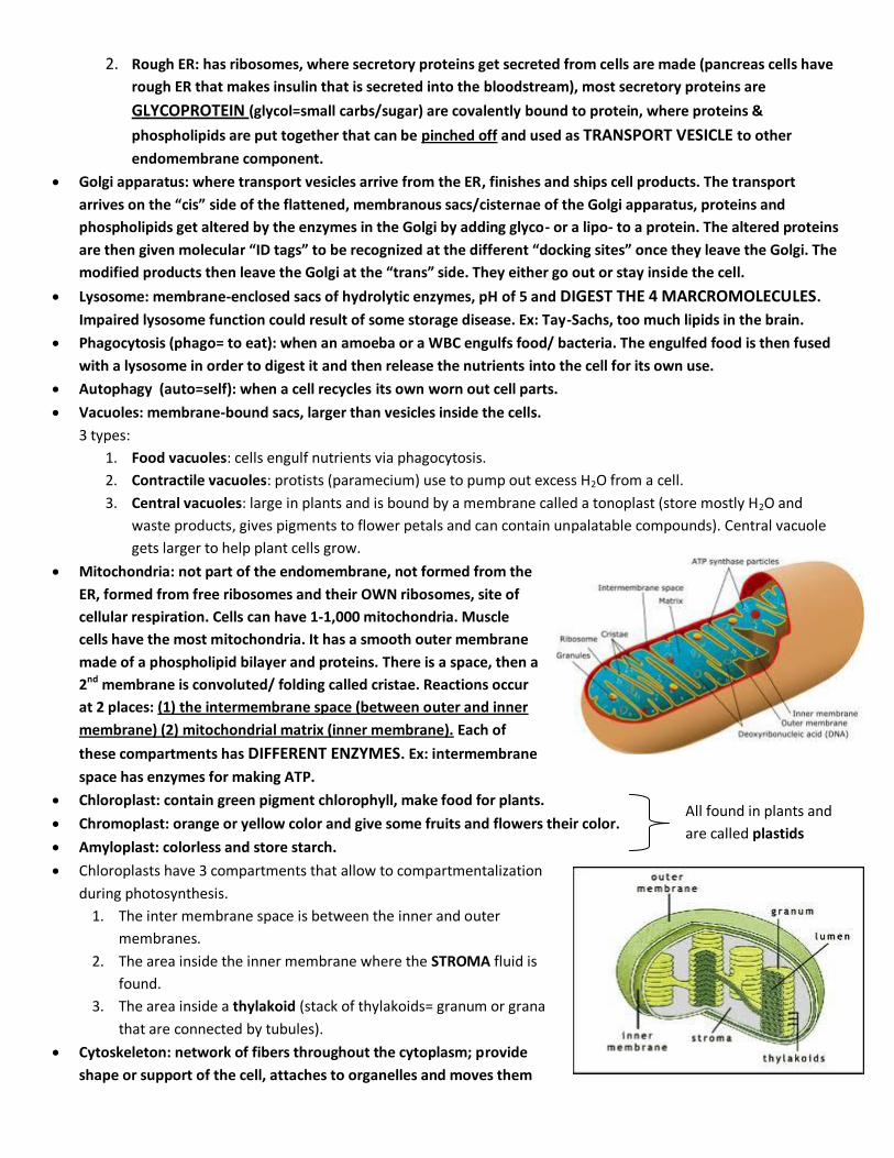
Mitochondria: not part of the endomembrane, not formed from the
ER, formed from free ribosomes and their OWN ribosomes, site of
cellular respiration. Cells can have 1-1,000 mitochondria. Muscle
cells have the most mitochondria. It has a smooth outer membrane
made of a phospholipid bilayer and proteins. There is a space, then a
2nd membrane is convoluted/ folding called cristae. Reactions occur
at 2 places: (1) the intermembrane space (between outer and inner
membrane) (2) mitochondrial matrix (inner membrane). Each of
these compartments has DIFFERENT ENZYMES. Ex: intermembrane
space has enzymes for making ATP.
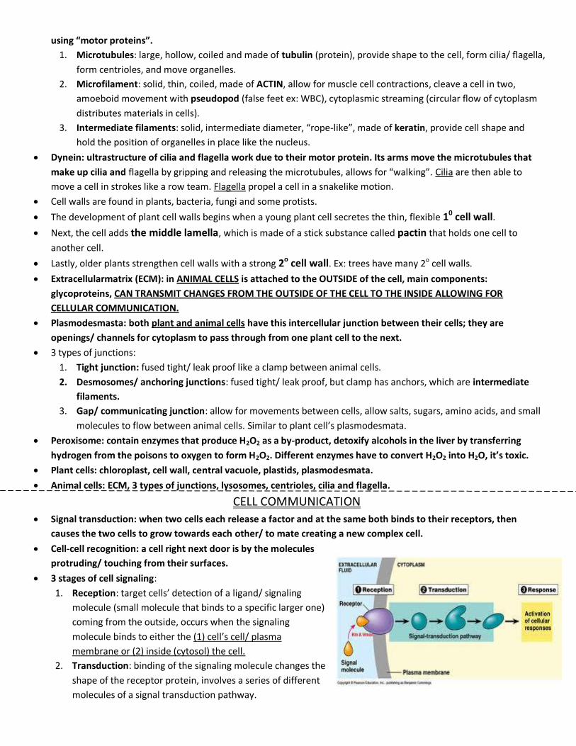
Chloroplast: contain green pigment chlorophyll, make food for plants.
Chromoplast: orange or yellow color and give some fruits and flowers their color.
Amyloplast: colorless and store starch.
Chloroplasts have 3 compartments that allow to compartmentalization
during photosynthesis.
1. The inter membrane space is between the inner and outer
membranes.
2. The area inside the inner membrane where the STROMA fluid is
found.
3. The area inside a thylakoid (stack of thylakoids= granum or grana
that are connected by tubules).
Cytoskeleton: network of fibers throughout the cytoplasm; provide
shape or support of the cell, attaches to organelles and moves them
All found in plants and
are called plastids
using “motor proteins”.
1. Microtubules: large, hollow, coiled and made of tubulin (protein), provide shape to the cell, form cilia/ flagella,
form centrioles, and move organelles.
2. Microfilament: solid, thin, coiled, made of ACTIN, allow for muscle cell contractions, cleave a cell in two,
amoeboid movement with pseudopod (false feet ex: WBC), cytoplasmic streaming (circular flow of cytoplasm
distributes materials in cells).
3. Intermediate filaments: solid, intermediate diameter, “rope-like”, made of keratin, provide cell shape and
hold the position of organelles in place like the nucleus.
Dynein: ultrastructure of cilia and flagella work due to their motor protein. Its arms move the microtubules that
make up cilia and flagella by gripping and releasing the microtubules, allows for “walking”. Cilia are then able to
move a cell in strokes like a row team. Flagella propel a cell in a snakelike motion.
Cell walls are found in plants, bacteria, fungi and some protists.
The development of plant cell walls begins when a young plant cell secretes the thin, flexible 10 cell wall.
Next, the cell adds the middle lamella, which is made of a stick substance called pactin that holds one cell to
another cell.
Lastly, older plants strengthen cell walls with a strong 2o cell wall. Ex: trees have many 2o cell walls.
Extracellularmatrix (ECM): in ANIMAL CELLS is attached to the OUTSIDE of the cell, main components:
glycoproteins, CAN TRANSMIT CHANGES FROM THE OUTSIDE OF THE CELL TO THE INSIDE ALLOWING FOR
CELLULAR COMMUNICATION.
Plasmodesmasta: both plant and animal cells have this intercellular junction between their cells; they are
openings/ channels for cytoplasm to pass through from one plant cell to the next.
3 types of junctions:
1. Tight junction: fused tight/ leak proof like a clamp between animal cells.
2. Desmosomes/ anchoring junctions: fused tight/ leak proof, but clamp has anchors, which are intermediate
filaments.
3. Gap/ communicating junction: allow for movements between cells, allow salts, sugars, amino acids, and small
molecules to flow between animal cells. Similar to plant cell’s plasmodesmata.
Peroxisome: contain enzymes that produce H2O2 as a by-product, detoxify alcohols in the liver by transferring
hydrogen from the poisons to oxygen to form H2O2. Different enzymes have to convert H2O2 into H2O, it’s toxic.
Plant cells: chloroplast, cell wall, central vacuole, plastids, plasmodesmata.
Animal cells: ECM, 3 types of junctions, lysosomes, centrioles, cilia and flagella.
CELL COMMUNICATION
Signal transduction: when two cells each release a factor and at the same both binds to their receptors, then
causes the two cells to grow towards each other/ to mate creating a new complex cell.
Cell-cell recognition: a cell right next door is by the molecules
protruding/ touching from their surfaces.
3 stages of cell signaling:
1. Reception: target cells’ detection of a ligand/ signaling
molecule (small molecule that binds to a specific larger one)
coming from the outside, occurs when the signaling
molecule binds to either the (1) cell’s cell/ plasma
membrane or (2) inside (cytosol) the cell.
2. Transduction: binding of the signaling molecule changes the
shape of the receptor protein, involves a series of different
molecules of a signal transduction pathway.
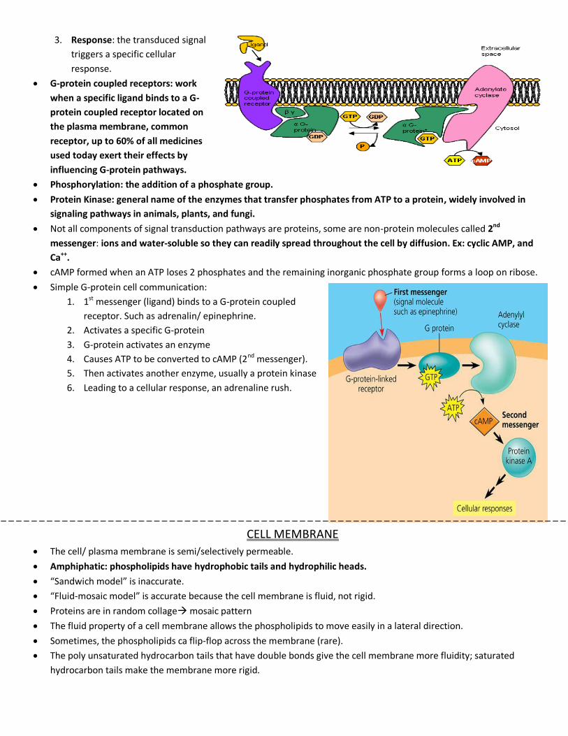
3. Response: the transduced signal
triggers a specific cellular
response.
G-protein coupled receptors: work
when a specific ligand binds to a G-
protein coupled receptor located on
the plasma membrane, common
receptor, up to 60% of all medicines
used today exert their effects by
influencing G-protein pathways.
Phosphorylation: the addition of a phosphate group.
Protein Kinase: general name of the enzymes that transfer phosphates from ATP to a protein, widely involved in
signaling pathways in animals, plants, and fungi.
Not all components of signal transduction pathways are proteins, some are non-protein molecules called 2nd
messenger: ions and water-soluble so they can readily spread throughout the cell by diffusion. Ex: cyclic AMP, and
Ca++.
cAMP formed when an ATP loses 2 phosphates and the remaining inorganic phosphate group forms a loop on ribose.
Simple G-protein cell communication:
1. 1st messenger (ligand) binds to a G-protein coupled
receptor. Such as adrenalin/ epinephrine.
2. Activates a specific G-protein
3. G-protein activates an enzyme
4. Causes ATP to be converted to cAMP (2nd messenger).
5. Then activates another enzyme, usually a protein kinase
6. Leading to a cellular response, an adrenaline rush.
CELL MEMBRANE
The cell/ plasma membrane is semi/selectively permeable.
Amphiphatic: phospholipids have hydrophobic tails and hydrophilic heads.
“Sandwich model” is inaccurate.
“Fluid-mosaic model” is accurate because the cell membrane is fluid, not rigid.
Proteins are in random collage mosaic pattern
The fluid property of a cell membrane allows the phospholipids to move easily in a lateral direction.
Sometimes, the phospholipids ca flip-flop across the membrane (rare).
The poly unsaturated hydrocarbon tails that have double bonds give the cell membrane more fluidity; saturated
hydrocarbon tails make the membrane more rigid.
High temp, cholesterol= hard; low temp, cholesterol= fluid.
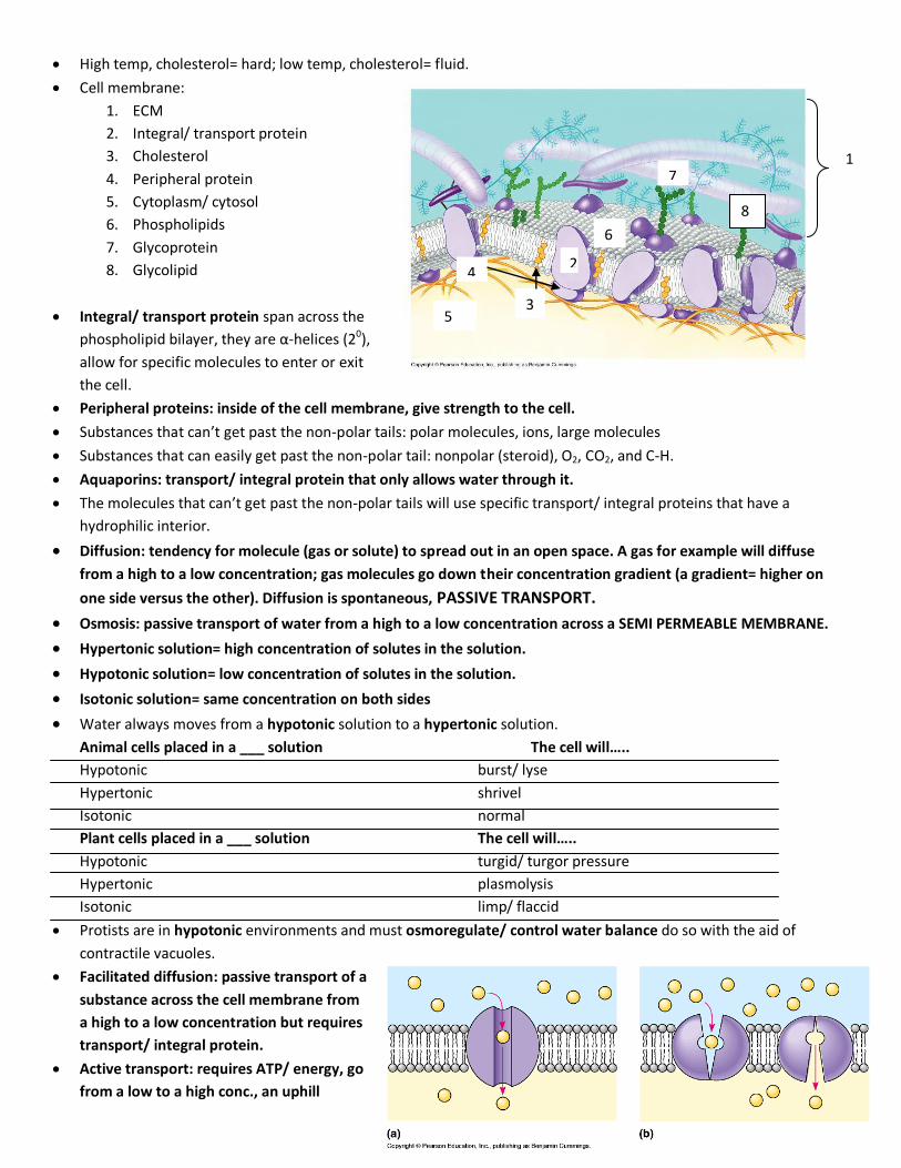
Cell membrane:
1. ECM
2. Integral/ transport protein
3. Cholesterol
4. Peripheral protein
5. Cytoplasm/ cytosol
6. Phospholipids
7. Glycoprotein
8. Glycolipid
Integral/ transport protein span across the
phospholipid bilayer, they are α-helices (20),
allow for specific molecules to enter or exit
the cell.
Peripheral proteins: inside of the cell membrane, give strength to the cell.
Substances that can’t get past the non-polar tails: polar molecules, ions, large molecules
Substances that can easily get past the non-polar tail: nonpolar (steroid), O2, CO2, and C-H.
Aquaporins: transport/ integral protein that only allows water through it.
The molecules that can’t get past the non-polar tails will use specific transport/ integral proteins that have a
hydrophilic interior.
Diffusion: tendency for molecule (gas or solute) to spread out in an open space. A gas for example will diffuse
from a high to a low concentration; gas molecules go down their concentration gradient (a gradient= higher on
one side versus the other). Diffusion is spontaneous, PASSIVE TRANSPORT.
Osmosis: passive transport of water from a high to a low concentration across a SEMI PERMEABLE MEMBRANE.
Hypertonic solution= high concentration of solutes in the solution.
Hypotonic solution= low concentration of solutes in the solution.
Isotonic solution= same concentration on both sides
Water always moves from a hypotonic solution to a hypertonic solution.
Animal cells placed in a ___ solution The cell will…..
Hypotonic burst/ lyse
Hypertonic shrivel
Isotonic normal
Plant cells placed in a ___ solution The cell will…..
Hypotonic turgid/ turgor pressure
Hypertonic plasmolysis
Isotonic limp/ flaccid
Protists are in hypotonic environments and must osmoregulate/ control water balance do so with the aid of
contractile vacuoles.
Facilitated diffusion: passive transport of a
substance across the cell membrane from
a high to a low concentration but requires
transport/ integral protein.
Active transport: requires ATP/ energy, go
from a low to a high conc., an uphill
1
2
3
4
5
6
7
8
reaction that goes against the concentration gradient. EX: 3 NA+ OUT, 2 K+ IN.
Inside of the cell is negative charged, low concentration; outside is positive charged, high concentration.
Plant uses proton pump.
Proton pump: pumps H+ ions out of the cells from a low to high concentration.
Cotransporter: a plant cell pumps H+ out of a cell. H+ then leaks back into the cell passively by facilitated diffusion
through a transport protein. It can bring back with small sugar, amino acids, or nutrients.
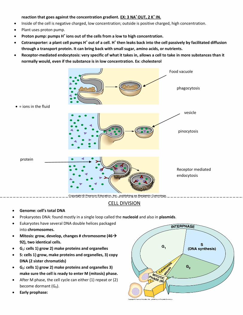
Receptor-mediated endocytosis: very specific of what it takes in, allows a cell to take in more substances than it
normally would, even if the substance is in low concentration. Ex: cholesterol
CELL DIVISION
Genome: cell’s total DNA
Prokaryotes DNA: found mostly in a single loop called the nucleoid and also in plasmids.
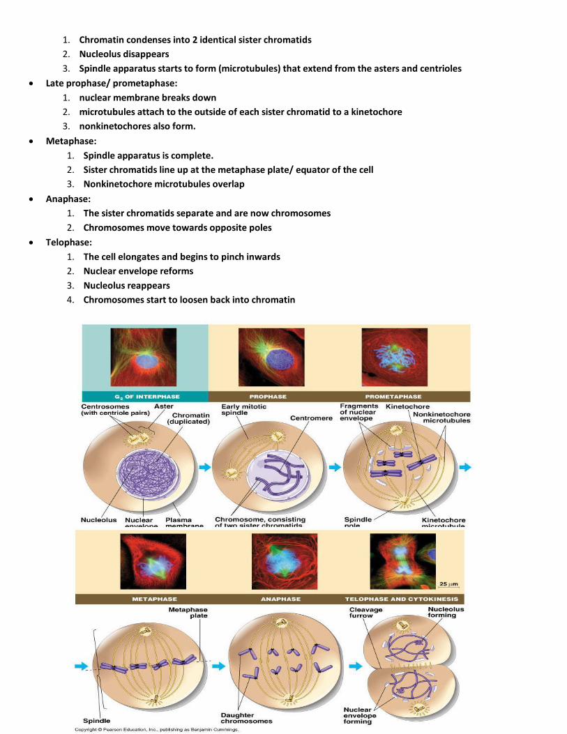
Eukaryotes have several DNA double helices packaged
into chromosomes.
Mitosis: grow, develop, changes # chromosome (46
92), two identical cells.
G1: cells 1) grow 2) make proteins and organelles
S: cells 1) grow, make proteins and organelles, 3) copy
DNA (2 sister chromatids)
G2: cells 1) grow 2) make proteins and organelles 3)
make sure the cell is ready to enter M (mitosis) phase.
After M phase, the cell cycle can either (1) repeat or (2)
become dormant (G0).
Early prophase:
Food vacuole
= ions in the fluid
vesicle
protein
phagocytosis
pinocytosis
Receptor mediated
endocytosis
1. Chromatin condenses into 2 identical sister chromatids
2. Nucleolus disappears
3. Spindle apparatus starts to form (microtubules) that extend from the asters and centrioles
Late prophase/ prometaphase:
1. nuclear membrane breaks down
2. microtubules attach to the outside of each sister chromatid to a kinetochore
3. nonkinetochores also form.
Metaphase:
1. Spindle apparatus is complete.
2. Sister chromatids line up at the metaphase plate/ equator of the cell
3. Nonkinetochore microtubules overlap
Anaphase:
1. The sister chromatids separate and are now chromosomes
2. Chromosomes move towards opposite poles
Telophase:
1. The cell elongates and begins to pinch inwards
2. Nuclear envelope reforms
3. Nucleolus reappears
4. Chromosomes start to loosen back into chromatin
Spindle apparatus includes:
1) Centrosomes: made up of a centriole pair (at a 900 angle) and microtubules in a “star” shape called aster.
Some of the aster’s microtubules will elongate by adding more of the protein tubulin to them.
2) Non-kinetochore microtubules: never attach to the kinetochores of sister chromatids; they cause cell to
lengthen.
3) Kinetochore microtubules: attach to the kinetochores of the sister chromatids during late
prophase/metaphase, pull the sisters apart and shorten as they get near the poles.
4) Centrioles: only in animal cells, get replicated during interphase.
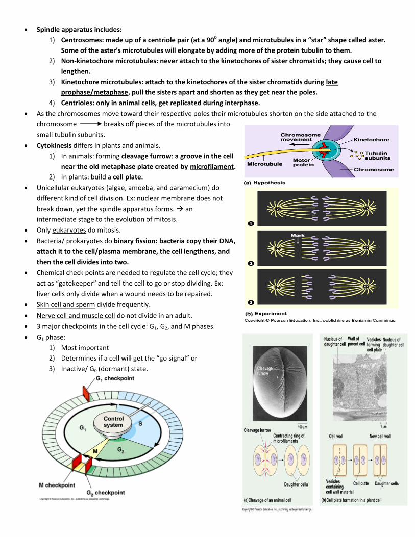
As the chromosomes move toward their respective poles their microtubules shorten on the side attached to the
chromosome breaks off pieces of the microtubules into
small tubulin subunits.
Cytokinesis differs in plants and animals.
1) In animals: forming cleavage furrow: a groove in the cell
near the old metaphase plate created by microfilament.
2) In plants: build a cell plate.
Unicellular eukaryotes (algae, amoeba, and paramecium) do
different kind of cell division. Ex: nuclear membrane does not
break down, yet the spindle apparatus forms. an
intermediate stage to the evolution of mitosis.
Only eukaryotes do mitosis.
Bacteria/ prokaryotes do binary fission: bacteria copy their DNA,
attach it to the cell/plasma membrane, the cell lengthens, and
then the cell divides into two.
Chemical check points are needed to regulate the cell cycle; they
act as “gatekeeper” and tell the cell to go or stop dividing. Ex:
liver cells only divide when a wound needs to be repaired.
Skin cell and sperm divide frequently.
Nerve cell and muscle cell do not divide in an adult.
3 major checkpoints in the cell cycle: G1, G2, and M phases.
G1 phase:
1) Most important
2) Determines if a cell will get the “go signal” or
3) Inactive/ G0 (dormant) state.
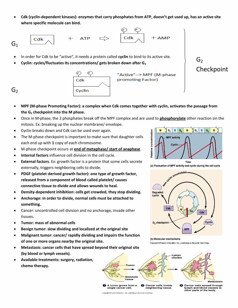
Cdk (cyclin-dependent kinases): enzymes that carry phosphates from ATP, doesn’t get used up, has an active site
where specific molecule can bind.
In order for Cdk to be “active”, it needs a protein called cyclin to bind to its active site.
Cyclin: cycles/fluctuates its concentrations/ gets broken down after G2
MPF (M-phase Promoting Factor): a complex when Cdk comes together with cyclin, activates the passage from
the G2 checkpoint into the M phase.
Once in M-phase, the 2 phosphates break off the MPF complex and are used to phosphorylate other reaction sin the
mitosis. Ex: breaking up the nuclear membrane/ envelope.
Cyclin breaks down and Cdk can be used over again.
The M-phase checkpoint is important to make sure that daughter cells
each end up with 1 copy of each chromosome.
M-phase checkpoint occurs at end of metaphase/ start of anaphase.
Internal factors influence cell division in the cell cycle.
External factors. Ex: growth factor is a protein that some cells secrete
externally, triggers neighboring cells to divide.
PDGF (platelet-derived growth factor): one type of growth factor,
released from a component of blood called platelet/ causes
connective tissue to divide and allows wounds to heal.
Density-dependent inhibition: cells get crowded, they stop dividing.
Anchorage: in order to divide, normal cells must be attached to
something.
Cancer: uncontrolled cell division and no anchorage, invade other
tissues.
Tumor: mass of abnormal cells
Benign tumor: slow dividing and localized at the original site
Malignant tumor: cancer/ rapidly dividing and impairs the function
of one or more organs nearby the original site.
Metastasis: cancer cells that have spread beyond their original site
(by blood or lymph vessels).
Available treatments: surgery, radiation,
chemo therapy.
G2
Checkpoint
G1
G2