Cell Segmentation: 50 Years Down the Road
8
Cell Segmentation: 50 Years Down the Road Erik Meijering IEEE Signal Processing Magazine, vol. 29, no. 5, September 2012, pp. 140–145 E ver since the establishment of cell theory in the early 19th century, which recognized the cell as the fundamental building unit of life, biologists have sought to explain the under- lying principles. Momentous discoveries were made in the course of many decades of research [1], but the quest to attain full understanding of cellular mechanisms and how to manip- ulate them to improve health, continues to the present day, with bigger budgets, more minds, and more sophisticated tools than ever before. One of the tools to which a great deal of the progress in cell biology can be attributed is light microscopy [2]. The field has come a long way since Antoni van Leeuwenhoek’s first steps in the 1670s toward improving and exploiting microscopic imaging for studying life at the cellular level. Not only do biologists nowadays have a plethora of differ- ent, complementary microscopic imaging techniques at their disposal, enabling them to visualize phenomena even way below the classical diffraction limit of light, advanced microscope systems also allow them to easily acquire very large numbers of images within just a matter of hours. The abundance, heterogeneity, dimensionality, and complexity of the data generated in modern imaging experiments rule out manual image management, processing, and analysis. Consequently, comput- erized techniques for performing these tasks have become of key importance for further progress in cell biology [3–6]. A central problem in many studies, and often regarded as the cornerstone of image analysis, is image segmentation. Specifically, since cellular morphology is an impor- tant phenotypic feature, indicative of the physiological state of a cell, and since the cell contour is often required for subsequent analysis of intracellular processes (zooming in to nanoscale), or of cell sociology (zooming out to millimeter scale), the problem of cell segmentation has received increasing attention in past years [7]. Here we reflect on how the field has evolved over the years and how past developments can be expected to extrapolate into the future. A VERY BRIEF HISTORY OF CELL ANALYSIS The first uses of computers for the analysis of cells date back more than half a century. Already in the mid-1950s, systems were developed to automate the classification of smears of exfoliated cells, with the ultimate aim to enable mass screening for cervical cancer. These systems applied thresholding-based decision rules to serial one-dimensional (1D) microscopic line scans of a spec- imen [8]. The 1960s witnessed the first examples of automated processing of two-dimensional (2D) images for the purpose of differential counting of white blood cells (leukocytes) according to their main classes based on simple colorimetric and morphological measurements [9]. Commer- cial systems for performing this routine clinical test hit the market around the mid-1970s and even contained multiple computer circuits to parallelize the tasks of analyzing the image of the previous
Transcript of Cell Segmentation: 50 Years Down the Road
Cell Segmentation: 50 Years Down the Road
Erik Meijering
IEEE Signal Processing Magazine, vol. 29, no. 5, September 2012, pp. 140–145
E ver since the establishment of cell theory in the early 19th century, which recognized thecell as the fundamental building unit of life, biologists have sought to explain the under-lying principles. Momentous discoveries were made in the course of many decades of
research [1], but the quest to attain full understanding of cellular mechanisms and how to manip-ulate them to improve health, continues to the present day, with bigger budgets, more minds, andmore sophisticated tools than ever before. One of the tools to which a great deal of the progress incell biology can be attributed is light microscopy [2]. The field has come a long way since Antonivan Leeuwenhoek’s first steps in the 1670s toward improving and exploiting microscopic imagingfor studying life at the cellular level. Not only do biologists nowadays have a plethora of differ-ent, complementary microscopic imaging techniques at their disposal, enabling them to visualizephenomena even way below the classical diffraction limit of light, advanced microscope systemsalso allow them to easily acquire very large numbers of images within just a matter of hours. Theabundance, heterogeneity, dimensionality, and complexity of the data generated in modern imagingexperiments rule out manual image management, processing, and analysis. Consequently, comput-erized techniques for performing these tasks have become of key importance for further progressin cell biology [3–6]. A central problem in many studies, and often regarded as the cornerstoneof image analysis, is image segmentation. Specifically, since cellular morphology is an impor-tant phenotypic feature, indicative of the physiological state of a cell, and since the cell contouris often required for subsequent analysis of intracellular processes (zooming in to nanoscale), orof cell sociology (zooming out to millimeter scale), the problem of cell segmentation has receivedincreasing attention in past years [7]. Here we reflect on how the field has evolved over the yearsand how past developments can be expected to extrapolate into the future.
A VERY BRIEF HISTORY OF CELL ANALYSISThe first uses of computers for the analysis of cells date back more than half a century. Alreadyin the mid-1950s, systems were developed to automate the classification of smears of exfoliatedcells, with the ultimate aim to enable mass screening for cervical cancer. These systems appliedthresholding-based decision rules to serial one-dimensional (1D) microscopic line scans of a spec-imen [8]. The 1960s witnessed the first examples of automated processing of two-dimensional(2D) images for the purpose of differential counting of white blood cells (leukocytes) according totheir main classes based on simple colorimetric and morphological measurements [9]. Commer-cial systems for performing this routine clinical test hit the market around the mid-1970s and evencontained multiple computer circuits to parallelize the tasks of analyzing the image of the previous
CELL SEGMENTATION: 50 YEARS DOWN THE ROAD 2
cell, while grabbing the image of the present cell, and at the same time locating the next cell inthe specimen [10]. These were also the times when the first computer assisted microscopes weredeveloped for tracing and morphological analysis of neuronal cells [11]. The advent of confocalmicroscope systems in the 1980s opened the door to three-dimensional (3D) cell image analysis.But it was not until the 1990s, when computers became powerful enough to handle 3D data, or evencomplex 2D data such as in histopathology [12], that the image processing and computer visioncommunities really began to take up the challenge. Over the past decades, literature on the subjecthas grown exponentially, with more than half of the bulk of papers appearing after the year 2000.Published cell image analysis methods have already been the basis of numerous studies involvingcell counting (numbers), the identification of cell types or cell phases (shapes), the quantificationof cell migration and interaction (morphodynamics), cellular sociology (tissue level organization),and intracellular structures (cell organization) [5].
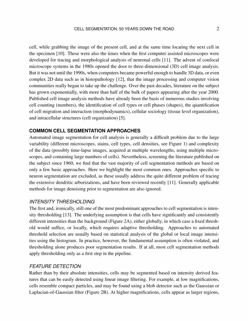
COMMON CELL SEGMENTATION APPROACHESAutomated image segmentation for cell analysis is generally a difficult problem due to the largevariability (different microscopes, stains, cell types, cell densities, see Figure 1) and complexityof the data (possibly time-lapse images, acquired at multiple wavelengths, using multiple micro-scopes, and containing large numbers of cells). Nevertheless, screening the literature published onthe subject since 1960, we find that the vast majority of cell segmentation methods are based ononly a few basic approaches. Here we highlight the most common ones. Approaches specific toneuron segmentation are excluded, as these usually address the quite different problem of tracingthe extensive dendritic arborizations, and have been reviewed recently [11]. Generally applicablemethods for image denoising prior to segmentation are also ignored.
INTENSITY THRESHOLDINGThe first and, ironically, still one of the most predominant approaches to cell segmentation is inten-sity thresholding [13]. The underlying assumption is that cells have significantly and consistentlydifferent intensities than the background (Figure 2A), either globally, in which case a fixed thresh-old would suffice, or locally, which requires adaptive thresholding. Approaches to automatedthreshold selection are usually based on statistical analysis of the global or local image intensi-ties using the histogram. In practice, however, the fundamental assumption is often violated, andthresholding alone produces poor segmentation results. If at all, most cell segmentation methodsapply thresholding only as a first step in the pipeline.
FEATURE DETECTIONRather than by their absolute intensities, cells may be segmented based on intensity derived fea-tures that can be easily detected using linear image filtering. For example, at low magnifications,cells resemble compact particles, and may be found using a blob detector such as the Gaussian orLaplacian-of-Gaussian filter (Figure 2B). At higher magnifications, cells appear as larger regions,
CELL SEGMENTATION: 50 YEARS DOWN THE ROAD 3
A
B
C
D
E
F
G
H
FIGURE 1: Cell images may vary widely, depending on the type of microscopy and staining used, as wellas the cell type and cell density. This makes the development of a generally applicable cell segmentationmethod a huge challenge. Shown here are various illustrative examples of cell images (with cell densityincreasing from left to right), acquired using bright-field microscopy (A-B), phase-contrast microscopy (C),differential interference contrast microscopy (D), and fluorescence microscopy (E-H). In the latter case, theuse of fluorescent dyes or proteins enables biologists to selectively label virtually any target of interest inthe cell or nucleus, such as the nucleoprotein histone involved in DNA folding (E), DNA binding proteinssuch as Rad18 or Rad54 (F), or cell adhesion proteins such as E-cadherin (G). The resulting additionalvariability in cell appearance further complicates the development of generic cell segmentation methods.Finally, whereas most cells are fairly spherical, some classes of cells may show quite different shape.Especially neurons (H) may extend far beyond the compact cell body.
but if their shapes are relatively invariant, a dedicated filter template could be derived from the im-ages. Alternatively, in more variable scenarios, edge detection (first-order differential filtering) orridge detection (second-order differential filtering) is often used, followed by some linking proce-dure [13]. Similar to thresholding, such filters alone usually do not produce definitive cell outlines,but may provide useful cues for subsequent steps in the pipeline.
MORPHOLOGICAL FILTERINGAnother popular class of filters are those from the field of mathematical morphology. Being non-linear, operators such as erosion, dilation, opening, and closing allow for the examination andmanipulation of geometrical and topological properties of objects in images, and are often used

CELL SEGMENTATION: 50 YEARS DOWN THE ROAD 4
A B C D
FIGURE 2: Examples of cell image segmentation based on the discussed approaches. The rows show, re-spectively, the input images, the automatically found cell contours (overlaid in green), and the correspond-ing labeled cell regions (arbitrary colors). (A) Cells that are fairly well separated and clearly brighter thanthe background are easily segmented using thresholding. Binary ultimate erosion and reconstruction wasused to split the few clumped cells. (B) Scenarios with higher cell densities and intensity variations requiremore sophisticated methods. The method used here involves graph-cuts based binarization, Laplacian-of-Gaussian based cell detection (see red dots), and marker based clustering (from [14] with permission). (C)Membrane stained images are ideally suited for watershed based segmentation. Grayscale morphologicalprefiltering was used both for background estimation (opening operation) and filling imperfectly stainedsegments (closing operation). (D) Studies of intracellular dynamic processes often result in images withsignificant intensity variations (in both space and time) and require robust cell segmentation and trackingmethods. The method used here is based on level sets [15]. All of these methods were specifically designedfor the given application and required careful parameter tuning.
in connection with cell segmentation. More complicated filters can be easily constructed by com-bination or successive application of such operators. A distinction must be made between binarymorphology and grayscale morphology [13]. The former is used mostly as a postprocessing stepto polish coarse segmentations (Figure 2A) while the latter is used mostly as a preprocessing stepto enhance or suppress specific image structures for segmentation (Figure 2C).

CELL SEGMENTATION: 50 YEARS DOWN THE ROAD 5
REGION ACCUMULATIONAn alternative approach to cell segmentation is to start from selected seed points in the image and toiteratively add connected points to form labeled regions. The most straightforward implementationof this idea is ordinary region growing, which works per neighborhood layer of connected pointsand, when applied directly to the image, assumes (and suffers from) a similar image model as inthe case of intensity thresholding. Hierarchical split-and-merge schemes, operating per resolutionlayer and using some uniformity predicate, have also been used occasionally. Another exampleis the watershed transform [13], the main segmentation approach from mathematical morphology,which works per intensity layer and requires an edge enhanced image (gradient magnitude), asit is commonly desired to have the watershed lines at the edges (Figure 2C). Though by far themost popular region accumulation approach, the watershed transform is infamous for producingoversegmentation, and usually requires further processing.
DEFORMABLE MODEL FITTINGThe final class of cell segmentation approaches mentioned here consists of procedures that fita deformable model to the image data (Figure 2D). Deformable models may be formulated eitherexplicitly, as a parametric contour (2D) or surface (3D), or implicitly, as the zero-level of a functionwith dimensionality (nD) one higher than the image to be segmented. Level sets have the advantagethat they naturally handle topological changes, such as cell division, and are therefore attractivefor cell tracking [16]. Initialized by a first, coarse segmentation, a deformable model is iterativelyevolved to minimize a predefined energy functional, which typically consists of image-based andshape-based terms, the latter of which allow the incorporation of prior knowledge to constrain theresults. The energy terms need to be carefully designed to avoid erroneous segmentation.
CELL SEGMENTATION APPROACHES ANALYZEDIt is interesting to analyze the usage of the different approaches to cell segmentation over the years(consult Figure 3 for the following discussion). Intensity thresholding, being the conceptuallysimplest and computationally cheapest of all, was the first approach to be used since the 1960s.Soon after, it was realized that differential features (in particular edges) could also be computedrelatively easily, and might provide useful information. The field of mathematical morphologystarted to develop around the same time, and its basic operators were first used in the late 1970sfor refining the results of thresholding based cell segmentation. During that same decade, allmentioned region accumulation approaches were conceived, and first examples of their usage in thecontext of cell image analysis appeared around the mid-1980s. Finally, the first deformable modelfitting approaches for image segmentation were introduced in the second half of the 1980s, andwere first applied to cell images in the early 1990s. Since then, miscellaneous other segmentationapproaches found their way into the field, with as yet limited usage. Examples include dynamicprogramming, graph cuts, active masks, support vector machines, tensor voting schemes, neuralnetworks, Markov random fields, and other concepts.

CELL SEGMENTATION: 50 YEARS DOWN THE ROAD 6
0
10
20
30
40
50
60
70
80
0%
10%
20%
30%
40%
50%
60%
70%
80%
90%
100%
Thresholds Features Morphology Regions Deformables Miscellany
Number of papers per lustrum
1960 2015*2010200520001995199019851980197519701965
FIGURE 3: Literature on cell image analysis shows an exponentially increasing interest in cell segmen-tation and the emergence of new approaches for this purpose. In total 250 journal papers describing cellsegmentation methods were analyzed for the present article. The top panel shows their time histogramusing lustrum bins (the last bin obviously contains only partial data, up to March 2012, as indicated by theasterisk). The distribution is representative of that of the total numbers of papers published on the subjectin the various periods. The bottom panel shows the breakdown of published methods per lustrum into sixmain classes of approaches (explained in the main text): intensity thresholding (blue), feature detection(red), morphological filtering (green), region accumulation (yellow), deformable model fitting (violet), andmiscellaneous approaches (magenta) that could not be classified as any of the former. Most methods use acombination of several approaches.
Several observations follow from the analysis of the literature on cell segmentation in thepast 50 years. First, most of the different approaches were originally developed for applicationsin other fields (computer vision, robotics, materials science, medical imaging), and were lateradopted for cell segmentation. This is remarkable, given the unique and unparalleled challengesin cell image analysis, which should provoke the development of original ideas. Second, eventhough new approaches are introduced once in a while, they seem to never fully replace old ones.Apparently, while none of them alone produces satisfactory results, they all continue to be useful

CELL SEGMENTATION: 50 YEARS DOWN THE ROAD 7
to some extent. Third, as a consequence, methods proposed in recent times are rarely based on asingle new concept, but are often merely new combinations of the discussed approaches, tailoredto a specific application. Rather than converging to a robust, unified solution, it thus seems thatthe field is diverging, and by now almost as many cell segmentation methods have been developedas there exist cell analysis problems [7]. However, the explosion of technical papers in the pastdecade suggests that the performance and applicability of these methods remain limited, and morepowerful methods will need to be developed.
THE FUTURE OF CELL SEGMENTATION?Reflecting on the developments in the field in the past 50 years, one wonders what to expect fromthe next 50 years of research. As early as 1966, researchers already exclaimed that “automation ofthe acquisition and interpretation of data in microscopy has been a focus of biomedical researchfor almost a decade”, and they concluded that “many facets of the problem appear to be well withinthe grasp of present-day technology”, leading them to anticipate that “modern large capacity, highspeed data facilities at last provide the ability to manipulate the hitherto unmanageable quantitiesof optical information contained within all but the simplest images” [9]. On one hand, it may feelembarrassing to admit that today, half a century down the road, very similar remarks still apply. Itseems as if from the very beginning, the “grasp of present-day technology” in the field of imageanalysis has held a firm position a few years ahead in the future, barely able to keep up with therate of progress in its application areas. While microscopic imaging, biological experimentation,and computer hardware development all underwent major revolutions in the past decades, mostcell image analysis methods are still based on textbook ingredients [13] from the early days. Onthe other hand, perhaps this is just a testimony to the fact that the ease with which humans cansee things in images is very deceptive, and computerizing this capacity is actually a notoriouslydifficult problem. The provisional solution has thus far been to isolate applications and to developa dedicated method for each. But the real challenge remains to design methods that are sufficientlygeneric to be easily trainable for a wide range of applications while consistently achieving highsensitivity and specificity in each case. Perhaps the rapidly increasing market share of alternativesegmentation approaches (Figure 3) signifies the beginning of a new era. Important catalysts for thedevelopment of more powerful methods will be improved availability and testability. The technicalliterature is full of alleged great methods, which were claimed to beat all previous methods for agiven application, but subsequently disappeared into oblivion because no one was able to use orreproduce them. Their availability in popular (open-source) image analysis platforms will alleviatethis problem and should be increasingly enforced before publication. But even then, methods maybe easily abused by others to “prove” superiority of their own methods. The organization of openchallenges based on standardized test data and criteria should suppress this practice and can beexpected to further accelerate progress in the field in the near future.
CELL SEGMENTATION: 50 YEARS DOWN THE ROAD 8
ACKNOWLEDGMENTSThe author thankfully acknowledges Gert van Cappellen, Jeroen Essers, Timo ten Hagen (ErasmusMC, Rotterdam, The Netherlands), Paul De Koninck (Laval University, Quebec, Canada), MichaelW. Davidson (Florida State University, Tallahassee, FL, USA), Thomas Lecuit (DevelopmentalBiology Institute, Marseilles, France), and the Robert H. Waterston lab (University of Washington,Seattle, WA, USA) for the sample cell images.
REFERENCES[1] B. Alberts, A. Johnson, J. Lewis, M. Raff, K. Roberts, and P. Walter, Molecular Biology of the Cell, 5th ed.,
Garland Science, New York, NY, 2007.[2] C. Vonesch, F. Aguet, J.-L. Vonesch, and M. Unser, “The colored revolution of bioimaging”, IEEE Signal
Processing Magazine, vol. 23, no. 3, pp. 20–31, May 2006.[3] H. Peng, “Bioimage informatics: A new area of engineering biology”, Bioinformatics, vol. 24, no. 17, pp. 1827–
1836, September 2008.[4] J. R. Swedlow and K. W. Eliceiri, “Open source bioimage informatics for cell biology”, Trends in Cell Biology,
vol. 19, no. 11, pp. 656–660, November 2009.[5] J. Rittscher, “Characterization of biological processes through automated image analysis”, Annual Review of
Biomedical Engineering, vol. 12, pp. 315–344, August 2010.[6] G. Danuser, “Computer vision in cell biology”, Cell, vol. 147, no. 5, pp. 973–978, November 2011.[7] E. Bengtsson, C. Wahlby, and J. Lindblad, “Robust cell image segmentation methods”, Pattern Recognition and
Image Analysis, vol. 14, no. 2, pp. 157–167, May 2004.[8] W. E. Tolles, “The Cytoanalyzer — An example of physics in medical research”, Transactions of the New York
Academy of Sciences, vol. 17, no. 3, pp. 250–256, January 1955.[9] J. M. S. Prewitt and M. L. Mendelsohn, “The analysis of cell images”, Annals of the New York Academy of
Sciences, vol. 128, no. 3, pp. 1035–1053, January 1966.[10] K. Preston, “Computer processing of biomedical images”, Computer, vol. 9, no. 5, pp. 54–68, May 1976.[11] E. Meijering, “Neuron tracing in perspective”, Cytometry Part A, vol. 77, no. 7, pp. 693–704, July 2010.[12] M. N. Gurcan, L. Boucheron, A. Can, A. Madabhushi, N. Rajpoot, and B. Yener, “Histopathological image
analysis: A review”, IEEE Reviews in Biomedical Engineering, vol. 2, pp. 147–171, December 2009.[13] Q. Wu, F. A. Merchant, and K. R. Castleman, Microscope Image Processing, Academic Press, Burlington, MA,
2008.[14] Y. Al-Kofahi, W. Lassoued, W. Lee, and B. Roysam, “Improved automatic detection and segmentation of cell
nuclei in histopathology images”, IEEE Transactions on Biomedical Engineering, vol. 57, no. 4, pp. 841–852,April 2010.
[15] O. Dzyubachyk, W. A. van Cappellen, J. Essers, W. J. Niessen, and E. Meijering, “Advanced level-set-basedcell tracking in time-lapse fluorescence microscopy”, IEEE Transactions on Medical Imaging, vol. 29, no. 3,pp. 852–867, March 2010.
[16] E. Meijering, O. Dzyubachyk, I. Smal, and W. A. van Cappellen, “Tracking in cell and developmental biology”,Seminars in Cell and Developmental Biology, vol. 20, no. 8, pp. 894–902, October 2009.