Cell population heterogeneity during growth of Bacillus ...
13
Cell population heterogeneity during growth of Bacillus subtilis Daniel B. Kearns 1,2 and Richard Losick 1,3 1 Department of Molecular and Cellular Biology, Harvard University, Cambridge, Massachusetts 02138, USA; 2 Department of Biology, Indiana University, Bloomington, Indiana 47405, USA We have discovered that cells of Bacillus subtilis at the mid-exponential phase of growth are a mixed population of two strikingly different cell types. One type is single swimming cells (or cell doublets) in which the transcription factor for motility, D , is active ( D ON). The other type is long chains of sessile cells in which D is inactive ( D OFF). The population is strongly biased toward D -ON cells by the action of a novel regulatory protein called SwrA. SwrA stimulates the transcription of a large operon (the flagellum/chemotaxis operon), which includes the genes for D and an activator of D -directed gene expression, SwrB. Cell population heterogeneity could enable B. subtilis to exploit its present location through the production of sessile cells as well as to explore new environmental niches through the generation of nomadic cells. [Keywords: Motility; bistability; SwrA; D ; swarming; flagella] Supplemental material is available at http://www.genesdev.org. Received September 12, 2005; revised version accepted October 27, 2005. Microbiologists have traditionally viewed populations of genetically identical bacteria as if they were homoge- neous. Increasingly, however, it has become apparent that under certain conditions certain populations of bac- teria are heterogeneous, consisting of mixtures of cells in decidedly distinct physiological or developmental states. For example, Escherichia coli cells can spontaneously enter a “persister” state in which some cells in the popu- lation temporarily stop growing and, as a consequence, escape the killing action of -lactam antibiotics (Balaban et al. 2004; Keren et al. 2004). Similarly, under condi- tions of nutrient limitation, cells of Bacillus subtilis generate a mixed population in which about half of the cells activate the master regulator for sporulation, Spo0A, and the remainder do not (Chung et al. 1994; González-Pastor et al. 2003; Fujita et al. 2005). The bio- logical significance of this example of population hetero- geneity is that cells that have activated Spo0A delay be- coming committed to spore formation by a process of cannibalism that involves the killing of sibling cells that have not activated the master regulator (González-Pastor et al. 2003) Yet another example of population heterogeneity is seen during the transition to stationary phase under con- ditions in which cells of B. subtilis enter a physiological state known as genetic competence. Competent cells cease growth and become capable of undergoing DNA- mediated transformation. During the transition to sta- tionary phase, only some cells in the population (10%– 20%) activate the master regulator for competence, ComK, and become competent (Hadden and Nester 1968; Haseltine-Cahn and Fox 1968; Haijema et al. 2001). In this manner only a fraction of the cells sacrifice their ability to grow in order to gain the benefit of DNA uptake. The remainder of the cells can continue to grow or alternatively respond to stationary phase in other ways such as the process of sporulation mentioned above. Here we report the discovery of a new and striking example of cell population heterogeneity that is seen during the exponential phase of growth. We show that growing cells of B. subtilis exist in one of two states with respect to the expression of genes involved in motility. Cells of B. subtilis swim by means of a flagellum, the protein components of which are partly encoded by a large (31-gene-long) operon (the fla/che operon) that in- cludes flagellum genes, genes involved in chemotaxis, and the gene for the alternative sigma factor D , the pen- ultimate member of the operon (Fig. 1; Helmann et al. 1988; Márquez et al. 1990; Zuberi et al. 1990; Albertini et al. 1991). The D factor, in turn, directs the transcription of the remaining genes involved in flagellum biosynthe- sis, which lie outside the operon. These include hag, the structural gene for the flagellar filament, and flgG and flgK, which encode major components of the flagellar hook–basal body (Mirel and Chamberlin 1989; Chen and Helmann 1994; Mirel et al. 1994; Serizawa et al. 2004). The D factor also directs the transcription of genes (lytC, lytD, and lytF) for autolytic enzymes that mediate 3 Corresponding author. E-MAIL [email protected]; FAX (617) 496-4642. Article and publication are at http://www.genesdev.org/cgi/doi/10.1101/ gad.1373905. GENES & DEVELOPMENT 19:3083–3094 © 2005 by Cold Spring Harbor Laboratory Press ISSN 0890-9369/05; www.genesdev.org 3083 Cold Spring Harbor Laboratory Press on November 22, 2021 - Published by genesdev.cshlp.org Downloaded from
Transcript of Cell population heterogeneity during growth of Bacillus ...

Cell population heterogeneityduring growth of Bacillus subtilisDaniel B. Kearns1,2 and Richard Losick1,3
1Department of Molecular and Cellular Biology, Harvard University, Cambridge, Massachusetts 02138, USA; 2Department ofBiology, Indiana University, Bloomington, Indiana 47405, USA
We have discovered that cells of Bacillus subtilis at the mid-exponential phase of growth are a mixedpopulation of two strikingly different cell types. One type is single swimming cells (or cell doublets) in whichthe transcription factor for motility, �D, is active (�D ON). The other type is long chains of sessile cells inwhich �D is inactive (�D OFF). The population is strongly biased toward �D-ON cells by the action of a novelregulatory protein called SwrA. SwrA stimulates the transcription of a large operon (the flagellum/chemotaxisoperon), which includes the genes for �D and an activator of �D-directed gene expression, SwrB. Cellpopulation heterogeneity could enable B. subtilis to exploit its present location through the production ofsessile cells as well as to explore new environmental niches through the generation of nomadic cells.
[Keywords: Motility; bistability; SwrA; �D; swarming; flagella]
Supplemental material is available at http://www.genesdev.org.
Received September 12, 2005; revised version accepted October 27, 2005.
Microbiologists have traditionally viewed populations ofgenetically identical bacteria as if they were homoge-neous. Increasingly, however, it has become apparentthat under certain conditions certain populations of bac-teria are heterogeneous, consisting of mixtures of cells indecidedly distinct physiological or developmental states.For example, Escherichia coli cells can spontaneouslyenter a “persister” state in which some cells in the popu-lation temporarily stop growing and, as a consequence,escape the killing action of �-lactam antibiotics (Balabanet al. 2004; Keren et al. 2004). Similarly, under condi-tions of nutrient limitation, cells of Bacillus subtilisgenerate a mixed population in which about half of thecells activate the master regulator for sporulation,Spo0A, and the remainder do not (Chung et al. 1994;González-Pastor et al. 2003; Fujita et al. 2005). The bio-logical significance of this example of population hetero-geneity is that cells that have activated Spo0A delay be-coming committed to spore formation by a process ofcannibalism that involves the killing of sibling cells thathave not activated the master regulator (González-Pastoret al. 2003)
Yet another example of population heterogeneity isseen during the transition to stationary phase under con-ditions in which cells of B. subtilis enter a physiologicalstate known as genetic competence. Competent cellscease growth and become capable of undergoing DNA-
mediated transformation. During the transition to sta-tionary phase, only some cells in the population (10%–20%) activate the master regulator for competence,ComK, and become competent (Hadden and Nester1968; Haseltine-Cahn and Fox 1968; Haijema et al.2001). In this manner only a fraction of the cells sacrificetheir ability to grow in order to gain the benefit of DNAuptake. The remainder of the cells can continue to growor alternatively respond to stationary phase in otherways such as the process of sporulation mentionedabove.
Here we report the discovery of a new and strikingexample of cell population heterogeneity that is seenduring the exponential phase of growth. We show thatgrowing cells of B. subtilis exist in one of two states withrespect to the expression of genes involved in motility.Cells of B. subtilis swim by means of a flagellum, theprotein components of which are partly encoded by alarge (31-gene-long) operon (the fla/che operon) that in-cludes flagellum genes, genes involved in chemotaxis,and the gene for the alternative sigma factor �D, the pen-ultimate member of the operon (Fig. 1; Helmann et al.1988; Márquez et al. 1990; Zuberi et al. 1990; Albertini etal. 1991). The �D factor, in turn, directs the transcriptionof the remaining genes involved in flagellum biosynthe-sis, which lie outside the operon. These include hag, thestructural gene for the flagellar filament, and flgG andflgK, which encode major components of the flagellarhook–basal body (Mirel and Chamberlin 1989; Chen andHelmann 1994; Mirel et al. 1994; Serizawa et al. 2004).The �D factor also directs the transcription of genes(lytC, lytD, and lytF) for autolytic enzymes that mediate
3Corresponding author.E-MAIL [email protected]; FAX (617) 496-4642.Article and publication are at http://www.genesdev.org/cgi/doi/10.1101/gad.1373905.
GENES & DEVELOPMENT 19:3083–3094 © 2005 by Cold Spring Harbor Laboratory Press ISSN 0890-9369/05; www.genesdev.org 3083
Cold Spring Harbor Laboratory Press on November 22, 2021 - Published by genesdev.cshlp.orgDownloaded from

the separation of sister cells after cell division (Fig. 1;Lazarevic et al. 1992; Margot et al. 1994, 1999; Blackmanet al. 1998).
We now show that growing cells of B. subtilis are amixed population in which �D is active (ON) in somecells and inactive (OFF) in other cells. When �D is ON,individual cells are motile, whereas when �D is OFF,cells grow as long sessile chains. We show that the popu-lation is strongly biased toward swimming cells by anovel regulatory protein called SwrA that stimulates thetranscription of the fla/che operon, which includes thegene for �D and the gene for a novel protein called SwrB(Fig. 1). We further show that SwrB is an activator of�D-directed gene expression and that biasing the cellpopulation to the �D-ON state requires high-level ex-pression of the genes for both �D and SwrB. Finally, wespeculate on the nature of the mechanism that switches�D activity ON or OFF and the biological significance ofthe alternative cell types.
Results
Growing cells exist in two alternative developmentalstates
Laboratory strains of B. subtilis, such as 168 and PY79,are traditionally recognized as existing in one of twomorphologically distinct forms (Fein 1979). One form ischaracterized by long chains of sessile cells, and this isthe predominant form seen during the exponential phaseof growth, as illustrated in Figure 2A. The other form isrepresented by single cells or cell doublets that are mo-tile. Motile cells represent a small proportion of thepopulation during growth but are known to becomeprevalent during the transition to stationary phase(Nishihara and Freese 1975).
While studying the wild B. subtilis strain 3610, wenoticed that the relative proportion of the two cell formswas dramatically different from that of the laboratorystrains. Instead of long chains of sessile cells, we princi-pally saw motile cells that were present as single cells orpairs (Fig. 2B). It is thought that the wild strain 3610 is anancestor to the laboratory strain PY79 and that duringdomestication various multicellular behaviors, such as
the capacity to form architecturally complex communi-ties of cells (biofilms) and to form rafts of swarming cellson surfaces, were lost (Branda et al. 2001; Kearns andLosick 2003). It now seems that the two strains also dif-fer with respect to their cell population biases betweenthe states of swimming and chaining.
Cell chaining and motility are controlled by the alter-native sigma factor �D, which governs motility and theproduction of autolysins involved in cell separation (Hel-mann et al. 1988; Márquez et al. 1990; Blackman et al.1998; Serizawa et al. 2004). We therefore wonderedwhether the difference between the long chains of sessilecells and the motile cells was reflected in the activity of�D. To monitor �D-directed gene transcription, we cre-ated a reporter construct (Phag–gfp) in which the gene forthe green fluorescent protein was fused to a strong pro-moter (Phag) for a gene (hag) under �D control (Mirel andChamberlin 1989). Indeed, the two cell types differedsharply with respect to the activity of the alternative �factor. Whereas single cells and cell doublets werebrightly fluorescent, little fluorescence was detected inthe cell chains. This was true both for the laboratorystrain (Fig. 3A), where cell chains were the predominantform, and for the wild strain (Fig. 3B), where motile cellswere the predominant form. As a control, cells mutantfor �D grew almost exclusively as long chains and failedto express the Phag–gfp reporter (Figs. 2K, 3K). We con-clude that sessile cells and motile cells represent alter-native developmental states that coexist during the ex-ponential phase of growth and differ with respect to �D-directed gene transcription.
SwrA biases cells to the swimming state
Next, we wanted to determine the basis for the strikingdifference between wild and laboratory strains in theproportions of cells in the two developmental states.Laboratory strains typically carry a frameshift mutationin swrA, a gene of unknown function that is required forswarming motility (Kearns et al. 2004; also referred to asswrAA, Calvio et al. 2005). We therefore wonderedwhether SwrA was responsible for the high bias towardmotile cells in the 3610 wild strain. Indeed, the intro-duction of a swrA mutation into 3610 caused a dramatic
Figure 1. The regulation and genetic organization of motility and autolysin genes in B. subtilis. Open arrows represent open readingframes. Gray arrows represent open reading frames encoding major components of the flagellum hook–basal body complex. Bent blackarrows represent promoters. Bent gray arrows represent the regulatory chain that governs transcription from the indicated promoters.
Kearns and Losick
3084 GENES & DEVELOPMENT
Cold Spring Harbor Laboratory Press on November 22, 2021 - Published by genesdev.cshlp.orgDownloaded from

increase in the proportion of cells in the sessile, chainingstate (Fig. 2C). Moreover, cell chains from the swrA mu-tant derivative of 3610 exhibited only a low level of ex-pression from the Phag–gfp reporter (Fig. 3C). Thus, theintroduction of a swrA mutation had caused the other-wise wild strain to closely resemble the laboratory strainPY79 both with respect to cell chaining and �D activity.As a control, a reporter construct (Phy-spank
C–gfp), inwhich the gene for the green fluorescent protein wasfused to a promoter (Phy-spank
C) that is constitutive andunder the control of �A, was introduced into the swrAmutant derivative of 3610. Figure 3L shows that thePhy-spank
C–gfp construct was expressed at similar levelsin single cells and in cell chains. Thus, the block in �D-directed gene transcription did not reflect a general de-fect in gene expression or a limitation in the capacity todetect green fluorescence in cell chains.
Finally, as evidence that the population bias was a fea-ture of cells in the exponential phase of growth, similarresults were obtained at early (OD600 < 0.3) and late(OD600 = 1.0) stages of the exponential phase of growth,and this was true both in the case of the wild type and inthe case of the swrA mutant (Supplementary Fig. S1).Also, to ensure that cells were truly in the exponentialphase of growth, the cultures of the experiment ofSupplementary Figure S1 were inoculated by a 20-folddilution from cell cultures that were themselves at themid-exponential phase of growth.
We conclude that B. subtilis exists in two alternativedevelopmental states during the exponential phase ofgrowth. The two states are governed by the activity of�D, being either ON or OFF. Cells in which �D activityis ON express autolysin and flagellum biosynthesisgenes and are characterized by being motile and under-going cell separation. Cells in which �D activity is OFF
fail to express these genes and grow as long, nonmotilechains. SwrA biases population heterogeneity in the di-rection of cells in which �D activity is ON. Thus, in theabsence of SwrA, the cells are chiefly in the �D-OFFstate, whereas in the presence of SwrA, cells are princi-pally in the �D-ON state.
Overexpression of swrA eliminates the lagin the transition to swarming
To investigate further the role of swrA in cell chaining,a strain was constructed in which swrA was overex-pressed as a consequence of being fused to a strong, �A-recognized promoter (Phy-spank). In contrast to the pre-dominance of chains observed for the swrA mutant andthe preponderance of swimming cells observed forstrains that were wild type for swrA, a strain in whichswrA was overexpressed generated few, if any, chains(Fig. 2D). Instead, the population consisted almost en-tirely of single, swimming cells that uniformly exhibiteda high level of �D-directed gene transcription (Fig. 3D). Inaddition, overexpression of swrA resulted in a strikingoverproduction of flagella (Fig. 4).
During swarming motility, B. subtilis cells becomehyperflagellated, resembling cells in which swrA hasbeen overexpressed (Kearns and Losick 2003; Senesi et al.2004). To test the consequences of varying SwrA levelson swarming motility, the overexpression construct de-scribed above in which swrA expression is under thecontrol of an IPTG-inducible promoter, was introducedinto a swrA mutant background, and the levels of swrAexpression were varied by adjusting the amount of in-ducer (IPTG) present in the liquid growth medium priorto transferring the cells to solid medium. Consistentwith the fact that swrA is required for swarming, the
Figure 2. Populations of B. subtilis cells con-sist of two morphologically distinct forms.Phase contrast micrographs were taken ofmid-exponential phase (OD600 = 1) cultures ofthe following strains: PY79 (A), 3610 (B),DS215 (C), DS526 (D), DS726 (E), DS934 (F),DS1488 (G), DS1489 (H), DS1512 (I), DS1539(J), and DS1543 (K). Bar, 5 µm. PY79 is a labo-ratory strain, and 3610 is a wild strain. Thestrains in C–K were derived from 3610. Thecell population distribution data presented inthe graph were obtained from cells repre-sented in the correspondingly labeled panelsof micrographs that had been treated with themembrane stain FM4-64 and visualized byfluorescence microscopy. Isolated single cellsand cells in pairs were enumerated and cat-egorized as “singlets and doublets.” Cells inchains of four or more individuals were enu-merated and categorized as “chains.” Finally,the number of cells in singlets and doubletsand the number of cells in chains were di-vided by the total number of cells and con-verted into a percentage. At least 800 cellswere counted for each strain.
B. subtilis heterogeneity during growth
GENES & DEVELOPMENT 3085
Cold Spring Harbor Laboratory Press on November 22, 2021 - Published by genesdev.cshlp.orgDownloaded from

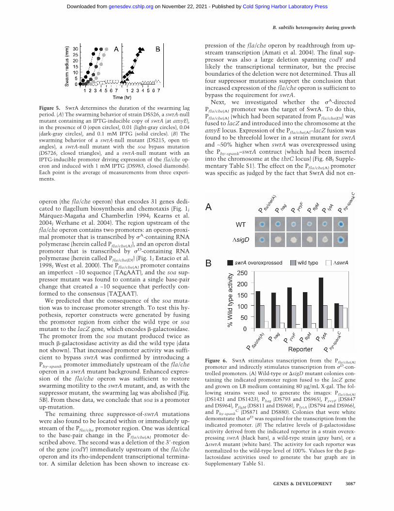
resulting strain (DS526) failed to exhibit surface motilityon swarm agar plates in the absence of inducer (Fig. 5A).However, when grown in liquid medium fortified withIPTG, the ability of the cells to swarm when subse-quently tested on solid medium was found to have beenrestored (Fig. 5A). Furthermore, whereas swarming inwild-type cells is preceded by a lag period that is depen-dent on the number of cells used in the inoculum(Kearns and Losick 2003; Shimada et al. 2004), theswarming lag of DS526 was found to be dependent on theIPTG concentration. Indeed, at the highest levels of in-ducer, the swarming lag was completely abolished (Fig.5A). Based on the observation that overexpression ofswrA both abolishes the swarming lag and causes hyper-flagellation, it seems possible that high levels of SwrAbypass both the cell density and surface requirements for
entry into the swarming state and produce differentiated“swarm cells” in liquid medium.
A suppressor mutation bypasses the role of SwrAin swimming and swarming
To investigate the mechanism by which SwrA acts, welooked for extragenic suppressors of the swarming defectin SwrA mutants. To do so, we took advantage of therequirement for swrA in surface motility to select formutants that had acquired the capacity to swarm in thepresence of a null mutation of the gene. When a swrA-null mutant was inoculated in the center of a swarm agarplate, the cells initially grew as a tight central colony andfailed to completely colonize the surface of the plate.However, after 48 h, flares of cells that had regained theability to swarm emerged from the central colony. Cellsfrom four independently isolated suppressor mutantswere clonally purified.
The results obtained with one such suppressor of swrA(soa) mutant were as follows. The mutant exhibited aphenotype that resembled that observed when swrA wasoverexpressed: The suppressor mutant swarmed withouta lag (Fig. 5B), grew predominantly as single motile cells(Fig. 2E), and was hyperflagellated in liquid medium(Fig. 4), and �D-directed gene transcription was uniformamong cells in the population (Fig. 3E). That the soasuppressor mutant retained the original swrA-null mu-tation was confirmed by polymerase chain reaction(PCR) analysis.
The soa suppressor mutation was found to residewithin the promoter region (Pfla/che) upstream of a large
Figure 3. �D-directed gene expression is subject to an ON/OFFswitch that is influenced by SwrA and SwrB. Fluorescence mi-crographs were taken of mid-exponential phase (OD600 = 1)cultures of the following strains: DS901 (A), DS908 (B), DS909(C), DS933 (D), DS973 (E), DS934 (F), DS1488 (G), DS1489(H), DS1512 (I), DS1539 (J), DS1543 (K), and DS910 (L). Allstrains contained Phag-gfp except DS910 (L), which containedPhy-spank
C-gfp. Membranes were stained with FM4-64 andfalse-colored in red. GFP signals were false-colored in green. Bar,5 µm.
Figure 4. SwrA overexpression results in hyperflagellation.Flagellum staining was conducted on mid-exponential-phasecells grown in the presence of 1 mM IPTG for the following: theswrA-overexpressing strain DS526 (A), the wild-type strain3610 (B), the swrA mutant DS215 (C), and the swrA soa sup-pressor mutant strain DS726 (D). Bar, 2 µm.
Kearns and Losick
3086 GENES & DEVELOPMENT
Cold Spring Harbor Laboratory Press on November 22, 2021 - Published by genesdev.cshlp.orgDownloaded from
operon (the fla/che operon) that encodes 31 genes dedi-cated to flagellum biosynthesis and chemotaxis (Fig. 1;Márquez-Magaña and Chamberlin 1994; Kearns et al.2004; Werhane et al. 2004). The region upstream of thefla/che operon contains two promoters: an operon-proxi-mal promoter that is transcribed by �A-containing RNApolymerase (herein called Pfla/che(A)), and an operon distalpromoter that is transcribed by �D-containing RNApolymerase (herein called Pfla/che(D)) (Fig. 1; Estacio et al.1998; West et al. 2000). The Pfla/che(A) promoter containsan imperfect −10 sequence (TAcAAT), and the soa sup-pressor mutant was found to contain a single base-pairchange that created a −10 sequence that perfectly con-formed to the consensus (TATAAT).
We predicted that the consequence of the soa muta-tion was to increase promoter strength. To test this hy-pothesis, reporter constructs were generated by fusingthe promoter region from either the wild type or soamutant to the lacZ gene, which encodes �-galactosidase.The promoter from the soa mutant produced twice asmuch �-galactosidase activity as did the wild type (datanot shown). That increased promoter activity was suffi-cient to bypass swrA was confirmed by introducing aPhy-spank promoter immediately upstream of the fla/cheoperon in a swrA mutant background. Enhanced expres-sion of the fla/che operon was sufficient to restoreswarming motility to the swrA mutant, and, as with thesuppressor mutant, the swarming lag was abolished (Fig.5B). From these data, we conclude that soa is a promoterup-mutation.
The remaining three suppressor-of-swrA mutationswere also found to be located within or immediately up-stream of the Pfla/che promoter region. One was identicalto the base-pair change in the Pfla/che(A) promoter de-scribed above. The second was a deletion of the 3�-regionof the gene (codY) immediately upstream of the fla/cheoperon and its rho-independent transcriptional termina-tor. A similar deletion has been shown to increase ex-
pression of the fla/che operon by readthrough from up-stream transcription (Amati et al. 2004). The final sup-pressor was also a large deletion spanning codY andlikely the transcriptional terminator, but the preciseboundaries of the deletion were not determined. Thus allfour suppressor mutations support the conclusion thatincreased expression of the fla/che operon is sufficient tobypass the requirement for swrA.
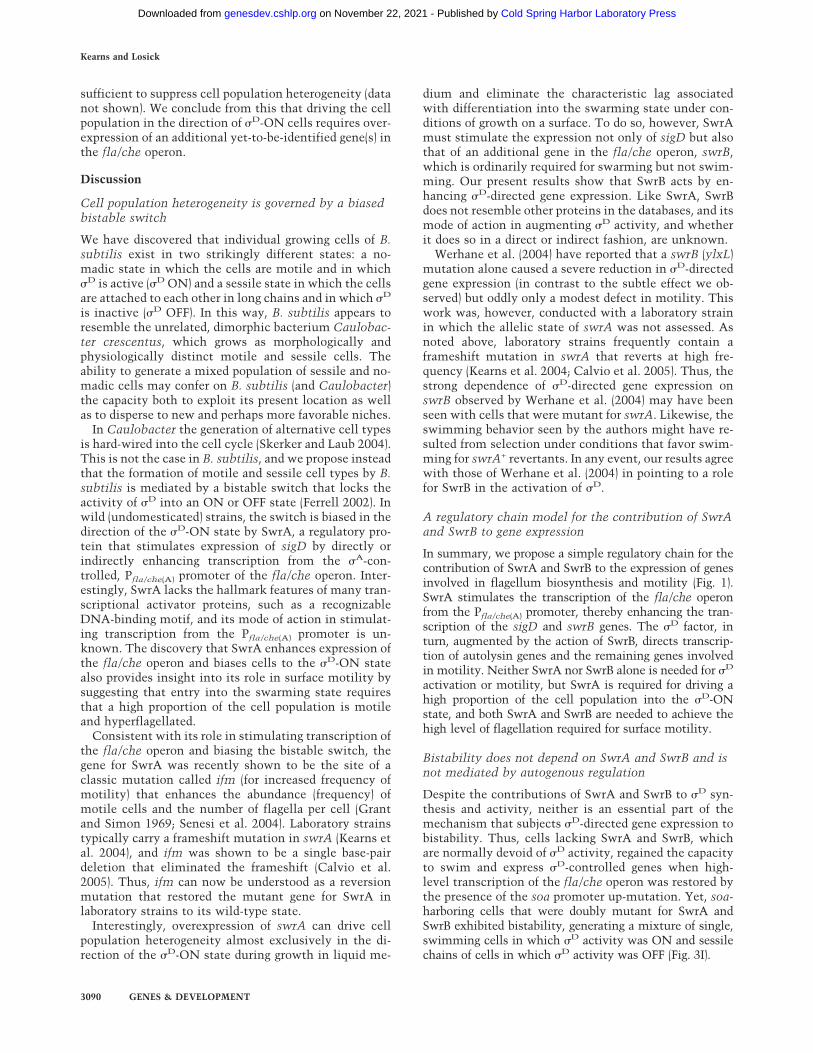
Next, we investigated whether the �A-directedPfla/che(A) promoter was the target of SwrA. To do this,Pfla/che(A) (which had been separated from Pfla/che(D)) wasfused to lacZ and introduced into the chromosome at theamyE locus. Expression of the Pfla/che(A)–lacZ fusion wasfound to be threefold lower in a strain mutant for swrAand ∼50% higher when swrA was overexpressed usingthe Phy-spank–swrA contruct (which had been insertedinto the chromosome at the thrC locus) (Fig. 6B; Supple-mentary Table S1). The effect on the Pfla/che(A) promoterwas specific as judged by the fact that SwrA did not en-
Figure 5. SwrA determines the duration of the swarming lagperiod. (A) The swarming behavior of strain DS526, a swrA-nullmutant containing an IPTG-inducible copy of swrA (at amyE),in the presence of 0 (open circles), 0.01 (light-gray circles), 0.04(dark-gray circles), and 0.1 mM IPTG (solid circles). (B) Theswarming behavior of a swrA-null mutant (DS215, open tri-angles), a swrA-null mutant with the soa bypass mutation(DS726, closed triangles), and a swrA-null mutant with anIPTG-inducible promoter driving expression of the fla/che op-eron and induced with 1 mM IPTG (DS983, closed diamonds).Each point is the average of measurements from three experi-ments.
Figure 6. SwrA stimulates transcription from the Pfla/che(A)
promoter and indirectly stimulates transcription from �D-con-trolled promoters. (A) Wild-type or �sigD mutant colonies con-taining the indicated promoter region fused to the lacZ geneand grown on LB medium containing 80 µg/mL X-gal. The fol-lowing strains were used to generate the images: Pfla/che(A)
(DS1421 and DS1423), Phag (DS793 and DS965), PyvyF (DS847and DS964), PflgM (DS811 and DS968), PlytA (DS794 and DS966),and Phy-spank
C (DS871 and DS880). Colonies that were whitedemonstrate that �D was required for the transcription from theindicated promoter. (B) The relative levels of �-galactosidaseactivity derived from the indicated reporter in a strain overex-pressing swrA (black bars), a wild-type strain (gray bars), or a�swrA mutant (white bars). The activity for each reporter wasnormalized to the wild-type level of 100%. Values for the �-ga-lactosidase activities used to generate the bar graph are inSupplementary Table S1.
B. subtilis heterogeneity during growth
GENES & DEVELOPMENT 3087
Cold Spring Harbor Laboratory Press on November 22, 2021 - Published by genesdev.cshlp.orgDownloaded from

hance transcription from another promoter transcribedby �A-containing RNA polymerase (Phy-spank
C) (Fig. 6B).We conclude that SwrA acts as a positive regulator of thefla/che operon by stimulating transcription from thePfla/che(A) promoter.
SwrA and SigD direct the transcription of partiallyoverlapping sets of genes
To compare the patterns of SwrA- and �D-directed genetranscription on a genome-wide basis, we carried out twokinds of transcriptional profiling experiments. In one,RNA from mid-exponential-phase cells of a swrA mu-tant was compared with RNA from mid-exponential-phase cells in which swrA was overexpressed. In theother, RNA from mid-exponential-phase cells of a sigDmutant was compared with RNA from mid-exponential-phase cells in which sigD was overexpressed. The resultsshow that the patterns of SwrA- and �D-activated tran-scription overlapped extensively in that SwrA stimu-lated the expression of many of the members of the �D
regulon (Table 1; Supplementary Table S2). That SwrAstimulates transcription of �D-controlled genes was con-firmed by the use of lacZ fusions to four promoters(Phag, PyvyF, PflgM, and PlytA) known to be under the di-rect control of the alternative � factor (Fig. 6; Mirel andChamberlin 1989; Lazarevic et al. 1992; Mirel et al. 1994;Estacio et al. 1998).
A simple explanation for these results is that SwrAstimulates transcription from Pfla/che(A) and thereby in-directly enhances �D-directed transcription by promot-ing transcription through sigD, which is the penultimategene in the fla/che operon (Fig. 1). Despite the presenceof the Pfla/che(D) promoter, transcription of the fla/cheoperon was only weakly enhanced by �D, not meetingthe 2.5-fold threshold of significance set in our microar-ray experiments. However, this result was not unex-pected as Pfla/che(D) is much weaker than Pfla/che(A) andwas known from earlier work to make only a minor con-tribution to transcription of the fla/che operon (West etal. 2000). This was confirmed in our present inves-tigation by the use of a Pfla/che(D)–lacZ fusion, whichgenerated 88 Miller units of activity, as compared to aPfla/che(A)–lacZ fusion, which generated 515 Miller unitsof activity (Supplementary Table S1).
As the overexpression of swrA stimulates the expres-sion of sigD, we wondered whether the overexpression ofsigD alone would be sufficient to bias the population inthe direction of �D-ON cells. Unlike the case for swrAoverexpression in which Phag–gfp was uniformly ex-pressed in almost all cells in the population (Fig. 3D),overexpression of sigD was not sufficient to produce auniform population of cells in the �D-ON state (Figs. 2F,3F). From this we conclude that an additional gene(s)under SwrA control contributes to biasing cells to the�D-ON state. Genes within the fla/che operon werelikely candidates as the soa suppressor up-regulated theexpression of this operon and strongly biased the popu-lation to the �D-ON state.
SwrB also contributes to �D-directed gene expression
In previous work we identified a gene called swrB that isrequired for swarming but not swimming motility(Kearns et al. 2004). The swrB gene is located immedi-ately downstream of, and is contranscribed with, sigD inthe fla/che operon (Fig. 1; Werhane et al. 2004). In light ofour results with SwrA, we wondered whether SwrBmight also contribute to �D-directed gene expressionduring growth. To answer this question, we monitored�D-directed gene expression using both the Phag–gfp fu-sion/reporter and a �D-dependent lacZ reporter. In theabsence of SwrB, we observed only a slight reduction inthe number of cells in the population in which �D wasactive and there was a slight decrease in overall �D ac-tivity (Figs. 2G, 3G; Supplementary Table S1). Strikingly,however, �D activity was almost completely abolishedin cells that were doubly mutant for SwrA and SwrB(Figs. 2H, 3H; Supplementary Table S1). Unlike the swrAmutation, the swrB mutation did not affect expressionfrom the Pfla/che(A) promoter (Supplementary Table S1).Evidently, SwrB does contribute to �D-directed gene ex-pression, but the contribution of SwrB is chiefly maskedby the contribution of SwrA.
The simplest interpretation of these results is thatSwrB is largely dispensable for �D-directed gene expres-sion under conditions in which �D is abundant due tohigh levels of sigD transcription. However, under condi-tions in which SwrA is absent and therefore �D levels arelow, SwrB plays a critical role in �D-directed gene ex-pression. In keeping with this interpretation, the soa pro-moter up-mutation, which bypasses the dependence ofthe Pfla/che(A) promoter on SwrA, restored �D-directedgene expression to a swrA swrB double mutant (Figs. 2I,3I). In this case, the soa mutation allowed sigD (and therest of the fla/che operon) to be transcribed at a highlevel in the absence of SwrA, which is consistent withthe hypothesis that at high levels of �D, the role of SwrBin �D-directed gene expression is dispensable.
Biasing cell population heterogeneity requires SwrB
As noted above, overexpressing swrA or bypassing swrAby means of a promoter up-mutation (soa) suppressedheterogeneity, resulting in a cell population that was al-most exclusively in the �D-ON state (Fig. 3D). Yet, over-expressing sigD alone (by use of the Phy-spank–sigD con-struct) failed to do so (Fig. 3F). Evidently, suppressing cellpopulation heterogeneity requires enhanced transcrip-tion not only of sigD but also of an additional gene(s) inthe fla/che operon. An appealing candidate for this geneis swrB, as swrB lies immediately downstream of, and iscotranscribed with, sigD, and hence SwrA and soa wouldbe expected to enhance its transcription as well. In sup-port of this hypothesis, the promoter up-mutation soawas unable to generate a largely homogeneous popula-tion of �D-ON cells when the fla/che operon was mutantfor swrB (Figs. 2I, 3I). We conclude that driving the popu-lation in the direction of �D-ON cells requires high lev-els of both �D and of its activator, SwrB. Simultaneousoverexpression of sigD and swrB alone was not, however,
Kearns and Losick
3088 GENES & DEVELOPMENT
Cold Spring Harbor Laboratory Press on November 22, 2021 - Published by genesdev.cshlp.orgDownloaded from

Table 1. Genes activated by SwrA and SigD
GeneSwrAratioa
SigDratiob Functionc Gene
SwrAratioa
SigDratiob Functionc
Genes activated by SwrA Genes activated by SwrA and SigDcheA 4.1 < fla/che (chemotaxis) acoA 4.9 6.2 Acetoin biosynthesischeB 5.9 < fla/che (chemotaxis) appA 4.5 2.9 Oligopeptide-binding proteincheC 6.3 < fla/che (chemotaxis) cheD 6.2 3.0 fla/che (chemotaxis)cheY 8.1 < fla/che (chemotaxis) cheV 6.5 14.3 ChemotaxisflgB 7.2 < fla/che (basal body) cheW 9.3 3.6 fla/che (chemotaxis)flgC 8.4 < fla/che (basal body) comGG 3.0 3.5 CompetenceflgE 11.4 < fla/che (hook) comS 3.4 4.7 Competence regulatorflhB 12.0 < fla/che (flagellum associated) flgG 4.1 2.6 Flagellum basal body (flhO)flhF 11.1 < fla/che (flagellum associated) flgK 5.6 4.5 Flagellum hookfliE 9.0 < fla/che (hook–basal body, minor) flgL 6.3 5.2 Flagellum hookfliF 7.4 < fla/che (basal body) flgM 3.6 2.6 �D anti-sigma factorfliG 9.2 < fla/che (motor switch) flhA 6.9 2.9 fla/che (flagellum associated)fliI 9.3 < fla/che (ATP synthase) flhP 3.1 2.6 Flagellum basal body (minor)fliJ 3.1 < fla/che (flagellum assembly) fliD 5.1 6.9 Flagellum hookfliK 6.2 < fla/che (hook assembly) fliH 5.1 2.7 fla/che (flagellum assembly)fliL 3.1 < fla/che (flagellum assembly) fliS 7.4 6.8 Flagellar proteinfliM 9.6 < fla/che (motor switch) fliT 5.4 7.5 Flagellar proteinfliP 2.8 < fla/che (flagellum assembly) gerD 5.7 6.0 Germination proteinfliQ 8.7 < fla/che (flagellum assembly) glgD 8.6 28.4 Glycogen biosynthesisfliR 10.7 < fla/che (flagellum assembly) hag 11.0 34.6 FlagellinfliY 9.4 < fla/che (motor switch) hemAT 10.0 60.7 MCPfliZ 7.7 < fla/che (flagellum assembly) lytA 5.0 2.8 Autolysin productionsigD 6.6 32.6d fla/che (�D) lytB 5.5 4.2 Autolysin productionswrB 8.0 < fla/che (swarming motility) lytD 9.2 18.1 AutolysinylxF 3.8 < fla/che (unknown) lytF 4.8 5.4 AutolysinylxG 10.4 < fla/che (hook assembly) mcpA 11.4 11.5 MCP
mcpB 9.8 5.6 MCPacoL 4.8 < Acetoin biosynthesis mcpC 4.7 3.8 MCParaR 4.3 < Transcriptional regulator motA 5.6 3.8 Flagellar motor componentcotA 7.7 < Spore coat protein motB 10.0 43.6 Flagellar motor componentlytC 3.5 < Autolysin phrF 4.6 3.9 Cell–cell signaling peptiderodA 3.5 < Cell shape protein tasA 2.7 3.3 Biofilm matrix componentycgM 5.8 < Similar to proline oxidase tlpA 5.4 3.0 MCP homologycgN 5.3 < Similar to dehydrogenase tlpB 13.8 6.4 MCP homologydcL 3.0 < Similar to integrase tlpC 10.5 7.8 MCP homologydcM 3.1 < Similar to prophage immunity ydeQ 5.3 5.7 Similar to oxidoreductaseyfiV 2.8 < Transcriptional regulator ydfQ 6.9 12.7 Similar to thioredoxinykuU 2.7 < Similar to peroxiredoxin yesT 3.1 3.8 Similar to acetyltransferaseyoaH 30.0 < MCP yfmS 12.3 8.2 MCP homologyolD 8.4 < Unknown yfmT 6.0 5.7 Similar to dehydrogenaseyprB 3.1 < Unknown yfnH 5.8 27.1 Similar to dehydrogenaseytlQ 2.9 < Unknown yhxD 4.3 6.3 Similar to dehydrogenaseyusE 2.7 < Similar to thioredoxin yisS 4.6 6.3 Similar to dehydrogenaseyvbY 6.2 < Unknown yolA 6.5 16.7 UnknownyvyF 3.6 < Unknown yolC 7.9 24.2 UnknownywjF 3.2 < Similar to iron-sulfur reductase yrvJ 3.4 3.1 Similar to dehydrogenaseywrO 3.4 < Similar to oxidoreductase yscB 5.1 2.8 Unknown
yvaB 3.9 3.8 Similar to dehydrogenaseyvaQ 7.6 10.8 MCP homologyvbX 7.9 15.5 Similar to chitinaseyviE 4.2 5.1 UnknownyvyC 7.7 36.8 UnknownyvyG 6.1 12.6 UnknownyvzB 7.7 13.6 Similar to flagellinywtD 6.1 4.0 Similar to murien hydrolase
aThe SwrA ratio is signal strength of RNA purified from cells overexpressing SwrA (DS526 + 1 mM IPTG) divided by the signal strengthof RNA purified from cells mutant for swrA (DS215). Values are the average of five replicates. (<) A value < 2.5.bThe SigD ratio is signal strength of RNA purified from cells overexpressing SigD (DS880 + 1 mM IPTG) divided by the signal strengthof RNA purified from cells mutant for sigD (DS323). Values are the average of 4 replicates. (<) A value < 2.5.c(fla/che) Members of the fla/che operon. (MCP) methyl-accepting chemotaxis protein.dsigD value is artificial because the sigD gene itself was overexpressed as part of the experiment.
B. subtilis heterogeneity during growth
GENES & DEVELOPMENT 3089
Cold Spring Harbor Laboratory Press on November 22, 2021 - Published by genesdev.cshlp.orgDownloaded from
sufficient to suppress cell population heterogeneity (datanot shown). We conclude from this that driving the cellpopulation in the direction of �D-ON cells requires over-expression of an additional yet-to-be-identified gene(s) inthe fla/che operon.
Discussion
Cell population heterogeneity is governed by a biasedbistable switch
We have discovered that individual growing cells of B.subtilis exist in two strikingly different states: a no-madic state in which the cells are motile and in which�D is active (�D ON) and a sessile state in which the cellsare attached to each other in long chains and in which �D
is inactive (�D OFF). In this way, B. subtilis appears toresemble the unrelated, dimorphic bacterium Caulobac-ter crescentus, which grows as morphologically andphysiologically distinct motile and sessile cells. Theability to generate a mixed population of sessile and no-madic cells may confer on B. subtilis (and Caulobacter)the capacity both to exploit its present location as wellas to disperse to new and perhaps more favorable niches.
In Caulobacter the generation of alternative cell typesis hard-wired into the cell cycle (Skerker and Laub 2004).This is not the case in B. subtilis, and we propose insteadthat the formation of motile and sessile cell types by B.subtilis is mediated by a bistable switch that locks theactivity of �D into an ON or OFF state (Ferrell 2002). Inwild (undomesticated) strains, the switch is biased in thedirection of the �D-ON state by SwrA, a regulatory pro-tein that stimulates expression of sigD by directly orindirectly enhancing transcription from the �A-con-trolled, Pfla/che(A) promoter of the fla/che operon. Inter-estingly, SwrA lacks the hallmark features of many tran-scriptional activator proteins, such as a recognizableDNA-binding motif, and its mode of action in stimulat-ing transcription from the Pfla/che(A) promoter is un-known. The discovery that SwrA enhances expression ofthe fla/che operon and biases cells to the �D-ON statealso provides insight into its role in surface motility bysuggesting that entry into the swarming state requiresthat a high proportion of the cell population is motileand hyperflagellated.
Consistent with its role in stimulating transcription ofthe fla/che operon and biasing the bistable switch, thegene for SwrA was recently shown to be the site of aclassic mutation called ifm (for increased frequency ofmotility) that enhances the abundance (frequency) ofmotile cells and the number of flagella per cell (Grantand Simon 1969; Senesi et al. 2004). Laboratory strainstypically carry a frameshift mutation in swrA (Kearns etal. 2004), and ifm was shown to be a single base-pairdeletion that eliminated the frameshift (Calvio et al.2005). Thus, ifm can now be understood as a reversionmutation that restored the mutant gene for SwrA inlaboratory strains to its wild-type state.
Interestingly, overexpression of swrA can drive cellpopulation heterogeneity almost exclusively in the di-rection of the �D-ON state during growth in liquid me-
dium and eliminate the characteristic lag associatedwith differentiation into the swarming state under con-ditions of growth on a surface. To do so, however, SwrAmust stimulate the expression not only of sigD but alsothat of an additional gene in the fla/che operon, swrB,which is ordinarily required for swarming but not swim-ming. Our present results show that SwrB acts by en-hancing �D-directed gene expression. Like SwrA, SwrBdoes not resemble other proteins in the databases, and itsmode of action in augmenting �D activity, and whetherit does so in a direct or indirect fashion, are unknown.
Werhane et al. (2004) have reported that a swrB (ylxL)mutation alone caused a severe reduction in �D-directedgene expression (in contrast to the subtle effect we ob-served) but oddly only a modest defect in motility. Thiswork was, however, conducted with a laboratory strainin which the allelic state of swrA was not assessed. Asnoted above, laboratory strains frequently contain aframeshift mutation in swrA that reverts at high fre-quency (Kearns et al. 2004; Calvio et al. 2005). Thus, thestrong dependence of �D-directed gene expression onswrB observed by Werhane et al. (2004) may have beenseen with cells that were mutant for swrA. Likewise, theswimming behavior seen by the authors might have re-sulted from selection under conditions that favor swim-ming for swrA+ revertants. In any event, our results agreewith those of Werhane et al. (2004) in pointing to a rolefor SwrB in the activation of �D.
A regulatory chain model for the contribution of SwrAand SwrB to gene expression
In summary, we propose a simple regulatory chain for thecontribution of SwrA and SwrB to the expression of genesinvolved in flagellum biosynthesis and motility (Fig. 1).SwrA stimulates the transcription of the fla/che operonfrom the Pfla/che(A) promoter, thereby enhancing the tran-scription of the sigD and swrB genes. The �D factor, inturn, augmented by the action of SwrB, directs transcrip-tion of autolysin genes and the remaining genes involvedin motility. Neither SwrA nor SwrB alone is needed for �D
activation or motility, but SwrA is required for driving ahigh proportion of the cell population into the �D-ONstate, and both SwrA and SwrB are needed to achieve thehigh level of flagellation required for surface motility.
Bistability does not depend on SwrA and SwrB and isnot mediated by autogenous regulation
Despite the contributions of SwrA and SwrB to �D syn-thesis and activity, neither is an essential part of themechanism that subjects �D-directed gene expression tobistability. Thus, cells lacking SwrA and SwrB, whichare normally devoid of �D activity, regained the capacityto swim and express �D-controlled genes when high-level transcription of the fla/che operon was restored bythe presence of the soa promoter up-mutation. Yet, soa-harboring cells that were doubly mutant for SwrA andSwrB exhibited bistability, generating a mixture of single,swimming cells in which �D activity was ON and sessilechains of cells in which �D activity was OFF (Fig. 3I).
Kearns and Losick
3090 GENES & DEVELOPMENT
Cold Spring Harbor Laboratory Press on November 22, 2021 - Published by genesdev.cshlp.orgDownloaded from

A distinctive feature of the bistable switch governing�D activity is that it is manifest during the exponentialphase of growth. Two other examples of bistableswitches in B. subtilis, those governing the activation ofthe master regulators for competence (ComK) (Maamarand Dubnau 2005; Smits et al. 2005) and sporulation(Spo0A) (Veening et al. 2005), operate under conditionsin which cells are entering stationary phase. In contrast,cell population heterogeneity with respect to the state of�D activity was seen under (presumably) steady-stateconditions in which all cells in the population were ac-tively growing.
The bistable switch for �D also differs from those forComK and Spo0A in that it does not appear to operate bymeans of autogenous regulation. Compelling evidenceindicates that bistability for the activation of ComK andSpo0A is governed by positive feedback loops in whichthe transcription factors stimulate the expression oftheir own structural genes (directly in the case of formerand indirectly in the case of latter) (Maamar and Dubnau2005; Smits et al. 2005; Veening et al. 2005). This isapparently not the case for �D as we now explain. First,although the fla/che operon is preceded by a �D-con-trolled promoter, Pfla/che(D) is much weaker than theSwrA-controlled Pfla/che(A) promoter (West et al. 2000;Supplementary Table S1) and is unlikely to contribute sig-nificantly to the overall level of expression of sigD (andswrB). Indeed, and as demonstrated by gene microarray ex-periments, overexpression of sigD had little effect on tran-scription of the fla/che operon (Table 1). Second, it waspreviously shown that deletion of Pfla/che(D) has no conse-quence on motility (West et al. 2000). We have confirmedand extended this observation by building a mutation thatentirely removed Pfla/che(D) (see Supplemental Material)and found that it had little or no effect on motility or bi-stability (Figs. 2J, 3J). Third, gene microarray experimentsfailed to reveal evidence for any other �D-controlled pro-moter in the operon driving expression of sigD (and swrB)(data not shown). Evidently, then, bistability does not oc-cur by an autogenous control mechanism in which �D
stimulates transcription of its own structural gene.
A hook–basal body model for bistability
What then is the basis for the bistable switch if, as wesurmise, bistability occurs at a step downstream of thetranscription of the sigD gene? One possibility is that theswitch operates at the level of the synthesis or activity of�D by a positive feedback loop involving the assembly ofthe flagellum basal body. It is known from work withSalmonella that the activity of the � factor for flagellumbiosynthesis (called �F in Gram-negative bacteria) is de-pendent on the assembly of the flagellum basal body bya pathway involving the anti-sigma factor FlgM (Komeda1986; Hughes et al. 1993; Kutsukake 1994; Karlinsey etal. 2000). We speculate that an analogous pathway (in-deed, possibly involving the B. subtilis homolog of FlgM,which is known to be an antagonist of �D) (Caramori etal. 1996) sets up a self-reinforcing cycle that stimulatesthe synthesis and/or activity of �D. Many of the genes for
the basal body (those in the fla/che operon) are not under�D control, but two major hook–basal body genes, flgGand flgK, lie outside of the operon and are transcribed ina �D-dependent manner as judged by gene microarrayanalysis (Fig. 1; Table 1; Zuberi et al. 1991; Kubori et al.1997; Serizawa et al. 2004). Thus, �D-directed transcrip-tion of flgG and flgK would stimulate basal body forma-tion, which, in turn, would stimulate the synthesis and/or activity of �D. We note that suppression of bistabilityrequired overexpression not only of the genes for �D andSwrB but also of additional unidentified gene(s) in thefla/che operon. We speculate that this additional gene(s)could be one or more of the other hook–basal body genesnear the 5�-end of the fla/che operon (Fig. 1). Finally, thehigher-order assembly of the basal body would introducecooperativity into the bistable switch, thereby helping toachieve sharp ON/OFF control of �D activity.
In addition to the bistable switch, �D activation is sub-ject to a phase-variation control mechanism. As we haveseen, the bistable switch is strongly biased by the action ofthe product of swrA, which is subject to a phase-variationswitch that is thought to operate by means of “slippedstrand” mispairing during replication (Henderson et al.1999; Kearns et al. 2004). Thus, the capacity to exist inalternative motile and sessile life styles is subject to twolevels of stochastic control, a high-frequency bistable (andpresumably epigenetic) switch that ensures that the popu-lation is a mixture of two cell types and a low-frequencyphase variation (genetic) switch that biases the proportionof the two cell types in one direction or the other.
Lastly, we speculate that the population bias betweenswimming and chaining cells may be influenced by en-vironmental conditions. Under some conditions, wildstrains of B. subtilis are capable of swarming in a robustmanner on surfaces, a behavior that involves the forma-tion of groups of motile cells that are joined together inassemblies called rafts. Here we have demonstrated thatSwrA biases the population in favor of motile cells,which, in turn, contribute to raft formation and swarm-ing motility. Under other conditions, wild strains arecapable of forming architecturally complex communi-ties (biofilms) in which long chains of nonmotile cellsare tightly bundled together through the formation of anextracellular matrix (Branda et al. 2001, 2004; Kearns etal. 2005). It will be interesting to determine whethersome proteins required for biofilm formation serve tobias the cell population in favor of chaining. Opposingmechanisms that increase the proportion of swimmingcells or chaining cells may be of central importance indetermining which multicellular behavior, swarming orbiofilm formation, is adopted for a particular environment.
Materials and methods
Strains and growth conditions
B. subtilis PY79, 3610, and derivatives thereof were grown at37°C in Luria-Bertani (LB) (10 g of tryptone, 5 g of yeast extract,5 g of NaCl per liter) broth or LB plates supplemented with 1.5%Bacto agar. When appropriate, antibiotics were included at the
B. subtilis heterogeneity during growth
GENES & DEVELOPMENT 3091
Cold Spring Harbor Laboratory Press on November 22, 2021 - Published by genesdev.cshlp.orgDownloaded from
following concentrations: 10 µg/mL tetracycline, 100 µg/mLspectinomycin, 5 µg/mL chloramphenicol, 5 µg/mL kanamycin,and 1 µg/mL erythromycin plus 25 µg/mL lincomycin (mls).Isoproyl �-D-thiogalactopyranoside (IPTG, Sigma) or 5-bromo-4-chloro-3-indolyl �-D-galactopyranoside (X-Gal, Sigma) wasadded to either liquid or solid medium at the indicated concen-tration when appropriate.
Microscopy
For phase contrast images, cells were grown to 1 OD600, and 5µL of culture was spotted on a glass microscope slide and im-mobilized with a poly-L-lysine-treated glass coverslip. Sampleswere observed under phase contrast at 1000× magnification us-ing an Olympus BX60 microscope and recorded with Meta-morph image capture software (Universal Imaging).
For fluorescence microscopy, 1.0 mL of broth culture was har-vested at 1.0 OD600, and washed twice in 1.0 mL of phosphate-buffered saline (137 mM NaCl, 2.7 mM KCl, 10 mM Na2HPO4,and 2 mM KH2PO4), PBS buffer. The suspension was pelleted,resuspended in 50 µL of PBS buffer containing 1 µg/mL FM4-64(Molecular Probes), and incubated for 5 min at room temperature.Three microliters of suspension were placed on a microscope slideand immobilized with a poly-L-lysine-treated coverslip. Fluores-cence microscopy was performed with an Olympus BX60 micro-scope with Mercury lamp. Fluorescent signals were viewed usingthe phase contrast objective UplanF1 100× and visualized using aU-WIBA filter cube (GFP, excitation filter 460–490 nm, barrierfilter 515–550 nm) or a U-WG filter cube (FM4-64, excitation filter510–550 nm, barrier filter >590 nm). Images were captured, false-colored, and overlaid using Metamorph software, exported as TIFFfiles, and adjusted with Adobe Photoshop software.
Flagella were stained according to Mayfield and Inniss (1977).Stain was prepared by mixing 10 parts mordant (2 g of tannicacid, 10 mL of 5% phenol, 10 mL of saturated aqueousAlKO8S2 · 12 H2O) with 1 part stain (12% crystal violet in etha-nol). Three microliters of 1.0 OD600 broth-grown cells were ap-plied to a microscope slide and covered with a 22 mm × 40 mmcoverslip. The slide was propped vertically, and 10 µL of dye wasapplied to the top edge of the coverslip to stain the sample bycapillary action. Samples were observed at 1000× magnificationunder phase contrast using an Olympus BX60 microscope andrecorded with Metamorph image capture software.
Swarm expansion assay
Cells were grown to mid-log phase at 37°C in LB broth and resus-pended to 10 OD600 in PBS buffer containing 0.5% India ink (Hig-gins). Freshly prepared LB containing 0.7% agar (25 mL/plate) wasdried for 30 min in a laminar flow hood, centrally inoculated with10 µL of the cell suspension, dried for another 10 min, and incu-bated at 37°C. The India ink demarked the origin of the colony,and swarm radii were measured relative to the origin. For consis-tency, an axis was drawn on the back of the plate, and swarm radiimeasurements were taken along this transect.
Strain construction
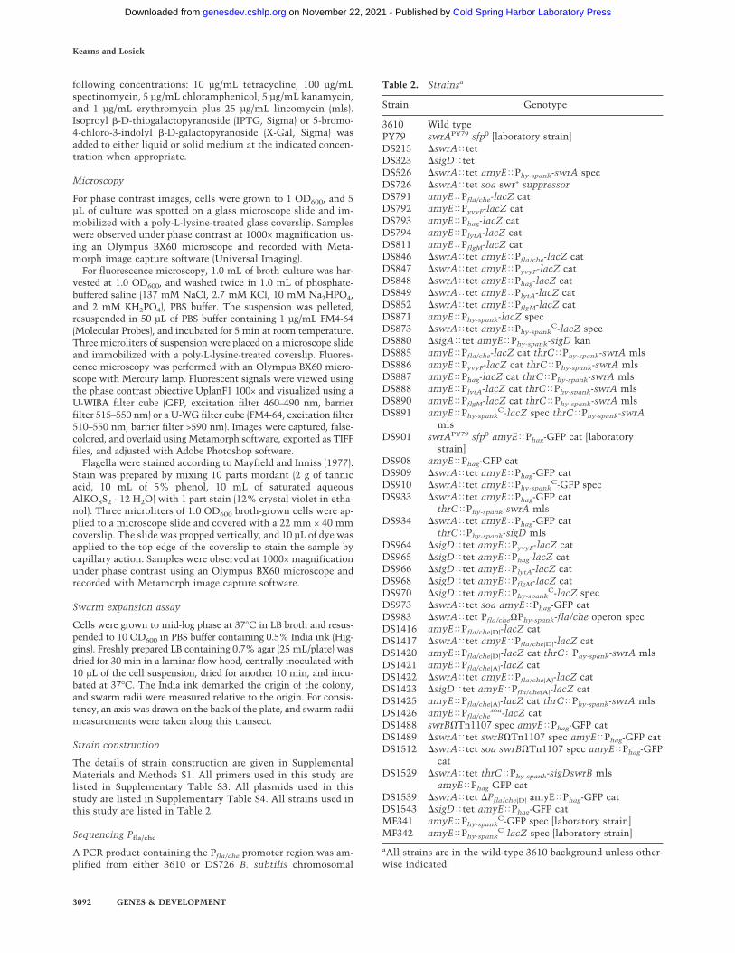
The details of strain construction are given in SupplementalMaterials and Methods S1. All primers used in this study arelisted in Supplementary Table S3. All plasmids used in thisstudy are listed in Supplementary Table S4. All strains used inthis study are listed in Table 2.
Sequencing Pfla/che
A PCR product containing the Pfla/che promoter region was am-plified from either 3610 or DS726 B. subtilis chromosomal
Table 2. Strainsa
Strain Genotype
3610 Wild typePY79 swrAPY79 sfp0 [laboratory strain]DS215 �swrA�tetDS323 �sigD�tetDS526 �swrA�tet amyE�Phy-spank-swrA specDS726 �swrA�tet soa swr+ suppressorDS791 amyE�Pfla/che-lacZ catDS792 amyE�PyvyF-lacZ catDS793 amyE�Phag-lacZ catDS794 amyE�PlytA-lacZ catDS811 amyE�PflgM-lacZ catDS846 �swrA�tet amyE�Pfla/che-lacZ catDS847 �swrA�tet amyE�PyvyF-lacZ catDS848 �swrA�tet amyE�Phag-lacZ catDS849 �swrA�tet amyE�PlytA-lacZ catDS852 �swrA�tet amyE�PflgM-lacZ catDS871 amyE�Phy-spank-lacZ specDS873 �swrA�tet amyE�Phy-spank
C-lacZ specDS880 �sigA�tet amyE�Phy-spank-sigD kanDS885 amyE�Pfla/che-lacZ cat thrC�Phy-spank-swrA mlsDS886 amyE�PyvyF-lacZ cat thrC�Phy-spank-swrA mlsDS887 amyE�Phag-lacZ cat thrC�Phy-spank-swrA mlsDS888 amyE�PlytA-lacZ cat thrC�Phy-spank-swrA mlsDS890 amyE�PflgM-lacZ cat thrC�Phy-spank-swrA mlsDS891 amyE�Phy-spank
C-lacZ spec thrC�Phy-spank-swrAmls
DS901 swrAPY79 sfp0 amyE�Phag-GFP cat [laboratorystrain]
DS908 amyE�Phag-GFP catDS909 �swrA�tet amyE�Phag-GFP catDS910 �swrA�tet amyE�Phy-spank
C-GFP specDS933 �swrA�tet amyE�Phag-GFP cat
thrC�Phy-spank-swrA mlsDS934 �swrA�tet amyE�Phag-GFP cat
thrC�Phy-spank-sigD mlsDS964 �sigD�tet amyE�PyvyF-lacZ catDS965 �sigD�tet amyE�Phag-lacZ catDS966 �sigD�tet amyE�PlytA-lacZ catDS968 �sigD�tet amyE�PflgM-lacZ catDS970 �sigD�tet amyE�Phy-spank
C-lacZ specDS973 �swrA�tet soa amyE�Phag-GFP catDS983 �swrA�tet Pfla/che�Phy-spank-fla/che operon specDS1416 amyE�Pfla/che(D)-lacZ catDS1417 �swrA�tet amyE�Pfla/che(D)-lacZ catDS1420 amyE�Pfla/che(D)-lacZ cat thrC�Phy-spank-swrA mlsDS1421 amyE�Pfla/che(A)-lacZ catDS1422 �swrA�tet amyE�Pfla/che(A)-lacZ catDS1423 �sigD�tet amyE�Pfla/che(A)-lacZ catDS1425 amyE�Pfla/che(A)-lacZ cat thrC�Phy-spank-swrA mlsDS1426 amyE�Pfla/che
soa-lacZ catDS1488 swrB�Tn1107 spec amyE�Phag-GFP catDS1489 �swrA�tet swrB�Tn1107 spec amyE�Phag-GFP catDS1512 �swrA�tet soa swrB�Tn1107 spec amyE�Phag-GFP
catDS1529 �swrA�tet thrC�Phy-spank-sigDswrB mls
amyE�Phag-GFP catDS1539 �swrA�tet �Pfla/che(D) amyE�Phag-GFP catDS1543 �sigD�tet amyE�Phag-GFP catMF341 amyE�Phy-spank
C-GFP spec [laboratory strain]MF342 amyE�Phy-spank
C-lacZ spec [laboratory strain]
aAll strains are in the wild-type 3610 background unless other-wise indicated.
Kearns and Losick
3092 GENES & DEVELOPMENT
Cold Spring Harbor Laboratory Press on November 22, 2021 - Published by genesdev.cshlp.orgDownloaded from
DNA using the primers codYF/PflacheR. The Pfla/che PCR prod-uct was then sequenced using either primer individually.
�-Galactosidase assay
One milliliter of cells were harvested from a mid-log phase(OD600 ∼ 1.0) culture grown in LB broth shaken at 37°C, har-vested, and resuspended in an equal volume of Z buffer (40 mMNaH2PO4, 60 mM Na2HPO4, 1 mM MgSO4, 10 mM KCl, 38mM �-mercaptoethanol). To each sample, lysozyme was add-ed to a final concentration of 0.2 mg/mL and incubated at30°C for 15 min. Each sample was diluted appropriately in500 µL of Z buffer and the reaction was started with 100 mL of4 mg/mL 2-nitrophenyl �-D-galactopyranoside (in Z buffer)and stopped with 250 mL of 1 M Na2CO3. The OD420 of thereaction mixtures was recorded, and the �-galactosidase-specific activity was calculated according to the equation:[OD420/(time × OD600)] × dilution factor × 1000.
Transcriptional profiling assay
Total RNA was isolated from mid-exponential-phase culturesgrown in LB broth supplemented with 1 mM IPTG. RNA wasisolated using the hot acid/phenol method (protocol availableat http://mcb.harvard.edu/losick/fawcettpaper/RNAprep.htm).RNA purified from strain DS215 (�swrA�tet) was comparedwith strain DS526 (�swrA�tet amyE�Phy-spank–swrA cat), andRNA purified from DS323 (�sigD�tet) was compared withstrain DS880 (�sigD�tet amyE�Phy-spank–sigD cat). A total of20 µg of RNA from each strain was used to apply to the array.The volume of each RNA sample was brought up to 14 µL usingDEPC-treated H2O and 1 µL of 1 mg mL−1 of random primer wasadded. The samples were incubated at 70°C for 10 min and thenincubated on ice for 2 min. To the appropriate tube, 2 µL of[Cy5]dUTP and [Cy3]dUTP were added along with 12 µL ofreaction mix (2.5× Invitrogen First Strand buffer, 25 mM DTT,2 mM ATP, 2 mM CTP, 2 mM GTP, 0.8 mM TTP, 100 U ofRoche Protector RNase inhibitor). After addition of [Cy5]dUTPand [Cy3]dUTP, the reactions were shielded from light for therest of the procedure. To the reaction mixture, 200 U of Super-Script II RNase H-free reverse transcriptase (Invitrogen) wereadded. The tubes were incubated at room temperature for 10min, then incubated at 42°C for 2 h, and finally incubated at70°C for 15 min. After incubating the reactions, 3 U of RNase H(Invitrogen) was added, and the mixtures were placed at 37°C for20 min. The RNA samples to be compared were combined, anda QIAGEN PCR purification kit was used to purify the cDNAprobes. A Speedvac was then applied to the samples until 5 µLof probe remained. To the remaining probe, 20 µg of yeasttRNA, 5× SSC, 0.5% SDS, and 33% formamide was added. Thereactions were heated to 100°C for 1 min, spun briefly, andapplied to an oligo array containing >99% of the annotated pro-tein-coding genes of the B. subtilis genome (Britton et al. 2002).Prior to hybridization, the slides were rinsed in rinsing solution(0.2% SDS) for 4 min and washed twice in ddH2O for 2 mineach. The slides were then blocked for 15 min using aldehydeblocking solution (1.0 g of NaBH4, 0.75× PBS, 25% ethanol), andthe rinse and washing steps were repeated. The slides were driedbefore applying probe. Hybridizations were performed overnightin CMT-hybridization chambers (Corning) submerged in a wa-terbath at 42°C. After hybridization, unbound probe waswashed off the slides by rinsing for 1 min in wash buffer 1 (2×SSC and 0.2% SDS), 1 min in wash buffer 2 (2× SSC), and 1 minin wash buffer 3 (0.2× SSC). Finally, the slides were dried andscanned on a GenePix 4000B scanner (Axon Instruments). Im-ages were processed and analyzed with GenePix 4.0 software
(Axon Instruments, Inc.). Spots containing low signals were ex-cluded from further analysis. In our final data set, we includedonly genes in which there was a 2.5-fold change in expression,as seen using data from at least three replicates of the transcrip-tional profiling experiment.
Acknowledgments
We thank K. Blair, P. Eichenberger, M. Fujita, C. Fuqua, D.Rudner, and A.L. Sonenshein for reagents, constructs, and as-sistance; E. Quordokus and Y. Brun for use of their microscopefor the experiment of Supplementary Figure S1; and D. Dubnau,M. Fujita, M. Laub, D. Rudner, and L. Shapiro for critical com-ments on the manuscript. This work was supported by an NIHNational Research Service Award GM66612 to D.B.K. and aNational Institutes of Health grant GM18568 to R.L.
References
Albertini, A.M., Caramori, T., Crabb, W.D., Scoffone, F., andGalizzi, A. 1991. The flaA locus of Bacillus subtilis is part ofa large operon coding for flagellar structures, motility func-tions, and an ATPase-like polypeptide. J. Bacteriol. 173:3573–3579.
Amati, G., Bisicchia, P., and Galizzi, A. 2004. DegU-P repressesexpression of the motility fla-che operon in Bacillus subtilis.J. Bacteriol. 186: 6003–6014.
Balaban, N.Q., Merrin, J., Chait, R., Kowalik, L., and Leibler, S.2004. Bacterial persistence as a phenotypic switch. Science305: 1622–1625.
Blackman, S.A., Smith, T.J., and Foster, S.J. 1998. The role ofautolysins during vegetative growth of Bacillus subtilis 168.Microbiology 144: 73–82.
Branda, S.S., González-Pastor, J.E., Ben-Yehuda, S., Losick, R.,and Kolter, R. 2001. Fruiting body formation by Bacillussubtilis. Proc. Natl. Acad. Sci. 98: 11621–11626.
Branda, S.S., Gonzalez-Pastor, J.E., Dervyn, E., Ehrlich, D.,Losick, R., and Kolter, R. 2004. Genes involved in the for-mation of structured multicellular communities by Bacillussubtilis. J. Bacteriol. 186: 3970–3979.
Britton, R.A., Eichenberger, P., González-Pastor, J.E., Fawcett,P., Monson, R., Losick, R., and Grossman, A.D. 2002. Ge-nome-wide analysis of the stationary-phase � factor (�-H)regulon of Bacillus subtilis. J. Bacteriol. 184: 4881–4890.
Calvio, C., Celandroni, F., Ghelardi, E., Amati, G., Salvetti, S.,Ceciliani, F., Galizzi, A., and Senesi, S. 2005. Swarming dif-ferentiation and swimming motility in Bacillus subtilis arecontrolled by swrA, a newly identified dicistronic operon. J.Bacteriol. 187: 5356–5366.
Caramori, T., Barillà, D., Nessi, C., Sacchi, L., and Galizzi, A.1996. Role of FlgM in �D-dependent gene expression in Ba-cillus subtilis. J. Bacteriol. 178: 3113–3118.
Chen, L. and Helmann, J.D. 1994. The Bacillus subtilis �D-dependent operon encoding the flagellar proteins FliD, FliS,and FliT. J. Bacteriol. 176: 3093–3101.
Chung, J.D., Stephanopoulos, G., Ireton, K., and Grossman,A.D. 1994. Gene expression in single cells of Bacillus sub-tilis: Evidence that a threshold mechanism controls the ini-tiation of sporulation. J. Bacteriol. 176: 1977–1984.
Estacio, W., Santa Anna-Arriola, S., Adedipe, M., and Márquez-Magaña, L.M. 1998. Dual promoters are responsible for tran-scription initiation of the fla/che operon in Bacillus subtilis.J. Bacteriol. 180: 3548–3555.
Fein, J.E. 1979. Possible involvement of bacterial autolytic en-
B. subtilis heterogeneity during growth
GENES & DEVELOPMENT 3093
Cold Spring Harbor Laboratory Press on November 22, 2021 - Published by genesdev.cshlp.orgDownloaded from
zymes in flagellar morphogenesis. J. Bacteriol. 137: 933–946.Ferrell Jr., J.E. 2002. Self-perpetuating states in signal transduc-
tion: Positive feedback, double-negative feedback and bista-bility. Curr. Opin. Cell Biol. 14: 140–148.
Fujita, M., González-Pastor, J.E., and Losick, R. 2005. High- andlow-threshold genes in the Spo0A regulon of Bacillus subti-lis. J. Bacteriol. 187: 1357–1368.
González-Pastor, J.E., Hobbs, E.C., and Losick, R. 2003. Canni-balism by sporulating bacteria. Science 301: 510–513.
Grant, G.F. and Simon, M.I. 1969. Synthesis of bacterial flagellaII. PBS1 transduction of flagella-specific markers in Bacillussubtilis. J. Bacteriol. 99: 116–124.
Hadden, C. and Nester, E.W. 1968. Purification of competentcells in the Bacillus subtilis transformation system. J. Bac-teriol. 95: 876–885.
Haijema, B.-J., Hahn, J., Haynes, J., and Dubnau, D. 2001. AComGA-dependent checkpoint limits growth during the es-cape from competence. Mol. Microbiol. 40: 52–64.
Haseltine-Cahn, F. and Fox, M.S. 1968. Fractionation of trans-formable bacteria from competent cultures of Bacillus sub-tilis on renografin gradients. J. Bacteriol. 95: 867–875.
Helmann, J.D., Márquez, L.M., and Chamberlin, M.J. 1988.Cloning, sequencing, and disruption of the Bacillus subtilis�28 gene. J. Bacteriol. 170: 1568–1574.
Henderson, I.R., Owen, P., and Nataro, J.P. 1999. Molecularswitches—The ON and OFF of bacterial phase variation.Mol. Microbiol. 33: 919–932.
Hughes, K.T., Gillen, K.L., Semon, M.J., and Karlinsey, J.E.1993. Sensing structural intermediates in bacterial flagellarassembly by export of a negative regulator. Science262: 1277–1280.
Karlinsey, J.E., Tanaka, S., Bettenworth, V., Yamaguchi, S.,Boos, W., Aizawa, S.-I., and Hughes, K.T. 2000. Completionof the hook–basal body complex of the Salmonella typhimu-rium flagellum is coupled to FlgM secretion and fliC tran-scription. Mol. Microbiol. 37: 1220–1231.
Kearns D.B. and Losick, R. 2003. Swarming motility in undo-mesticated Bacillus subtilis. Mol. Microbiol. 49: 581–590.
Kearns, D.B., Chu, F., Rudner, R., and Losick, R. 2004. Genesgoverning swarming in Bacillus subtilis and evidence for aphase variation mechanism controlling surface motility.Mol. Microbiol. 52: 357–369.
Kearns, D.B., Chu, F., Branda, S.S., Kolter, R., and Losick, R.2005. A master regulator for biofilm formation by Bacillussubtilis. Mol. Microbiol. 55: 739–749.
Keren, I., Shah, D., Spoering, A., Kaldalu, N., and Lewis, K. 2004.Specialized persister cells and the mechanism of multidrug tol-erance in Escherichia coli. J. Bacteriol. 186: 8172–8180.
Komeda, Y. 1986. Transcriptional control of flagellar genes inEscherichia coli K-12. J. Bacteriol. 168: 1315–1318.
Kubori, T., Okumura, M., Kobayashi, N., Nakamura, D.,Iwakura, M., and Aizawa, S.-I. 1997. Purification and char-acterization of the flagellar hook–basal body complex of Ba-cillus subtilis. Mol. Microbiol. 24: 399–410.
Kutsukake, K. 1994. Excretion of the anti-sigma factor througha flagellar substructure couples flagellar gene expressionwith flagellar assembly in Salmonella typhimurium. Mol.Gen. Genet. 243: 605–612.
Lazarevic, V., Margot, P., Soldo, B., and Karamata, D. 1992. Se-quencing and analysis of the Bacillus subtilis lytRABC di-vergon: A regulatory unity encompassing the structuralgenes of the N-acetylmuramoyl-L-alanine amidase and itsmodifier. J. Gen. Microbiol. 138: 1949–1961.
Maamar, H. and Dubnau, D. 2005. Bistability in the Bacillussubtilis K-state (competence) system requires a positive
feedback loop. Mol. Microbiol. 56: 615–624.Margot, P., Mauël, C., and Karamata, D. 1994. The gene of the
N-acetylglucosaminidase, a Bacillus subtilis 168 cell wallhydrolase not involved in vegetative cell autolysis. Mol. Mi-crobiol. 12: 535–545.
Margot, P., Pagni, M., and Karamata, D. 1999. Bacillus subtilis168 gene lytF encodes a �-D-glutamate-meso-diaminopime-late muropeptidase expressed by the alternative � factor �D.Microbiology 145: 57–65.
Márquez, L.M., Helmann, J.D., Ferrari, E., Parker, H.M., Ordal,G.W., and Chamberlin, M.J. 1990. Studies of �D-dependentfunctions in Bacillus subtilis. J. Bacteriol. 172: 3435–3443.
Márquez-Magaña, L.M. and Chamberlin, M.J. 1994. Character-ization of the sigD transcription unit of Bacillus subtilis. J.Bacteriol. 176: 2427–2434.
Mayfield, C.I. and Inniss, W.E. 1977. A rapid, simple method forstaining bacterial flagella. Can. J. Microbiol. 23: 1311–1313.
Mirel, D.B. and Chamberlin, M.J. 1989. The Bacillus subtilisflagellin gene (hag) is transcribed by the �28 form of RNApolymerase. J. Bacteriol. 174: 3095–3101.
Mirel, D.B., Lauer, P., and Chamberlin, M.J. 1994. Indentifica-tion of flagellar synthesis regulatory and structural genes ina �D-dependent operon of Bacillus subtilis. J. Bacteriol.176: 4492–4500.
Nishihara T. and Freese, E. 1975. Motility of Bacillus subtilisduring growth and sporulation. J. Bacteriol. 123: 366–371.
Senesi, S., Ghelardi, E., Celandroni, F., Salvetti, S., Parisio, E.,and Galizzi, A. 2004. Surface-associated flagellum formationand swarming differentiation in Bacillus subtilis are con-trolled by the ifm locus. J. Bacteriol. 186: 1158–1164.
Serizawa, M., Yamamoto, H., Yamaguchi, H., Yasutaro, F., Ko-bayashi, K., Ogasawara, N., and Sekiguchi, J. 2004. System-atic analysis of SigD-regulated genes in Bacillus subtilis byDNA microarray and Northern blotting analyses. Gene329: 125–136.
Shimada, H., Ikeda, T., Wakita, J., Itoh, H., Kurosu, S., Hira-matsu, F., Nakatsuchi, M., Yamazaki, Y., Matsuyama, T.,and Matsushita, M. 2004. Dependence of local cell densityon concentric ring colony formation by bacterial species Ba-cillus subtilis. J. Phys. Soc. Japan 73: 1082–1089.
Skerker, J.M. and Laub, M.T. 2004. Cell-cycle progression andthe generation of asymmetry in Caulobacter crescentus.Nat. Rev. Microbiol. 2: 325–327.
Smits, W.K., Eschevins, C.C., Susanna, K.A., Bron, S., Kuipers,O.P., and Hamoen, L.W. 2005. Stripping Bacillus: ComKauto-stimulation is responsible for the bistable response incompetence development. Mol. Microbiol. 56: 604–614.
Veening, J.-W., Hamoen, L.W., and Kuipers, O.P. 2005. Phos-phatases modulate the bistable sporulation gene expressionpattern in Bacillus subtilis. Mol. Microbiol. 56: 1481–1494.
Werhane, H., Lopez, P., Mendel, M., Zimmer, M., Ordal, G.W.,and Márquez-Magaña, L.M. 2004. The last gene of the fla/che operon in Bacillus subtilis, ylxL, is required for maximal�D function. J. Bacteriol. 186: 4025–4029.
West, J.T., Estacio, W., and Márquez-Magana, L. 2000. Relativeroles of the fla/che PA, PD-3, and PsigD promoters in regulat-ing motility and sigD expression in Bacillus subtilis. J. Bac-teriol. 182: 4841–4848.
Zuberi, A.R., Ying, C., Weinreich, M.R., and Ordal, G.W. 1990.Transcriptional organization of a cloned chemotaxis locus ofBacillus subtilis. J. Bacteriol. 172: 1870–1876.
Zuberi, A.R., Ying, C., Bischoff, D.S., and Ordal, G.W. 1991.Gene–protein relationships in the flagellar hook–basal bodycomplex of Bacillus subtilis: Sequences of the flgB, flgC,flgG, fliE, and fliF genes. Gene 101: 23–31.
Kearns and Losick
3094 GENES & DEVELOPMENT
Cold Spring Harbor Laboratory Press on November 22, 2021 - Published by genesdev.cshlp.orgDownloaded from

10.1101/gad.1373905Access the most recent version at doi: 19:2005, Genes Dev.
Daniel B. Kearns and Richard Losick
Bacillus subtilisCell population heterogeneity during growth of
Material
Supplemental
http://genesdev.cshlp.org/content/suppl/2005/11/30/19.24.3083.DC1
References
http://genesdev.cshlp.org/content/19/24/3083.full.html#ref-list-1
This article cites 51 articles, 29 of which can be accessed free at:
License
ServiceEmail Alerting
click here.right corner of the article or
Receive free email alerts when new articles cite this article - sign up in the box at the top
Cold Spring Harbor Laboratory Press
Cold Spring Harbor Laboratory Press on November 22, 2021 - Published by genesdev.cshlp.orgDownloaded from