1 Economic Models and Economic Policies Nico van der Windt 5 December 2005.
We’d like to invite you to join us in enjoying this Best of Cell collection, an inspiring look back at some of this year’s notable stories.
We have selected the papers presented here based on a number of criteria. First and foremost is our vision for the scope and
breadth of Cell. We’ve included papers that we were exceptionally excited about when they first came in, ones that the team found
itself talking about in the hallways of the office and at the proverbial water cooler, and papers that the reviewers were extremely
enthusiastic about (often but not always the same ones!). The selection is also influenced by which papers were most highly read.
Looking at the papers with the greatest number of downloads gives a sense for which papers caught the eye of a large swath of
the scientific community. Of course, this measure is heavily slanted towards articles published in the beginning of the year, so we
took efforts to “control” for that. We’ve also included a selection of Leading Edge Reviews and SnapShots that we hope will pique
your interest and curiosity.
Cell was conceived of and continues to be a journal representing the broad interests of the biology community. Over the years, thel
boundaries of this community have expanded to welcome chemists, physicists, clinicians, and a host of other researchers in the
spirit of collaboration and cross-pollination of ideas. Cell’s scope has grown with the community, and you’ll see that reflected in this
collection. For example, we are excited to see an increase in the number of papers that we publish with direct relevance to human
biology, with articles on adult human neurogenesis, modeling recent human evolution in mice, and deriving human embryonic
stem cells with new cloning techniques. Clearly, any list like this must also omit many important and valuable papers, but hopefully
this collection gives you a flavor of some of the standout moments of the year.
Of course, all of this great science would not be featured in Cell if it were not for the support of the scientists who submit their l
best work for consideration, provide expertise as advisors and peer reviewers, serve on our editorial board, and read the journal
and share our enthusiasm for exciting biology. Cell is first and foremost a journal of, by, and for scientists. Thank you all for your
contributions.
We hope that you will enjoy reading this special collection, and we welcome your feedback on how we are doing at the journal
(you can also access this collection online at www.cell.com/bestof, where you can see freely available digital editions of other Cell
Press journals’ “Best of…” collections). Please feel free to leave a comment at Cell.com on a paper that has caught your interest,
or drop us a line at [email protected] or email one of the editors directly. We are always happy to hear from you.
We hope that you have had a fruitful 2013, and we look forward to working with you in 2014 and beyond. We are particularly
excited to be celebrating our 40th anniversary in 2014! It will be a year full of celebration, and we hope you’ll join us online at Cell.
com, in the print issues, and in person at conferences as we take stock of the exciting journey we’ve been on since 1974.
Emilie, Elena, Karen, Robert, Lara, Rosy, Steve, Sri, Jiaying, Mirna, and João
Foreword
(continued)
Articles
Reviews
Cell in the News
2014 Preview
i
ii
Lessons from the Cancer Genome
The Hallmarks of Aging
lincRNAs: Genomics, Evolution, and Mechanisms
The Regulation of Cell Size
Master Transcription Factors and Mediator Establish
Super-Enhancers at Key Cell Identity Genes
One-Step Generation of Mice Carrying Mutations in
Multiple Genes by CRISPR/Cas-Mediated Genome
Engineering
Human Embryonic Stem Cells Derived by Somatic Cell
Nuclear Transfer
Dynamics of Hippocampal Neurogenesis in Adult
Humans
Levi A. Garraway and Eric S. Lander
Carlos López-Otín, Maria A. Blasco, Linda Partridge, Manuel Serrano, and Guido Kroemer
Igor Ulitsky and David P. Bartel
Alison C. Lloyd
Warren A. Whyte, David A. Orlando, Denes Hnisz, Brian J. Abraham, Charles Y. Lin, Michael H. Kagey,Peter B. Rahl, Tong Ihn Lee, and Richard A. Young
Haoyi Wang, Hui Yang, Chikdu S. Shivalila, Meelad M. Dawlaty, Albert W. Cheng, Feng Zhang,and Rudolf Jaenisch
Masahito Tachibana, Paula Amato, Michelle Sparman, Nuria Marti Gutierrez, Rebecca Tippner-Hedges, Hong Ma,Eunju Kang, Alimujiang Fulati, Hyo-Sang Lee, Hathaitip Sritanaudomchai, Keith Masterson, Janine Larson, Deborah Eaton, Karen Sadler-Fredd, David Battaglia, David Lee, Diana Wu, Jeffrey Jensen, Phillip Patton, Sumita Gokhale, Richard L. Stouffer, Don Wolf, and Shoukhrat Mitalipov
Kirsty L. Spalding, Olaf Bergmann, Kanar Alkass, Samuel Bernard, Mehran Salehpour, Hagen B. Huttner, Emil Boström,Isabelle Westerlund, Céline Vial, Bruce A. Buchholz, Göran Possnert, Deborah C. Mash, Henrik Druid, and Jonas Frisén
Best of 2013
Reprogramming Adult Schwann Cells to Stem Cell-like
Cells by Leprosy Bacilli Promotes Dissemination of
Infection
Toshihiro Masaki, Jinrong Qu, Justyna Cholewa-Waclaw,Karen Burr, Ryan Raaum, and Anura Rambukkana

Modeling Recent Human Evolution in Mice by Expression
of a Selected EDAR Variant
Yana G. Kamberov, Sijia Wang, Jingze Tan, Pascale Gerbault,Abigail Wark, Longzhi Tan, Yajun Yang, Shilin Li, Kun Tang, Hua Chen, Adam Powell, Yuval Itan, Dorian Fuller, Jason Lohmueller, Junhao Mao, Asa Schachar, Madeline Paymer, Elizabeth Hostetter, Elizabeth Byrne, Melissa Burnett, Andrew P. McMahon, Mark G. Thomas, Daniel E. Lieberman, Li Jin, Clifford J. Tabin, Bruce A. Morgan, and Pardis C. Sabeti
Posttranscriptional Control of T Cell Effector Function by
Aerobic Glycolysis
Chih-Hao Chang, Jonathan D. Curtis, Leonard B. Maggi, Jr., Brandon Faubert, Alejandro V. Villarino, David O’Sullivan, Stanley Ching-Cheng Huang, Gerritje J.W. van der Windt, Julianna Blagih, Jing Qiu, Jason D. Weber, Edward J. Pearce, Russell G. Jones, and Erika L. Pearce
Genome-wide Chromatin State Transitions Associated
with Developmental and Environmental Cues
Jiang Zhu, Mazhar Adli, James Y. Zou, Griet Verstappen,Michael Coyne, Xiaolan Zhang, Timothy Durham, Mohammad Miri, Vikram Deshpande, Philip L. De Jager, David A. Bennett, Joseph A. Houmard, Deborah M. Muoio, Tamer T. Onder, Ray Camahort, Chad A. Cowan, Alexander Meissner, Charles B. Epstein, Noam Shoresh, and Bradley E. Bernstein
SnapShots
Selective Autophagy
Transcription Regulation: Pausing
Mass Spectrometry for Protein and Proteome Analyses
The Intestinal Crypt
p38 MAPK Signaling
p38 MAPK Substrates
Pathobiology of Alzheimer’s Disease
Class I PI3K Isoform Signaling
Inflammasomes
Single-Molecule Fluorescence
Meiyan Jin, Xu Liu, and Daniel J. Klionsky
George Fromm, Daniel A. Gilchrist, and Karen Adelman
Alexander Leitner and Ruedi Aebersold
Hans Clevers and Eduard Batlle
Natalia Trempolec, Natalia Dave-Coll, and Angel R. Nebreda
Natalia Trempolec, Natalia Dave-Coll, and Angel R. Nebreda
Dennis J. Selkoe
Ralph Fritsch and Julian Downward
Maninjay K. Atianand, Vijay A. Rathinam, and Katherine A. Fitzgerald
Susanta K. Sarkar, Ambika Bumb, Maria Mills,and Keir C. Neuman
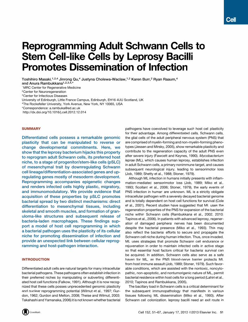
Reprogramming Adult Schwann Cells toStem Cell-like Cells by Leprosy BacilliPromotes Dissemination of InfectionToshihiro Masaki,1,2,4 Jinrong Qu,4 Justyna Cholewa-Waclaw,1,2 Karen Burr,2 Ryan Raaum,4
and Anura Rambukkana1,2,3,4,*1MRC Center for Regenerative Medicine2Center for Neuroregeneration3Center for Infectious DiseasesUniversity of Edinburgh, Little France Campus, Edinburgh, EH16 4UU Scotland, UK4The Rockefeller University, York Avenue, New York, NY 10065, USA
*Correspondence: [email protected]://dx.doi.org/10.1016/j.cell.2012.12.014
SUMMARY
Differentiated cells possess a remarkable genomicplasticity that can be manipulated to reverse orchange developmental commitments. Here, weshow that the leprosy bacterium hijacks this propertyto reprogram adult Schwann cells, its preferred hostniche, to a stage of progenitor/stem-like cells (pSLC)of mesenchymal trait by downregulating Schwanncell lineage/differentiation-associated genes and up-regulating genes mostly of mesoderm development.Reprogramming accompanies epigenetic changesand renders infected cells highly plastic, migratory,and immunomodulatory. We provide evidence thatacquisition of these properties by pSLC promotesbacterial spread by two distinct mechanisms: directdifferentiation to mesenchymal tissues, includingskeletal and smooth muscles, and formation of gran-uloma-like structures and subsequent release ofbacteria-laden macrophages. These findings sup-port a model of host cell reprogramming in whicha bacterial pathogen uses the plasticity of its cellularniche for promoting dissemination of infection andprovide an unexpected link between cellular reprog-ramming and host-pathogen interaction.
INTRODUCTION
Differentiated adult cells are natural targets for many intracellular
bacterial pathogens. Thesepathogensoften establish infection in
their preferred niches by manipulating or subverting differenti-
ated host cell functions (Falkow, 1991). Although it is now recog-
nized that these cells posses unprecedented genomic plasticity
and nuclear reprogramming potential (Wilmut et al., 1997; Gur-
don, 1962; Gurdon and Melton, 2008; Theise and Wilmut, 2003;
Takahashi andYamanaka, 2006) it is not knownwhether bacterial
pathogens have coevolved to leverage such host cell plasticity
for their advantage. Among differentiated cells, Schwann cells,
the glial cells of the adult peripheral nervous system (PNS) that
are comprisedofmyelin-forming andnon-myelin-formingpheno-
types (Jessen andMirsky, 2005), show remarkable plasticity and
contribute to the regeneration capacity of the adult PNS even
after severe injury (Fawcett and Keynes, 1990). Mycobacterium
leprae (ML), which causes human leprosy, establishes infection
in adult Schwann cells, a primary nonimmune target, and causes
subsequent neurological injury, leading to sensorimotor loss
(Job, 1989; Shetty et al., 1988; Stoner, 1979).
Although ML infection in humans initially presents with inflam-
mation-mediated sensorimotor loss (Job, 1989; Miko et al.,
1993; Scollard et al., 2006; Stoner, 1979), the early events of
PNS infection in human are unknown. ML is a strictly obligate
intracellular pathogen with a severely decayed bacterial genome
and is totally dependent on host cell functions for survival (Cole
et al., 2001). Recent studies have suggested that ML uses the
regeneration properties of the PNS for expansion of the bacterial
niche within Schwann cells (Rambukkana et al., 2002, 2010;
Tapinos et al., 2006). In patients with advanced leprosy, regener-
ation of damaged peripheral nerves has been documented
despite the bacterial presence (Miko et al., 1993). This may
also reflect the bacterial efforts to secure and propagate the
Schwann cell niche during human infection. Thus, once invaded,
ML uses strategies that promote Schwann cell endurance or
rejuvenation in order to maintain infected cells in active stage
so that essential host factors critical for bacterial survival can
be acquired. In addition, Schwann cells also serve as a safe
haven for ML, as the PNS blood-nerve barrier protects ML
from host immune assault (Job, 1989; Stoner, 1979). Such favor-
able conditions, which are assisted with the nontoxic, noncyto-
pathic, non-apoptotic, and nontumorigenic nature of ML, permit
bacterial residencewithin host cells for a long period (Lahiri et al.,
2010; Tapinos and Rambukkana, 2005).
The bacillary load in Schwann cells is a critical determinant for
the subsequent immunopathology that manifests in various
tissues following ML dissemination (Miko et al., 1993). After
Schwann cell colonization, leprosy bacilli need an exit route in
Cell 152, 51–67, January 17, 2013 ª2013 Elsevier Inc. 51
BA
D E F
G
H I
C
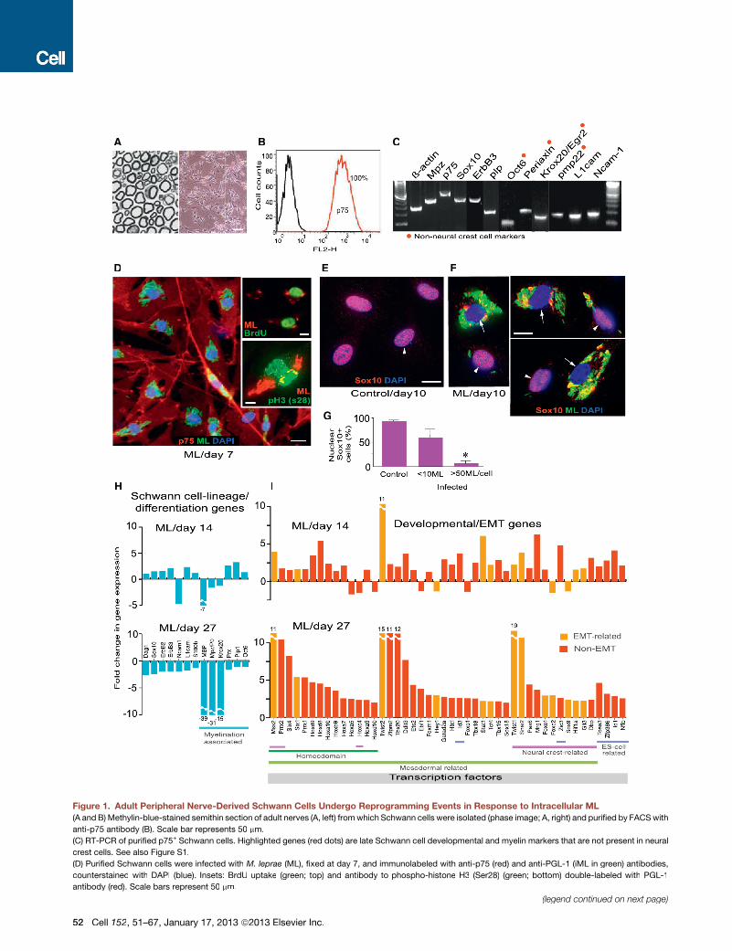
Figure 1. Adult Peripheral Nerve-Derived Schwann Cells Undergo Reprogramming Events in Response to Intracellular ML
(A and B) Methylin-blue-stained semithin section of adult nerves (A, left) fromwhich Schwann cells were isolated (phase image; A, right) and purified by FACSwith
anti-p75 antibody (B). Scale bar represents 50 mm.
(C) RT-PCR of purified p75+ Schwann cells. Highlighted genes (red dots) are late Schwann cell developmental and myelin markers that are not present in neural
crest cells. See also Figure S1.
(D) Purified Schwann cells were infected with M. leprae (ML), fixed at day 7, and immunolabeled with anti-p75 (red) and anti-PGL-1 (iML in green) antibodies,
counterstained with DAPI (blue). Insets: BrdU uptake (green; top) and antibody to phospho-histone H3 (Ser28) (green; bottom) double-labeled with PGL-1
antibody (red). Scale bars represent 50 mm.
(legend continued on next page)
52 Cell 152, 51–67, January 17, 2013 ª2013 Elsevier Inc.
order to successfully infect other tissues and transmit infection.
In leprosy patients, disseminated ML could be seen in several
tissues, including skeletal muscles and smooth muscles (Pear-
son et al., 1970; Job, 1989; Kaur et al., 1981; Scollard et al.,
2006; Werneck et al., 1999). Also, the involvement of skeletal
muscles in human leprosy is considered secondary due to
peripheral neuropathy with the obvious peripheral-nerve inner-
vations of skeletal muscles (Pearson et al., 1970; Werneck
et al., 1999). However, it is unknown how initial colonization of
ML in Schwann cells subsequently leads to the spread of infec-
tion to other tissues.
In this study, we show that leprosy bacteria trigger reprogram-
ming of adult Schwann cells to a stage of progenitor/stem-like
cells (pSLC) with migratory and immunomodulatory properties
that promote bacterial dissemination. Reprogrammed cells facil-
itate bacterial spread by two distinct mechanisms—by direct
differentiation to mesenchymal tissues, skeletal muscles, and
smooth muscles and by contributing to form granuloma-like
structures (GLS) that subsequently release bacteria-laden
macrophages. Our findings present an unexpected link between
cellular reprogramming and host-pathogen interaction and thus
direct to potential new therapeutic strategies for combating
infections with pathogenic bacteria.
RESULTS
Schwann Cells Derived from Adult Peripheral NervesUndergo Reprogramming Events in Response toIntracellular MLWe recapitulated the colonization of ML and subsequent early
molecular events in adult Schwann cells by infecting primary
Schwann cells isolated from adult mouse peripheral nerves.
We isolated Schwann cells from adult wild-type and Sox2-
green fluorescent protein (GFP) transgenic mice and purified
by fluorescence-activated cell sorting (FACS) with antibody to
Schwann cell-surface marker p75 NTR or GFP expression under
the control of Sox2 promoter, respectively (Figures 1A–1C, 2A,
and 2B and Figures S1 and S3A available online). Single-cell-
derived Schwann cells were generated from FACS-sorted cells
and characterized extensively (Figure S1). All were positive for
the markers for late Schwann cell development and myelina-
tion-associated genes (Figures 1C and S1). Most of these differ-
entiation markers are not present in neural crest stem cells and
do not appear until around the time of birth (Jessen and Mirsky,
2005; Finzsch et al., 2010). Therefore, they represent mature de-
differentiated Schwann cells. Moreover, karyotyping of these
cells showed intact chromosome numbers and no evidence of
translocation, suggesting their genomic stability (Figure S1).
As expected, these dedifferentiated adult Schwann cells are
highly susceptible toML infection with rapid bacterial engulfment
with >90% efficiency (Figures 1D, 2Ac, and S1). We performed
extensive genome-wide transcriptome profiling with Affymetrix
mouse genechips and subsequent RT-PCR and quantitative
PCR (qPCR) analyses. Strikingly, infected Schwann cells com-
pared to uninfected/control Schwann cells, which were main-
tainedunder identical experimental conditions, showedupregula-
tion of numerous genes of embryonic development, transcription,
chromatin remodeling, cell signaling, chemokines, and cell divi-
sion-cycle/DNA replication (Figures 1I and S2). The latter was
consistent with a moderate increase of S phase cells in infected
cultures; despite the presence of numerous intracellular ML
(iML), infected cells showed BrdU uptake and phospho-histone
H3 (S28) positive nuclei,which represent theS andmitotic phases
of the cell cycle, respectively (Figure 1D, insets). Strikingly, ML
infection was followed by the removal or export of Sox10 from
Schwann cell nuclei (Figures 1E–1G). Whereas, similar to unin-
fected/control cells, infected Schwann cells that carry fewer iML
maintained Sox10 exclusively in nuclei, high bacterial load
invariably caused the removal of nuclear Sox10, which might
affect the regulation of its key target Schwann cell genes,
including myelin gene Mpz (Figures 1E–1G) (Finzsch et al., 2010;
Jessen andMirsky, 2005;Weider et al., 2012). Indeed, downregu-
lation of myelin genes was found in infected Schwann cells over
time (Figure 1H). Sox10 is a master regulator of Schwann cell
homeostasis, identity, and myelin maintenance as well as differ-
entiation, with nuclear Sox10 being required to recruit chro-
matin-remodeling complexes (Finzsch et al., 2010; Weider et al.,
2012). Therefore, bacterial-induced removal of Sox10 from nuclei
is likely to perturb normal Sox10-mediated transcriptional events.
Together, these findings set the stage for a potential reprogram-
ming or change in Schwann cell fate in response to iML.
Mesenchymal Transition and Induction of EMT-likeProgram in Adult Schwann CellsThe fate of iML-induced effects on Schwann cells was examined
by gene-expression analyses over a 4 week period. Strikingly,
only infected cells gradually ‘‘turn off’’ Schwann cell differentia-
tion/myelination- and lineage-associated genes and ‘‘turn on’’
numerous developmental genes, comprising mostly the meso-
derm development, including homeodomain/Hox, EMT, and
neural crest-related genes (Figures 1H, 1I, and S2); at day 27
post-infection, most of the Schwann cell lineage and differentia-
tion/myelination genes, but not negative regulators of myelina-
tion like Sox2 and c-Jun (Jessen and Mirsky, 2005), were down-
regulated (Figures 1H and S2), suggesting that iML gradually
shuts down the Schwann cell differentiation program. Figure 1I
shows the selected key developmental-regulated transcription
factor (DRTF) genes (with known functions; Table S1).
A major tissue-remodeling program that is central to early
mesoderm development during embryogenesis is EMT (Polyak
and Weinberg, 2009). Interestingly, master regulators of EMT,
Twist1 and 2, Snail2, and Msx2, which are capable of inducing
(E–G) Expression of nuclear Sox10 in control (E) and infected (F) Schwann cells at day 10 post-infection; antibodies to Sox10 (red) and PGL-1 (iML; green),
counterstained with DAPI (blue) (F). Shown in (F) are selected representative Deltavision images from infected cultures with high (arrows) and low bacterial loads
(arrowheads). (G) Quantification of nuclear Sox10 in control and infected Schwann cells with iML per cell. Scale bars represent 10 mm.
(H and I) Gene array analyses at days 14 and 27 post-infection. Schwann cell lineage/differentiation-associated genes (H) and development- and EMT-associated
transcription factor (TF) genes (I) are shown as fold change in expression.
See also Figure S2 and Table S1.
Cell 152, 51–67, January 17, 2013 ª2013 Elsevier Inc. 53
A
B
C D E
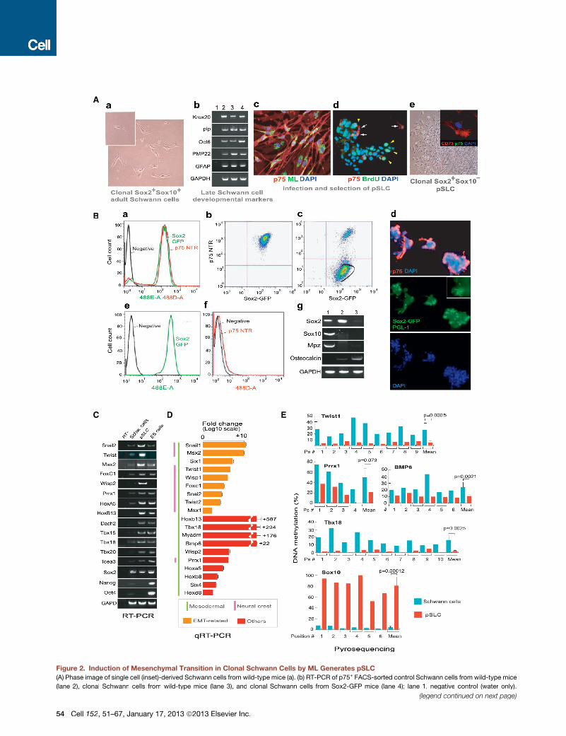
Figure 2. Induction of Mesenchymal Transition in Clonal Schwann Cells by ML Generates pSLC
(A) Phase image of single cell (inset)-derived Schwann cells from wild-type mice (a). (b) RT-PCR of p75+ FACS-sorted control Schwann cells from wild-type mice
(lane 2), clonal Schwann cells from wild-type mice (lane 3), and clonal Schwann cells from Sox2-GFP mice (lane 4); lane 1, negative control (water only).
(legend continued on next page)
54 Cell 152, 51–67, January 17, 2013 ª2013 Elsevier Inc.
EMT in epithelial cells (Mani et al., 2008) (Table S1), were among
the highly upregulated transcription factor (TF) genes in ML-in-
fected Schwann cells (Figures 1I and S2). The observed cell-
fate changes in Schwann cells are complex as infection involves
upregulation of multiple developmental and functional genes
(Figures 1H, 1I, and S2; Table S1), and these events are not asso-
ciated with tumor-suppressor genes like p53 or Schwann cell
tumor-associated gene NF1 (Figures S2A and S3A).
Our findings also suggest that reprogramming requires a high
number of intracellular bacteria (>50 iML/cell with >90% infec-
tion efficiency) and a long-term incubation; we could not find
any significant change in gene expression or the removal of
nuclear Sox10 at a much lower rate of infection (<10 iML/cell;
Figures 1E–1G). Shortage or lack of such optimum conditions
appears to weaken or fail to induce transcriptional changes.
For example, the observed changes in gene expression are
highly significant with live ML, as compared to irradiated ML;
bacterial components like PGL-1 did not show reprogramming
events, despite their capacity to induce rapid demyelination
upon their extracellular binding to myelinated Schwann cells
(Rambukkana et al., 2002). This appears to be due to high inva-
sion capacity of live ML and thus the long-term exposure of
Schwann cells to a high number of iML. In addition, incubation
of Schwann cells with other mycobacteria like Mycobacterium
smegmatis did not lead to reprogramming (data not shown).
Bacterially Reprogrammed Schwann Cells ExhibitProperties Similar to pSLCTo isolate reprogrammed cells from the infected cell population,
we transferred clonal-infected Schwann cells to mesenchymal
stem cell medium (Mscm), which is selective for mesenchymal
stem cells (MSC) from bone marrow cells (Li et al., 2009). In
Mscm, only reprogrammed Schwann cells showed a high prolif-
eration index as demonstrated by BrdU uptake (Figure 2Ad);
>90% of these BrdU+ cells were negative for Schwann cell
lineage markers p75 and Sox10 as well as other mature
phenotypes (Figure 2A). We used this property to select a
p75� population by FACS sorting. Analyses of these p75� cells
by microarrays, RT-PCR, qPCR, immunofluorescence (IF), and
FACS revealed the loss of all Schwann cell lineage/myelin-
specific markers and acquisition of various mesodermal and
neural crest markers (Figures 2C, 2D, S3A, and S3B). Thus, we
concluded that iML converted Sox2+/p75+/Sox10+ Schwann
cells to Sox2+/p75�/Sox10� reprogrammed cells with loss of
Schwann cell markers (Figures 2B and S3A), which we referred
to as pSLC because of their stem cell characteristics (see below).
Further characterization revealed the expression of CD73, CD44,
Sca-1, and Cd29 MSC markers but the absence of hematopoi-
etic markers CD45, CD34, and c-kit (Figure S3H). pSLC also
acquired highly migratory properties as evident by their migra-
tion through connective tissues when administered into injured
muscles (Figures S3F and S3G).
ReprogrammingRemovesSchwannCell Lineage/MyelinRegulator Sox10 and Maintains Stem Cell Marker Sox2Conversion of parent Schwann cells to pSLC led to the loss of
Schwann cell master regulator Sox10 but maintained Sox2. To
track Sox2 expression during reprogramming, we generated
clonal Schwann cells isolated from adult Sox2-GFP mice (Eminli
et al., 2008). Clonal cells in Schwann cell media maintained both
Sox10 and other mature Schwann cell markers, whereas in-
fected cells lost Sox10 and other Schwann cell markers and
subsequently changed into pSLC when maintained in Mscm
(Figures 2A and 2B). Infected Schwann cells continued to
maintain GFP (Sox2) expression but upon reprogramming lost
expression of p75, Sox10, and other mature markers (Figures
2Ba–2Bd). When pSLC were maintained in Mscm, clusters of
infected GFP+ cells with both p75� and p75+ were frequently
observed (Figure 2Bd). When p75� reprogrammed cells in
Mscm were isolated by FACS sorting, they continued to express
GFP (Sox2) despite the loss of lineage/mature Schwann cell
markers (Figures 2Bc–2Bg). However, GFP/Sox2 expression
disappeared when pSLC were subjected to bone differentiation
(Pittenger et al., 1999), which in contrast upregulated bone
marker osteocalcin (Figures 2Bg and 3C), further underscoring
stem cell characteristics of pSLC. Tracking the GFP expression
from parent GFP+/Sox2+/p75+/Sox10+ Schwann cells to GFP+/
Sox2+/p75�/Sox10� pSLC at the clonal level, we confirmed
that pSLC are derived from Schwann cells but not from any
progenitor cell type within the Schwann cell preparation.
Reprogramming Schwann Cells to pSLC AccompaniesEpigenetic ChangesWe next examined whether this change in cell fate is epigeneti-
cally regulated. We analyzed DNA methylation at 5 methylcyto-
sine in cytosine guanine dinucleotide (CpG) in the promoter
regions of selected genes that are expressed and repressed in
pSLC as compared to parent Schwann cells. Methylation status
was assessed by bisulfite pyrosequencing, which allows quanti-
tative determination of the extent of methylation of each cytosine
(c) Infected clonal wild-type Schwann cells at day 7 (PGL-1+ML, green; p75+ Schwann cells, red). (d) BrdU uptake (green arrowheads) of day 27-infected Schwann
cells maintained in mesenchymal stem cell media (Mscm); anti-p75 antibody in red (arrows), and nuclei (DAPI) in blue. (e) Phase image of p75� FACS-sorted cells
that express CD73 (e-inset). Magnification 203; Inset (e): 40�.
(B) FACS analysis of GFP+(Sox2+)/p75+ Schwann cells isolated from adult Sox2-GFP mice (a). Clonal GFP+/p75+ cells derived from FACS-sorted Schwann cells
(b) were infected with ML for 4 weeks (also see Figure S1M) and transferred to Mscm, and Sox2+/p75� population was gated for FACS (c). (d) IF analysis of
aggregated infected cells maintained in Mscm before FACS; p75+ (red) and DAPI (blue). Inset shows PGL-1+ ML as green rods. (e and f) FACS analysis of sorted
cells showing GFP+/Sox2+ (e) and p75� (f) cells, which are referred to as pSLC. (g) RT-PCR of control Schwann cells (lane 1), pSLC (lane 2), and pSLC in bone
differentiation media (lane 3). Magnification (d): 103.
(C) RT-PCR of embryonic/developmental marker genes in pSLC as compared to control Schwann cells and mouse ESCs. See also Figure S3.
(D) qPCR for differential expression of selected embryonic/developmental marker genes in pSLC relative to control Schwann cells. Data are mean ± standard
error of the mean (SEM) from three samples.
(E) Cell-fate change of Schwann cells to pSLC accompanies changes in DNAmethylation statuses of promoter regions of key developmental/EMT and Schwann
cell-lineage genes as analyzed by pyrosequencing.
Cell 152, 51–67, January 17, 2013 ª2013 Elsevier Inc. 55
A B
C D E
F I
LK
P Q
J M
G
H
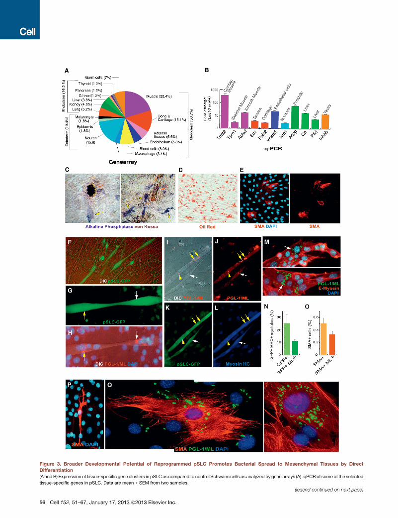
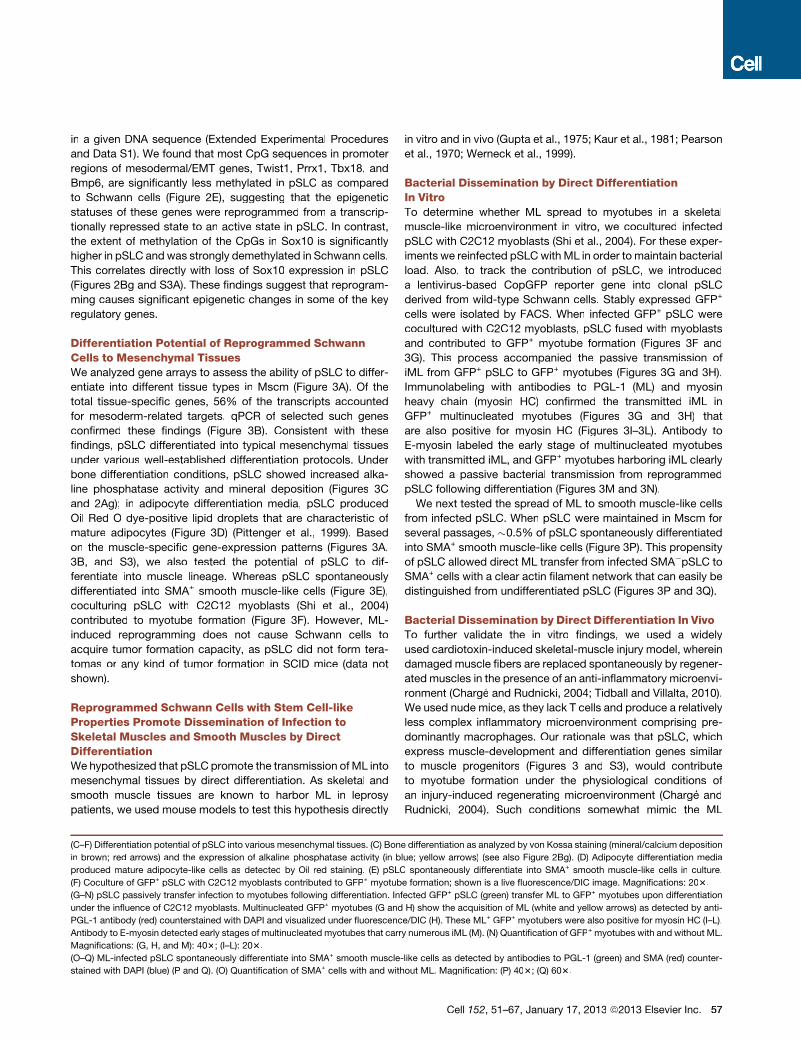
Figure 3. Broader Developmental Potential of Reprogrammed pSLC Promotes Bacterial Spread to Mesenchymal Tissues by Direct
Differentiation(A and B) Expression of tissue-specific gene clusters in pSLC as compared to control Schwann cells as analyzed by gene arrays (A). qPCR of some of the selected
tissue-specific genes in pSLC. Data are mean ± SEM from two samples.
(legend continued on next page)
56 Cell 152, 51–67, January 17, 2013 ª2013 Elsevier Inc.
in a given DNA sequence (Extended Experimental Procedures
and Data S1). We found that most CpG sequences in promoter
regions of mesodermal/EMT genes, Twist1, Prrx1, Tbx18, and
Bmp6, are significantly less methylated in pSLC as compared
to Schwann cells (Figure 2E), suggesting that the epigenetic
statuses of these genes were reprogrammed from a transcrip-
tionally repressed state to an active state in pSLC. In contrast,
the extent of methylation of the CpGs in Sox10 is significantly
higher in pSLC and was strongly demethylated in Schwann cells.
This correlates directly with loss of Sox10 expression in pSLC
(Figures 2Bg and S3A). These findings suggest that reprogram-
ming causes significant epigenetic changes in some of the key
regulatory genes.
Differentiation Potential of Reprogrammed SchwannCells to Mesenchymal TissuesWe analyzed gene arrays to assess the ability of pSLC to differ-
entiate into different tissue types in Mscm (Figure 3A). Of the
total tissue-specific genes, 56% of the transcripts accounted
for mesoderm-related targets. qPCR of selected such genes
confirmed these findings (Figure 3B). Consistent with these
findings, pSLC differentiated into typical mesenchymal tissues
under various well-established differentiation protocols. Under
bone differentiation conditions, pSLC showed increased alka-
line phosphatase activity and mineral deposition (Figures 3C
and 2Ag); in adipocyte differentiation media, pSLC produced
Oil Red O dye-positive lipid droplets that are characteristic of
mature adipocytes (Figure 3D) (Pittenger et al., 1999). Based
on the muscle-specific gene-expression patterns (Figures 3A,
3B, and S3), we also tested the potential of pSLC to dif-
ferentiate into muscle lineage. Whereas pSLC spontaneously
differentiated into SMA+ smooth muscle-like cells (Figure 3E),
coculturing pSLC with C2C12 myoblasts (Shi et al., 2004)
contributed to myotube formation (Figure 3F). However, ML-
induced reprogramming does not cause Schwann cells to
acquire tumor formation capacity, as pSLC did not form tera-
tomas or any kind of tumor formation in SCID mice (data not
shown).
Reprogrammed Schwann Cells with Stem Cell-likeProperties Promote Dissemination of Infection toSkeletal Muscles and Smooth Muscles by DirectDifferentiationWe hypothesized that pSLC promote the transmission of ML into
mesenchymal tissues by direct differentiation. As skeletal and
smooth muscle tissues are known to harbor ML in leprosy
patients, we used mouse models to test this hypothesis directly
in vitro and in vivo (Gupta et al., 1975; Kaur et al., 1981; Pearson
et al., 1970; Werneck et al., 1999).
Bacterial Dissemination by Direct DifferentiationIn VitroTo determine whether ML spread to myotubes in a skeletal
muscle-like microenvironment in vitro, we cocultured infected
pSLC with C2C12 myoblasts (Shi et al., 2004). For these exper-
iments we reinfected pSLCwith ML in order to maintain bacterial
load. Also, to track the contribution of pSLC, we introduced
a lentivirus-based CopGFP reporter gene into clonal pSLC
derived from wild-type Schwann cells. Stably expressed GFP+
cells were isolated by FACS. When infected GFP+ pSLC were
cocultured with C2C12 myoblasts, pSLC fused with myoblasts
and contributed to GFP+ myotube formation (Figures 3F and
3G). This process accompanied the passive transmission of
iML from GFP+ pSLC to GFP+ myotubes (Figures 3G and 3H).
Immunolabeling with antibodies to PGL-1 (ML) and myosin
heavy chain (myosin HC) confirmed the transmitted iML in
GFP+ multinucleated myotubes (Figures 3G and 3H) that
are also positive for myosin HC (Figures 3I–3L). Antibody to
E-myosin labeled the early stage of multinucleated myotubes
with transmitted iML, and GFP+ myotubes harboring iML clearly
showed a passive bacterial transmission from reprogrammed
pSLC following differentiation (Figures 3M and 3N).
We next tested the spread of ML to smooth muscle-like cells
from infected pSLC. When pSLC were maintained in Mscm for
several passages, �0.5% of pSLC spontaneously differentiated
into SMA+ smooth muscle-like cells (Figure 3P). This propensity
of pSLC allowed direct ML transfer from infected SMA�pSLC to
SMA+ cells with a clear actin filament network that can easily be
distinguished from undifferentiated pSLC (Figures 3P and 3Q).
Bacterial Dissemination by Direct Differentiation In VivoTo further validate the in vitro findings, we used a widely
used cardiotoxin-induced skeletal-muscle injury model, wherein
damaged muscle fibers are replaced spontaneously by regener-
ated muscles in the presence of an anti-inflammatory microenvi-
ronment (Charge and Rudnicki, 2004; Tidball and Villalta, 2010).
We used nude mice, as they lack T cells and produce a relatively
less complex inflammatory microenvironment comprising pre-
dominantly macrophages. Our rationale was that pSLC, which
express muscle-development and differentiation genes similar
to muscle progenitors (Figures 3 and S3), would contribute
to myotube formation under the physiological conditions of
an injury-induced regenerating microenvironment (Charge and
Rudnicki, 2004). Such conditions somewhat mimic the ML
(C–F) Differentiation potential of pSLC into various mesenchymal tissues. (C) Bone differentiation as analyzed by von Kossa staining (mineral/calcium deposition
in brown; red arrows) and the expression of alkaline phosphatase activity (in blue; yellow arrows) (see also Figure 2Bg). (D) Adipocyte differentiation media
produced mature adipocyte-like cells as detected by Oil red staining. (E) pSLC spontaneously differentiate into SMA+ smooth muscle-like cells in culture.
(F) Coculture of GFP+ pSLC with C2C12 myoblasts contributed to GFP+ myotube formation; shown is a live fluorescence/DIC image. Magnifications: 203.
(G–N) pSLC passively transfer infection to myotubes following differentiation. Infected GFP+ pSLC (green) transfer ML to GFP+ myotubes upon differentiation
under the influence of C2C12 myoblasts. Multinucleated GFP+ myotubes (G and H) show the acquisition of ML (white and yellow arrows) as detected by anti-
PGL-1 antibody (red) counterstained with DAPI and visualized under fluorescence/DIC (H). These ML+ GFP+ myotubers were also positive for myosin HC (I–L).
Antibody to E-myosin detected early stages of multinucleated myotubes that carry numerous iML (M). (N) Quantification of GFP+ myotubes with and without ML.
Magnifications: (G, H, and M): 403; (I–L): 203.
(O–Q) ML-infected pSLC spontaneously differentiate into SMA+ smooth muscle-like cells as detected by antibodies to PGL-1 (green) and SMA (red) counter-
stained with DAPI (blue) (P and Q). (O) Quantification of SMA+ cells with and without ML. Magnification: (P) 403; (Q) 603.
Cell 152, 51–67, January 17, 2013 ª2013 Elsevier Inc. 57

infection-associated inflammation in muscles and thus com-
mensurate with the muscle pathology documented in the lepro-
matous form of leprosy in patients with high bacterial load (Gupta
et al., 1975; Werneck et al., 1999).
We first injected cardiotoxin into the tibialis anterior (TA)
muscles of nude mice, and the muscle injury, inflammation,
and repair process were confirmed by histological studies and
immunolabeling (data not shown). GFP+ pSLC containing ML
were then injected into preinjured TAmuscles, and their distribu-
Figure 4. Redifferentiation of Reprog-
rammed pSLC Contributes to Passive
Bacterial Transfer to Skeletal Muscles and
Smooth Muscles In Vivo
(A–D) Transverse frozen sections of TA muscles
injected with ML-infected GFP+ pSLC after
10 days. Deltavision images showed GFP+ pSLC
(green) either fused with myofibers (A and B) or
migrating in between muscle fibers (C and D).
Antibodies to adult myosin detected muscle fibers
(red in A and B and blue in D), and laminin
demarcated individual muscle fibers (red in C).
Antibodies to PGL-1 detected ML (in red; arrow)
associated closely with muscle fibers (D). Aster-
isks in all figures denote individual myofibers.
Magnifications: 603.
(E–J) Infected GFP+ pSLC were incorporated into
regenerating muscles at 3 weeks post-injection.
GFP+ myofibers are positive for adult myosin
(E and F) and are demarcated by laminin (G and H).
pSLC incorporation passively transmitted ML
to myofibers as detected by PGL-1+ ML (red;
arrows) in GFP+myofibers (I and J). Magnifications
(Deltavision images): 403.
(K and L) Bacterial presence within the skeletal
muscle fibers (asterisks) was confirmed by acid-
fast mycobacterial staining (red). Arrows show
clumps of rod-shaped ML in several individual
muscle fibers. Magnifications: 603.
(M–P) Administered infected GFP+ pSLC differ-
entiate into SMA+ smooth muscles in the dermal
area. Antibody to SMA (red) colocalizes with
differentiated GFP+pSLC (green; arrowheads);
merged image is shown in (O). Magnifications:
(M–O) 403. (P) Higher magnification (1003)
showed the PGL+ML (red; arrows) within SMA+
smooth muscles (blue) near dermal vessels.
tion was examined at different time inter-
vals. Antibody staining to adult myosin
clearly showed the intimate interaction
and early fusion process of ML-laden
GFP+ cells around the muscle fibers at
the injury site after day 10 (Figures 4A–
4D). The engrafted GFP+ pSLC were
detected within muscle milieu at days 14
and 21 either as fused to resident regen-
erating fibers or fully integrated into
smaller diameter myofibers that express
adult myosin and are demarcated by lam-
inin in the basal lamina (Figures 4E–4H).
Anti-PGL-1 antibody staining that specif-
ically identifies ML (Ng et al., 2000) confirmed the bacterial pres-
ence within fully integrated GFP+ myofibers (Figures 4I and 4J).
Specific acid-fast mycobacterial staining further confirmed rod-
shaped ML within myofibers (Figures 4K and 4L). In contrast to
pSLC, injection of nonreprogrammed infected GFP+ Schwann
cells (infection only for 2 days) to injured TA muscles failed to
transmit infection to regeneratingmuscle fibers (data not shown).
Similarly, we also found that administered infectedGFP+ pSLC
can differentiate into SMA+ smooth muscles (or myofibroblasts),
58 Cell 152, 51–67, January 17, 2013 ª2013 Elsevier Inc.

and ML can be detected within SMA+ smooth muscles in the
dermal area of muscle-skin interphase and associated blood
vessels (Figures 4M–4P). In contrast, infected GFP+ Schwann
cells (nonreprogrammed cells infected for 2 days only) neither
differentiated into smooth muscles nor contributed to transfer-
ring infection to SMA+ cells (data not shown). Together, these
results suggest that under the influence of injury and inflamma-
tory tissue microenvironment, infected pSLC could elaborate
their migratory capacity and differentiate into myofibers or
smooth muscles and passively transmit ML to these tissues
that are favorable for sequestration of infection.
Reprogrammed pSLC Acquire ImmunomodulatoryPropertiesWe examined whether reprogrammed pSLC also secrete immu-
nomodulatory factors in a way similar to that of MSCs. To test
this, we first used mouse chemokine/cytokine protein arrays to
analyze proteins released into serum/supplement-free condi-
tioned media (CM) from pSLC. Figure 5A shows the quantitative
analysis of differentially expressed soluble proteins released by
pSLC. Strikingly, pSLC produced a range of chemokines, cyto-
kines, growth/survival factors, soluble adhesion receptors, and
tissue-remodeling factors to varying degrees (Figures 5A and
S4). These findings also validate the MSC-like functional proper-
ties of pSLC because almost all chemokines/cytokines that are
known to be produced by MSC are also released by pSLC (Fig-
ure 5A) (Hoogduijn et al., 2010; Klopp et al., 2011). Although
a fewer number of these proteins are also released by control/
uninfected Schwann cells, the fold increase of their expression
is several fold higher in pSLC (Figures 5A and S4), suggesting
a greater potential of pSLC to participate in multiple immuno-
modulatory functions. Interestingly, once reprogrammed, pSLC
continued to secrete soluble factors regardless of bacterial pres-
ence, as complete removal of ML from pSLC (with no detectable
ML genomic DNA) continued to produce almost the same
soluble proteins (Figures 5A, inset and S4).
pSLC-Derived Soluble Factors Mediate MacrophageSurvival and MigrationWe next examined the functional effects of pSLC-derived immu-
nomodulatory factors on tissue macrophages. As a source, we
used freshly isolated mouse peritoneal macrophages, which
comprised >85% F4/80+ and �100% CD68+ macrophages.
When incubated with pSLC-CM alone, both F4/80+ and CD68+
macrophages were maintained >14 days, whereas the majority
of macrophages inmedium alone did not survive or lifted off early
in cultures (Figure 5B). Macrophages maintained in pSLC-CM
showed a firm attachment and elongated morphology with
strong F4/80 expression, suggesting possible maturation of
macrophages in pSLC-CM. pSLC-CM also influenced increased
survival of macrophages with minimum apoptosis (Figure 5C).
However, pSLC-CM did not increase S phase cells significantly
(data not shown).
Consistent with the chemokine/cytokine production, pSLC-
CM attracted a significant number of macrophages when a cell
migration assay was performed with the Trans-well system
(Figures 5D–5F). Migration capacity was further increased in
the presence of live pSLC, implying that freshly secreted pSLC
factors attracted a higher number of macrophages (Figures 5E
and 5F). Although pSLC-CM-induced macrophage migration
was slightly decreased (<10%) in the presence of a standard
dose (200–1000 ng) of broader CC-chemokine inhibitor, CCI
(recombinant viral CCI-Fc chimera), which has shown to be
effective in other studies (Buatois et al., 2010), a high dose of
nontoxic concentrations of CCI (5000 ng) was required to
produce a significant inhibition of migration (Figure 5G). Because
pSLC produce an array of soluble proteins (Figures 5A and S4),
our data suggest that a collective effect of immune factors, but
not CC-chemokine alone, is required to exert effective macro-
phage chemotactic ability.
Reprogrammed pSLC Promote Bacterial Disseminationvia Macrophages In VivoWe reasoned that a nude-mouse muscle-injury model with
a predominantly macrophage-containing inflammatory microen-
vironment would be useful to directly test whether pSLC pro-
mote bacterial dissemination via macrophages. To ensure that
all injected pSLC express GFP and all bacteria are inside the
cells, strongly positive GFP+ pSLC were FACS sorted and rein-
fected with ML 2 days before cell injection to the preinjured TA
muscles. Analyzing the fate of pSLC and iML at weeks 1, 2,
and 3 post-injection revealed that almost all GFP+ cells migrate
further away from the site of injection after 1 week and continue
to spread over time along the interstitial connective tissues in
the perimysium and to the skeletal muscle-dermal interphase
(SkMDIP) at week 3 post-injection (Figures 6A, S5, and S6). Strik-
ingly, iML were no longer confined to pSLC as most of iML were
detected within recipient non-GFP cells (Figures 6Aa, 6Ab, 6Ah,
and 6Aj). These data indicate that iMLwere efficiently transferred
from donor GFP+ pSLC to GFP� recipient tissue cells in vivo.
Both anti-PGL-1 antibody and acid-fast mycobacterial staining
confirmed these findings (Figures 6Aj and S5). These ML+
GFP� cells were identified as endogenous macrophages as
>90% of them were positive for macrophage markers F4/80,
CD11b, CD68, and CD206 (Figures 6Ac–6Aj and S5).
Inflammatory conditions in cardiotoxin-induced TA muscles
are known to bring a sequential appearance of macrophage
subpopulations, initially with CD68+ M1 subtype peaks at week
2 followed by a high CD163/CD206 M2 population that overlaps
within the M1 population at week 2–3 post-injury (Tidball and Vil-
lalta, 2010). These established conditions allowed us to correlate
GFP+ pSLC distribution with infiltrated macrophage subtypes
in injured muscles in a timely manner. Indeed, our findings
with serial tissue sections from infected GFP+ pSLC engrafted
mice at week 3 post-infection showed that CD68+, CD163+,
CD206+, or F4/80+ cells in the interstitial tissues were codistrib-
uted with GFP+ pSLC. Double immunolabeling with an antibody
to PGL-1 and macrophage markers confirmed iML within these
macrophages (Figures 6Aj, S5K, and S5L). Once ML transferred
from pSLC to macrophages, they appeared to transmit infection
to uninfected macrophages and thus facilitate ML spread via
these tissue macrophages along the connective tissues in
between muscle fibers and SkMDIP. Figures 6Aa, S5, and S6
clearly show examples illustrating the presence of iML within
numerous non-GFP recipient cells along the perimysium/con-
nective tissues and SkMDIP. Analysis of serial tissue sections
Cell 152, 51–67, January 17, 2013 ª2013 Elsevier Inc. 59
BA
C
D E
F
G
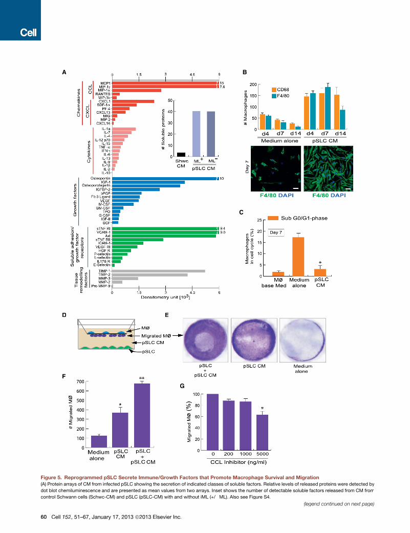
Figure 5. Reprogrammed pSLC Secrete Immune/Growth Factors that Promote Macrophage Survival and Migration
(A) Protein arrays of CM from infected pSLC showing the secretion of indicated classes of soluble factors. Relative levels of released proteins were detected by
dot blot chemiluminescence and are presented as mean values from two arrays. Inset shows the number of detectable soluble factors released from CM from
control Schwann cells (Schwc-CM) and pSLC (pSLC-CM) with and without iML (+/�ML). Also see Figure S4.
(legend continued on next page)
60 Cell 152, 51–67, January 17, 2013 ª2013 Elsevier Inc.
strongly suggests ML transfer from pSLC to both M1 and M2
macrophages (Figures 6 and S5).
pSLC Contribute to Granuloma-like Formation In Vivo:Role in Bacterial DisseminationStrikingly, infected pSLC that had migrated to the loosely
associated connective tissue areas within SkMDIP formed orga-
nized aggregates together with macrophages (Figures 6B and
S6). These organized aggregates resemble typical granulomas
seen in mycobacterial infections, both in murine models and
humans (Flynn and Chan, 2001; Bold and Ernst, 2009; Modlin
and Rea, 1988). Analyzing the cellular distribution of these GLS
revealed that pSLC contribute to a major cellular mass highly
organized into a spiral form of GFP+ cells and interposed
between infiltrated macrophages in both the core area and the
periphery of GLS (Figure 6B). Interestingly, a distinct distribution
of macrophage subpopulation was observed within these GLS:
the exteriors of the GLS are strongly positive for F4/80 (F4/
80+high) and CD206 populations, whereas the core areas are
weakly positive for F4/80 (F4/80+low) but strongly positive for
CD68+ cells with tightly packed multinucleated macrophages
(Figures 6Ba and S7). This phenotypic distribution of macro-
phages with F4/80+high/CD206+high and F4/80+low/CD68+high
corresponds to M1 and M2 subtypes, respectively (Figures 6B,
S6, and S7) (Mosser and Edwards, 2008). Anti-PGL-1 and Fite’s
staining (modified acid-fast mycobacterial staining) in serial
sections clearly showed that macrophages in both core and
periphery of GLS harbor large numbers of iML (Figures 6B, S5,
and S6). In contrast, we were unable to detect such GLS when
infected GFP+ Schwann cells were administered under similar
experimental conditions.
Release of ML-laden Macrophages from theGranulomasWe found a significant shift in distribution of macrophages when
analyzed across granuloma-containing tissues within SkMDIP.
Figures 6B and 6C illustrate the distribution of pSLC and macro-
phages and their bacterial contents within the GLS in serial
sections taken from 250–300 mm apart tissue areas. In one end
of the GLS-bearing tissues, CD68+ cells with a high number of
iML were organized into the core area surrounded by GFP+
pSLC and bacterial laden F4/80+high/CD206+high macrophages
in the exterior, whereas in the other end, both CD68+ and
CD206+ macrophages were found migrating out of the tapered
aggregates of pSLC carrying iML with them (Figures 6B, 6C,
and S6). These data collectively suggest not only the transfer
of ML from pSLC tomacrophages but also themigration of these
bacterial-laden macrophages from the organized GLS.
In Vitro Granuloma-like Formation by pSLC andMacrophagesNext we developed an in vitromodel of GLS.We found that pSLC
tend to form aggregates at high density, and no leaked bacte-
ria were found outside the aggregates (Figure 7A). Real-time
microscopy revealed that uninfected macrophages migrated
into infected GFP+ pSLC aggregates when peritoneal macro-
phages were added (Figure 7B). Most of these macrophages
reached the core area of the GLS as early as 6 hr and 18 hr
and acquired ML from pSLC; bacterial transfer from some in-
fected pSLC to macrophages was also observed adjacent to
the GLS (Figures 7B). Some macrophages were also found to
contain GFP+ pSLC cell debris with the bacteria in them, sug-
gesting the phagocytosis of dead or apoptotic GFP+ pSLC by
macrophages (Figure 7B). However, only �0.04% of TUNEL-
positive cells were detected in these cell aggregates. Over
time, an increasing number of macrophages continued to
migrate and form larger GLS, and at this point, most of the ML
were transferred to macrophages (Figure 7C). These organized
GLS then gradually started to disintegrate and subsequently
release ML-laden macrophages (Figure 7D). Quantification of
bacterial transfer from pSLC to macrophages showed an effec-
tive transmission of infection to macrophages over time (Fig-
ure 7E). These findings suggest that chemoattractants released
from pSLC aggregates may have provided the right signals for
macrophage migration and GLS formation. Further studies on
dissection of regulatory signaling pathways involved may estab-
lish new links between host cell reprogramming and innate
phase immune responses during infection.
DISCUSSION
In this study, we provide evidence that leprosy bacteria perturb
dynamic mechanisms that normally preserve the lineage com-
mitment of adult Schwann cells and hence change the Schwann
cell fate to pSLCwithmesenchymal characteristics that promote
bacterial dissemination. The findings describe an unexpected
but natural exploitation of adult Schwann cell plasticity by ML
during infection. Once invaded, ML gradually ‘‘turn off’’ Schwann
cell lineage/differentiation-associated genes/TFs and ‘‘turn on’’
numerous embryonic/developmental genes and TFs of meso-
derm and neural crest. Such alterations in gene expression are
likely to disrupt the stoichiometry of transcriptional regulators,
leading to reprogramming of infected cell nuclei.
Of particular interest is the induction of TFs of the homeodo-
main/Hox family and EMT in Schwann cells in response to ML.
It is known that the fate of somatic cells can be altered by forced
expressions of both Hox and EMT genes (Mani et al., 2008).
(B) Soluble factors secreted by pSLC promote macrophage survival. Quantification of adherent F4/80+ and CD68+ macrophages maintained in pSLC-CM and
media alone for up to 14 days is shown (B, top panel). IF of adherent F4/80+macrophages in the presence of pSLC-CM (right) andmedia alone (left) at day 7 is also
shown (B, bottom panel). Magnifications: 203.
(C) Macrophages in indicated culture conditions in the sub G0/G1 cell-cycle phase that corresponds to apoptotic cells, as analyzed with propidium iodide. Data
are presented as mean ± SEM from three experiments; *p < 0.001.
(D–G) pSLC secretory factors promote macrophage migration. (D) Schematic for the macrophage transwell migration assay in response to CM or media con-
taining viable pSLC. (E) Crystal violet dye staining of migrated macrophages through the membrane under the influence of indicated conditions. (F) Quantification
of migrated macrophages. (G) Effect of CC-chemokine inhibitor on macrophage migration in response to pSLC-CM. The data shown in (F) and (G) are presented
as mean ± SEM from three experiments; *p < 0.01, **p < 0.001.
Cell 152, 51–67, January 17, 2013 ª2013 Elsevier Inc. 61
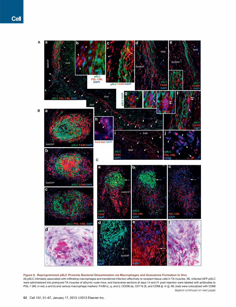
Figure 6. Reprogrammed pSLC Promote Bacterial Dissemination via Macrophages and Granuloma Formation In Vivo
(A) pSLC intimately associated with infiltrating macrophages and transferred infection effectively to recipient tissue cells in TA muscles. ML-infected GFP-pSLC
were administered into preinjured TA muscles of athymic nude mice, and transverse sections at days 14 and 21 post-injection were labeled with antibodies to
PGL-1 (ML in red; a and b) and various macrophage markers: F4/80 (c, g, and i), CD206 (e), CD11b (f), and CD68 (j). In (j), ML (red) were colocalized with CD68
(legend continued on next page)
62 Cell 152, 51–67, January 17, 2013 ª2013 Elsevier Inc.
ML-infected cells upregulated several Hox and key EMT-associ-
ated TFs, including themaster regulators of EMT, Twist and Snail
(Polyak andWeinberg, 2009; Mani et al., 2008). Demethylation of
the promoter region of Twist1 in reprogrammed cells further
suggests that the change in cell fate accompanies the change
in epigenetic status. Reprogramming that follows the silencing
of Sox10, the master regulator of Schwann cell lineage and
differentiation/myelination (Finzsch et al., 2010; Jessen and Mir-
sky, 2005), directly correlates with significant DNAmethylation of
Sox10 in pSLC. On the other hand, no change in DNA methyla-
tion in the Sox2 promoter region (data not shown) suggests the
continuous expression of Sox2 in both parent Schwann cells
and pSLC. Thus, the silencing of Sox10 and maintaining or
induction of Sox2 may be critical for Schwann cell reprogram-
ming during infection.
In contrast to Sox2+/Sox10�pSLC, nerve injury-induceddedif-
ferentiated Schwann cells maintain Sox2+/Sox10+ and other
Schwann cell markers (Le et al., 2005). Safeguarding the
Schwann cell lineage markers, particularly Sox10, is critical for
an effective differentiation towards myelination during the
peripheral nerve regeneration process (Le et al., 2005; Finzsch
et al., 2010). This phenotypic difference together with the acqui-
sition of numerous mesoderm developmental genes distin-
guishes pSLC from nerve injury-induced dedifferentiated
Schwann cells. On the other hand, our previous studies have
shown that the initial interaction of extracellular ML with myelin-
ated Schwann cells induces demyelination, which in turn gener-
ates similar dedifferentiated cells (Rambukkana et al., 1997,
1998, 2002; Tapinos et al., 2006). ML appear to use this demye-
lination strategy to generate dedifferentiated Schwann cells as
they are highly susceptible to invasion and favorable for bacterial
colonization (Rambukkana, 2010). In the present study, we
mimicked such conditions by directly isolating dedifferentiated
Schwann cells from adult mouse peripheral nerves and showed
that, once invaded, iML reprogram these Schwann cells to
pSLC. It remains to be determined which mechanisms or
signaling are involved in this bacterial-driven cell reprogramming.
The important role of Erk1/2 MAPK signaling in inducing
demyelination without immune responses or lesions was first
demonstrated with leprosy bacteria as a model (Tapinos et al.,
2006). This finding was recently confirmed with inducible Raf-
kinase transgenic mice in which Erk1/2 activation in myelinated
Schwann cells was shown to induce demyelination and inflam-
matory responses (Napoli et al., 2012). Interestingly, the latter
activates p75, whereas reprogramming Schwann cells to pSLC
results in the loss of p75 (Figure 2). Also, unlike Erk1/2-induced
demyelination where transiently appearing inflammatory cells
promote peripheral nerve regeneration, pSLC acquired sustain-
able secretion of numerous macrophage chemoattractants even
after complete removal of ML. This capacity may facilitate con-
tinuous attraction of macrophages to pSLC in many tissues
during the early dissemination process. Although ML use Erk1/
2 signaling for Schwann cell manipulation (Tapinos and Rambuk-
kana, 2005; Tapinos et al., 2006), Erk1/2 alone does not appear
to contribute to reprogramming as the pharmacological in-
hibition of Erk1/2 did not abrogate the reprogramming events
(data not shown). This further suggests that the activation of
Erk1/2 alone does not cause Schwann cell reprogramming.
Although the lineage through which infected Schwann cells
are converted to pSLC is not known, this change may have
important benefits for a bacterium like ML that depends totally
on host cell functions for survival (Cole et al., 2001). Conversion
of Schwann cells to pSLC with mesenchymal characteristics
sets the stage for ML to use the reprogrammed cells as a vehicle
to spread the infection to distal tissues such as skeletal and
smooth muscles. We propose two major mechanisms by which
ML may use reprogrammed cells to promote bacterial dissemi-
nation (Figure 7H). First, ML take advantage of mesenchymal
stem cell-like properties of pSLC to migrate and spontaneously
differentiate into skeletal and smooth muscles under inflamma-
tory conditions and thus passively transmit infection to these
tissues. Second, using immunomodulatory properties of pSLC,
ML may use reprogrammed cells to create a secondary niche
by recruiting macrophages for further bacterial expansion and
dissemination.
Importantly, our findings in vivo showed that pSLC contribute
to macrophage granuloma formation, one of the pathologic
hallmarks of mycobacterial infections in murine models and
in patients with both leprosy and tuberculosis (Modlin and
Rea, 1988; Flynn and Chan, 2001). Although mycobacterial
granulomas are considered to be essential for containment of
infection, recent studies in zebrafish have suggested that macro-
phage granulomas may also promote mycobacterial dissemina-
tion during early infection (Davis and Ramakrishnan, 2009).
However, unlike other pathogenic mycobacteria, ML use adult
Schwann cells as primary nonimmune tissue cells for initial colo-
nization (Stoner, 1979). Once colonized, ML take full advantage
of Schwann cell plasticity to convert infected cells to pSLC
(blue; arrowheads). Endogenous non-GFP tissue cells that have taken up ML are shown with arrowheads (a) and are predominantly comprised of infiltrated
macrophages of M1 and M2 phenotypes (e, i, and j). ML localization is confirmed by acid-fast staining (red) in parallel sections (h). Insets in (e), (f), and (g) (arrows
and arrowheads) show the formation of early smaller macrophage GLS. SkM: skeletal muscles (asterisks); SkMDIP: skeletal muscle-dermal interphase.
Magnifications: (a, d, e, f, and I) 103; (b, c, g, and h) 403.
(B) pSLC contribute to the formation of typical macrophage GLS in SkMDIP. Migrated pSLC formed organized GLS with M2 and M1 macrophages that
show distinct distribution and morphological features, as analyzed by antibodies to F4/80 (a) or CD206 (see also Figure S7) and CD68 (b), respectively. The
distribution of ML (red) within GLS was localized in parallel sections with antibody to PGL-1 (c) and by Fite’s staining (in purple), which detects mycobacteria in
infected tissues (d; also see Figure S7). Note the strongly F4/80+ cells in the periphery and fused and multinucleated CD68+ cells in the core of GLS. Magnifi-
cations are 203.
(C) Disintegration and release of bacterial-laden macrophages from granulomas. Sections distant from the granulomas (as in B) within the same tissues show
disintegration of both CD68 (a) and CD206 (e) macrophages from tapered GFP+ pSLC aggregates (a–c) and the emigrating macrophages carrying large numbers
ofML as detected by anti-PGL-1 (red; b and c) and Fite’s staining (f). Arrows in the top and bottom panels show the pSLCwithmuch fewer iML and high content of
acid-fast+ ML in migrating macrophages, respectively. Magnifications: (a–c) 103; (d–f) 403.
See also Figures S5, S6, and S7.
Cell 152, 51–67, January 17, 2013 ª2013 Elsevier Inc. 63
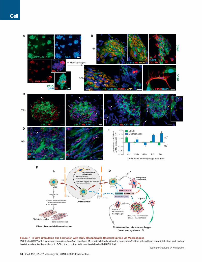
Figure 7. In Vitro Granuloma-like Formation with pSLC Recapitulates Bacterial Spread via Macrophages
(A) Infected GFP+ pSLC form aggregates in culture (top panel) andML confined strictly within the aggregates (bottom left) and form bacterial clusters (red; bottom
insets), as detected by antibody to PGL-1 (red; bottom left), counterstained with DAPI (blue).
(legend continued on next page)
64 Cell 152, 51–67, January 17, 2013 ª2013 Elsevier Inc.
with the capacity to produce chemoattractants and trophic
factors,which in turnpromotemacrophage recruitment, bacterial
transfer, and survival of infected macrophages (Figures 5, 6, and
7H). Interestingly, some of the immune factors/chemokines
released from pSLC are also known to foster granuloma forma-
tion (Qiu et al., 2001; Chiu et al., 2004). Collectively, these events
may facilitate pSLC to recruit macrophages and contribute to
form GLS. Our in vivo and in vitro analyses provide further
evidence that ML-laden M1 and M2 macrophages in the granu-
lomas contributed to spread the infection. Recent work showing
that the viruses used for reprogramming skin fibroblasts to plurip-
otent stem cells (Takahashi and Yamanaka, 2006) can promote
theefficiencyof nuclear reprogrammingvia activationof common
microbial-induced innate immune responses (Lee et al., 2012)
conceptually supports our study. Finally, our findings may have
far-reaching implications not only for host-pathogen interactions
but also for understanding the basic biology of adult-tissue cell
plasticity, reprogramming, and tissue regeneration.
EXPERIMENTAL PROCEDURES
Primary Adult Schwann Cell Cultures
Detailed experimental protocols for isolation and characterization of
mouse primary Schwann cells are described in the Extended Experimental
Procedures.
Infection with ML
Schwann cells were infected with ML according to our previous protocol (Ng
et al., 2000; Extended Experimental Procedures). In vivo-grown ML derived
from nude-mouse footpads were prepared as described previously (Truman
and Krahenbuhl, 2001; Lahiri et al., 2010), and both viable and nonviable ML
were provided by the Laboratory Research Branch of NHDP (Baton Rouge,
LA, USA).
Mice
These studies employed 4- to 6-week-old young adult CD-1 (ICR) (strain code:
022) mice (Charles-River Laboratory), GFP mice that constitutively express
eGFP (strain: C57BL/6-Tg [ACTB-EGFP] 1Osb/J, stock: 003291; Jackson
lab [Bar Harbor, ME, USA]), 6- to 8-week-old CD-1/nude mice (Charles-River),
and 4- to 6-week-old NOD-SCID mice (Jackson Lab). Sox2/GFP mice (Eminli
et al., 2008) were kindly provided by R. Jaenisch (Whitehead Institute, MIT,
Cambridge, MA, USA). Animals were maintained at the Rockefeller University
and University of Edinburgh animal facilities according to institutionally
approved protocols.
Flow Cytometry, Immunolabeling, Antibodies, and Microscopy
Flow cytometry, primary Schwann cell sorting, generation of clonal cells,
immunolabeling with primary antibodies, and imaging (Confocal, Deltavision,
and Time-lapse live-cell imaging) are described in the Extended Experimental
Procedures. Antibodies to Sox10, ML PGL-1, and Oct6 were gifts from
M. Wagner, A. Kolk, and D. Meijer respectively. A list of other antibodies
used is available in Table S2.
Schwann Cell Infection with ML and Reprogramming Protocol
FACS-sorted p75+ Schwann cells or clonal Schwann cells generated from
wild-type mice or Sox2-GFP mice were infected with ML as described (Ng
et al., 2000; Tapinos and Rambukkana, 2005). For the isolation of reprog-
rammed cells, infected cells were transferred to mesenchymal stem cell
media that allowed the selection of a pSLC population based on the cell-fate
change from p75+/Sox10+ cells to p75�/Sox10�. These cells were FACS
sorted with p75 antibody and subjected to detailed characterization (Extended
Experimental Procedures). pSLC were also transduced with CopGFP-CDH-
MSCV-cG reporter vector (System Biosciences, CA, USA) to obtain stable
CopGFP-expressing pSLC (referred to as GFP+ pSLC).
Gene and Protein Expression Analyses
Microarray, protein array, and qPCR analyses are detailed in the Extended
Experimental Procedures and Table S3.
pSLC Differentiation to Mesenchymal Tissues
We employed established protocols for bone and adipocyte differentiation
from mesenchymal stem cells (Pittenger et al., 1999). Myogenic differentiation
with C2C12 myoblasts was performed as described (Shi et al., 2004). Detailed
descriptions are available in the Extended Experimental Procedures.
In Vivo GFP+ pSLC Transplantation to Skeletal Muscles and
Dissemination of Infections
CD-1/nude mice (6–8 weeks, Charles-River) were preinjured with cardio-
toxin-1 24 hr prior to the administration of ML-infected GFP+ pSLC or GFP+
Schwann cells. Each indicated experiment was performed with 5–6 mice per
experimental group by repeating at least 3–4 times. Experimental details are
in the Extended Experimental Procedures.
Macrophage-pSLC Cocultures, Migration Assays, and In Vitro
Granuloma Formation
Detailed descriptions of experimental procedures of macrophage isolation
and cocultures and in vitro granuloma formation are described in the Extended
Experimental Procedures.
SUPPLEMENTAL INFORMATION
Supplemental Information includes Extended Experimental Procedures, seven
figures, three tables, and one data file and can be found with this article online
at http://dx.doi.org/10.1016/j.cell.2012.12.014.
ACKNOWLEDGMENTS
We thank IanWilmut and Emil Gotschlich for critical reading of the manuscript.
We are grateful to Emil Gotschlich, Paul Nurse, Michael Young, Vincent Fi-
schetti, Thomas Sakmar (Rockefeller University), John Savill, Jonathan Sackl,
Nick Hastie (University of Edinburgh), and James Krahenbuhl (National Han-
sen’s Disease Programs; NHDP) for their support during the initial and final
phases of this study. Our thanks to Richard Truman (NHDP) for providing Fite’s
(B–E) Macrophages were added to infected pSLC aggregates, incubated for 6 hr, 18 hr (B), 72 hr (C), and 96 hr (D), and labeled with antibodies to PGL-1 and
macrophage markers F4/80 and CD11b. Shown are confocal images after 3D reconstructions (B); arrows (top panel) indicate phagocytosed ML-infected pSLC
(green; ML in red) by macrophages (blue) within GLS. At 18 hr, a high number of ML (green) was found in macrophages (blue) that penetrated into GLS (B, bottom
left panels). Occasional TUNEL+ cells (red; 0.04%) were also found in these GLS (nuclei are shown in yellow). Bacterial transfer from GFP+ pSLC to F4/80+
macrophages (red) was also seen outside GLS (B, bottom right). (C) At 72 hr, more macrophages (blue) were incorporated into GLS, and ML were taken up by
these macrophages (arrows); note less PGL+ML in pSLC (left and middle panels) and departure of some macrophages carrying ML with them; yellow arrows
depict macrophages carrying both pSLC debries and ML (right panels). (D) Disintegration of GLS and the release of ML-laden macrophages (arrows).
(E) Correlation coefficients of bacterial transfer from pSLC to macrophages over time based on bacterial presence or absence (+/�ML) in pSCL (green) and in
macrophages (blue).
(F) The proposed model: Schwann cells in the adult PNS infected with ML undergo a reprogramming process that converts Schwann cells to pSLC that promote
bacterial dissemination. BNB: blood nerve barrier.
Scale bars, (A) 5 mm; (B–D) 20 mm.
Cell 152, 51–67, January 17, 2013 ª2013 Elsevier Inc. 65
staining of infectedmouse tissue sections; members of the core facilities at the
Rockefeller University and the University of Edinburgh for their invaluable
assistance; Joe Dybaas, Jennifer Smith, Clara Eastby, Paola Basilico, and
Maxmilien Grandclaudon (Rambukkana laboratory) for their participation and
technical assistance; and members of Genome Exploration and EpigenDx
for technical assistance and data analysis. Mycobacterium leprae were
provided by The Laboratory Research Branch of NHDP, Baton Rouge, LA
with funding from The American Leprosy Missions and the Hospitaler Order
of St. Lazarus of Jerusalem. This work was funded in part by grants fromNIAID
(AI45816) and NINDS (NS45187), The Order of MALTALEP Foundation, The
Rockefeller University, and the University of Edinburgh.
Received: July 9, 2012
Revised: October 31, 2012
Accepted: December 10, 2012
Published: January 17, 2013
REFERENCES
Bold, T.D., and Ernst, J.D. (2009). Who benefits from granulomas, mycobacte-
ria or host? Cell 136, 17–19.
Buatois, V., Fagete, S., Magistrelli, G., Chatel, L., Fischer, N., Kosco-Vilbois,
M.H., and Ferlin, W.G. (2010). Pan-CC chemokine neutralization restricts sple-
nocyte egress and reduces inflammation in a model of arthritis. J. Immunol.
185, 2544–2554.
Charge, S.B., and Rudnicki, M.A. (2004). Cellular and molecular regulation of
muscle regeneration. Physiol. Rev. 84, 209–238.
Chiu, B.C., Freeman, C.M., Stolberg, V.R., Hu, J.S., Komuniecki, E., and Chen-
sue, S.W. (2004). The innate pulmonary granuloma: characterization and
demonstration of dendritic cell recruitment and function. Am. J. Pathol. 164,
1021–1030.
Cole, S.T., Eiglmeier, K., Parkhill, J., James, K.D., Thomson, N.R., Wheeler,
P.R., Honore, N., Garnier, T., Churcher, C., Harris, D., et al. (2001). Massive
gene decay in the leprosy bacillus. Nature 409, 1007–1011.
Davis, J.M., and Ramakrishnan, L. (2009). The role of the granuloma in expan-
sion and dissemination of early tuberculous infection. Cell 136, 37–49.
Eminli, S., Utikal, J., Arnold, K., Jaenisch, R., and Hochedlinger, K. (2008).
Reprogramming of neural progenitor cells into induced pluripotent stem cells
in the absence of exogenous Sox2 expression. Stem Cells 26, 2467–2474.
Falkow, S. (1991). Bacterial entry into eukaryotic cells. Cell 65, 1099–1102.
Fawcett, J.W., and Keynes, R.J. (1990). Peripheral nerve regeneration. Annu.
Rev. Neurosci. 13, 43–60.
Finzsch, M., Schreiner, S., Kichko, T., Reeh, P., Tamm, E.R., Bosl, M.R.,
Meijer, D., and Wegner, M. (2010). Sox10 is required for Schwann cell identity
and progression beyond the immature Schwann cell stage. J. Cell Biol. 189,
701–712.
Flynn, J.L., and Chan, J. (2001). Immunology of tuberculosis. Annu. Rev.
Immunol. 19, 93–129.
Gupta, J.C., Jesupadam, T., Gupta, M.C., andGupta, D.K. (1975). A histopath-
ologic study of striated muscle biopsies in leprosy. Int. J. Lepr. Other Myco-
bact. Dis. 43, 348–355.
Gurdon, J.B. (1962). Adult frogs derived from the nuclei of single somatic cells.
Dev. Biol. 4, 256–273.
Gurdon, J.B., and Melton, D.A. (2008). Nuclear reprogramming in cells.
Science 322, 1811–1815.
Hoogduijn, M.J., Popp, F., Verbeek, R., Masoodi, M., Nicolaou, A., Baan, C.,
and Dahlke, M.H. (2010). The immunomodulatory properties of mesenchymal
stem cells and their use for immunotherapy. Int. Immunopharmacol. 10, 1496–
1500.
Jessen, K.R., andMirsky, R. (2005). The origin and development of glial cells in
peripheral nerves. Nat. Rev. Neurosci. 6, 671–682.
Job, C.K. (1989). Nerve damage in leprosy. Int. J. Lepr. Other Mycobact. Dis.
57, 532–539.
Kaur, S., Malik, A.K., and Kumar, B. (1981). Pathologic changes in striated
muscles in leprosy. Lepr. India 53, 52–56.
Klopp, A.H., Gupta, A., Spaeth, E., Andreeff, M., and Marini, F., 3rd. (2011).
Concise review: Dissecting a discrepancy in the literature: do mesenchymal
stem cells support or suppress tumor growth? Stem Cells 29, 11–19.
Lahiri, R., Randhawa, B., and Krahenbuhl, J.L. (2010). Infection of mouse
macrophages with viable Mycobacterium leprae does not induce apoptosis.
J. Infect. Dis. 201, 1736–1742.
Le, N., Nagarajan, R., Wang, J.Y., Araki, T., Schmidt, R.E., and Milbrandt, J.
(2005). Analysis of congenital hypomyelinating Egr2Lo/Lo nerves identifies
Sox2 as an inhibitor of Schwann cell differentiation and myelination. Proc.
Natl. Acad. Sci. USA 102, 2596–2601.
Lee, J., Sayed, N., Hunter, A., Au, K.F.,Wong,W.H., Mocarski, E.S., Pera, R.R.,
Yakubov, E., and Cooke, J.P. (2012). Activation of innate immunity is required
for efficient nuclear reprogramming. Cell 151, 547–558.
Li, Y., Chen, S., Yuan, J., Yang, Y., Li, J., Ma, J., Wu, X., Freund, M., Pollok, K.,
Hanenberg, H., et al. (2009). Mesenchymal stem/progenitor cells promote the
reconstitution of exogenous hematopoietic stem cells in Fancg-/- mice in vivo.
Blood 113, 2342–2351.
Mani, S.A., Guo, W., Liao, M.J., Eaton, E.N., Ayyanan, A., Zhou, A.Y., Brooks,
M., Reinhard, F., Zhang, C.C., Shipitsin, M., et al. (2008). The epithelial-
mesenchymal transition generates cells with properties of stem cells. Cell
133, 704–715.
Miko, T.L., Le Maitre, C., and Kinfu, Y. (1993). Damage and regeneration of
peripheral nerves in advanced treated leprosy. Lancet 342, 521–525.
Modlin, R.L., and Rea, T.H. (1988). Immunopathology of leprosy granulomas.
Springer Semin. Immunopathol. 10, 359–374.
Mosser, D.M., and Edwards, J.P. (2008). Exploring the full spectrum of macro-
phage activation. Nat. Rev. Immunol. 8, 958–969.
Napoli, I., Noon, L.A., Ribeiro, S., Kerai, A.P., Parrinello, S., Rosenberg, L.H.,
Collins, M.J., Harrisingh, M.C., White, I.J., Woodhoo, A., and Lloyd, A.C.
(2012). A central role for the ERK-signaling pathway in controlling
Schwann cell plasticity and peripheral nerve regeneration in vivo. Neuron 73,
729–742.
Ng, V., Zanazzi, G., Timpl, R., Talts, J.F., Salzer, J.L., Brennan, P.J., and Ram-
bukkana, A. (2000). Role of the cell wall phenolic glycolipid-1 in the peripheral
nerve predilection of Mycobacterium leprae. Cell 103, 511–524.
Pearson, J.M., Rees, R.J., and Weddell, A.G. (1970). Mycobacterium leprae in
the striated muscle of patients with leprosy. Lepr. Rev. 41, 155–166.
Pittenger, M.F., Mackay, A.M., Beck, S.C., Jaiswal, R.K., Douglas, R., Mosca,
J.D., Moorman, M.A., Simonetti, D.W., Craig, S., and Marshak, D.R. (1999).
Multilineage potential of adult human mesenchymal stem cells. Science 284,
143–147.
Polyak, K., and Weinberg, R.A. (2009). Transitions between epithelial and
mesenchymal states: acquisition of malignant and stem cell traits. Nat. Rev.
Cancer 9, 265–273.
Qiu, B., Frait, K.A., Reich, F., Komuniecki, E., and Chensue, S.W. (2001).
Chemokine expression dynamics in mycobacterial (type-1) and schistosomal
(type-2) antigen-elicited pulmonary granuloma formation. Am. J. Pathol. 158,
1503–1515.
Rambukkana, A. (2010). Usage of signaling in neurodegeneration and
regeneration of peripheral nerves by leprosy bacteria. Prog. Neurobiol. 91,
102–107.
Rambukkana, A., Salzer, J.L., Yurchenco, P.D., and Tuomanen, E.I. (1997).
Neural targeting of Mycobacterium leprae mediated by the G domain of the
laminin-alpha2 chain. Cell 88, 811–821.
Rambukkana, A., Yamada, H., Zanazzi, G., Mathus, T., Salzer, J.L.,
Yurchenco, P.D., Campbell, K.P., and Fischetti, V.A. (1998). Role of alpha-
dystroglycan as a Schwann cell receptor for Mycobacterium leprae. Science
282, 2076–2079.
Rambukkana, A., Zanazzi, G., Tapinos, N., and Salzer, J.L. (2002). Contact-
dependent demyelination by Mycobacterium leprae in the absence of immune
cells. Science 296, 927–931.
66 Cell 152, 51–67, January 17, 2013 ª2013 Elsevier Inc.
Scollard, D.M., Adams, L.B., Gillis, T.P., Krahenbuhl, J.L., Truman, R.W., and
Williams, D.L. (2006). The continuing challenges of leprosy. Clin. Microbiol.
Rev. 19, 338–381.
Shetty, V.P., Antia, N.H., and Jacobs, J.M. (1988). The pathology of early
leprous neuropathy. J. Neurol. Sci. 88, 115–131.
Shi, D., Reinecke, H., Murry, C.E., and Torok-Storb, B. (2004). Myogenic fusion
of human bone marrow stromal cells, but not hematopoietic cells. Blood 104,
290–294.
Stoner, G.L. (1979). Importance of the neural predilection of Mycobacterium
leprae in leprosy. Lancet 2, 994–996.
Takahashi, K., and Yamanaka, S. (2006). Induction of pluripotent stem cells
from mouse embryonic and adult fibroblast cultures by defined factors. Cell
126, 663–676.
Tapinos, N., and Rambukkana, A. (2005). Insights into regulation of human
Schwann cell proliferation by Erk1/2 via a MEK-independent and p56Lck-
dependent pathway from leprosy bacilli. Proc. Natl. Acad. Sci. USA 102,
9188–9193.
Tapinos, N., Ohnishi, M., and Rambukkana, A. (2006). ErbB2 receptor tyrosine
kinase signaling mediates early demyelination induced by leprosy bacilli. Nat.
Med. 12, 961–966.
Theise, N.D., andWilmut, I. (2003). Cell plasticity: flexible arrangement. Nature
425, 21.
Tidball, J.G., and Villalta, S.A. (2010). Regulatory interactions between muscle
and the immune system during muscle regeneration. Am. J. Physiol. Regul.
Integr. Comp. Physiol. 298, R1173–R1187.
Truman, R.W., and Krahenbuhl, J.L. (2001). Viable M. leprae as a research
reagent. Int. J. Lepr. Other Mycobact. Dis. 69, 1–12.
Weider, M., Kuspert, M., Bischof, M., Vogl, M.R., Hornig, J., Loy, K., Kosian, T.,
Muller, J., Hillgartner, S., Tamm, E.R., et al. (2012). Chromatin-remodeling
factor Brg1 is required for Schwann cell differentiation and myelination. Dev.
Cell 23, 193–201.
Werneck, L.C., Teive, H.A., and Scola, R.H. (1999). Muscle involvement in
leprosy. Study of the anterior tibial muscle in 40 patients. Arq. Neuropsiquiatr.
57(3B), 723–734.
Wilmut, I., Schnieke, A.E., McWhir, J., Kind, A.J., and Campbell, K.H. (1997).
Viable offspring derived from fetal and adult mammalian cells. Nature 385,
810–813.
Cell 152, 51–67, January 17, 2013 ª2013 Elsevier Inc. 67