Cell Membranes. Objectives Describe the function of the plasma membrane Describe the fluid mosaic...
47
Cell Membranes
-
Upload
ralf-mclaughlin -
Category
Documents
-
view
225 -
download
3
Transcript of Cell Membranes. Objectives Describe the function of the plasma membrane Describe the fluid mosaic...
Cell Membranes
Objectives
• Describe the function of the plasma membrane • Describe the fluid mosaic model of membrane
structure • Explain how hydrophobic interactions determine
membrane structure and function • Describe how proteins are arranged in
membranes and how they contribute to membrane functioning
Objectives Cont.
• Describe those factors which affect membrane permeability
• Define diffusion; explain what causes it and why it's a spontaneous process
• Explain passive transport and the factors that regulate its rate
• Explain why a concentration gradient across a membrane represents potential energy

Objectives Cont.
• Define osmosis and be able to predict the direction of water movement based upon differences in solute concentration
• Explain how transport proteins are similar to enzymes
• Provide a general description of the process of facilitated diffusion
• Explain how active transport differs from diffusion
Objectives Cont.
• Explain the mechanisms by which a membrane potential or electrochemical gradient may be established
• Explain (in very general terms) how potential energy stored as electrochemical gradients is used to transport substances across the membrane
• Explain how very large molecules are transported across the cell membrane
Membranes
1. Fluid Mosaic model of membrane construction
2. Behavior of phospolipids in H2O is due to their amphipathic structure and tendency to form bi-layers
3. Phospholipids have fluid like behavior a. Fluid portion of fluid mosaic model
b. Phospholipids and proteins can (and do) move side to side
c. Flip-flopping is possible, though occurs rarely
d. Demonstrated by mouse/human hybrid membrane
4. Tend to form vesicles rather than free ends
a. Can reseal to form intact membranes
b. Allows membranes to fuse with other membranes1) Responsible for endo- and exocytosis
5. Selectively permeable
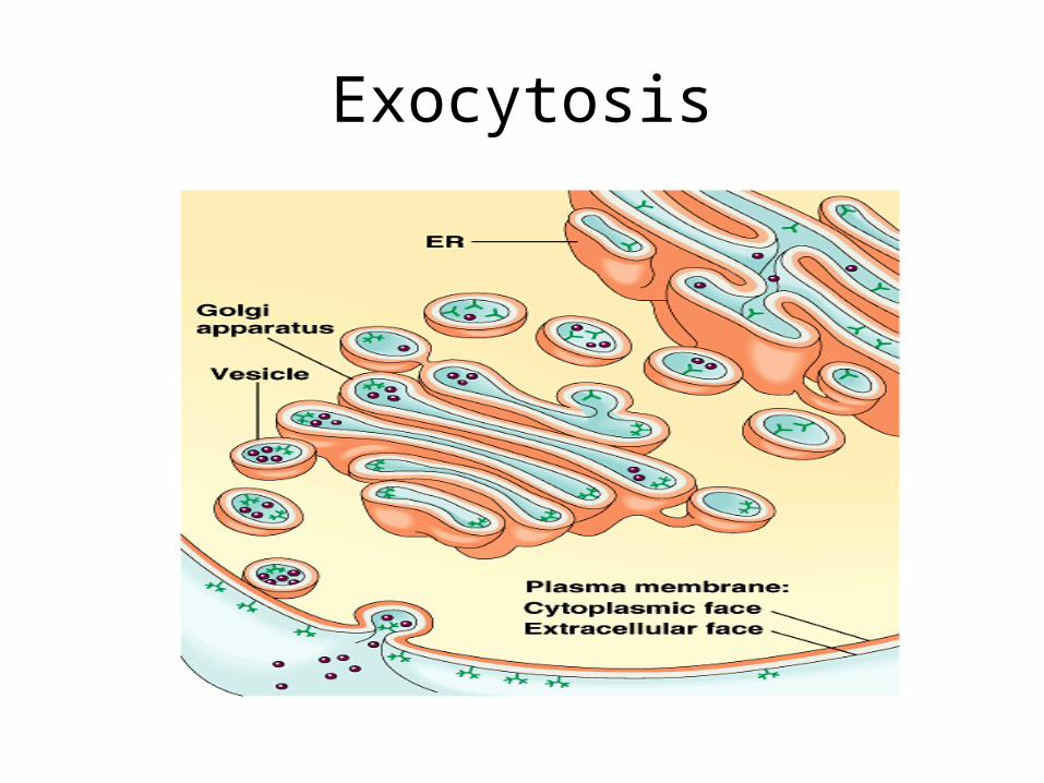
Exocytosis
Membrane Proteins
1. Proteins provide specialization2. Two types: Peripheral and integral
a. Peripheral proteins are those that are easily removed (held by non-covalent linkages)
b. Integral proteins are tightly linked1) May be buried2) May completely penetrate a bi-layer (ex. Aquaporins allow
H2O to move through freely)
3. The two faces of the membrane (inner and outer) are asymmetrical with respect to their protein composition

Membrane Protein Functions
1. Cell adhesion
2. Protein channelsa. Channels to allow certain molecules through
b. eg. aquaporins
3. Transport proteinsa. Move molecules from one side to the other
b. Requires energy
c. eg. glucose

4. Signal receptor proteins
5. Attachment site for cytoskeleton and soluble enzymes
6. ATPase active transport pumpsa. Moves against a concentration gradient
b. Requires energy
c. eg. Sodium-potassium pump (Na+-K+ pump)
7. Enzymes

• The proteins in the plasma membrane may provide a variety of major cell functions.
Fig. 8.9

Other Membrane Components
1. Carbohydratesa. Short, 15 or fewer sugar units (oligosaccharide)b. Can covalently bond to membrane components (glyco means
presence of carbohydrate)1) Lipids= glycolipids2) Proteins=glycoproteins
2. Cell “markers”a. Allow for cell recognition
1) Necessary for sorting cells into tissues and organs in embryos2) Basis for rejection of foreign cells
3. Cholesterola. Reduces membrane fluidity as temp increasesb. Increases membrane fluidity as temp decreases
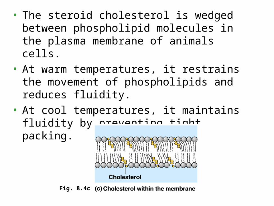
• The steroid cholesterol is wedged between phospholipid molecules in the plasma membrane of animals cells.
• At warm temperatures, it restrains the movement of phospholipids and reduces fluidity.
• At cool temperatures, it maintains fluidity by preventing tight packing.
Fig. 8.4c

• To work properly with active enzymes and appropriate permeability, membrane must be fluid, about as fluid as salad oil.
• Cells can alter the lipid composition of membranes to compensate for changes in fluidity caused by changing temperatures.– For example, cold-adapted organisms, such as
winter wheat, increase the percentage of unsaturated phospholipids in the autumn.
– This allows these organisms to prevent their membranes from solidifying during winter.

Activity
• Each Table, make your own diagram of a cell membrane
• Be sure to include all of the components of the cell membrane and explain the function of each component

Copyright © 2002 Pearson Education, Inc., publishing as Benjamin Cummings
Fig. 8.6
Movement Across Membranes
1. A membrane’s composition results in selective permeability
a. small molecules and ions moves across the plasma membrane in both directions
1) For example, sugars, amino acids, and other nutrients enter a muscle cell and metabolic waste products leave
2) The cell absorbs oxygen and expels carbon dioxide3) It also regulates concentrations of inorganic ions, like Na+,
K+, Ca2+, and Cl-, by shuttling them across the membrane
2. However, substances do not move across the barrier indiscriminately; membranes are selectively permeable
Permeability
1. Permeability depends on the interaction of a molecule with the hydrophobic core of the membrane.
a. Hydrophobic molecules, can dissolve in the lipid bilayer and cross easily
1) hydrocarbons2) CO23) O2
b. Ions and polar molecules pass through with difficulty.1) small molecules, like water, and larger critical molecules,
like glucose and other sugars.2) Ions, whether atoms or molecules, also have difficulties
penetrating the hydrophobic core. c. Proteins can assist and regulate the transport of ions and
polar molecules.
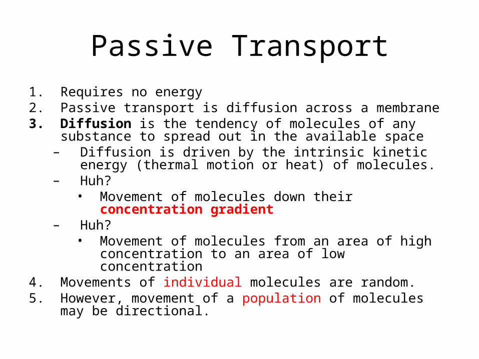
Passive Transport1. Requires no energy2. Passive transport is diffusion across a membrane3. Diffusion is the tendency of molecules of any substance to
spread out in the available space– Diffusion is driven by the intrinsic kinetic energy (thermal
motion or heat) of molecules. – Huh?
• Movement of molecules down their concentration gradient
– Huh?• Movement of molecules from an area of high concentration
to an area of low concentration4. Movements of individual molecules are random.5. However, movement of a population of molecules may be
directional.

• Diffusion is passive transport because it requires no energy from the cell to make it happen
• Diffusion is the movement of molecules down their concentration gradient– The concentration gradient represents potential
energy and drives diffusion.• However, because membranes are selectively
permeable, the interactions of the molecules with the membrane play a role in the diffusion rate
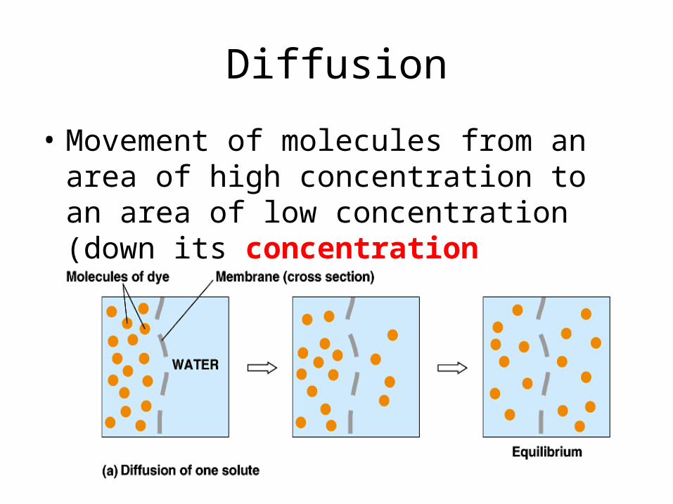
Diffusion
• Movement of molecules from an area of high concentration to an area of low concentration (down its concentration gradient)
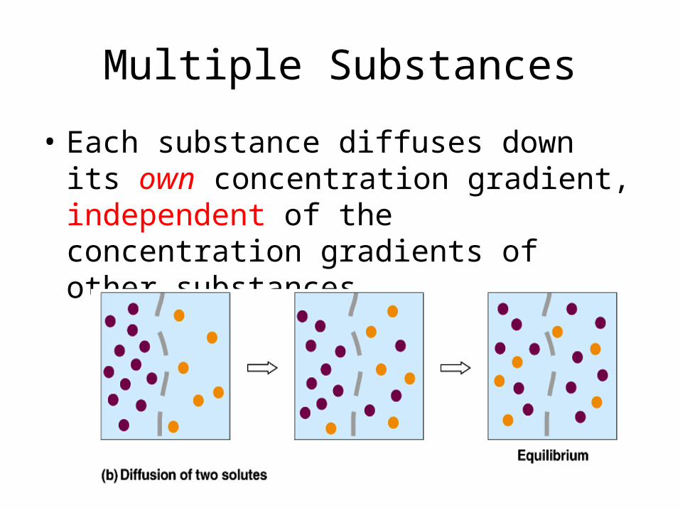
Multiple Substances
• Each substance diffuses down its own concentration gradient, independent of the concentration gradients of other substances
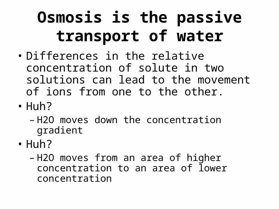
Osmosis is the passive transport of water
• Differences in the relative concentration of solute in two solutions can lead to the movement of ions from one to the other.
• Huh?– H2O moves down the concentration gradient
• Huh?– H2O moves from an area of higher
concentration to an area of lower concentration
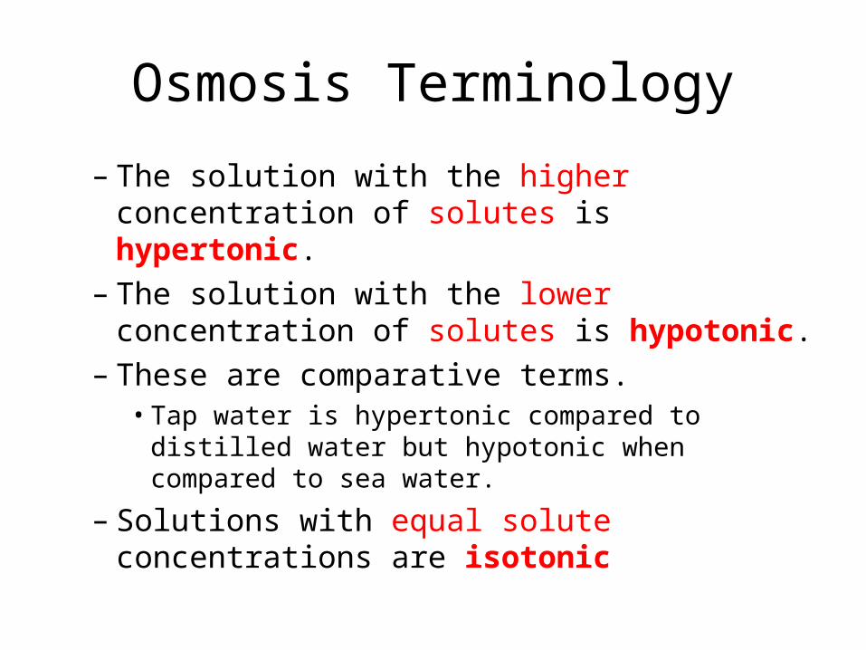
Osmosis Terminology
– The solution with the higher concentration of solutes is hypertonic.
– The solution with the lower concentration of solutes is hypotonic.
– These are comparative terms.• Tap water is hypertonic compared to distilled water
but hypotonic when compared to sea water.
– Solutions with equal solute concentrations are isotonic

• Imagine that two sugar solutions differing in concentration are separated by a membrane that will allow water through, but not sugar.
• The hypertonic solution has a lower water concentration than the hypotonic solution.– More of the water molecules in the hypertonic
solution are bound up in hydration shells around the sugar molecules, leaving fewer unbound water molecules.

• Unbound water molecules will move from the hypotonic solution where they are abundant to the hypertonic solution where they are rarer.
• This diffusion of water across a selectively permeable membrane is a special case of passive transport called osmosis.
• Osmosis continues until the solutions are isotonic.
Copyright © 2002 Pearson Education, Inc., publishing as Benjamin Cummings
Fig. 8.11
This is Important!
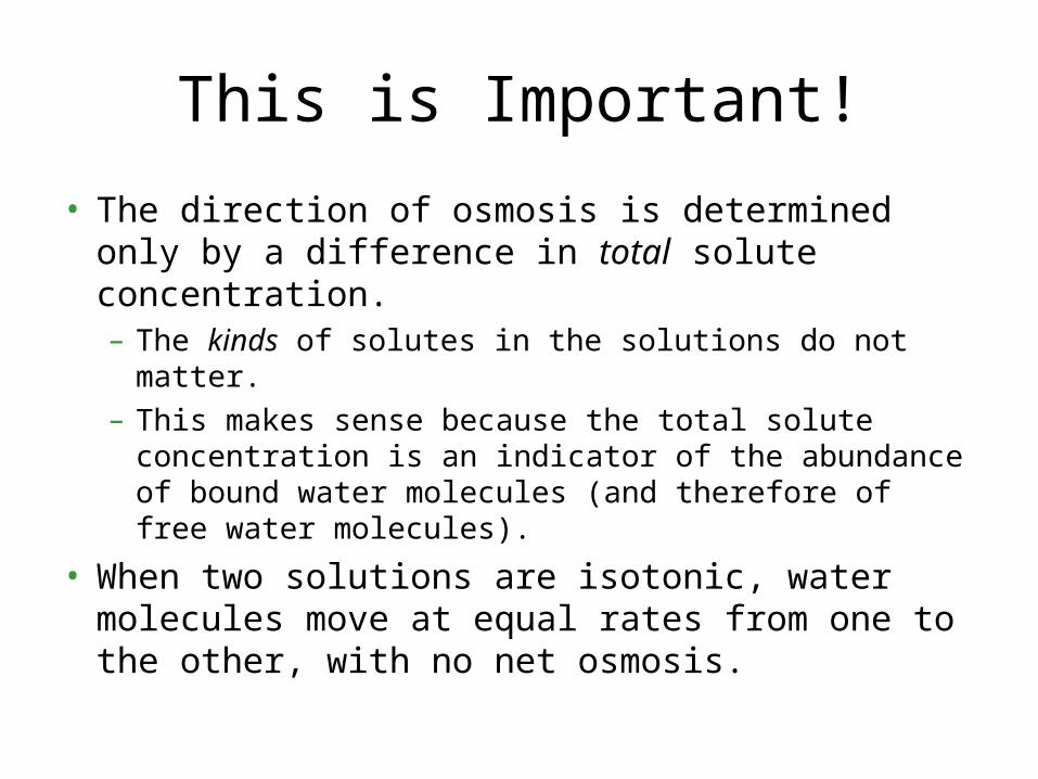
• The direction of osmosis is determined only by a difference in total solute concentration.– The kinds of solutes in the solutions do not matter.– This makes sense because the total solute
concentration is an indicator of the abundance of bound water molecules (and therefore of free water molecules).
• When two solutions are isotonic, water molecules move at equal rates from one to the other, with no net osmosis.

Cell survival depends on balancing water uptake and loss
• An animal cell immersed in an isotonic environment experiences no net movement of water across its plasma membrane.– Water flows across the membrane, but at the
same rate in both directions.– The volume of the cell is stable.

• The same cell is a hypertonic environment will loose water, shrivel, and probably die.
• A cell in a hypotonic solution will gain water, swell, and burst.
Fig. 8.12
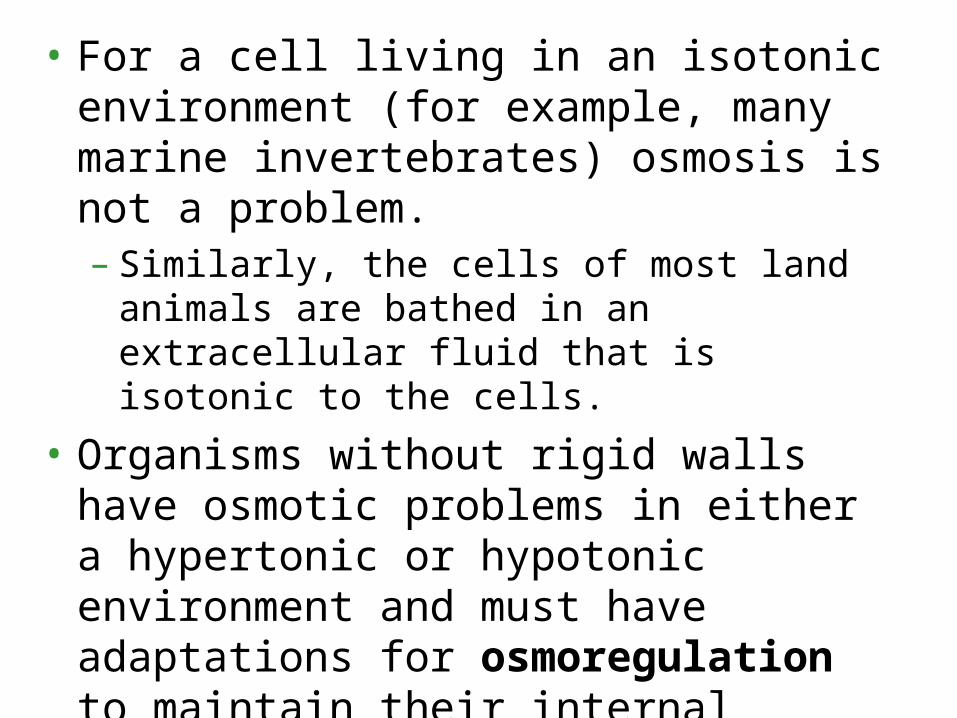
• For a cell living in an isotonic environment (for example, many marine invertebrates) osmosis is not a problem.– Similarly, the cells of most land animals are
bathed in an extracellular fluid that is isotonic to the cells.
• Organisms without rigid walls have osmotic problems in either a hypertonic or hypotonic environment and must have adaptations for osmoregulation to maintain their internal environment.
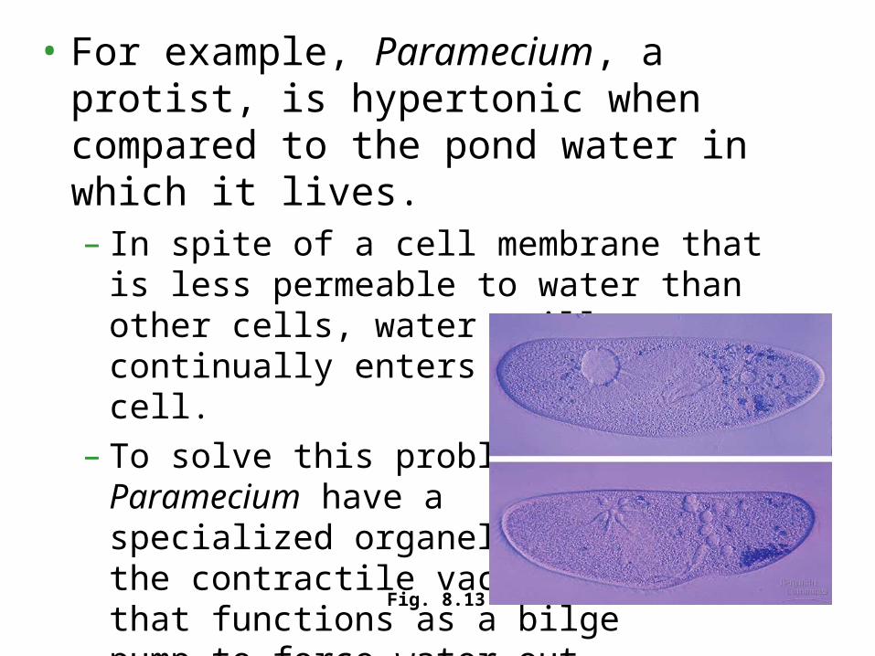
• For example, Paramecium, a protist, is hypertonic when compared to the pond water in which it lives.– In spite of a cell membrane that is less
permeable to water than other cells, water still continually enters the Paramecium cell.
– To solve this problem, Paramecium have a specialized organelle, the contractile vacuole, that functions as a bilge pump to force water out of the cell.
Fig. 8.13
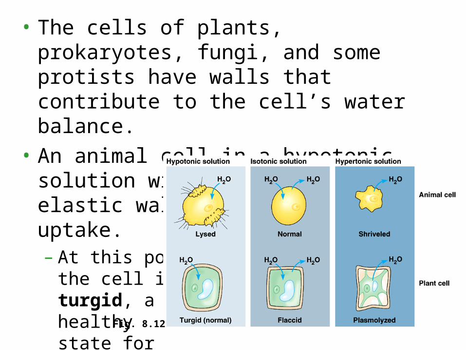
• The cells of plants, prokaryotes, fungi, and some protists have walls that contribute to the cell’s water balance.
• An animal cell in a hypotonic solution will swell until the elastic wall opposes further uptake.– At this point
the cell is turgid, a healthy state for most plant cells. Fig. 8.12

• Turgid cells contribute to the mechanical support of the plant.
• If a cell and its surroundings are isotonic, there is no movement of water into the cell and the cell is flaccid and the plant may wilt.
Fig. 8.12

• In a hypertonic solution, a cell wall has no advantages.
• As the plant cell looses water, its volume shrinks.
• Eventually, the plasma membrane pulls away from the wall.
• This plasmolysis is usually lethal.
Fig. 8.12

Specific proteins facilitate passive transport of water and selected solutes
• Many polar molecules and ions diffuse passively with the help of integral transport proteins that span the membrane.
• The passive movement of molecules down its concentration gradient via a transport protein is called facilitated diffusion
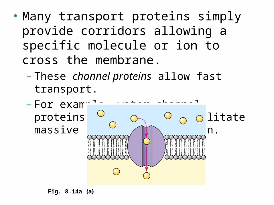
• Many transport proteins simply provide corridors allowing a specific molecule or ion to cross the membrane.– These channel proteins allow fast transport.– For example, water channel proteins,
aquaprorins, facilitate massive amounts of diffusion.
Fig. 8.14a

• Some channel proteins, gated channels, open or close depending on the presence or absence of a physical or chemical stimulus.
• The chemical stimulus is usually different from the transported molecule.
• For example, when neurotransmitters bind to specific gated channels on the receiving neuron, these channels open.
– This allows sodium ions into a nerve cell.– When the neurotransmitters are not present, the
channels are closed
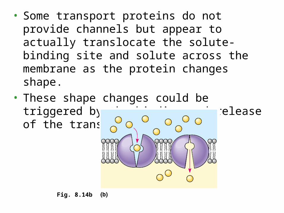
• Some transport proteins do not provide channels but appear to actually translocate the solute-binding site and solute across the membrane as the protein changes shape.
• These shape changes could be triggered by the binding and release of the transported molecule.
Fig. 8.14b

Active transport
• pumping of solutes against their gradients• This active transport requires the cell to
expend its own metabolic energy.• Active transport is critical for a cell to maintain its
internal concentrations of small molecules that would otherwise diffuse across the membrane.
• performed by specific proteins embedded in the membranes.
• ATP supplies the energy for most active transport.
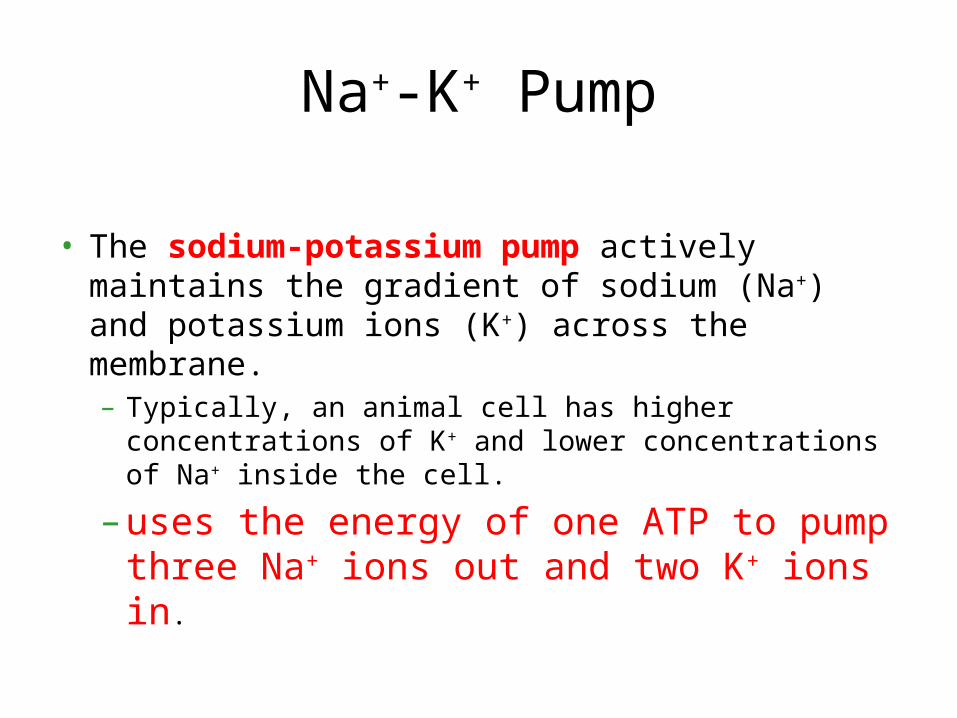
Na+-K+ Pump
• The sodium-potassium pump actively maintains the gradient of sodium (Na+) and potassium ions (K+) across the membrane.– Typically, an animal cell has higher concentrations of
K+ and lower concentrations of Na+ inside the cell.
– uses the energy of one ATP to pump three Na+ ions out and two K+ ions in.
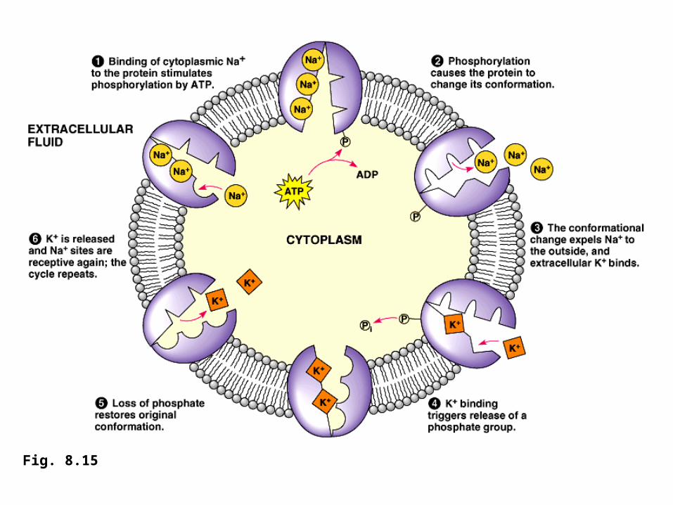
Fig. 8.15

Copyright © 2002 Pearson Education, Inc., publishing as Benjamin Cummings
Fig. 8.16 Both diffusion and facilitated diffusion are forms of passive transport of molecules down their concentration gradient, while active transport requires an investment of energy to move molecules against their concentration gradient.

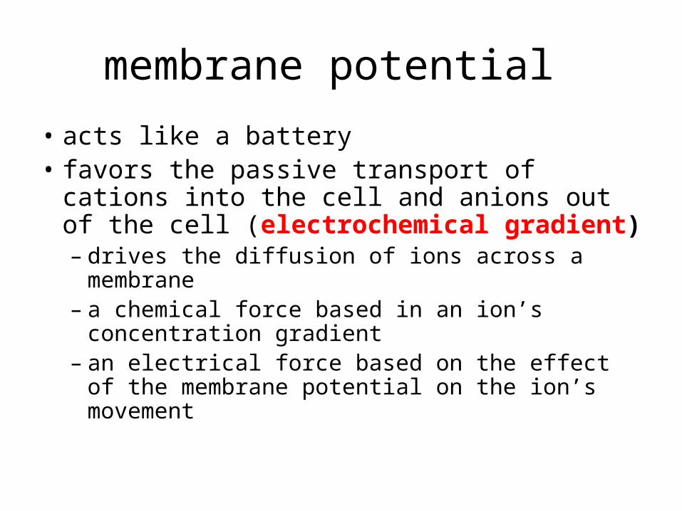
• All cells maintain a voltage across their plasma membranes.– The cytoplasm of a cell is negative in charge
compared to the extracellular fluid because of an unequal distribution of cations and anions on opposite sides of the membrane.
– This voltage is the membrane potential
Some ion pumps generate voltage across membranes
Copyright © 2002 Pearson Education, Inc., publishing as Benjamin Cummings
membrane potential
• acts like a battery• favors the passive transport of cations into
the cell and anions out of the cell (electrochemical gradient)– drives the diffusion of ions across a
membrane– a chemical force based in an ion’s
concentration gradient – an electrical force based on the effect of the
membrane potential on the ion’s movement

• In plants, bacteria, and fungi, a proton pump is the major electrogenic pump, actively transporting H+ out of the cell.
• Protons pumps in the cristae of mitochondria and the thylaloids of chloroplasts, concentrate H+ behind membranes.
• These electrogenic pumps store energy that can be accessed for cellular work.
Copyright © 2002 Pearson Education, Inc., publishing as Benjamin Cummings
Fig. 8.17

Cotransport
• a membrane protein couples the transport of two solutes
• A single ATP-powered pump that transports one solute can indirectly drive the active transport of several other solutes through cotransport via a different protein.
• As the solute that has been actively transported diffuses back passively through a transport protein, its movement can be coupled with the active transport of another substance against its concentration gradient.