Cell infiltrative hydrogel fibrous scaffolds for ... infiltrative hydrogel fibrous scaffolds for...
12
Full length article Cell infiltrative hydrogel fibrous scaffolds for accelerated wound healing Xin Zhao a,d,e,1 , Xiaoming Sun h,1 , Lara Yildirimer a,1 , Qi Lang a , Zhi Yuan (William) Lin a , Reila Zheng a , Yuguang Zhang h , Wenguo Cui a,g,⇑ , Nasim Annabi a,f,⇑ , Ali Khademhosseini a,b,c,⇑ a Biomaterials Innovation Research Center, Division of Biomedical Engineering, Department of Medicine, Brigham and Women’s Hospital, Harvard Medical School, Boston, MA 02139, USA b Wyss Institute for Biologically Inspired Engineering, Harvard University, Boston, MA 02115, USA c Department of Physics, King Abdulaziz University, Jeddah 21569, Saudi Arabia d School of Life Science and Technology, Xi’an Jiaotong University of Medicine, Xi’an, Shaanxi 710049, China e Bioinspired Engineering and Biomechanics Center, Xi’an Jiaotong University, Shaanxi 710049, China f Department of Chemical Engineering, Northeastern University, Boston, MA 02115, USA g Department of Orthopedics, The First Affiliated Hospital of Soochow University, Orthopedic Institute, Soochow University, Suzhou, Jiangsu 215006, China h Department of Plastic and Reconstructive Surgery, Shanghai Ninth People’s Hospital Affiliated to Shanghai Jiaotong University, Shanghai 200011, China article info Article history: Received 17 June 2016 Received in revised form 21 October 2016 Accepted 3 November 2016 Available online xxxx Keywords: Gelatin methacryloyl Photocrosslinkable hydrogel Water retention Soft elasticity Wound healing abstract Development of natural protein-based fibrous scaffolds with tunable physical properties and biocompat- ibility is highly desirable to construct three-dimensional (3D), fully cellularized scaffolds for wound heal- ing. Herein, we demonstrated a simple and effective technique to construct electrospun 3D fibrous scaffolds for accelerated wound healing using a photocrosslinkable hydrogel based on gelatin methacry- loyl (GelMA). We found that the physical properties of the photocrosslinkable hydrogel including water retention, stiffness, strength, elasticity and degradation can be tailored by changing the light exposure time. We further observed that the optimized hydrogel fibrous scaffolds which were soft and elastic could support cell adhesion, proliferation and migration into the whole scaffolds, facilitating regeneration and formation of cutaneous tissues within two weeks. Such tunable characteristics of the fibrous GelMA scaf- folds distinguished them from other reported substrates developed for reconstruction of wound defects including glutaraldehyde-crosslinked gelatin or poly (lactic-co-glycolic acid) (PLGA), whose physical and chemical properties were difficult to modify to allow cell infiltration into the 3D scaffolds for tissue regeneration. We anticipate that the ability to become fully cellularized will make the engineered GelMA fibrous scaffolds suitable for widespread applications as skin substitutes or wound dressings. Statement of Significance In present study, we are able to generate three dimensional photocrosslinkable gelatin (GelMA)-based fibrous scaffolds with tunable physical and biological properties by using a combined photocrosslink ing/electrospinning approach. The developed GelMA fibrous scaffolds can not only support cell viability and cell adhesion, but also facilitate cell migration and proliferation, accelerating regeneration and for- mation of cutaneous tissues. In addition, the physical properties of the engineered fibrous GelMA hydro- gel including water retention capability, mechanical properties and biodegradability can be tuned to accommodate different patients’ needs, making it a promising candidate for skin tissue engineering. Ó 2016 Acta Materialia Inc. Published by Elsevier Ltd. All rights reserved. 1. Introduction Skin wounds may be subdivided into those caused by acute trauma, burns and surgical procedures, or those caused by long- term diseases such as diabetes. Healing of such wounds depends on an intricate interplay between cellular factors as well as the surrounding extracellular matrix (ECM) [1]. Tissue engineering strategies seek to accelerate wound healing by providing the http://dx.doi.org/10.1016/j.actbio.2016.11.017 1742-7061/Ó 2016 Acta Materialia Inc. Published by Elsevier Ltd. All rights reserved. ⇑ Corresponding authors at: Biomaterials Innovation Research Center, Division of Biomedical Engineering, Department of Medicine, Brigham and Women’s Hospital, Harvard Medical School, Boston, MA 02139, USA. E-mail addresses: [email protected] (W. Cui), [email protected] (N. Annabi), [email protected] (A. Khademhosseini). 1 Co-first authors. Acta Biomaterialia xxx (2016) xxx–xxx Contents lists available at ScienceDirect Acta Biomaterialia journal homepage: www.elsevier.com/locate/actabiomat Please cite this article in press as: X. Zhao et al., Cell infiltrative hydrogel fibrous scaffolds for accelerated wound healing, Acta Biomater. (2016), http://dx. doi.org/10.1016/j.actbio.2016.11.017
Transcript of Cell infiltrative hydrogel fibrous scaffolds for ... infiltrative hydrogel fibrous scaffolds for...

Acta Biomaterialia xxx (2016) xxx–xxx
Contents lists available at ScienceDirect
Acta Biomaterialia
journal homepage: www.elsevier .com/locate /actabiomat
Full length article
Cell infiltrative hydrogel fibrous scaffolds for accelerated wound healing
http://dx.doi.org/10.1016/j.actbio.2016.11.0171742-7061/� 2016 Acta Materialia Inc. Published by Elsevier Ltd. All rights reserved.
⇑ Corresponding authors at: Biomaterials Innovation Research Center, Division ofBiomedical Engineering, Department of Medicine, Brigham and Women’s Hospital,Harvard Medical School, Boston, MA 02139, USA.
E-mail addresses: [email protected] (W. Cui), [email protected](N. Annabi), [email protected] (A. Khademhosseini).
1 Co-first authors.
Please cite this article in press as: X. Zhao et al., Cell infiltrative hydrogel fibrous scaffolds for accelerated wound healing, Acta Biomater. (2016), htdoi.org/10.1016/j.actbio.2016.11.017
Xin Zhao a,d,e,1, Xiaoming Sun h,1, Lara Yildirimer a,1, Qi Lang a, Zhi Yuan (William) Lin a, Reila Zheng a,Yuguang Zhang h, Wenguo Cui a,g,⇑, Nasim Annabi a,f,⇑, Ali Khademhosseini a,b,c,⇑aBiomaterials Innovation Research Center, Division of Biomedical Engineering, Department of Medicine, Brigham and Women’s Hospital, Harvard Medical School, Boston, MA02139, USAbWyss Institute for Biologically Inspired Engineering, Harvard University, Boston, MA 02115, USAcDepartment of Physics, King Abdulaziz University, Jeddah 21569, Saudi Arabiad School of Life Science and Technology, Xi’an Jiaotong University of Medicine, Xi’an, Shaanxi 710049, ChinaeBioinspired Engineering and Biomechanics Center, Xi’an Jiaotong University, Shaanxi 710049, ChinafDepartment of Chemical Engineering, Northeastern University, Boston, MA 02115, USAgDepartment of Orthopedics, The First Affiliated Hospital of Soochow University, Orthopedic Institute, Soochow University, Suzhou, Jiangsu 215006, ChinahDepartment of Plastic and Reconstructive Surgery, Shanghai Ninth People’s Hospital Affiliated to Shanghai Jiaotong University, Shanghai 200011, China
a r t i c l e i n f o
Article history:Received 17 June 2016Received in revised form 21 October 2016Accepted 3 November 2016Available online xxxx
Keywords:Gelatin methacryloylPhotocrosslinkable hydrogelWater retentionSoft elasticityWound healing
a b s t r a c t
Development of natural protein-based fibrous scaffolds with tunable physical properties and biocompat-ibility is highly desirable to construct three-dimensional (3D), fully cellularized scaffolds for wound heal-ing. Herein, we demonstrated a simple and effective technique to construct electrospun 3D fibrousscaffolds for accelerated wound healing using a photocrosslinkable hydrogel based on gelatin methacry-loyl (GelMA). We found that the physical properties of the photocrosslinkable hydrogel including waterretention, stiffness, strength, elasticity and degradation can be tailored by changing the light exposuretime. We further observed that the optimized hydrogel fibrous scaffolds which were soft and elastic couldsupport cell adhesion, proliferation and migration into the whole scaffolds, facilitating regeneration andformation of cutaneous tissues within two weeks. Such tunable characteristics of the fibrous GelMA scaf-folds distinguished them from other reported substrates developed for reconstruction of wound defectsincluding glutaraldehyde-crosslinked gelatin or poly (lactic-co-glycolic acid) (PLGA), whose physical andchemical properties were difficult to modify to allow cell infiltration into the 3D scaffolds for tissueregeneration. We anticipate that the ability to become fully cellularized will make the engineeredGelMA fibrous scaffolds suitable for widespread applications as skin substitutes or wound dressings.
Statement of Significance
In present study, we are able to generate three dimensional photocrosslinkable gelatin (GelMA)-basedfibrous scaffolds with tunable physical and biological properties by using a combined photocrosslinking/electrospinning approach. The developed GelMA fibrous scaffolds can not only support cell viabilityand cell adhesion, but also facilitate cell migration and proliferation, accelerating regeneration and for-mation of cutaneous tissues. In addition, the physical properties of the engineered fibrous GelMA hydro-gel including water retention capability, mechanical properties and biodegradability can be tuned toaccommodate different patients’ needs, making it a promising candidate for skin tissue engineering.
� 2016 Acta Materialia Inc. Published by Elsevier Ltd. All rights reserved.
1. Introduction
Skin wounds may be subdivided into those caused by acutetrauma, burns and surgical procedures, or those caused by long-term diseases such as diabetes. Healing of such wounds dependson an intricate interplay between cellular factors as well as thesurrounding extracellular matrix (ECM) [1]. Tissue engineeringstrategies seek to accelerate wound healing by providing the
tp://dx.

2 X. Zhao et al. / Acta Biomaterialia xxx (2016) xxx–xxx
wound bed with an artificial matrix into which cells such as dermalfibroblasts can migrate to proliferate and form new tissues. Theideal dermal substitute consists of a flexible and durable materialwhich demonstrates biocompatibility and non-antigenicity whenin contact with the wounded host tissue. The most commonlydeveloped skin substitutes include hydrogels [2], sponges [3] andelectrospun mats [4]. Electrospun substitutes exhibit several fun-damental features for rapid and functional wound healing whichrender them superior to hydrogels or sponges. Their ECM-likenanofibrous architectures provides a high surface-to-volume ratiowhich not only enables maximal cell-material interactions andmaterial-mediated signaling, but also promotes rapid hemostasis.Additionally, the nanoporous nature of these mats aids in nutrientand waste exchange whilst creating a barrier to external pathogens[5,6].
Despite these advantages, nanoporous structures limit threedimensional (3D) cellular infiltration [7]. To solve this, severalattempts at increasing pore sizes of fibrous scaffolds have beenmade. The main methods involve the co-electrospinning ofsacrificial fibers or the addition of porogens which can be leachedout after electrospinning. For example, co-electrospinning ofwater-soluble poly (ethylene oxide) (PEO) and water-insolublepoly (e-caprolactone) (PCL) fibers, the former of which act as thesacrificial component, resulted in increased pore sizes of the finalPCL scaffolds [8]. Addition of salt particles onto the collecting platewhile fibers were being electrospun and subsequent dissolution ofthese particles in water also resulted in increased pore sizes in aPCL based scaffold [9]. While these approaches allowed for the for-mation of larger pore sizes and improved cellular infiltration, theyreduced the mechanical properties and dimensional stability ofsuch mats, limiting their tissue engineering applications. It is,therefore, crucial to develop a strategy to synthesize fully cellular-ized, yet mechanically reliable scaffolds to feasibly regeneratedamaged skin.
One such approach is to facilitate cell infiltration by tuning thephysical properties of the electrospun nanofibers themselves with-out changing the fiber or pore diameter. It has been suggested thatthere is an essential link between the stiffness of a scaffold and itsability to promote cell adhesion, proliferation, infiltration, and dif-ferentiation [10–13]. Cell migration into a soft matrix has beenfound to be faster compared to that of a stiff scaffold [14,15]. Thisis because fibers with low stiffness result in recruitment of nearbyfibers by active cells, which increases ligand density on cell sur-faces and stimulates cell adhesion as well as associated signalingpathways, thus facilitating cell migration [13].
Electrospun fibers are commonly made of synthetic polymerswith tunable mechanical and degradation properties. However,the lack of cell-recognition signals renders them less than idealfor skin regeneration applications. Biopolymers including collagen[16], gelatin [17–21], fibrinogen [22] and elastin [23] are promisingscaffold materials for tissue regeneration owing to their abilities toimprove cell-material interactions. Furthermore, their innatehydrophilicity renders them particularly amenable to retainingan appropriate balance of moisture in the wound bed to aid thehealing process [24]. Additionally, the ability to absorb large quan-tities of water maintains the scaffold’s mechanical compliance andelasticity, which is important for skin substitutes due to the highdegree of tension experienced by skin tissues. To ensure that thestructure of these hydrophilic scaffolds remains stable in aqueousenvironments, crosslinking is required. However, crosslinking withglutaraldehyde [25] or other agents [26] may induce toxicity issuesor compromise the scaffold’s mechanical compliance and elasticity[27]. Also, controlling factors such as mechanical, degradation, andbiological properties of the electrospun protein-based scaffoldsstill remains a challenge. The primary amino acid sequence andthe protein conformation dictate the mechanical and degradation
Please cite this article in press as: X. Zhao et al., Cell infiltrative hydrogel fibroudoi.org/10.1016/j.actbio.2016.11.017
properties of the end-proteins, neither of which may be effectivelycontrolled [28]. The aim of this project is thus to fabricate electro-spun natural protein-based fibrous scaffolds with tunable physicalproperties including water retention, mechanical and degradationcharacteristics. The engineered scaffolds should also be able to pro-mote cell adhesion, proliferation and infiltration and thus to form a3D, fully cellularized, ECM-like constructs to facilitate woundhealing.
Here, by incorporating reactive methacryloyl groups intogelatin, we synthesized a photocrosslinkable gelatin prepolymercalled GelMA. Electrospinning of the resulting polymer formedphotocrosslinkable GelMA hydrogel fibers, which exhibitedhydrophilicity and biocompatibility due to their protein domainas well as tunable physical properties (e.g., mechanical properties)due to their methacryloyl domain. Modification of gelatin with thereactive methacryloyl groups further introduced the spatial andtemporal benefits of radical photocrosslinking, endowing the elec-trospun GelMA fibers with controllable physical propertiesrequired to aid wound healing. Through simple adjustment of thelight exposure time, we could precisely tailor the physical proper-ties of the resulting scaffolds including water permeability/reten-tion, mechanics, and degradation kinetics. Such tunable featuresdistinguished the engineered scaffold from frequently usedsubstrates like glutaraldehyde-crosslinked gelatin or poly (lactic-co-glycolic acid) (PLGA) (controls in this study). By mimickingthe structure of the native ECM, GelMA scaffolds were proved toeffectively aid wound regeneration by guiding cellular processes,making them ideal candidates for skin regeneration.
2. Materials and methods
2.1. Synthesis of GelMA
GelMA was synthesized using methods previously described[2]. In brief, 10.0 g of type A porcine skin gelatin (Sigma-Aldrich,St. Louis, MO) was dissolved in 100 mL Dulbecco’s phosphatebuffered saline (DPBS) (Invitrogen, San Diego, CA) at 60 �C underconstant stirring. 8.0 mL of methacrylic anhydride was added at arate of 0.5 mL�min�1. After a 3 h reaction, 1:5 diluted DPBS wasadded to stop the reaction and the mixture was subject to dialysis(MWCO 12-14,000, Fisher Scientific) in distilled water for 1 weekto remove salts as well as other small molecule impurities [29].Then, the solution was freeze-dried for 1 week and the resultantwhite porous foam was stored at �80 �C until later use. 1H NMRspectroscopy was used to assess the degree of methacryloyl mod-ification; at room temperature, the spectra were obtained at600 MHz frequency in deuterium oxide using a Varian INOVANMR spectrometer. The degree of substitution was quantifiedusing the method described by Hoch et al. [30]. Briefly, in eachspectrum, the proton signal that belonged to aromatic amino acids(d = 7.0–7.5 ppm) was used as a reference. The area under thesignal curves was integrated using proton signal from the methy-lene groups (d = 2.7–2.9 ppm) in the vicinities of lysine amino acid.The degree of substitution was calculated using this formula:1-(integrated signal of lysine in GelMA/integrated signal of lysinein gelatin).
2.2. Fabrication of electrospun fibrous mats
2.2.1. Fabrication of electrospun PLGA fibers500 mg of PLGA (50 kDa, 75:25) was completely dissolved in
1 mL of hexafluoro-2-propanol (HFIP, Sigma-aldrich, St. Louis,MO). The solution was then filled into a syringe and kept in asyringe pump attached to a high voltage device (Glassman Highvoltage, Inc. TX). An aluminium foil collector plate (30 cm � 30 cm
s scaffolds for accelerated wound healing, Acta Biomater. (2016), http://dx.

X. Zhao et al. / Acta Biomaterialia xxx (2016) xxx–xxx 3
square plate) was used on the anode. The working distance was setto be 10–15 cm between the syringe tip and the collector plate,with the voltage between the source and the collector plate setat 20 kV. The solution was pumped out at 4 mL�h�1, and 1 g ofPLGA was used to spin each sheet.
2.2.2. Fabrication of electrospun gelatin fibersElectrospinning of gelatin fibers used the same procedure as
described above except that the electrospinning solution consistedof 100 mg of gelatin A from porcine skin in 1 mL of HFIP. After elec-trospinning, the gelatin was chemically crosslinked using glu-taraldehyde (10 mL�500 mL�1 ethanol) overnight. Finally, theexcessive glutaraldehyde was quenched using glycine(7.5 g�500 mL�1 distilled water). The electrospun mats were rinsedin distilled water 5 times for 10 min each and then air-driedovernight.
2.2.3. Fabrication of electrospun GelMA fibersElectrospinning GelMA fibers followed the same protocol as
that of gelatin fibers. The electrospun GelMA sheets were pho-tocrosslinked by crosslinking the methacryloyl groups upon UVlight exposure for different lengths of time. To prepare the pho-tocrosslinking solution, 1.0 g of 2-hydroxy-40-(2-hydroxyethoxy)v-2-methylpropiophenone (photo-initiator, Irgacure 2959, Sigma-Aldrich, St. Louis, MO) was added to 10 mL of ethanol in theabsence of light and stirred until completely dissolved. Uncros-slinked electrospun GelMA fibers were immersed in the ethanolsolution containing 10% photo-initiator for 2 h and exposed to365 nm UV light (Blak-Ray B-100 Series high powered UV lamp(B-100A) at 10 cm working distance for 2 min (GelMA-2), 6 min(GelMA-6) and 10 min (GelMA-10), respectively. The UV crosslink-ing method was similar to what was used previously by Burdick’sgroup: UV crosslinking the electrospun Norbornene-functionalizedhyaluronic acid fibers [31]. To confirm the crosslinking of the pho-tocrosslinked electrospun GelMA fibers, the crosslinked electro-spun mats were immersed in deionized water for 24 h to removethe uncrosslinked GelMA and dried in air until obtaining a constantweight. We observed that photocrosslinked GelMA fibers did notdissolve in 50 �C water whereas the uncrosslinked GelMA fibersdissolved immediately in the presence of water (see the video pro-vided in the Supporting information). The weight of the electro-spun fibrous mats before and after water immersion wasmeasured using analytical electronic balance (Mettler ToledoAL204). The percentage of the weight of the crosslinked, undis-solved GelMA after water immersion relative to the initial dryweight of the GelMA samples before water immersion (Wc%, whichis correlated with the degree of crosslinking) was calculated usingthe following equation [32,33]:
Wc% ¼ WW
Wd� 100% ð1Þ
where Wd is the initial dry weight of the photocrosslinked samplebefore water immersion and WW is the dry weight of the pho-tocrosslinked sample after water immersion for 24 h.
2.3. Physical characterization
2.3.1. MorphologyMorphology of the electrospun fibrous scaffolds before and
after immersion in distilled water was examined using scanningelectron microscope (SEM). After thorough rinsing, all the electro-spun fibers before and after immersion in water for 24 h at 37 �Cwere subject to incubation in 1% osmium tetroxide, followed bydehydrations in graded ethanol series as well as drying at criticalpoint with a Polaron E3000 critical point dryer (Quorum Technolo-gies Inc., Guelph, ON, Canada). Sputter coating was conducted at
Please cite this article in press as: X. Zhao et al., Cell infiltrative hydrogel fibroudoi.org/10.1016/j.actbio.2016.11.017
2.2 kV for 2 min using a Polaron SC7640 sputter coater beforeimaging with a Hitachi S-800 SEM (Hitachi High Technologies,Pleasanton, CA).
2.3.2. Water permeability, water retention and water vaporpermeability
Electrospun scaffolds were cut to fit into 24-well plates,weighed, and then incubated in 1 mL of distilled water at 37 �Cfor 24 h. Scaffolds were then blotted dry, weighed and subjectedto the following examinations:
i. Water permeabilityThe setup for permeability experiments was based on the work
of Sell et al. [34]. Briefly, the scaffolds were placed into separatewater-tight specimen mounts. A stop/start valve was used to facil-itate turning the water on and off, and connected between theinflow tube and the specimen mount. The inflow reservoir waspositioned near-horizontally at the top of the inflow tube. Oncefilled, the water level was maintained at 100–105 cm. The smalldifference of 5 cm was due to the negligible error contribution inpermeability as per Darcy’s Law. One h prior to water permeabilityexperiments, electrospun scaffolds were soaked in distilled water.The thickness of the swollen scaffold was measured with a caliper.The tubing was filled with distilled water, and the fibrous scaffoldsloaded into the specimen mount with a cross-sectional area of0.7 cm2. The initial volume on the pipette was recorded, and thevalve was opened to allow free water flow. The volume of the pip-ette was recorded again after 5 min which was determined to bethe cut-off time after which water flow decreased for all samples.Each material type was tested at least 4 times.
In accordance to Darcy’s Law, permeability (k) was calculatedusing the formula below:
k ¼ Qgh=Ftp ð2Þwhere k is the permeability of the scaffold in Darcy’s units (D); Q isthe volume of fluid that permeated the scaffold at time t; g is thefluid viscosity (in this case, 0.89 cp for water at 25 �C); h is the scaf-fold thickness; F is the cross sectional area of the scaffold that isperpendicular to fluid flow direction; p is the applied pressure head[34].
ii. Water retentionThe weight of the samples before and after 24 h immersion in
distilled water was recorded as WD and W0, respectively. Sampleswere then left in air at room temperature and the weight (Wt) wasmeasured at pre-determined time points [35]. The water sorptionSR at time t was calculated according to the following equation:
SR ¼ Wt �WD
WD� 100% ð3Þ
iii. Water vapor permeabilityTo examine the water vapor permeability of the nanofibers, the
dry PLGA, gelatin and GelMA nanofibers were separately stuck onthe ends of 17 � 60 mm (O.D. � H) Glass Threaded Vials (Fisher,MA) containing 6 mL of DPBS. The vials were placed at 37 �C andweighed every 24 h to determine the water loss [34].
2.3.3. Tensile and cyclic testingTensile properties of the electrospun fibers were tested by using
an Instron 5542 mechanical tester. Prior to uniaxial tensile testingexperiments, the electrospun fibrous sheets were cut into rectan-gular shapes (30 mm � 10 mm) and soaked in distilled water for24 h. The sample was secured between opposing clamps whichwere approximately 20 mm apart from each other. For tensile test-ing, the samples were stretched until failure at 10 mm�min�1. For
s scaffolds for accelerated wound healing, Acta Biomater. (2016), http://dx.

4 X. Zhao et al. / Acta Biomaterialia xxx (2016) xxx–xxx
cyclic tensile testing, the sample was stretched to a maximum of5 mm, then relaxed to its original length, at 10 cycles per minutefor 30 cycles.
2.3.4. DegradationCut round samples of 2 cm diameter were incubated at 37 �C
with 1 mL 0.02 units collagenase mL�1 of DPBS for a month [36].At pre-determined time points, the DPBS was removed and thesamples were washed twice with distilled water, lyophilized, andweighed. The following equation was used to calculate the per-centage degradation (D%):
D% ¼ W0 �Wt
W0� 100% ð4Þ
where W0 is the initial sample dry weight and Wt is the dry weightafter time t.
2.4. Biological characterization
2.4.1. Cell cultureMinimum Essential Medium (MEM, Invitrogen, San Diego), sup-
plemented with 10% fetal bovine serum (FBS) (Life Technologies,NY), 1% L-glutamine (Life Technologies, NY), and 1% penicillin/streptomycin (Life Technologies, NY), was used to culture skin der-mal fibroblasts (BJ-6s, Lonza) at 37 �C and 5% CO2. Cells were keptin tissue culture polystyrene (TCP) flasks and passaged 1:6 when a70% confluence was reached.
BJ-6s cells were then seeded onto the surfaces of PLGA, gelatin,GelMA nanofibers and glass (control) at a seeding density of5 � 104 cells�cm�2 for cell viability, adhesion and proliferationstudies.
2.4.2. Cell viabilityCell viability was determined using Live/Dead� Viability Kit
(Life Technologies, NY). Calcein AM and ethidium homodimer-1were first diluted in DPBS at concentrations of 0.5 lL�mL�1 and2 lL�mL�1, respectively. At 1, 4, and 7 days, samples were washed3� in DPBS and incubated in the mixture for 15 min at 37 �C. Allsamples were imaged immediately without mounting using a Zeissfluorescence microscope (Zeiss, NY).
2.4.3. Cell adhesionTo examine cell adhesion on samples at days 1, 4, and 7, phal-
loidin (Alexa Fluor 488, Invitrogen, San Diego, CA) and DAPI(Sigma, St Louis, MO) were used to stain F-actin and cell nuclei,respectively, as per manufacturer’s instructions. Briefly, following3�wash in DPBS, samples were sequentially subject to 30-min fix-ation in 4% paraformaldehyde (PFA), 20-min permeabilization in0.1% Triton X-100, and 45-min blocking in 1% BSA. Then thephalloidin solution (1:40 dilution in DPBS) was added followedby a 45-min incubation at 37 �C. Finally, DAPI solution (1:1000dilution in DPBS) was added for 5-min incubation at 37 �C. Cellimaging was performed using a Zeiss fluorescence microscope; cellnumber and area were quantified using NIH ImageJ software.
2.4.4. Cell proliferationCell proliferation was evaluated using PicoGreen� DNA quan-
tification assay (Life Technologies, NY). At each time point (0, 1, 4and 7 days), the samples were lysed for 2 h with 500 lL of50 lg�mL�1 proteinase k at 37 �C, and pelleted by centrifugationat 18,000g at 4 �C for 10 min. After removal of the supernatant,the samples were diluted in 100 lL 1�TE buffer and then incu-bated in dark with 100 lL PicoGreen� working solution (1:200dilution in TE buffer) for 5 min at room temperature. 96-wellplates (Thermo Fisher Scientific, Waltham, MA) were used forfluorescence measurement at excitation and emission wavelengths
Please cite this article in press as: X. Zhao et al., Cell infiltrative hydrogel fibroudoi.org/10.1016/j.actbio.2016.11.017
of 485 nm and 520 nm, respectively, using a microplate reader(Fluostar, Ortenberg, Germany). The standard curve was createdby mixing 100 lL of each DNA concentration (2 lmol�L�1,200 nmol�L�1, 20 nmol�L�1, 2 nmol�L�1, 0 nmol�L�1) with 100 lLof PicoGreen� solution (1:200) in a 96-well plate and incubatingat room temperature for 5 min. Fluorescence of the standard curvemixture was measured as described above.
2.5. Animal study
2.5.1. Mouse dorsal model of wound healing and ethics80 healthy female ICR mice, weighed 35–40 g, were recruited
from the Shanghai Slac Laboratory Animal Co. Ltd. (Shanghai,China). Ethical clearance was obtained beforehand from the Com-mittee of Experimental Animal Administration of Shanghai NinthPeople’s Hospital Affiliated to Shanghai Jiaotong University Schoolof Medicine. During the course of the study, animals weremaintained under specific pathogen-free conditions (SPF) and fedad libitum, while complying with the international ethics guideli-nes and the National Institutes of Health Guide concerning the Careand Use of Laboratory Animals.
Mouse dorsal wound model was used as previously describedfor wound healing study [37,38]. In brief, animals were anes-thetized and operations were performed, both under sterile condi-tions. One circular full-thickness skin wound (8-mm diameter) wascreated on the back of each mouse, while normal skin (2 mm) waspeeled around each wound to prevent the implanted scaffolds frombeing dislodged. The epidermis, dermis and perichondrium in eachwound were completely excised. Mice were randomly assigned tofour groups and received treatment with control (no treatment),PLGA, gelatin and GelMA-10 electrospun scaffolds, respectively.Mice were returned to their cages after recovering from anesthesia.
2.5.2. Macroscopic evaluation and wound healing rateWound healing was carefully monitored and mice were
sacrificed 0, 7, 14, and 21 days after operation (n = 5). The woundhealing rate in each group was calculated as the ratio of the areaof healed wounds compared to the original area of wound.
2.5.3. Morphological analysis of the wound tissueIn each group, wounds were subject to excisions with a 5-mm
margin of surrounding intact tissue and were then cut in halves.After 24 h of immersion in 4% formalin, they were embedded inparaffin, and subsequently cross-sectioned along the tissue. Finally,hematoxylin-eosin (H&E) and modified Masson’s trichrome wereused to stain the samples for examining re-epithelialization andcollagen deposition, respectively [4].
2.5.4. qRT-PCR analysis21 days after implantation, total RNA from the regenerated tis-
sues was prepared using Trizol Reagent (Thermo Scientific, Wal-tham, MA) according to the manufacturer’s instructions. RNAintegrity was confirmed using optical density absorption ratioOD260 nm/OD280 nm between 1.8 and 2.0. cDNA was synthesizedusing RNA PCR kit protocol (Thermo Scientific, Waltham, MA). Theprimer sequences are shown in Table S1. In triplicates, quantitativepolymerase chain reaction (qPCR) amplification was performedusing SYBR Green PCR reagent system. Real-time qPCR was con-ducted in the following steps: initial 10-min denaturation at95 �C and 40 cycles of PCR, each lasting for 15 s at 95 �C and then45 s at 60 �C. Endogenous control GAPDH was used for normaliza-tion. Relative quantification was conducted by applying the previ-ously described comparative 2�DDCt method.
s scaffolds for accelerated wound healing, Acta Biomater. (2016), http://dx.
X. Zhao et al. / Acta Biomaterialia xxx (2016) xxx–xxx 5
2.6. Statistical analysis
All experiments had 5 replicates unless otherwise stated. In IBMSPPS Statistics 19 for Windows, ANOVA with a post hoc Dunn orBonferroni test was used to analyze the data. p < 0.05 was consid-ered statistically significant.
3. Results
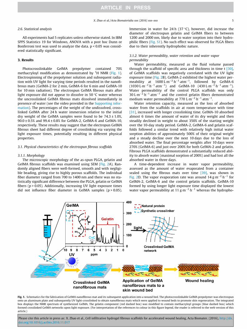
Photocrosslinkable GelMA prepolymer contained 70%methacryloyl modification as demonstrated by 1H NMR (Fig. 1).Electrospinning of the prepolymer solution and subsequent radia-tion with UV light for varying time periods resulted in the nanofi-brous mats (GelMA-2 for 2 min, GelMA-6 for 6 min and GelMA-10for 10 min radiation). The electrospun GelMA fibrous mats afterlight exposure did not appear to dissolve in 50 �C water whereasthe uncrosslinked GelMA fibrous mats dissolved immediately inpresence of water (see the video provided in the Supporting infor-mation). The percentages of the weight of the undissolved, cross-linked GelMA after 24 h water immersion relative to the initialdry weight of the GelMA samples were found to be 74.3 ± 1.0%,90.0 ± 0.5% and 99.4 ± 0.8% for GelMA-2, GelMA-6 and GelMA-10,respectively. These results may suggest that the electropun GelMAfibrous sheet had different degree of crosslinking via varying thelight exposure times, potentially resulting in different physicalproperties.
3.1. Physical characteristics of the electrospun fibrous scaffolds
3.1.1. MorphologyThe microscopic morphology of the as-spun PLGA, gelatin and
GelMA fibrous scaffolds was examined using SEM (Fig. 2A). Ran-domly aligned fibers were well-formed, smooth and with negligi-ble beading, giving rise to highly porous scaffolds. The individualfiber diameter ranged from 700 to 1400 nm and there was no sta-tistically significant difference between the PLGA, gelatin or GelMAfibers (p > 0.05). Additionally, increasing UV light exposure timesdid not influence fiber diameter in GelMA samples (p > 0.05).
Fig. 1. Schematics for the fabrication of GelMA nanofibrous mat and its subsequent appliconto an aluminum plate and subsequently UV light-crosslinked to obtain nanofibrous mabox displays the NMR spectrum of synthesized GelMA. The gelatin component (red daformed crosslinked GelMA networks upon light exposure. (For interpretation of the referarticle.)
Please cite this article in press as: X. Zhao et al., Cell infiltrative hydrogel fibroudoi.org/10.1016/j.actbio.2016.11.017
Immersion in water for 24 h (37 �C), however, did increase thediameter of electrospun gelatin and GelMA fibers to between1200 and 2000 nm, likely due to water sorption into their hydro-philic fibers (Fig. S1). No such effect was observed for PLGA fibersdue to their inherently hydrophobic nature.
3.1.2. Water permeability, water retention and water vaporpermeability
Water permeability, measured as the fluid volume passedthrough the scaffold of specific area and thickness in time t [30],of GelMA scaffolds was negatively correlated with the UV lightexposure time (Fig. 2B). GelMA-2 exhibited the highest water per-meability at 1600 L�m�2�h�1�atm�1, followed by GelMA-6(1030 L�m�2�h�1�atm�1) and GelMA-10 (430 L�m�2�h�1�atm�1).Water permeability of the control PLGA scaffolds was only30 L�m�2�h�1�atm�1 and the control gelatin fibers demonstratedmedium water permeability at 770 L�m�2�h�1�atm�1.
Water retention capacity, measured as the loss of absorbedwater from the scaffolds in air at room temperature with time[31], increased with longer crosslinking time. GelMA-10 absorbedalmost 6 times the amount of water of its dry weight and thensteadily declined in weight to about 350% of the starting weightover the 10-day study period. GelMA-2, GelMA-6 and gelatin scaf-folds followed a similar trend with relatively high initial watersorption abilities of approximately 500% of their original weightand a steady decline over the next 10 days due to the loss ofabsorbed water. The final percentage weights after 10 days were270% (GelMA-6) and just over 200% for both GelMA-2 and gelatin.Fibrous PLGA scaffolds demonstrated a substantially reduced abil-ity to absorb water (maximal sorption of 200%) and had lost all theabsorbed water in three days.
A time-dependent increase in water vapor permeability,assessed as the amount of water evaporated from a containersealed using the fibrous mats over time [39], was shown inFig. 2D. The vapor evaporation rate was around 14 g�m�2�h�1 forGelMA-2, GelMA-6 and the control gelatin scaffolds. GelMA-10formed by using longer light exposure time displayed the lowestwater vapor permeability at 11 g�m�2�h�1 whereas the hydropho-
ation into a wound bed. The photocrosslinkable GelMA prepolymer was electrospunts which were applied to wound beds to promote skin regeneration. The integratedshed box) was modified to contain methacryloyl groups (blue dashed box) whichences to colour in this figure legend, the reader is referred to the web version of this
s scaffolds for accelerated wound healing, Acta Biomater. (2016), http://dx.
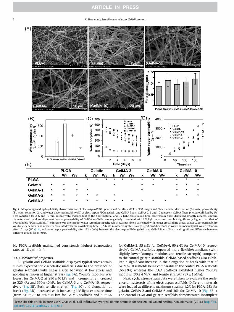
Fig. 2. Morphology and hydrophilicity characterization of electrospun PLGA, gelatin and GelMA scaffolds. SEM images and fiber diameter distribution (A), water permeability(B), water retention (C) and water vapor permeability (D) of electrospun PLGA, gelatin and GelMA fibers. GelMA-2, 6 and 10 represent GelMA fibers photocrosslinked by UVlight radiation for 2, 6 and 10 min, respectively. Independent of the fiber material and UV light-crosslinking time, electrospun fibers displayed smooth surfaces, uniformdiameters and random alignment. Water permeability of GelMA scaffolds was negatively correlated with UV light exposure time but significantly higher than that ofhydrophobic PLGA scaffolds. The inverse was the case for water retention capacity which was positively correlated with longer crosslinking times. Water vapor permeabilitywas time-dependent and inversely correlated with the crosslinking time. E) A table summarizing statistically significant difference in water permeability (k), water retentionafter 10 days (Wr) [14], and water vapor permeability after 192 h (Wv), between the electrospun PLGA, gelatin and GelMA fibers. ⁄Statistical significant difference betweendifferent groups for p < 0.05.
6 X. Zhao et al. / Acta Biomaterialia xxx (2016) xxx–xxx
bic PLGA scaffolds maintained consistently highest evaporationrates at 18 g�m�2�h�1.
3.1.3. Mechanical propertiesAll gelatin and GelMA scaffolds displayed typical stress-strain
curves expected for viscoelastic materials due to the presence ofgelatin segments with linear elastic behavior at low stress andnon-linear region at higher stress (Fig. 3A). Young’s modulus waslowest for GelMA-2 at 290 ± 40 kPa and incrementally increasedto 325 kPa and 350 ± 40 kPa for GelMA-6 and GelMA-10, respec-tively (Fig. 3B). Both tensile strength (Fig. 3C) and elongation atbreak (Fig. 3D) increased with increasing UV light exposure time(from 310 ± 20 to 360 ± 40 kPa for GelMA scaffolds and 50 ± 6%
Please cite this article in press as: X. Zhao et al., Cell infiltrative hydrogel fibroudoi.org/10.1016/j.actbio.2016.11.017
for GelMA-2, 55 ± 5% for GelMA-6, 60 ± 4% for GelMA-10, respec-tively). GelMA scaffolds appeared more flexible/compliant (withslightly lower Young’s modulus and tensile strength) comparedto the control gelatin scaffolds. GelMA-based scaffolds also exhib-ited a significant increase in the elongation at break with that ofGelMA-10 scaffolds being comparable to the control PLGA scaffolds(66 ± 9%) whereas the PLGA scaffolds exhibited higher Young’smodulus (30 ± 4 MPa) and tensile strength (37 ± 1 MPa).
Next, cyclic stress-strain data were taken to evaluate the resili-ence or hysteresis of the electrospun scaffolds. Different materialswere loaded at different maximum strains: 1.2% for PLGA, 25% forgelatin, GelMA-2 and GelMA-6 and 30% for GelMA-10 (Fig. 3E-I).The control PLGA and gelatin scaffolds demonstrated incomplete
s scaffolds for accelerated wound healing, Acta Biomater. (2016), http://dx.

Fig. 3. Mechanical characteristics of engineered PLGA, gelatin and GelMA fibers. Representative stress-strain curves of electrospun PLGA, gelatin and GelMA scaffolds (A).Young’s modulus (B), tensile strength (C) and elongation at break (D) obtained from the stress-strain curves of the electrospun PLGA, gelatin and GelMA scaffolds. Anincreasing trend in all three parameters was observed with increasing crosslinking time of the GelMA electrospun scaffolds, although the difference between different groupsis statistically insignificant. Cyclic stress-strain curves of the electrospun PLGA (E), gelatin (F), GelMA-2 (G), GelMA-6 (H) and GelMA-10 (I) scaffolds. Scaffolds exhibitedincomplete recovery from tensile strains which was most pronounced for the PLGA and gelatin controls. ⁄Significant difference between GelMA-2, GelMA-6, GelMA-10 andthe control PLGA scaffolds. ⁄⁄Significant difference between GelMA-2, GelMA-6, GelMA-10 and the control gelatin scaffolds.
X. Zhao et al. / Acta Biomaterialia xxx (2016) xxx–xxx 7
recovery from tensile strains of 1.2% and 25%, respectively, whilstthe stress-strain curves of GelMA scaffolds, overlapped and werereproducible although slight hysteresis and dissipated energy wereobserved during the cycle (Fig. 3G-I).
3.1.4. DegradationMass loss of the scaffolds was determined via immersing them
in collagenase solution (0.02 units collagenase mL�1 of PBS) over aperiod of 28 days (Fig. S2). Mass loss of GelMA was found to benegatively correlated with the UV light crosslinking times.GelMA-2 exhibited the most pronounced percentage mass losswhich was statistically significant compared to the mass loss ofall other scaffolds (p < 0.05). An initial burst of mass loss of almost15% over the first 3 days was observed, followed by a more gradualloss to 90% of the initial weight by day 28. GelMA-6 and 10 exhib-ited slightly higher degradation (�70%) compared to the controlgelatin (�60%) scaffolds after 28 days of immersion in collagenasesolution possibly due to the decreased proportion of crosslinkerscompared to that in glutaraldehyde-crosslinked gelatin, makingthe material more hydrophilic and sensitive to degradation[40,41]. The property of biodegradation of GelMA is beneficial forskin regeneration as it can respond to the dynamic wound environ-ment to regulate skin regeneration [42]. Additionally, the degrada-tion product of GelMA, methacryloylated amino acid derivatives ormethacrylic acid, are small molecules and relatively non-toxic,which can easily be excreted directly or after entry and exit fromvarious metabolic pathways [43,44]. As expected, PLGA exhibitedslowest degradation (8%), which may inhibit wound healing.
To conclude, the above results demonstrated that the waterpermeability/retention, mechanical and degradation properties ofthe GelMA scaffolds could be readily tuned upon variation of thelight exposure time. Increase in the crosslinking time in the GelMAscaffolds resulted in increased water retention, stiffness, strength,elongation at break and elasticity but decreased water permeabil-ity and degradation possibly due to the increased crosslinking den-sity. GelMA scaffolds with shorter crosslinking time exhibitedmore comparable properties with the control gelatin scaffoldsalthough they are slightly less stiff. GelMA-10 scaffolds with long-est crosslinking time demonstrated physical properties betweenthe control gelatin and PLGA scaffolds except that they appearedless stiff than both gelatin and PLGA scaffolds. They possessed
Please cite this article in press as: X. Zhao et al., Cell infiltrative hydrogel fibroudoi.org/10.1016/j.actbio.2016.11.017
increased elongation at break compared to the control gelatin scaf-folds as well as improved water permeability and water retentioncompared to the control PLGA scaffolds. In the following studies,GelMA-10 with the longest crosslinking time and probably highestcrosslinking density was selected because it is more comparablewith natural skin in terms of the capability to retain water, elastic-ity, strength and elongation at break.
3.2. In vitro biological properties of the electrospun scaffolds
Viability of fibroblasts was evaluated by quantifying the liveand dead cells adhered to the 3D fibrous scaffolds(Fig. 4A and B). Live/dead analysis revealed that fibroblasts wereviable on all three scaffolds (PLGA, gelatin, GelMA-10) with over75% viability after 1 day and 90% after 4 and 7 days of culture. Cellproliferation was quantified using the Picogreen� DNA quantifica-tion kit. It was found that the number of cells grown on the scaf-folds increased with time. On days 4 and 7, cells were mostabundant on the GelMA-10 scaffolds, followed by gelatin and PLGAscaffolds (Fig. 4C, see Fig. 5 for more details). Fibroblasts grown onGelMA-10 and control gelatin surfaces appeared more elongatedwith higher numbers of filamentous projections compared to thecells grown on the PLGA (Fig. S3).
Cell migration was quantified using the mean migration depth,defined as the average depth of the scaffolds at which cells weredetected, starting from the scaffold surface (Fig. 5A). It was foundthat cells seeded on the GelMA-10 scaffolds migrated deepest com-pared to the control PLGA or gelatin scaffolds. After 7 days of cul-ture, the cells seeded onto the GelMA-10 scaffolds migrated tothe full depth of the electrospun fibrous mats (100 lm) whereasthose on the gelatin scaffolds only migrated to approximately halfdepth of the fibrous mats (60 lm) (Fig. 5B). PLGA showed the low-est cell migration depth (40 lm) compared to gelatin or GelMAscaffolds.
3.3. In vivo wound healing
All wounds experienced gradual healing without infection, butaccelerated wound closure was found to be mediated by GelMA-10 scaffolds. On day 7 post surgery, the wounds treated using dif-ferent electrospun fibrous scaffolds appeared similar. At day 14,GelMA-10 and gelatin scaffolds resulted in 80% wound closure,
s scaffolds for accelerated wound healing, Acta Biomater. (2016), http://dx.

Fig. 4. In vitro studies on the engineered fibrous PLGA, gelatin and GelMA-10 scaffolds. Representative live/dead images based on viability test (A), quantification of cellviability (B), and cell proliferation on the electrospun scaffolds measured using PicoGreen� DNA quantification kit (C). Green fluorescent cells are living cells and redfluorescent cells indicate dead cells (scale bar = 100 lm). Most cells were alive when cultured on all scaffolds and showed a time-dependent increase in cell number.⁄⁄Significant difference between GelMA-10 and PLGA scaffolds. ⁄Significant difference between GelMA-10 and gelatin scaffolds. These results indicate that all PLGA, gelatinand GelMA-10 scaffolds have good cyto-compatibility. In particular, GelMA-10 scaffolds supported highest cell proliferation.
8 X. Zhao et al. / Acta Biomaterialia xxx (2016) xxx–xxx
which was significantly greater than the 60% wound closureobserved in PLGA and control groups which received no scaffolds(p < 0.05). After 3 weeks, those wounds closed using GelMA-10scaffolds were completely healed although a linear scar was left(Fig. 6A). The remaining wound area relative to the original woundarea was the smallest for the GelMA-10 (0%), followed by gelatin(5%), PLGA (10%) and control (15%) (Fig. S4).
Please cite this article in press as: X. Zhao et al., Cell infiltrative hydrogel fibroudoi.org/10.1016/j.actbio.2016.11.017
3.3.1. Histologic analysis and re-epithelializationThe H&E staining images showed that the granulation tissue
was present after 7 days of healing in all scaffolds (see examplein Fig. 6B). After 14 days, the granulation tissue in the woundbed was gradually replaced with new tissues in the GelMA-10 scaf-folds whereas it could be observed in all other samples. For gelatinand GelMA-10 samples, re-epithelialization was observed after
s scaffolds for accelerated wound healing, Acta Biomater. (2016), http://dx.

Fig. 5. Cell migration into the electrospun scaffolds. Representative images of cell migration into different scaffolds stained using phalloidin (Alexa Fluor 488) for cell filament(green) (A). Scale bar = 50 lm. Quantification of cell migration depth into different electrospun fibers over time. Cells cultured on GelMA-10 scaffolds demonstrably migrateddeepest compared to the controls with cell penetration approaching 100% of the scaffold’s depth at day 7 (B). ⁄⁄Significant difference between the GelMA-10 and PLGAscaffolds. ⁄Significant difference between GelMA-10 and gelatin scaffolds.
X. Zhao et al. / Acta Biomaterialia xxx (2016) xxx–xxx 9
14 days. After 21 days of healing, intact epidermis of 100 lm thick-ness was observed for both the gelatin and GelMA scaffoldswhereas the resultant epidermis in PLGA or control samplesappeared immature (lack of stratum corneum).
3.3.2. Collagen depositionMasson’s trichrome staining was used to highlight the collagen
deposition in the wounds treated using different scaffolds. Dermalcollagen fibers of PLGA and no-treatment control groups were rel-atively disorganized and sparse, whereas fibers of gelatin andGelMA-10 groups were bundled and arranged in a regular fashion,in particular for the GelMA-10 group. Over time, collagen fiberswere increasingly deposited in all groups. On day 21 post-operatively, more collagen fibers could be observed in the samplestreated with gelatin and GelMA-10 (Fig. 6C). Collagen depositionwas further quantified using qPCR to evaluate the stage of tissuerepair. On day 21, expression of collagen I and collagen III of theGelMA-10 group was significantly higher than other groups
Please cite this article in press as: X. Zhao et al., Cell infiltrative hydrogel fibroudoi.org/10.1016/j.actbio.2016.11.017
(p < 0.05). The relative expression of collagen I over collagen III ofthe GelMA-10 groups was significantly higher than the controland PLGA groups (p < 0.05) (Fig. 6D).
4. Discussion
In the present study, we demonstrated a simple and effectivetechnique to construct 3D cell infiltrative hydrogel fibrous scaf-folds for accelerated wound healing using a photocrosslinkablegelatin-based hydrogel, GelMA. Gelatin was first subject tometharyloyl modification and electrospun into nanofibrous matsand then crosslinked upon UV exposure. Dermal fibroblasts BJ-6cells were then cultured on the GelMA-10 nanofibrous scaffoldsto examine the in vitro biocompatibility of the GelMA scaffolds. Amurine dorsal wound healing model was used to investigate theskin regeneration potential of the GelMA-10 hydrogel fibers. PLGAand glutaraldehyde-crosslinked gelatin fibrous mats were used as
s scaffolds for accelerated wound healing, Acta Biomater. (2016), http://dx.

Fig. 6. In vivo wound healing using PLGA, gelatin and GelMA-10 scaffolds. Images of wound beds healed using different electrospun fibrous scaffolds (A). Histologicalappearance of wounds harvested on day 7, 14 and 21 of each group. Wounds treated with GelMA-10 scaffolds displayed faster replacement of granulation tissue with newtissues by day 14 compared to the control wounds. By day 21, only wounds treated with either GelMA-10 or gelatin scaffolds exhibited re-epithelialization, whilst untreatedand PLGA-treated wounds appeared immature due to a lack of a stratum corneum layer. GT: granulation tissue; NT: neo-tissue; RE: re-epithelization (B). Masson’s staining ofwounds of each group harvested on day 7, 14 and 21. Collagen fibers of wounds treated with GelMA-10 scaffolds appeared bundled and regularly arranged compared to therelatively more disorganized collagen layer found in other groups (C). qRT-PCR analysis of the relative mRNA levels of Collagen I and Collagen III after 21 days of operationusing different scaffolds. The relative expression of Collagen I to Collagen III increased most significantly in the GelMA-10-treated wounds, indicating optimal wound healing(D). ⁄p < 0.05 compared to the control group. ⁄⁄p < 0.05 compared between its counterpart group.
10 X. Zhao et al. / Acta Biomaterialia xxx (2016) xxx–xxx
controls to help evaluate the skin regeneration potential of thedeveloped GelMA hydrogel fibers [45–47].
The ability of tissue engineered scaffolds to absorb significantamount of fluid whilst maintaining a moist and hydrated woundbed is of critical importance for skin regeneration [30,33]. Ourresults showed that, in general, water permeability decreasedand water retention capacity increased with longer UV exposuretimes due to the increased crosslinking density which resulted inmore water molecules becoming trapped within the network. Sim-ilarly, water vapor permeability was the lowest with GelMA withhighest crosslinking density (crosslinked for longest time), sug-gesting that hydrophilic and highly crosslinked scaffolds are better
Please cite this article in press as: X. Zhao et al., Cell infiltrative hydrogel fibroudoi.org/10.1016/j.actbio.2016.11.017
able to maintain hydration of the wound bed compared to thehydrophobic PLGA controls.
Besides taking up wound exudates, skin substitutes mustdemonstrate suitable physical properties to accommodate differ-ent types of skin wounds. GelMA scaffolds were demonstrated tobe readily tuned by changing UV light exposure times, which, ifincrementally increased, resulted in increasing crosslinking den-sity. This, in turn, was associated with higher material stiffnessand strength. In our study, Young’s moduli of the GelMA scaffoldsfell well within the range reported in the literature for native skinwhich varies from 10 kPa to 50 MPa [48,49]. Whilst the stiffnessand strength did indeed increase with longer light exposure time,
s scaffolds for accelerated wound healing, Acta Biomater. (2016), http://dx.

X. Zhao et al. / Acta Biomaterialia xxx (2016) xxx–xxx 11
this did not negatively influence the scaffold’s elongation at breakand elasticity compared to the gelatin control. The elongation atbreak and elasticity is conferred by increased chain length anddecreased proportion of crosslinkers (see the representative chem-ical structure in Fig. S5). Additionally, both the control PLGA andgelatin scaffolds demonstrated softening behaviour after repeatedexposure to cyclic stress, indicating a lack of viscoelasticity. WhilstGelMA scaffolds also experienced a degree of energy dissipation asevidenced by the slight hysteresis observed during the cycles,stress-strain curves overlapped significantly more and in a repro-ducible fashion. The soft elasticity is hypothesized to be due tothe soft chemical crosslinkers of GelMA [40,41]. GelMA-based scaf-folds thus appear to have an optimal crosslinking density whichgrants strength whilst maintaining the elasticity required for skinsubstitutes [40,41].
Mechanical properties such as soft elasticity is of fundamentalimportance for tissue engineered skin substitutes [50] as elasticdeformation of the scaffold material is known to regulate cellresponses via biomechanical transduction [51–53]. Our cell seed-ing results demonstrated that from day 1 onwards, GelMA-10 scaf-folds supported the adhesion and viability of a larger number ofcells compared to the control PLGA and gelatin scaffolds and byday 7, fully cellularized 3D tissue constructs were obtained. Thiswas aided by the capacity of cells to migrate throughout the entiredepth of the GelMA-10 scaffolds, which we hypothesized was dueto the GelMA fibers being less stiff than the control PLGA or gelatinfibers. Cells cultured on PLGA scaffolds showed the lowest migra-tion depth, likely due to higher stiffness as well as a lack of cellrecognition sites to guide cell migration [54]. Studies have con-firmed the importance of matrix compliance with respect to cellu-lar migratory capacities; soft 3D matrices encouraged faster cellmigration compared to stiff matrices. This is postulated to be dueto the ability of cells to sense their way further in a softer environ-ment which encourages cell migration [14,15]. Also, low fiber stiff-ness supports active recruitment of nearby fibers by cells, whichincreases ligand density on the cell surface and promotes cell adhe-sion as well as associated signaling to ultimately facilitate cellmigration [13]. It is worth noting that fibroblasts grown onGelMA-10 and gelatin surfaces appeared more elongated withhigher numbers of filamentous projections compared to cellsgrown on PLGA scaffolds. This was likely due to the presence of cellrecognition sites within ECM proteins [54].
Wound healing is a highly orchestrated event which encom-passes cell adhesion, proliferation as well as migration which even-tually leads to highly regulated tissue regeneration [13]. This wasdemonstrated in our in vivo skin defect models which showed thatcomplete wound closure and re-epithelialization was onlyobtained with implanted GelMA-10 scaffolds. In contrast, treat-ment with PLGA scaffolds had almost no beneficial effect whencompared to the no-treatment group with respect to wound clo-sure. This, again, is likely due to its hydrophobic nature which pre-vents critical 3D cell migration [55]. Another marker for woundhealing is collagen deposition. Collagen not only adds strength tothe wound during healing, but also encourages cell adhesion, pro-liferation and differentiation. Prior to collagen deposition, thewound is held close by fibrin-fibronectin clots which, however,provide only insufficient strength [56]. Our results indicated thatcollagen fibers laid down on GelMA-10 scaffolds were bundledand arranged in a more regular fashion compared to gelatin, indi-cating a role for GelMA fibrous hydrogel scaffolds in supportingskin remodelling. This was further corroborated by the relativelyhigher expression of collagen I over collagen III on these scaffolds,suggesting that the collagen III produced in the proliferative phasewas gradually replaced by the stronger collagen I. PLGA scaffolds aswell as no-treatment control wounds, on contrast, displayed onlysparse and relatively disorganized collagen fibers. In addition, ratio
Please cite this article in press as: X. Zhao et al., Cell infiltrative hydrogel fibroudoi.org/10.1016/j.actbio.2016.11.017
of collagen I over collagen III was significantly lower on these con-trol scaffolds. Collectively, these findings indicated that the GelMA-10 scaffolds are optimal for in vivo wound healing.
5. Conclusions
We present a simple and effective technique to construct 3D,fully cellularized ECM-like scaffolds for accelerated wound healingusing a hydrogel based on photocrosslinkable gelatin, GelMA. Dueto the benefits that photocrosslinkable gelatin can provide, includ-ing tailorable mechanical and degradation properties and the abil-ity to support cell adhesion, proliferation and migration, theresultant electrospun nanofibrous scaffolds can provide rapidregeneration and formation of cutaneous tissues within twoweeks. The physical and biological properties of GelMA can befine-tuned by altering light exposure time to accommodate differ-ent patients’ needs. GelMA is a particularly promising candidate forskin tissue engineering due to its combined advantages of photo-polymerizability and tunable water retention capability, mechani-cal properties, biodegradability as well as biocompatibility. Suchunique feature is in stark contrast with other candidates such asPLGA and glutaraldehyde-crosslinked gelatin, whose propertiescannot be easily modified. We envision that the GelMA hydrogelfibrous scaffolds will be suitable as wound dressings, skin substi-tutes, and substrate for in vitro skin model construction.
Acknowledgements
This work was supported in part by the National NaturalScience Foundation of China (51373112 and 81372073), JiangsuProvincial Special Program of Medical Science (BL2012004),Jiangsu Provincial Clinical Orthopedic Center, the Priority Aca-demic Program Development of Jiangsu Higher Education Institu-tions (PAPD).
Appendix A. Supplementary data
Supplementary data associated with this article can be found, inthe online version, at http://dx.doi.org/10.1016/j.actbio.2016.11.017.
References
[1] J.E. Janis, R.K. Kwon, D.H. Lalonde, A practical guide to wound healing, Plast.Reconstr. Surg. 125 (2010) 230–244.
[2] X. Zhao, Q. Lang, L. Yildirimer, Z.Y. Lin, W. Cui, N. Annabi, K.W. Ng, M.R.Dokmeci, A.M. Ghaemmaghami, A. Khademhosseini, Photocrosslinkablegelatin hydrogel for epidermal tissue engineering, Adv. Healthc. Mater. 5(2016) 108–118.
[3] H.M. Wang, Y.T. Chou, Z.H. Wen, Z.R. Wang, C.H. Chen, M.L. Ho, Novelbiodegradable porous scaffold applied to skin regeneration, PLoS One 8 (2013)e56330.
[4] X. Sun, L. Cheng, J. Zhao, R. Jin, B. Sun, Y. Shi, L. Zhang, Y. Zhang, W. Cui, BFGF-grafted electrospun fibrous scaffolds via poly(dopamine) for skin woundhealing, J. Mat. Chem. B 2 (2014) 3636–3645.
[5] R. Murugan, S. Ramakrishna, Nano-featured scaffolds for tissue engineering: areview of spinning methodologies, Tissue Eng. 12 (2006) 435–447.
[6] Q.P. Pham, U. Sharma, A.G. Mikos, Electrospinning of polymeric nanofibers fortissue engineering applications: a review, Tissue Eng. 12 (2006) 1197–1211.
[7] M.R. Ladd, T.K. Hill, J.J. Yoo, S.L. Lee, Electrospun nanofibers in tissueengineering, in: T. Lin (Ed.), Nanofibers – Production, Properties andFunctional Applications, In Tech, 2011.
[8] B.M. Baker, A.O. Gee, R.B. Metter, A.S. Nathan, R.A. Marklein, J.A. Burdick, R.L.Mauck, The potential to improve cell infiltration in composite fiber-alignedelectrospun scaffolds by the selective removal of sacrificial fibers, Biomaterials29 (2008) 2348–2358.
[9] J. Nam, Y. Huang, S. Agarwal, J. Lannutti, Improved cellular infiltration inelectrospun fiber via engineered porosity, Tissue Eng. 13 (2007) 2249–2257.
[10] A.J. Engler, S. Sen, H.L. Sweeney, D.E. Discher, Matrix elasticity directs stem celllineage specification, Cell 126 (2006) 677–689.
s scaffolds for accelerated wound healing, Acta Biomater. (2016), http://dx.
12 X. Zhao et al. / Acta Biomaterialia xxx (2016) xxx–xxx
[11] C.B. Khatiwala, S.R. Peyton, A.J. Putnam, Intrinsic mechanical properties of theextracellular matrix affect the behavior of pre-osteoblastic MC3T3-E1 cells,Am. J. Physiol. Cell Physiol. 290 (2006) 1640–1650.
[12] D.E. Discher, P. Janmey, Y.-L. Wang, Tissue cells feel and respond to thestiffness of their substrate, Science 310 (2005) 1139–1143.
[13] B.M. Baker, B. Trappmann, W.Y. Wang, M.S. Sakar, I.L. Kim, V.B. Shenoy, J.A.Burdick, C.S. Chen, Cell-mediated fibre recruitment drives extracellular matrixmechanosensing in engineered fibrillar microenvironments, Nat. Mater. 14(2015) 1262–1268.
[14] P. Soman, J.A. Kelber, J.W. Lee, T.N. Wright, K.S. Vecchio, R.L. Klemke, S. Chen,Cancer cell migration within 3D layer-by-layer microfabricatedphotocrosslinked PEG scaffolds with tunable stiffness, Biomaterials 33(2012) 7064–7070.
[15] A. Buxboim, I.L. Ivanovska, D.E. Discher, Matrix elasticity, cytoskeletal forcesand physics of the nucleus: how deeply do cells ’feel’ outside and in?, J Cell Sci.123 (2010) 297–308.
[16] H.M. Powell, D.M. Supp, S.T. Boyce, Influence of electrospun collagen onwound contraction of engineered skin substitutes, Biomaterials 29 (2008)834–843.
[17] Y.Z. Zhang, J. Venugopal, Z.M. Huang, C.T. Lim, S. Ramakrishna, Crosslinking ofthe electrospun gelatin nanofibers, Polymer 47 (2006) 2911–2917.
[18] K. Ulubayram, A.N. Cakar, P. Korkusuz, C. Ertan, N. Hasirci, EGF containinggelatin-based wound dressings, Biomaterials 22 (2001) 1345–1356.
[19] P.M. Neumann, B. Zur, Y. Ehrenreich, Gelatin-based sprayable foam as a skinsubstitute, J. Biomed. Mater. Res. 15 (1981) 9–18.
[20] T.W. Wang, H.C. Wu, Y.C. Huang, J.S. Sun, F.H. Lin, Biomimetic bilayeredgelatin-chondroitin 6 sulfate-hyaluronic acid biopolymer as a scaffold for skinequivalent tissue engineering, Artif. Organs 30 (2006) 141–149.
[21] S.B. Lee, Y.H. Kim, M.S. Chong, S.H. Hong, Y.M. Lee, Study of gelatin-containingartificial skin V: fabrication of gelatin scaffolds using a salt-leaching method,Biomaterials 26 (2005) 1961–1968.
[22] R. Imani, M. Rafienia, S.H. Emami, Synthesis and characterization ofglutaraldehyde-based crosslinked gelatin as a local hemostat sponge insurgery: an in vitro study, Bio-Med. Mater. Eng. 23 (2013) 211–224.
[23] E.D. Boland, J.A. Matthews, K.J. Pawlowski, D.G. Simpson, G.E. Wnek, G.L.Bowlin, Electrospinning collagen and elastin: preliminary vascular tissueengineering, Front. Biosci. 9 (2004) 1422–1432.
[24] P. Zahedi, I. Rezaeian, S.O. Ranaei-Siadat, S.H. Jafari, P. Supaphol, A review onwound dressings with an emphasis on electrospun nanofibrous polymericbandages, Polym. Adv. Technol. 21 (2010) 77–95.
[25] S. Zhong, W.E. Teo, X. Zhu, R. Beuerman, S. Ramakrishna, L.Y. Yung, Formationof collagen-glycosaminoglycan blended nanofibrous scaffolds and theirbiological properties, Biomacromolecules 6 (2005) 2998–3004.
[26] S.A. Sell, P.S. Wolfe, K. Garg, J.M. McCool, I.A. Rodriguez, G.L. Bowlin, The use ofnatural polymers in tissue engineering: a focus on electrospun extracellularmatrix analogues, Polymers 2 (2010) 522.
[27] E.A. Talman, D.R. Boughner, Glutaraldehyde fixation alters the internal shearproperties of porcine aortic heart valve tissue, Ann. Thorac. Surg. 60 (1995)369–373.
[28] R.J. Wade, E.J. Bassin, C.B. Rodell, J.A. Burdick, Protease-degradable electrospunfibrous hydrogels, Nat. Commun. 6 (2015) 6639.
[29] T. McOscar, Functionalization of Nanocellulouse Fibers for Use in RadicalReactions (PhD thesis), 2015.
[30] E. Hoch, C. Schuh, T. Hirth, G.M. Tovar, K. Borchers, Stiff gelatin hydrogels canbe photo-chemically synthesized from low viscous gelatin solutions usingmolecularly functionalized gelatin with a high degree of methacrylation, J.Mater. Sci. Mater. Med. 23 (2012) 2607–2617.
[31] R.J. Wade, E.J. Bassin, W.M. Gramlich, J.A. Burdick, Nanofibrous hydrogels withspatially patterned biochemical signals to control cell behavior, Adv. Mater. 27(2015) 1356–1362.
[32] A. Bigi, G. Cojazzi, S. Panzavolta, K. Rubini, N. Roveri, Mechanical and thermalproperties of gelatin films at different degrees of glutaraldehyde crosslinking,Biomaterials 22 (2001) 763–768.
[33] L. Yang, C.F.C. Fitié, K. Werf, M.L. Bennink, P.J. Dijkstra, J. Feijen, Mechanicalproperties of single electrospun collagen type I fibers, Biomaterials 29 (2008)955–962.
[34] S. Sell, C. Barnes, D. Simpson, G. Bowlin, Scaffold permeability as a means todetermine fiber diameter and pore size of electrospun fibrinogen, J. Biomed.Mater. Res. Part A 85 (2008) 115–126.
Please cite this article in press as: X. Zhao et al., Cell infiltrative hydrogel fibroudoi.org/10.1016/j.actbio.2016.11.017
[35] K.K. Nayak, P. Gupta, In vitro biocompatibility study of keratin/agar scaffold fortissue engineering, Int. J. Biol. Macromol. 81 (2015) 1–10.
[36] A.P. Kishan, R.M. Nezarati, C.M. Radzicki, A.L. Renfro, J.L. Robinson, M.E.Whitely, E.M. Cosgriff-Hernandez, In situ crosslinking of electrospun gelatinfor improved fiber morphology retention and tunable degradation, J. Mat.Chem. B 3 (2015) 7930–7938.
[37] L. Wu, G.F. Pierce, D.A. Ladin, L.L. Zhao, D. Rogers, T.A. Mustoe, Effects ofoxygen on wound responses to growth factors: Kaposi’s FGF, but not basic FGFstimulates repair in ischemic wounds, Growth Factors 12 (1995) 29–35.
[38] J.L. Xie, H.N. Bian, S.H. Qi, H.D. Chen, H.D. Li, Y.B. Xu, T.Z. Li, X.S. Liu, H.Z. Liang,B.R. Xin, Y. Huan, Basic fibroblast growth factor (bFGF) alleviates the scar of therabbit ear model in wound healing, Wound Repair Regen. 16 (2008) 576–581.
[39] Y. Shi, Y. Li, J. Wu, W. Wang, A. Dong, J. Zhang, A novel transdermal drugdelivery system based on self-adhesive Janus nanofibrous film with highbreathability and monodirectional water-penetration, J. Biomater. Sci. Polym.Ed. 25 (2014) 713–728.
[40] C.L. Dominique, A. Pascal, B.M. Martine, Biocompatibility of Elastomers,Polymeric Biomaterials, CRC Press, 2013, pp. 415–494.
[41] M. Szycher, A.M. Reed, Biostable polyurethane elastomers, Med. Device Tech. 3(1992) 42–51.
[42] J. Patterson, J.A. Hubbell, Enhanced proteolytic degradation of molecularlyengineered PEG hydrogels in response to MMP-1 and MMP-2, Biomaterials 31(2010) 7836–7845.
[43] A.S. Sawhney, C.P. Pathak, J.J. Rensburg, R.C. Dunn, J.A. Hubbell, Optimizationof photopolymerized bioerodible hydrogel properties for adhesion prevention,J Biomed. Mater. Res. 28 (1994) 831–838.
[44] X. Zhao, I. Olsen, H. Li, K. Gellynck, P.G. Buxton, J.C. Knowles, P.G. Buxton, J.C.Knowles, V. Salih, A.M. Young, Reactive calcium-phosphate-containing poly(ester-co-ether) methacrylate bone adhesives: chemical, mechanical andbiological considerations, Acta Biomater. 6 (2010) 845–855.
[45] M. Norouzi, I. Shabani, H.H. Ahvaz, M. Soleimani, PLGA/gelatin hybridnanofibrous scaffolds encapsulating EGF for skin regeneration, J. Biomed.Mater. Res. A 103 (2015) 2225–2235.
[46] Z.X. Meng, Y.S. Wang, C. Ma, W. Zheng, L. Li, Y.F. Zheng, Electrospinning ofPLGA/gelatin randomly-oriented and aligned nanofibers as potential scaffoldin tissue engineering, Mat. Sci. Eng. C 30 (2010) 1204–1210.
[47] H.M. Powell, S.T. Boyce, Fiber density of electrospun gelatin scaffolds regulatesmorphogenesis of dermal-epidermal skin substitutes, J. Biomed. Mater. Res. A84 (2008) 1078–1086.
[48] P.G. Agache, C. Monneur, J.L. Leveque, J. De Rigal, Mechanical properties andYoung’s modulus of human skin in vivo, Arch. Dermatol. Res. 269 (3) (1980)221–232.
[49] G. Jin, M.P. Prabhakaran, S. Ramakrishna, Stem cell differentiation to epidermallineages on electrospun nanofibrous substrates for skin tissue engineering,Acta Biomater. 7 (2011) 3113–3122.
[50] T.L. Yang, Chitin-based materials in tissue engineering: applications in softtissue and epithelial organ, Int. J. Mol. Sci. 12 (2011) 1936–1963.
[51] D.Y. Li, B. Brooke, E.C. Davis, R.P. Mecham, L.K. Sorensen, B.B. Boak, E. Eichwald,M.T. Keating, Elastin is an essential determinant of arterial morphogenesis,Nature 393 (1998) 276–280.
[52] G. Faury, S. Garnier, A.S. Weiss, J. Wallach, T. Fulop Jr., M.P. Jacob, R.P. Mecham,L. Robert, J. Verdetti, Action of tropoelastin and synthetic elastin sequences onvascular tone and on free Ca2+ level in human vascular endothelial cells, Circ.Res. 82 (1998) 328–336.
[53] D.Y. Li, G. Faury, D.G. Taylor, E.C. Davis, W.A. Boyle, R.P. Mecham, P. Stenzel, B.Boak, M.T. Keating, Novel arterial pathology in mice and humans hemizygousfor elastin, J Clin. Invest. 102 (1998) 1783–1787.
[54] T.S. Panetti, D.F. Hannah, C. Avraamides, J.P. Gaughan, C. Marcinkiewicz, A.Huttenlocher, D.F. Mosher, Extracellular matrix molecules regulate endothelialcell migration stimulated by lysophosphatidic acid, J. Thromb. Haemost. 2(2004) 1645–1656.
[55] D.R. Griffin, W.M. Weaver, P.O. Scumpia, D. Di Carlo, T. Segura, Acceleratedwound healing by injectable microporous gel scaffolds assembled fromannealed building blocks, Nat. Mater. 14 (2015) 737–744.
[56] G. Song, D.T. Nguyen, G. Pietramaggiori, S. Scherer, B. Chen, Q. Zhan, R. Ogawa,I.V. Yannas, A.J. Wagers, D.P. Orgill, G.F. Murphy, Use of the parabiotic model instudies of cutaneous wound healing to define the participation of circulatingcells, Wound Repair Regen. 18 (2010) 426–432.
s scaffolds for accelerated wound healing, Acta Biomater. (2016), http://dx.