Cell Host & Microbe Article - core.ac.uk · Cell Host & Microbe Article Modulation of RNA...
11
Cell Host & Microbe Article Modulation of RNA Polymerase II Phosphorylation Downstream of Pathogen Perception Orchestrates Plant Immunity Fangjun Li, 1,2,12 Cheng Cheng, 3,12 Fuhao Cui, 1,2,12 Marcos V.V. de Oliveira, 2,3,4,12 Xiao Yu, 2 Xiangzong Meng, 3 Aline C. Intorne, 2,4 Kevin Babilonia, 3,5 Maoying Li, 1,3 Bo Li, 2 Sixue Chen, 6 Xianfeng Ma, 7,8 Shunyuan Xiao, 7,8 Yi Zheng, 9 Zhangjun Fei, 9 Richard P. Metz, 10 Charles D. Johnson, 10 Hisashi Koiwa, 11 Wenxian Sun, 1 Zhaohu Li, 1 Gonc ¸ alo A. de Souza Filho, 4 Libo Shan, 2, * and Ping He 3, * 1 College of Agronomy and Biotechnology, China Agricultural University, Beijing 100193, China 2 Department of Plant Pathology & Microbiology, and Institute for Plant Genomics & Biotechnology, Texas A&M University, College Station, TX 77843, USA 3 Department of Biochemistry & Biophysics, and Institute for Plant Genomics & Biotechnology, Texas A&M University, College Station, TX 77843, USA 4 Center of Biosciences & Biotechnology, North Rio de Janeiro State University, 28013-602, Brazil 5 Department of Biology, University of Puerto Rico, Mayagu ¨ ez Campus, Mayagu ¨ ez, PR 00680, USA 6 Department of Biology, University of Florida, Gainesville, FL 32610, USA 7 Institute for Bioscience & Biotechnology Research, University of Maryland, Rockville, MD 20850 8 Department of Plant Science & Landscape Architecture, University of Maryland, College Park, MD 20742, USA 9 Boyce Thompson Institute for Plant Research, Cornell University, Ithaca, NY 14853, USA 10 Genomics and Bioinformatics Services, Texas A&M AgriLife Research, College Station, TX 77845, USA 11 Vegetable and Fruit Improvement Center, Department of Horticultural Sciences, Texas A&M University, College Station, TX 77843, USA 12 Co-first author *Correspondence: [email protected] (L.S.), [email protected] (P.H.) http://dx.doi.org/10.1016/j.chom.2014.10.018 SUMMARY Perception of microbe-associated molecular pat- terns (MAMPs) elicits host transcriptional reprog- ramming as part of the immune response. Although pathogen perception is well studied, the signaling networks orchestrating immune gene expression remain less clear. In a genetic screen for components involved in the early immune gene transcription reprogramming, we identified Arabidopsis RNA polymerase II C-terminal domain (CTD) phosphatase-like 3 (CPL3) as a negative regulator of immune gene expression. MAMP perception induced rapid and transient cyclin- dependent kinase C (CDKC)-mediated phosphory- lation of Arabidopsis CTD. The CDKCs, which are in turn phosphorylated and activated by a ca- nonical MAP kinase (MAPK) cascade, represent a point of signaling convergence downstream of multiple immune receptors. CPL3 directly dephos- phorylated CTD to counteract MAPK-mediated CDKC regulation. Thus, modulation of the phos- phorylation dynamics of eukaryotic RNA poly- merase II transcription machinery by MAPKs, CTD kinases, and phosphatases constitutes an essential mechanism for rapid orchestration of host immune gene expression and defense upon pathogen attacks. INTRODUCTION Plants and animals possess pattern recognition receptors (PRRs) to detect the presence of microbes by recognizing microbe-associated molecular patterns (MAMPs) (Boller and Felix, 2009). In plants, MAMPs are usually perceived by cell-sur- face-resident PRRs and elicit pattern-triggered immunity (PTI). Arabidopsis FLS2, a leucine-rich repeat receptor-like kinase (LRR-RLK), recognizes a conserved 22-aa peptide (flg22) from bacterial flagellin (Boller and Felix, 2009). Upon flagellin percep- tion, FLS2 rapidly associates with another LRR-RLK BAK1, thereby initiating downstream signaling (Chinchilla et al., 2007; Heese et al., 2007; Sun et al., 2013). BAK1 also heterodimerizes with several PRRs, including EFR (receptor for bacterial elonga- tion factor EF-Tu) (Roux et al., 2011) and AtPEPR1 (receptor for endogenous danger signal PEP1) (Postel et al., 2010). BIK1, a plasma-membrane-resident receptor-like cytoplasmic kinase in the FLS2/BAK1 complex, is rapidly phosphorylated upon flg22 perception to transduce intracellular signaling (Lin et al., 2014; Lu et al., 2010; Zhang et al., 2010). BIK1 is able to phosphorylate NADPH oxidase family member respiratory burst oxidase homolog D (RBOHD), thereby contributing to the production of reactive oxygen species (ROS) (Kadota et al., 2014; Li et al., 2014). Rapid activation of convergent MAP ki- nases (MAPKs) and calcium-dependent protein kinases down- stream of multiple PRRs is followed with the expression of MAMP-responsive genes (Tena et al., 2011). It appears that distinct MAMPs elicit massive overlapping transcriptional re- programming (Zipfel et al., 2006). Several transcription factors, especially members of WRKY and ERF families, could be directly 748 Cell Host & Microbe 16, 748–758, December 10, 2014 ª2014 Elsevier Inc.
Transcript of Cell Host & Microbe Article - core.ac.uk · Cell Host & Microbe Article Modulation of RNA...

Cell Host & Microbe
Article
Modulation of RNA Polymerase IIPhosphorylation Downstream of PathogenPerception Orchestrates Plant ImmunityFangjun Li,1,2,12 Cheng Cheng,3,12 Fuhao Cui,1,2,12 Marcos V.V. de Oliveira,2,3,4,12 Xiao Yu,2 Xiangzong Meng,3
Aline C. Intorne,2,4 Kevin Babilonia,3,5 Maoying Li,1,3 Bo Li,2 Sixue Chen,6 Xianfeng Ma,7,8 Shunyuan Xiao,7,8 Yi Zheng,9
Zhangjun Fei,9 Richard P. Metz,10 Charles D. Johnson,10 Hisashi Koiwa,11 Wenxian Sun,1 Zhaohu Li,1
Goncalo A. de Souza Filho,4 Libo Shan,2,* and Ping He3,*1College of Agronomy and Biotechnology, China Agricultural University, Beijing 100193, China2Department of Plant Pathology & Microbiology, and Institute for Plant Genomics & Biotechnology, Texas A&M University, College Station,
TX 77843, USA3Department of Biochemistry & Biophysics, and Institute for Plant Genomics & Biotechnology, Texas A&M University, College Station,TX 77843, USA4Center of Biosciences & Biotechnology, North Rio de Janeiro State University, 28013-602, Brazil5Department of Biology, University of Puerto Rico, Mayaguez Campus, Mayaguez, PR 00680, USA6Department of Biology, University of Florida, Gainesville, FL 32610, USA7Institute for Bioscience & Biotechnology Research, University of Maryland, Rockville, MD 208508Department of Plant Science & Landscape Architecture, University of Maryland, College Park, MD 20742, USA9Boyce Thompson Institute for Plant Research, Cornell University, Ithaca, NY 14853, USA10Genomics and Bioinformatics Services, Texas A&M AgriLife Research, College Station, TX 77845, USA11Vegetable and Fruit Improvement Center, Department of Horticultural Sciences, Texas A&M University, College Station,
TX 77843, USA12Co-first author*Correspondence: [email protected] (L.S.), [email protected] (P.H.)
http://dx.doi.org/10.1016/j.chom.2014.10.018
SUMMARY
Perception of microbe-associated molecular pat-terns (MAMPs) elicits host transcriptional reprog-ramming as part of the immune response.Although pathogen perception is well studied, thesignaling networks orchestrating immune geneexpression remain less clear. In a genetic screenfor components involved in the early immunegene transcription reprogramming, we identifiedArabidopsis RNA polymerase II C-terminal domain(CTD) phosphatase-like 3 (CPL3) as a negativeregulator of immune gene expression. MAMPperception induced rapid and transient cyclin-dependent kinase C (CDKC)-mediated phosphory-lation of Arabidopsis CTD. The CDKCs, which arein turn phosphorylated and activated by a ca-nonical MAP kinase (MAPK) cascade, represent apoint of signaling convergence downstream ofmultiple immune receptors. CPL3 directly dephos-phorylated CTD to counteract MAPK-mediatedCDKC regulation. Thus, modulation of the phos-phorylation dynamics of eukaryotic RNA poly-merase II transcription machinery by MAPKs,CTD kinases, and phosphatases constitutes anessential mechanism for rapid orchestration ofhost immune gene expression and defense uponpathogen attacks.
748 Cell Host & Microbe 16, 748–758, December 10, 2014 ª2014 Els
INTRODUCTION
Plants and animals possess pattern recognition receptors
(PRRs) to detect the presence of microbes by recognizing
microbe-associated molecular patterns (MAMPs) (Boller and
Felix, 2009). In plants, MAMPs are usually perceived by cell-sur-
face-resident PRRs and elicit pattern-triggered immunity (PTI).
Arabidopsis FLS2, a leucine-rich repeat receptor-like kinase
(LRR-RLK), recognizes a conserved 22-aa peptide (flg22) from
bacterial flagellin (Boller and Felix, 2009). Upon flagellin percep-
tion, FLS2 rapidly associates with another LRR-RLK BAK1,
thereby initiating downstream signaling (Chinchilla et al., 2007;
Heese et al., 2007; Sun et al., 2013). BAK1 also heterodimerizes
with several PRRs, including EFR (receptor for bacterial elonga-
tion factor EF-Tu) (Roux et al., 2011) and AtPEPR1 (receptor
for endogenous danger signal PEP1) (Postel et al., 2010). BIK1,
a plasma-membrane-resident receptor-like cytoplasmic kinase
in the FLS2/BAK1 complex, is rapidly phosphorylated upon
flg22 perception to transduce intracellular signaling (Lin et al.,
2014; Lu et al., 2010; Zhang et al., 2010). BIK1 is able to
phosphorylate NADPH oxidase family member respiratory
burst oxidase homolog D (RBOHD), thereby contributing to the
production of reactive oxygen species (ROS) (Kadota et al.,
2014; Li et al., 2014). Rapid activation of convergent MAP ki-
nases (MAPKs) and calcium-dependent protein kinases down-
stream of multiple PRRs is followed with the expression of
MAMP-responsive genes (Tena et al., 2011). It appears that
distinct MAMPs elicit massive overlapping transcriptional re-
programming (Zipfel et al., 2006). Several transcription factors,
especially members ofWRKY and ERF families, could be directly
evier Inc.

Cell Host & Microbe
RNAPII Phosphorylation Dynamics in Plant Immunity
phosphorylated by MAPKs and are potentially involved in de-
fense gene regulation (Meng and Zhang, 2013). Yet, the molec-
ular signaling networks leading to the rapid reprogramming of
immune genes have remained elusive.
Transcription of protein-coding genes in eukaryotes is intri-
cately orchestrated by RNA polymerase II (RNAPII), general tran-
scription factors, mediators, and gene-specific transcription
factors. The multisubunit RNAPII is evolutionarily conserved
from yeast to human. Its largest subunit Rpb1 contains a
carboxyl-terminal domain (CTD) consisting of conserved hepta-
peptide repeats with the consensus sequence Y1S2P3T4S5P6S7
(Buratowski, 2009). Thenumberof repeats varies from26 in yeast,
34 in Arabidopsis, to 52 in mammals (Hajheidari et al., 2013). The
combinatorial complexity of CTD posttranslational modifications
constitutes a ‘‘CTD code’’ that is ‘‘read’’ by CTD-binding proteins
to regulate the transcription cycle, modify chromatin structure,
and modulate RNA capping, splicing, and polyadenylation. In
particular, the CTD undergoes waves of Ser phosphorylation
and dephosphorylation events regulated by variousCTD kinases,
often members of cyclin-dependent kinases (CDKs), and phos-
phatases during transcription initiation, elongation, and termina-
tion. The interplay between different CTD kinases and phospha-
tases provides a means for coupling and coordinating specific
stages of transcription by recruiting other factors required for
proper gene expression (Buratowski, 2009).
Although both immune signaling mechanisms and CTD phos-
phorylation cycles in regulating gene transcription have been
studied separately,whether andhow theyareconnected remains
enigmatic. Here, we demonstrate that CTD phosphorylation dy-
namics play a key role in regulating host immunity. Arabidopsis
RNAPII CTD exhibits rapid and transient phosphorylation
dynamics upon perception of different MAMPs. Moreover,
biochemical and genetic analyses uncovered an immune gene
regulation circuit in Arabidopsis by MAPK-mediated phosphory-
lation of CTD kinasesCDKCs upon flagellin perception. A genetic
screen for components involved in the early immune gene tran-
scription reprogramming identified Arabidopsis CPL3 (CTD
phosphatase-like 3) as a negative regulator of immune gene
expression and immunity to pathogen infections. Arabidopsis
CPL3 is a homolog of yeast TFIIF-associating CTD phosphatase
(Fcp1) (Koiwa et al., 2002). The Arabidopsis genome encodes 4
CPLs, and CPL1 and CPL3 were uncovered from a genetic
screen for hyperinduction of plant abiotic stress response gene
RD29A promoter (Koiwa et al., 2002). Our biochemical analyses
indicate that CPL3 interacts with and preferentially dephosphor-
ylates Ser2 of RNAPII CTD to counterregulate MAPK/CDKC-
mediated CTD phosphorylation. Thus, our study suggests that
modulation of general transcription machinery phosphorylation
is a key feature of host immune response.
RESULTS
Enhanced Immune Gene Activation and DiseaseResistance in aggie1 and aggie3 MutantsTo elucidate the signaling networks regulating immune gene
activation, we developed a genetic screen with an ethyl metha-
nesulfonate (EMS)-mutagenized population of Arabidopsis
transgenic plants expressing a luciferase reporter gene under
the control of the FRK1 promoter (pFRK1::LUC). flg22-induced
Cell Host &
receptor-like kinase 1 (FRK1) is a specific and early marker
gene activated bymultipleMAMPs, likely downstream ofMAPKs
(Asai et al., 2002; He et al., 2006). A series of mutants with altered
pFRK1::LUC activity upon flg22 treatment were identified and
named as Arabidopsis genes governing immune gene expres-
sion (aggie). Two aggie mutants, aggie1 and aggie3 (aggie3
was found to be allelic with aggie1 after map-based cloning), ex-
hibited enhanced FRK1 promoter activity compared to wild-type
(WT) pFRK1::LUC transgenic plants at various time points after
flg22 treatment (Figures 1A and 1B). Notably, the aggie1 and
aggie3 mutants did not significantly activate the FRK1 promoter
(�0.8- to 2-fold) in the absence of flg22, suggesting specific
regulation of FRK1 expression by Aggie in immune signaling.
The aggie1 mutants also potentiated pFRK1::LUC activity in
response to elf18, an 18-aa peptide of bacterial EF-Tu, and
fungal chitin (Figure 1C; Figure S1A available online), suggesting
that the mutation in aggie1 likely occurs in a convergent compo-
nent downstream of multiple MAMP receptors. In addition, the
pFRK1::LUC activation by a nonadapted bacterium Pseudo-
monas syringae pv. tomatoDC3000 (Pst) hrcC, which is deficient
in delivery of type III effectors, and by a nonadaptive bacterium
P. syringae pv. phaseolicola NPS3121 was also enhanced in
aggie1 and aggie3 mutants (Figure 1D). The aggie1 and aggie3
mutants were more resistant to virulent Pst and P. syringae
pv. maculicola ES4326 (Psm) infections (Figures 1E, S1B, and
S1C). The bacterial population in aggie1 and aggie3 mutants
was about 5- to 10-fold less than that in WT plants 4 days post-
inoculation (dpi) with Psm (Figure 1E). A 10-fold reduction in bac-
terial growth was also observed in aggie1 mutant when Pst was
inoculated upon dipping inoculation (Figure S1B). The disease
symptom development was less pronounced in aggie1 mutant
than WT plants (Figure S1C). However, the flg22-induced
MAPK activation detected by an a-pERK antibody was not
affected in aggie1 mutant compared to WT plants (Figure 1F).
Similarly, the aggie1 mutant exhibited normal oxidative burst
and BIK1 phosphorylation in response to flg22 treatment (Fig-
ures 1G and S1D). These results suggest that Aggie1 functions
either downstream or independently of MAPK activation and
ROS production in FLS2 signaling.
Aggie1 and Aggie3 Encode a Plant CTDPhosphatase-Like ProteinGenetic analysis of F1 plants from a backcross of aggie1 to
pFRK1::LUC plants indicated that aggie1 is a recessive mutation
(Figure 2A). We crossed the aggie1 mutant (in the Col-0 back-
ground) with the Ler accession and mapped aggie1 to chromo-
some 2 between markers F4P9-3 and T1B8-2 that are 110 kb
apart (Figure S2A). Sequencing the individual genes within this
region identified a G-to-A mutation located 1,294 bp down-
stream of the predicted start codon of At2g33540. The mutation
occurred at the 30 splice site of the fourth intron, resulting in a
potential alternative 30 acceptor site located 43 bp downstream
(Figures 2B and S2B). The mutation in aggie3 was identified by
map-based cloning coupled with next-generation sequencing.
The aggie3 carries a G-to-A mutation at position 962 bp of
At2g33540, located at the 30 splice site of the second intron,
which results in one base pair shift of the 30 acceptor site. Thepredicted transcripts of aggie1 and aggie3 were confirmed by
Sanger sequencing of cDNA products (Figure S2B). At2g33540
Microbe 16, 748–758, December 10, 2014 ª2014 Elsevier Inc. 749
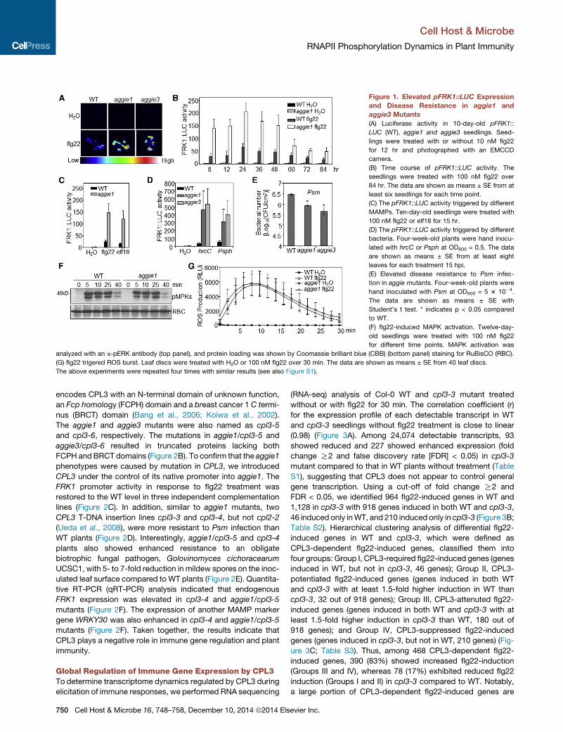
Figure 1. Elevated pFRK1::LUC Expression
and Disease Resistance in aggie1 and
aggie3 Mutants
(A) Luciferase activity in 10-day-old pFRK1::
LUC (WT), aggie1 and aggie3 seedlings. Seed-
lings were treated with or without 10 nM flg22
for 12 hr and photographed with an EMCCD
camera.
(B) Time course of pFRK1::LUC activity. The
seedlings were treated with 100 nM flg22 over
84 hr. The data are shown as means ± SE from at
least six seedlings for each time point.
(C) The pFRK1::LUC activity triggered by different
MAMPs. Ten-day-old seedlings were treated with
100 nM flg22 or elf18 for 15 hr.
(D) The pFRK1::LUC activity triggered by different
bacteria. Four-week-old plants were hand inocu-
lated with hrcC or Psph at OD600 = 0.5. The data
are shown as means ± SE from at least eight
leaves for each treatment 15 hpi.
(E) Elevated disease resistance to Psm infec-
tion in aggie mutants. Four-week-old plants were
hand inoculated with Psm at OD600 = 5 3 10�4.
The data are shown as means ± SE with
Student’s t test. * indicates p < 0.05 compared
to WT.
(F) flg22-induced MAPK activation. Twelve-day-
old seedlings were treated with 100 nM flg22
for different time points. MAPK activation was
analyzed with an a-pERK antibody (top panel), and protein loading was shown by Coomassie brilliant blue (CBB) (bottom panel) staining for RuBisCO (RBC).
(G) flg22 trigered ROS burst. Leaf discs were treated with H2O or 100 nM flg22 over 30 min. The data are shown as means ± SE from 40 leaf discs.
The above experiments were repeated four times with similar results (see also Figure S1).
Cell Host & Microbe
RNAPII Phosphorylation Dynamics in Plant Immunity
encodes CPL3 with an N-terminal domain of unknown function,
an Fcp homology (FCPH) domain and a breast cancer 1 C termi-
nus (BRCT) domain (Bang et al., 2006; Koiwa et al., 2002).
The aggie1 and aggie3 mutants were also named as cpl3-5
and cpl3-6, respectively. The mutations in aggie1/cpl3-5 and
aggie3/cpl3-6 resulted in truncated proteins lacking both
FCPH and BRCT domains (Figure 2B). To confirm that the aggie1
phenotypes were caused by mutation in CPL3, we introduced
CPL3 under the control of its native promoter into aggie1. The
FRK1 promoter activity in response to flg22 treatment was
restored to the WT level in three independent complementation
lines (Figure 2C). In addition, similar to aggie1 mutants, two
CPL3 T-DNA insertion lines cpl3-3 and cpl3-4, but not cpl2-2
(Ueda et al., 2008), were more resistant to Psm infection than
WT plants (Figure 2D). Interestingly, aggie1/cpl3-5 and cpl3-4
plants also showed enhanced resistance to an obligate
biotrophic fungal pathogen, Golovinomyces cichoracearum
UCSC1, with 5- to 7-fold reduction in mildew spores on the inoc-
ulated leaf surface compared to WT plants (Figure 2E). Quantita-
tive RT-PCR (qRT-PCR) analysis indicated that endogenous
FRK1 expression was elevated in cpl3-4 and aggie1/cpl3-5
mutants (Figure 2F). The expression of another MAMP marker
gene WRKY30 was also enhanced in cpl3-4 and aggie1/cpl3-5
mutants (Figure 2F). Taken together, the results indicate that
CPL3 plays a negative role in immune gene regulation and plant
immunity.
Global Regulation of Immune Gene Expression by CPL3To determine transcriptome dynamics regulated by CPL3 during
elicitation of immune responses, we performed RNA sequencing
750 Cell Host & Microbe 16, 748–758, December 10, 2014 ª2014 Els
(RNA-seq) analysis of Col-0 WT and cpl3-3 mutant treated
without or with flg22 for 30 min. The correlation coefficient (r)
for the expression profile of each detectable transcript in WT
and cpl3-3 seedlings without flg22 treatment is close to linear
(0.98) (Figure 3A). Among 24,074 detectable transcripts, 93
showed reduced and 227 showed enhanced expression (fold
change R2 and false discovery rate [FDR] < 0.05) in cpl3-3
mutant compared to that in WT plants without treatment (Table
S1), suggesting that CPL3 does not appear to control general
gene transcription. Using a cut-off of fold change R2 and
FDR < 0.05, we identified 964 flg22-induced genes in WT and
1,128 in cpl3-3 with 918 genes induced in both WT and cpl3-3,
46 induced only inWT, and 210 induced only in cpl3-3 (Figure 3B;
Table S2). Hierarchical clustering analysis of differential flg22-
induced genes in WT and cpl3-3, which were defined as
CPL3-dependent flg22-induced genes, classified them into
four groups: Group I, CPL3-required flg22-induced genes (genes
induced in WT, but not in cpl3-3, 46 genes); Group II, CPL3-
potentiated flg22-induced genes (genes induced in both WT
and cpl3-3 with at least 1.5-fold higher induction in WT than
cpl3-3, 32 out of 918 genes); Group III, CPL3-attenuted flg22-
induced genes (genes induced in both WT and cpl3-3 with at
least 1.5-fold higher induction in cpl3-3 than WT, 180 out of
918 genes); and Group IV, CPL3-suppressed flg22-induced
genes (genes induced in cpl3-3, but not in WT, 210 genes) (Fig-
ure 3C; Table S3). Thus, among 468 CPL3-dependent flg22-
induced genes, 390 (83%) showed increased flg22-induction
(Groups III and IV), whereas 78 (17%) exhibited reduced flg22
induction (Groups I and II) in cpl3-3 compared to WT. Notably,
a large portion of CPL3-dependent flg22-induced genes are
evier Inc.

Figure 2. Aggie1 and Aggie3 Encode CPL3,
a Regulator of Plant Immunity
(A) aggie1 is a recessive mutation. Ten-day-old F1seedlings derived from a cross between aggie1
and WT pFRK1::LUC were treated with 100 nM
flg22 for 12 hr. The data are shown as means ± SE
from at least five seedlings.
(B) The scheme of the CPL3 genomic DNA and
deduced protein domains. The top panel is a
schematic illustration of the CPL3 genomic DNA
with exons (solid box), intron (lines), and 30 UTR(open box). The stars indicate the mutations in
aggie1/cpl3-5 and aggie3/cpl3-6. The other panels
illustrate the protein domain structures of CPL3
and four truncated mutants.
(C) Complementation analysis. C1, C2, and C3 are
three independent transgenic lines of aggie1
complemented with pCPL3::CPL3. Ten-day-old
seedlings were treated with 100 nM flg22 for 12 hr.
The data are shown as means ± SE from at least
ten seedlings.
(D) The cpl3 mutants are more resistant to
bacterial infection. Four-week-old plants were
hand inoculated with Psm at OD600 = 5 3 10�4.
The data are shown as means ± SE with
Student’s t test. * indicates p < 0.05 compared
to WT.
(E) The cpl3 mutants are more resistant to
powdery mildew Golovinomyces cichoracearum
UCSC1. The pictures were taken at 10 dpi on
6-week-old plants. The numbers of spores per
mg tissue (31,000) are shown on the top of the
pictures.
(F) Endogenous FRK1 and WRKY30 expression. The 12-day-old seedlings were treated with 100 nM flg22 for qRT-PCR analysis. The data are shown
as means ± SE from three biological repeats with Student’s t test. * indicates p < 0.05 compared to WT.
The above experiments were repeated three times with similar results (see also Figure S2).
Cell Host & Microbe
RNAPII Phosphorylation Dynamics in Plant Immunity
classified to be associated with defense responses (Table S3
and S4). Enrichment analysis of Gene Ontology (GO) indicates
that among 180 CPL3-attenuted flg22-induced genes (Group
III), the frequency of genes associated with biotic stress, innate
immune response, response to bacterium and fungus, and
salicylic acid (SA)-mediated signaling pathway was significantly
enriched compared to the predicated frequency in the genome
(Figure 3D; Table S4). Many defense-related transcription
factors, such as WRKYs and ERFs, and RLKs were also over-
represented in Group III genes (Table S3). The genes encoding
flg22-activated MKK4 and MPK11 were highly induced in
cpl3-3 compared to WT. Interestingly, cpl3-3 also displayed
the increased expression of genes encoding PROPEP1 and
PROPEP3, the precursors of elicitor peptide PEPs, which func-
tion as endogenous damage-associated molecular pattern to
amplify danger signals during pathogen infection (Liu et al.,
2013). The elevated expression of several flg22-induced genes,
including At1g07160, At1g51920, At1g59860, and At2g17740 in
cpl3-3 and aggie1/cpl3-5 mutants, was confirmed with qRT-
PCR analysis (Figure 3E). Collectively, the data suggest that
CPL3 plays a negative role in regulating a large subset of
flg22-induced genes. Apparently, CPL3-regulated genes did
not show significant correlation with SA, ethylene, methyl
jasmonate (MeJA), and ABA-responsive genes (Figure S3).
Among 101 flg22 downregulated genes in WT and cpl3-3 (fold
changeR2 and FDR < 0.05), only two genes showed >2-fold dif-
Cell Host &
ference between WT and cpl3-3, suggesting that CPL3 does not
appear to significantly control flg22-reduced genes (Table S5).
CTD Phosphorylation Dynamics in PTI SignalingWith the FCPH domain, CPLs are hypothesized to regulate gene
transcription via modulating the phosphorylation status of
RNAPII CTD in the nucleus. When expressed in Arabidopsis pro-
toplasts, CPL3-GFP was observed in the nucleus, which is likely
mediated by a nuclear localization signal (NLS) at its N terminus
(Figure S4A). To reveal the potential involvement of CTD phos-
phorylation in plant immunity, we cloned CTD of Arabidopsis
RNAPII and expressed it in protoplasts. Significantly, flg22 treat-
ment induced a rapidmobility shift of CTD as early as 2min post-
treatment (Figure 4A). The flg22-induced mobility shift could be
removed by calf alkaline intestinal phosphatase (CIP) treatment
(Figure 4B), suggesting the involvement of phosphorylation in
flg22-induced CTDmodification. The elf18 and chitin treatments
also induced CTD mobility shift (Figure 4C). The flg22 treatment
also induced the mobility shift of CTD fused with NLS (NLS-CTD)
(Figure S4B).
Interestingly, flg22 treatment was able to induce CTD phos-
phorylation at Ser sites, as detected by specific antibodies
recognizing pSer2, pSer5, or pSer7 (Figure 4A). In addition,
flg22 treatment induced Ser2, Ser5, and Ser7 phosphoryla-
tion of endogenous CTD of RNAPII in Arabidopsis seedlings
treated with flg22 for different lengths of time (Figure 4D). The
Microbe 16, 748–758, December 10, 2014 ª2014 Elsevier Inc. 751

Figure 3. CPL3 Globally Regulates flg22-Induced Gene Expression
(A) Scatter plot with the expression of whole-genome transcripts between Col-0 (WT) and cpl3-3mutant. Twelve-day-old seedlings without treatment were used
for analysis. y axis indicates gene expression in cpl3-3, and x axis indicates gene expression in WT.
(B) Venn diagram of flg22-induced genes (fold change R2 and FDR < 0.05) in WT and/or cpl3-3 seedlings 30 min after 100 nM flg22 treatment.
(C) Heatmaps of CPL3-dependent flg22-induced genes. The four clusters are defined in the text.
(D) Enrichment of defense-related genes in CPL3-attenuted flg22-induced genes (Group III). The fold enrichment was calculated based on the frequency of genes
annotated to the term in Group III compared to their frequency in the genome.
(E) qRT-PCR analysis of CPL3-regulated genes. The data are shown as means ± SE from three biological replicates with Student’s t test. * indicates p < 0.05;
** indicates p < 0.01 compared to WT. See also Figure S3 and Tables S1, S2, S3, S4, and S5.
Cell Host & Microbe
RNAPII Phosphorylation Dynamics in Plant Immunity
phosphorylation intensity peaked between 10 and 30 min and
gradually declined 60 min after treatment. The phosphorylation
modification was confirmed by CIP treatment, which diminished
the signal detected with a-pSer2 antibody (Figure S4C).
Together, these data indicate that the rapid and transient phos-
phorylation of CTD upon flg22 perception may constitute an
important step in plant immune signaling.
MAPKs Phosphorylate CDKCs in flg22 SignalingCPL3 is predicted to be a CTD Ser2 phosphatase since its yeast
homolog Fcp1 preferentially dephosphorylates Ser2 (Koiwa
et al., 2002). CTD Ser2 is phosphorylated by cyclin-dependent
kinases CDK9 and CDK12 in mammals (Bartkowiak et al.,
2010). We examined the potential involvement of Arabidopsis or-
thologs CDKC;1 and CDKC;2 in CTD phosphorylation (Cui et al.,
2007). The flg22 treatment induced a mobility shift of CDKC;1
(Figure 5A), which could be removed by phosphatase CIP treat-
ment (Figure 5B), suggesting that CDKC;1 was phosphorylated
upon flg22 treatment. CDKC;2 exhibited multiple bands in the
absence of flg22 treatment (Figure 5A). Consistent with the acti-
vation of CDKs by cyclins, coexpression of CYCT1;3, the partner
752 Cell Host & Microbe 16, 748–758, December 10, 2014 ª2014 Els
of CDKCs (Cui et al., 2007), also resulted in two bands of
CDKC;1, similar to that after flg22 treatment (Figure S5A). The
data suggest that Arabidopsis CDKCs are activated upon flg22
perception.
MAPK cascades act as a convergent point mediating multiple
MAMP-triggered signaling. The flg22-induced mobility shift of
CDKC;1 was blocked by a MAPK inhibitor PD184161 (Figures
5C and S5B) and by coexpression of MAPK-specific phospha-
tase MKP (Figures 5D and S5B), suggesting that activation
of MAPKs by flg22 leads to CDKC phosphorylation. MPK3,
MPK4, MPK6, and MPK11 have been shown to be activated
by flg22 treatment (Meng and Zhang, 2013). We tested whether
these MAPKs could directly phosphorylate CDKCs. The
FLAG epitope-tagged MAPK was expressed in protoplasts (Fig-
ure S5C), activated by flg22 treatment, and then immunoprecip-
itated with a-FLAG antibody for an in vitro kinase assay with
GST-CDKC as a substrate. The flg22-activated MPK3 complex
strongly phosphorylated both GST-CDKC;1 and GST-CDKC;2
(Figure 5E). Consistently, the in vitro kinase assay with purified
MPK proteins also indicated that activated MPK3 directly
phosphorylated GST-CDKC;1 and GST-CDKC;2 (Figures 5F
evier Inc.

Figure 4. MAMPs Induce CTD Phosphoryla-
tion
(A) flg22 induces the mobility shift and phosphor-
ylation of CTD in protoplasts. Protoplasts were
expressed with CTD-HA and stimulated with
100 nM flg22 for �2 to 60 min. The proteins were
analyzed by western blot (WB) with a-HA, a-pSer2,
a-pSer5, a-pSer7, or a-pERK antibody. The protein
loading is shown by WB with a-Histone H3 anti-
body.
(B)CIP treatment removesCTD-HAmobility shift. The
flg22-stimulated protoplasts expressing CTD-HA
were treated with 30 units of CIP at 37�C for 1 hr.
(C) Different MAMPs induce CTD mobility shift in protoplasts. The CTD-HA transfected protoplasts were treated with 100 nM flg22 or elf18 or 50 mg/ml chitin
for 5 min.
(D) flg22 induces endogenous CTD phosphorylation in Arabidopsis seedlings. Twelve-day-old seedlings were treated with 100 nM flg22 for indicated times, and
the phosphorylation was detected with specific antibodies. The protein loading was shown by WB with a-CTD antibody.
The above experiments were repeated at least three times with similar results (see also Figure S4).
Cell Host & Microbe
RNAPII Phosphorylation Dynamics in Plant Immunity
and S5D). Activated MPK6 also phosphorylated GST-CDKC;1
and GST-CDKC;2 in vitro (Figure 5F), suggesting potential
redundancy of MPK3 and MPK6 in controlling CDKC activity.
Notably, MPK3 and MPK6 interacted with CDKC;1 and
CDKC;2, but not CYCT1;3, in protoplast coimmunoprecipitation
(coIP) assays (Figures 5G, S5E, and S5F). Sequence analysis of
CDKCs identified two potential MAPK phosphorylation sites
(Ser/Thr followed by Pro): S94 and S259 (Sorensson et al.,
2012). Exchange of S94, but not S259, to Ala blocked flg22-
induced CDKC;1 mobility shift and suppressed CDKC;2 mobility
shift (Figure S5G). Importantly, the CDKC;1S94A and CDKC;2S94A
mutants were no longer phosphorylated by activated MPK3 and
MPK6 in vitro (Figure 5F). Furthermore, mass spectrometry (MS)
analyses identified the phosphorylation of S94 in both CDKC;1
and CDKC;2 phosphorylated by MPK3 or MPK6 (Figures 5H,
S5H, S5I, and S5J). The data support that MPK3 and MPK6
phosphorylate CDKCs at S94.
CDKCs Are MAPK-Activated CTD KinasesTo test whether the flg22-inducedMAPK cascade could activate
CDKC kinase activity toward CTD, we first activated MBP-
CDKCs with MPK3 immunoprecipitated from flg22-treated pro-
toplasts. The activated MBP-CDKC proteins were used for a
kinase assay with GST-CTD as a substrate. The phosphorylation
of CTD was detected by a-pSer2, a-pSer5, and a-pSer7 anti-
bodies. MPK3-activated CDKC;1 or CDKC;2 phosphorylated
CTD at three Ser residues (Figures 6A and S6A). Both
CDKC;1S94A and CDKC;2S94A, which were no longer able to be
phosphorylated by MAPKs, were compromised in their ability
to phosphorylate CTD (Figure 6B). Similar with the activation of
CDKs by cyclins, immunoprecipitated CDKCs with CYCT1;3
could also phosphorylate CTD at Ser2, Ser5, and Ser7 residues
(Figures 6C and 6D). These data indicate that CDKCs are
authentic CTD kinases and that their activity is regulated by an
flg22-induced MAPK cascade.
CPL3 Dephosphorylates CDKC-Mediated CTDPhosphorylationWe next examined whether CPL3 possesses CTD phosphatase
activity. We purified MBP fusion proteins of full-length CPL3,
N-terminal CPL3 (CPL3N), and C-terminal CPL3 (CPL3C)
and performed in vitro phosphatase assays with GST-CTD
Cell Host &
that was phosphorylated by CDKC;1/CYCT1;3 (Figure 6C) or
CDKC;2/CYCT1;3 (Figures 6D and S6B). CPL3 was able to de-
phosphorylate pSer2, pSer5, and pSer7 in vitro with a prefer-
ence for pSer2 (Figure 6C, 6D, and S6B). CPL3C with both the
FCPH and BRCT domains, but not FCPH alone nor CPL3N,
carries the phosphatase activity that completely dephosphory-
lated CTD pSer2 and partially dephosphorylated pSer5 and
pSer7 (Figures 6C and 6D). Our results suggested that both
FCPH and BRCT domains are required for CPL3 phosphatase
activity. We further tested the potential interaction between
CPL3 and CTD. An in vitro pull-down assay indicated that
GST-CTD could pull down CPL3. The interaction between
CPL3 and CTD was markedly enhanced when CTD was phos-
phorylated (Figure 6E). Further analyses with different domains
of CPL3 indicated that both FCPH and BRCT domains were
required for the interaction (Figures S6C and S6D). The interac-
tion between CPL3C and CTD was confirmed with yeast two-
hybrid assay (Figure 6F). These results are consistent with the
finding that yeast Fcp1 predominantly dephosphorylates pSer2
and requires both FCPH and BRCT domains for its catalytic ac-
tivity (Ghosh et al., 2008).
Structural and mutational analyses of yeast Fcp1 defined
essential conserved residues within the FCPH domain (Ghosh
et al., 2008). We mutated some of these conserved resi-
dues in CPL3, including D933 (corresponding to Fcp1D258),
K1046 (Fcp1K280), and D1064D1065 (Fcp1D297D298) to
Ala, and tested their phosphatase activity. All these mutants
were still able to interact with CTD in an in vitro pull-down
assay (Figure S6E). D933A and D1064AD1065A (DD1064AA)
rendered a complete loss of CPL3 catalytic activity (Fig-
ure 6G). Similarly, expression of WT CPL3C, but not D933A
mutant, reduced flg22-induced pFRK1::LUC induction in
cpl3-4 mutant protoplasts, suggesting the functional impor-
tance of D933 (Figure S6F). Surprisingly, the K1046A mutation
did not affect CPL3 activity (Figure 6G). Given that the corre-
sponding residue K280 in Fcp1 was suggested to participate
in transition-state stabilization by binding to acylphosphate
intermediate during CTD dephosphorylation (Ghosh et al.,
2008), our data suggest that K1046 in CPL3 might not be
essential for stabilizing the transition state and that CPL3
has a similar but distinct requirement for catalytic activity
compared to Fcp1.
Microbe 16, 748–758, December 10, 2014 ª2014 Elsevier Inc. 753

Figure 5. MAPKs Phosphorylate CDKCs in
Flagellin Signaling
(A) flg22 induces CDKC mobility shift in protoplasts.
Protoplasts were expressed with CDKC;1-HA or
CDKC;2-HAand treatedwith100nMflg22 for15min.
Total proteinswere separated in a regular 10%SDS-
PAGEgel (top) orsupplementedwith50mMphos-tag
(Wako chemicals USA, Inc.) (middle). The loading
control with a-H3 is shown on the bottom.
(B) CDKC;1 mobility shift was removed by CIP
treatment. Total proteins were separated in a SDS-
PAGE gel supplemented with 50 mMphos-tag (top).
(C) CDKC;1 mobility shift is blocked by MKK in-
hibitor PD184161 in protoplasts. A total of 7.5 mM
PD184161 was added 1 hr before flg22 treatment.
(D) MAPK phosphatase (MKP) blocks CDKC;1
mobility shift. Protoplasts were coexpressed with
CDKC;1-HA and MKP. In (C) and (D), top is a reg-
ular SDS-PAGE and middle is phos-tag gel.
(E) MPK3 phosphorylates GST-CDKC;1 and GST-
CDKC;2 fusion proteins. FLAG epitope-tagged
MPKs were expressed in protoplasts treated with
100 nM flg22 and immunoprecipitated for in vitro
kinase assay with GST-CDKC;1 or GST-CDKC;2 as
substrate. The reactions with myelin basic protein
and GST protein as substrates are shown as
controls. The protein loading of substrates is
shown by CBB staining.
(F) MPKs phosphorylate CDKCs at S94 in vitro. The
recombinant HIS-MPK3 and HIS-MPK6 proteins
were activated by constitutively active MKK5DD and used to phosphorylate GST-CDKCs and their S94A mutants. The phosphorylation was detected by
autoradiograph, and the protein loading is shown by CBB staining. * in (E) and (F) indicates the expected position of GST-CDKC.
(G) CDKC;1 and CDKC;2 interact with MPK3 by coIP assay. FLAG epitope-tagged CDKC and HA epitope-tagged MPK3 were coexpressed in Col-0 protoplasts.
The proteins were immunoprecipitated with a-FLAG agarose beads, immune-blotted with a-HA or a-FLAG antibody. The input of MPK3 and CDKCs is shown
by WB.
(H) CDKC;1 S94 is phosphorylated by MPK3 as shown with MS analysis. The graph indicates the sequence of a doubly charged peptide ion at m/z 469.72 that
matches to EIVTpSPGR of CDKC;1.
The above experiments (except MS assay) were repeated three times with similar results (see also Figure S5).
Cell Host & Microbe
RNAPII Phosphorylation Dynamics in Plant Immunity
We further compared Ser2, Ser5, and Ser7 phosphorylation
level of endogenous CTD inWT and cpl3-4 seedlings. The overall
Ser2 phosphorylation level was enhanced in the cpl3-4 mutant
compared to WT plants with and without flg22 treatment (Fig-
ure 6H). In contrast, the level of Ser5 and Ser7 phosphorylation
appears to be similar in WT and cpl3-4 seedlings (Figure 6H).
This is consistent with in vitro analysis that CPL3 preferentially
dephosphorylates Ser2 of CTD.
CDKCs Positively Regulate Plant Innate ImmunityWe tested the potential involvement of CDKCs in CTD phos-
phorylation and plant immunity. The cdkc;2-1 (SALK_149280)
mutant has undetectable full-length transcripts (Figures S7A
and S7B), whereas the available T-DNA insertion lines of
cdkc;1 (SALK_148550 & SALK_091405) remained similar tran-
script levels as WT plants. We generated CDKC;1 RNAi plants
with reduced expression of CDKC;1 transcripts (Figure S7C).
Consistent with CDKCs being CTD kinases, the flg22-induced
phosphorylation of Ser2, Ser5, or Ser7 of CTD was reduced
in cdkc;1RNAi and cdkc;2 mutant plants (Figures 7A and
S7D). Similarly, the cdkc;1RNAi and cdkc;2 plants showed
reduced induction of MAMP marker genes, including FRK1,
WRKY30, PP2C (At1g07160), and At1g51920 upon flg22
treatment compared to WT plants (Figure 7B). In addition, the
cdkc;1RNAi and cdkc;2 mutant plants were more susceptible
754 Cell Host & Microbe 16, 748–758, December 10, 2014 ª2014 Els
to virulent Pst infection than WT plants as indicated by a 5-
to 8-fold increase of bacterial growth in the mutants (Figure 7C).
The bacterial growth of type-III-deficient Pst hrcC was also
increased in the cdkc;1RNAi or cdkc;2 mutants (Figure 7C).
The increased growth of Psm was also observed in cdkc;2
mutant (Figure S7E). CDKC;1 and CDKC;2 play redundant roles
in plant growth and development and resistance to virus infec-
tions (Cui et al., 2007). Therefore, we generated double mutants
by silencing CDKC;1 in the cdkc;2 mutant using virus-induced
gene silencing (VIGS). The cdkc;2/cdkc;1VIGS plants displayed
wrinkled leaves 3 weeks after VIGS and exhibited retarded
growth 4 weeks after VIGS (Figure S7F). We performed the
pathogen infection assays before the growth defects clearly
appeared. Similar with RNAi plants, the cdkc;1VIGS plants
showed reduced induction of FRK1 and WRKY30 upon flg22
treatment, and the reduction seemed to be further exacerbated
in cdkc;2/cdkc;1VIGS plants compared to plants silenced with
a vector control (Figure S7G). Consistently, the cdkc;1VIGS
mutant plants were more susceptible to Pst infection with
about 5- to 10-fold increase of bacterial growth than control
plants (Figure 7D). Notably, bacterial population in the cdkc;2/
cdkc;1VIGS plants was about 100-fold higher than that in
control plants (Figure 7D). Together, these data indicate that
CDKC;1 and CDKC;2 are positive regulators in plant immunity
to bacterial infections.
evier Inc.

Figure 6. CPL3 Is a CTD Phosphatase
(A) MPK3-activated CDKCs induce GST-CTD
phosphorylation in vitro. MPK3-HA was ex-
pressed in protoplasts treated with 100 nM flg22,
immunoprecipitated with a-HA agarose beads,
and incubated with MBP-CDKC proteins. The
phosphorylated CDKC proteins were collected
by centrifugation as supernatant (MPK3-HA
conjugated beads were in pellets) and used to
phosphorylate GST-CTD. The CTD phosphory-
lation was analyzed by WB with specific anti-
bodies.
(B) CDKC;1S94A and CDKC;2S94A reduce the ability
to phosphorylate GST-CTD.
(C) CPL3 dephosphorylates CDKC;1-activated
GST-CTD in vitro. CDKC;1-HA and CYCT1;
3-HA were expressed in protoplasts and
immunoprecipitated to phosphorylate GST-CTD.
The phosphorylated GST-CTD was dephos-
phorylated by MBP-CPL3N (N), MBP-CPL3C
(C), and MBP-FCPH. CTD phosphorylation
was detected by WB with specific anti-
bodies. CTD loading is shown by WB with a-GST
antibody.
(D) CPL3 dephosphorylates CDKC;2-activated
GST-CTD in vitro.
(E) CPL3 interacts with GST-CTD in vitro. Pull-
down assay was performed by incubating MBP-
CPL3 together with glutathione beads containing
CTD or phosphorylated CTD (pCTD). The HA-
tagged CPL3 proteins were detected with an a-HA WB after glutathione bead pull-down (PD). The input control is shown by WB.
(F) CTD and CPL3C interact in yeast. The interactions between pGADT7-CTD and pGBDT7-CPL3C, pGBDT7-CPL3N, and pGBDT7-FCPH were tested in SD
medium without His, Leu, and Trp (SD-H-L-T) supplemented with 1 mM 3AT. EV is the empty vector pGADT7.
(G) CPL3CD933A and CPL3CDD1064AA lose phosphatase activity.
(H) Enhanced CTD Ser2 phosphorylation in cpl3-4mutant seedling. One-week-old seedlings of WT and cpl3-4were treated with or without 1 mMflg22 for 30 min.
The above experiments were repeated three times with similar results (see also Figure S6).
Cell Host & Microbe
RNAPII Phosphorylation Dynamics in Plant Immunity
To further investigate the involvement of MPK3 and MPK6 in
flg22-induced CTD and CDKC phosphorylation, we generated
estradiol-inducible MPK6 RNAi transgenic plants in mpk3
mutant. The protein level of MPK6 was markedly reduced in
mpk3/MPK6RNAi plants after estradiol treatment (Figure S7H).
Importantly, the flg22-induced CTD Ser2, Ser5, and Ser7
phosphorylation was diminished in the mpk3/MPK6RNAi
plants compared to WT plants (Figure 7E). Furthermore, the
flg22-induced CDKC;1 mobility shift was blocked in the mpk3/
MPK6RNAi plants (Figure 7F), providing genetic evidence
of MPK3 and MPK6 in flg22-induced CDKC;1 and CTD
phosphorylation.
DISCUSSION
Wehave demonstrated that the phosphorylation dynamics of the
RNAPII core transcription machinery is regulated by the evolu-
tionarily conserved kinases and phosphatases in response to
pathogen attacks. Specifically, we revealed a MAMP-induced
phosphorylation relay emanating from a MAPK cascade down-
stream of multiple PRRs to CDKCs and RNAPII CTD that acti-
vates the transcription machinery for immune gene expression.
We also elucidated dephosphorylation of RNAPII CTD by CPL3
phosphatase as a counterregulatory mechanism to fine-tune
transcriptional reprogramming for appropriate immune re-
sponses. Thus, both CDKCs and CPL3 target RNAPII CTD and
Cell Host &
oppositely regulate plant immune gene expression and disease
resistance to bacterial and fungal pathogens (Figure 7G).
Considering the conservation of MAPKs and CTD kinases and
phosphatases among eukaryotes, the immune signaling circuits
identified in this study may represent an evolutionarily conver-
gent regulatory mechanism that eukaryotic cells use to promptly
respond to extracellular stimuli.
As an element of the core transcription machinery, RNAPII is
subject to complex regulation that ensures proper processing
of nascent mRNAs. The phosphorylation dynamics of RNAPII
CTD has been extensively studied, in particular in yeast, and
many CTD binding proteins and transcription events are associ-
ated with specific CTD phosphorylation patterns (Buratowski,
2009). A universal CTD cycle has been proposed to orchestrate
the transcription of virtually all genes through complex interplay
between different kinases, phosphatases, and other modifying
enzymes (Bataille et al., 2012). However, the manner in which
specific cellular responses regulate CTD phosphorylation dy-
namics, which in turn controls gene expression, has been amys-
tery. We show here that MAMP perception markedly induces
rapid and transient CTD phosphorylation on various Ser residues
in its heptad repeats. This phosphorylation is achieved by a
direct phosphorylation relay from a MAPK cascade to CDKCs
and counterregulated by the CPL3 phosphatase. The data sug-
gest an important role of MAPK-mediated RNAPII CTD phos-
phorylation in transcriptional regulation of plant immune genes.
Microbe 16, 748–758, December 10, 2014 ª2014 Elsevier Inc. 755

Figure 7. CDKCs Positively Regulate Plant Immunity
(A) CTD phosphorylation in cdkc mutants. One-week-old seedlings of WT, cdkc;1RNAi (cdkc;1), and cdkc;2 were treated with or without 1 mM flg22 for 30 min.
(B) flg22-induced gene expression. WT and cdkc seedlings were treated with 1 mM flg22 for 1 hr.
(C) Bacterial growth in cdkc plants. WT and cdkc plants were hand inoculated with Pst or hrcC at OD600 = 5 3 10�4.
(D) Bacterial growth in CDKC VIGS-silenced plants. The data in (B)–(D) are shown as mean ± SE from three independent repeats with Student’s t test. * indicates
p < 0.05, and ** indicates p < 0.01 compared to WT.
(E) MPK3/6 are required for flg22-induced CTD phosphorylation. Protoplasts isolated fromWT, andmpk3/MPK6RNAi plants pretreated with 5 mM estradiol were
treated with 100 nM flg22 for 15 min.
(F) CDKC;1mobility shift was partially blocked inmpk3/MPK6RNAi plants. Protoplasts were expressedwith CDKC;1-HA and treatedwith 100 nMflg22 for 15min.
Total proteins were separated in a 8% SDS-PAGE gel supplemented with 50 mM phos-tag.
(G) A model of RNAPII CTD phosphorylation dynamics in plant immunity. Upon flg22 perception by the FLS2/BAK1/BIK1 receptor complex, rapid activation of
MAPK cascade phosphorylates and activates CDKCs, which further phosphorylate the tail of RNAPII CTD heptapeptide. The phosphorylation status of CTD is
counterregulated by CPL3 (Aggie1) through direct dephosphorylation of the Ser residues on CTD. The phosphorylation dynamics of CTD serves as a ‘‘regulatory
code’’ to recruit gene specific transcription factors (TFs) and orchestrate immune gene transcription.
The above experiments were repeated three times with similar results (see also Figure S7).
Cell Host & Microbe
RNAPII Phosphorylation Dynamics in Plant Immunity
CPL3 belongs to a member of multigene family in Arabidopsis
(Koiwa et al., 2002, 2006). Only CPL3 and CPL4 contain both
FCPH and BRCT domains, whereas CPL1 and CPL2 contain
FCPH and double-stranded RNA-binding domains. The cpl1
mutants display altered expression of cold-, salt-, ABA-, and os-
motic-stress-inducible genes, and cpl3 mutants show specific
alternation to ABA responses (Jiang et al., 2013; Koiwa et al.,
2002). Despite lacking the BRCT domain, CPL1 could specif-
ically dephosphorylate Ser5 of Arabidopsis CTD (Koiwa et al.,
2004). In addition, CPL1 dephosphorylates RNA binding protein
HYPONASTIC LEAVES 1 (HYL1) to regulate processing and
strand selection during plant miRNA biogenesis (Manavella
et al., 2012). CPL3 is a prototype Fcp1 phosphatase containing
both FCPH phosphatase catalytic domain and BRCT domain.
756 Cell Host & Microbe 16, 748–758, December 10, 2014 ª2014 Els
Our results support that CPL3 is a genuine CTD Ser2 phospha-
tase and requires both FCPH and BRCT domains for its activity,
which is similar to yeast Fcp1. However, distinct from Fcp1,
CPL3 is not an essential gene. CPL4, the closest homolog of
CPL3, is likely an essential gene involved in plant vegetative
growth and development (Bang et al., 2006). Therefore, it ap-
pears that CPL4 is involved in the regulation of RNAPII activity
for general transcription, whereas CPL3 specifically regulates
RNAPII activity during plant stress responses. It remains un-
known how CPL3 activity is regulated upon MAMP treatment.
It is possible that CPL3 could be activated by upstream kinases
or other regulators in MAMP signaling. Additionally, CPL3 may
interact with specific transcription factors to regulate immune
gene expression.
evier Inc.

Cell Host & Microbe
RNAPII Phosphorylation Dynamics in Plant Immunity
CDKs are often activated by phosphorylation of conserved Thr
residues within the T-loops by CDK-activating kinases. A plant-
specific CDK, CDKF;1 phosphorylates the T-loops of CDKDs
and activates their kinase activity toward CTD (Hajheidari et al.,
2012). Here, we identified the activation of CDKCs by flg22-
induced MAPK cascade. In vivo and in vitro data provide
evidence that MPK3 directly phosphorylates CDKC;1 and
CDKC;2 and activates CDKC kinase activity toward CTD. In
fission yeast, CTD Ser2 phosphorylation is elevated under nitro-
gen starvation condition, which depends on a stress-responsive
MAPK pathway (Sukegawa et al., 2011). In human T cell receptor
and phorbal ester signaling, MAPKs are involved in phosphory-
lation of CDK9 to enhance HIV transcription (Mbonye et al.,
2013). Apparently, activation of CDKs by MAPK cascades might
be a conserved mechanism for the regulation of RNAPII CTD
phosphorylation.
EXPERIMENTAL PROCEDURES
Plant Growth, Generation of pFRK1::LUC Transgenic Plants, and
Mutant Screens
Arabidopsis accession Col-0, Ler, pFRK1::LUC WT and mutant (aggie)
transgenic plants, cpl2-2 (SALK_059753), cpl3-3 (SALK_094720C), cpl3-4
(SALK_051322C), and cdkc;2-1 (SALK_149280C) were grown in pots contain-
ing soil (Metro Mix 360) in a growth room at 23�C, 60% relative humidity and
75 mE m�2s�1 light with a 12 hr photoperiod. To detect CTD phosphorylation
and gene induction, 1-week-old or 12-day-old seedlings grown on ½ MS me-
dium were transferred to water for overnight and then treated with 100 nM
flg22 for indicated time points.
The pFRK1::LUC construct in protoplast transient expression vector (He
et al., 2006) was subcloned into a binary vector pCB302 and introduced into
Arabidopsis Col-0 plants. The transgenic plants were selected with Basta
resistance and analyzed with flg22-induced pFRK1::LUC expression. The
seeds of homozygous pFRK1::LUC transgenic plants were mutagenized by
0.4% EMS. Approximately 6,000 M2 seedlings were grown on liquid ½ MS
medium for 14 days, transferred to water for overnight, and then treated
with 10 nM flg22. The individual seedlings were transferred to each well of
96-well plate 12 hr after flg22 treatment and sprayed with 0.2 mM luciferin.
The plate was put in the dark for 20 min, and the bioluminescence signal
was read by a luminometer (Perkin Elmer). The putative mutants were trans-
ferred to solid ½ MS medium for additional 10 days and then transferred to
soil to set seeds.
Plasmid Construction
TheCTD,CDKC;1,CDKC;2,CYCT1;3,CPL3,CPL3N,CPL3C, andCPL3FCPH
genes were amplified from Col-0 cDNA library with primers containing BamHI
or NcoI at N terminus and StuI at C terminus and introduced into a plant
expression vector pHBT with an HA or FLAG epitope-tag at C terminus.
The point mutations ofCDKC;1S94A,CDKC;1S259A,CDKC;2S94A,CDKC;2S259A,
and different CPL3Cmutants were generated by site-directed mutagenesis. A
400-bp fragment ofCDKC;1was amplified from Col-0 cDNAwith primers con-
taining EcoRI at N terminus and KpnI at C terminus and inserted into VIGS
pYL156 (pTRV-RNA2) vector by EcoRI and KpnI digestion. The CDKC;1
RNAi construct was obtained from Dr. Z. Chen (Cui et al., 2007). The primers
for cloning and point mutations were listed in the Supplemental Experimental
Procedures. The CDKC;1 and CDKC;2 were subcloned into a modified GST
fusion protein expression vector pGEX4T-1 (Pharmacia) or a modified
pMAL-c2 vector (New England BioLabs) with BamHI and StuI digestion, and
different CPL3 constructs were subcloned into a modified pMAL-c2 vector
with SpeI or BamHI and StuI digestion. The recombinant CDKC and CPL3
fusion protein expression vectors were introduced into E. coli strain BL21
(DE3). Expression of fusion proteins and affinity purification were performed
with standard protocol. TheCPL3N,CPL3C, and FCPH genes were subcloned
into a modified pGBKT7 vector (Clontech) with SpeI or BamHI and StuI
digestion, and the CTD gene was subcloned into a modified pGADT7 vector
Cell Host &
(Clontech) with BamHI and StuI digestion for yeast two-hybrid assay. The
constructs of GST-CTD, pCPL3::CPL3, MAPK fusion protein, and protoplast
expression vectors were reported previously (He et al., 2006; Koiwa et al.,
2002, 2004). The MAPK-specific phosphatase (MKP) was cloned from mouse
(Kovtun et al., 1998).
Pathogen infections, NGS, RNA-seq, kinase assay, CTD phosphorylation
and dephosphorylation, VIGS, coIP, andMS assays are listed in Supplemental
Experimental Procedures.
SUPPLEMENTAL INFORMATION
Supplemental Information includes seven figures, seven tables, and Supple-
mental Experimental Procedures and can be found with this article online at
http://dx.doi.org/10.1016/j.chom.2014.10.018.
AUTHOR CONTRIBUTIONS
F.L. performed biochemical assays of CPL3, CTD, CDKC, andMAPK activities
and analyzed CDKC plants; C.C performed phenotypic analysis and cloning of
aggie1, RNA-seq analysis, and VIGS assays; F.C. performed screen, pheno-
typic analysis, cloning, and NGS of aggie3; M.O. sequenced candidate genes
of Aggie1 and performed RNA-seq analysis and CDKC plants; X.Y. generated
mpk6RNAi plants and performed CDKC and CTD phosphorylation in these
plants; X.M. (X. Meng) performed MAPK phosphorylation on CDKCs; A.I.
sequenced candidate genes of Aggie1; K.B. screened aggie1; M.L. performed
Y2H assay; B.L. generated mpk6RNAi plants and validated MAPK inhibitor
andMKP; S.C. performedMSassay; X.M. (X.Ma) and S.X. performed powdery
mildew assay; Y.Z. and Z.F. analyzed RNA-seq data; R.M. and C.J. performed
NGS; H.K. provided reagents; W.S., Z.L., and G.S.F. analyzed data; L.S and
P.H. generated mutant population, designed experiments, analyzed data,
and wrote the paper with input from other authors.
ACKNOWLEDGMENTS
We thank Salk Institute and ABRC for the Arabidopsis T-DNA insertion lines,
Dr. Zhixiang Chen for CDKC and CYCT fusion protein and RNAi constructs,
Dr. Shuqun Zhang for MPK fusion protein constructs, Dr. Keiko Torii for
pTK103 vector, Mr. Ning Zhu for technical assistance of MS analysis, and
Drs. Greg Martin and Paul de Figueiredo for critical reading of the manuscript.
The work was supported by NIH (R01GM092893) and NSF (IOS-1252539) to
P.H and NIH (R01GM097247) and the Robert A. Welch Foundation (A-1795)
to L.S. and NSF (MCB-0950459) to H.K. The NGS was supported by Texas
AgriLife Genomics Seed Grant. F.L. and F.C. were partially supported byChina
Scholarship Council. M.V.V.O. and A.C.I. were partially supported by Rio de
Janeiro State Research Foundation (FAPERJ), Brazil. K.B. was supported by
NSF REU program. The authors have declared no conflict of interests.
Received: April 29, 2014
Revised: August 22, 2014
Accepted: October 24, 2014
Published: November 26, 2014
REFERENCES
Asai, T., Tena, G., Plotnikova, J., Willmann, M.R., Chiu, W.L., Gomez-Gomez,
L., Boller, T., Ausubel, F.M., and Sheen, J. (2002). MAP kinase signalling
cascade in Arabidopsis innate immunity. Nature 415, 977–983.
Bang, W., Kim, S., Ueda, A., Vikram, M., Yun, D., Bressan, R.A., Hasegawa,
P.M., Bahk, J., and Koiwa, H. (2006). Arabidopsis carboxyl-terminal domain
phosphatase-like isoforms share common catalytic and interaction domains
but have distinct in planta functions. Plant Physiol. 142, 586–594.
Bartkowiak, B., Liu, P., Phatnani, H.P., Fuda, N.J., Cooper, J.J., Price, D.H.,
Adelman, K., Lis, J.T., and Greenleaf, A.L. (2010). CDK12 is a transcription
elongation-associated CTD kinase, the metazoan ortholog of yeast Ctk1.
Genes Dev. 24, 2303–2316.
Bataille, A.R., Jeronimo, C., Jacques, P.E., Laramee, L., Fortin, M.E., Forest,
A., Bergeron, M., Hanes, S.D., and Robert, F. (2012). A universal RNA
Microbe 16, 748–758, December 10, 2014 ª2014 Elsevier Inc. 757
Cell Host & Microbe
RNAPII Phosphorylation Dynamics in Plant Immunity
polymerase II CTD cycle is orchestrated by complex interplays between
kinase, phosphatase, and isomerase enzymes along genes. Mol. Cell 45,
158–170.
Boller, T., and Felix, G. (2009). A renaissance of elicitors: perception of
microbe-associated molecular patterns and danger signals by pattern-recog-
nition receptors. Annu. Rev. Plant Biol. 60, 379–406.
Buratowski, S. (2009). Progression through the RNA polymerase II CTD cycle.
Mol. Cell 36, 541–546.
Chinchilla, D., Zipfel, C., Robatzek, S., Kemmerling, B., Nurnberger, T., Jones,
J.D., Felix, G., andBoller, T. (2007). A flagellin-induced complex of the receptor
FLS2 and BAK1 initiates plant defence. Nature 448, 497–500.
Cui, X., Fan, B., Scholz, J., and Chen, Z. (2007). Roles of Arabidopsis cyclin-
dependent kinase C complexes in cauliflower mosaic virus infection, plant
growth, and development. Plant Cell 19, 1388–1402.
Ghosh, A., Shuman, S., and Lima, C.D. (2008). The structure of Fcp1, an
essential RNA polymerase II CTD phosphatase. Mol. Cell 32, 478–490.
Hajheidari, M., Farrona, S., Huettel, B., Koncz, Z., and Koncz, C. (2012).
CDKF;1 and CDKD protein kinases regulate phosphorylation of serine resi-
dues in the C-terminal domain of Arabidopsis RNA polymerase II. Plant Cell
24, 1626–1642.
Hajheidari, M., Koncz, C., and Eick, D. (2013). Emerging roles for RNA
polymerase II CTD in Arabidopsis. Trends Plant Sci. 18, 633–643.
He, P., Shan, L., Lin, N.C., Martin, G.B., Kemmerling, B., Nurnberger, T., and
Sheen, J. (2006). Specific bacterial suppressors of MAMP signaling upstream
of MAPKKK in Arabidopsis innate immunity. Cell 125, 563–575.
Heese, A., Hann, D.R., Gimenez-Ibanez, S., Jones, A.M., He, K., Li, J.,
Schroeder, J.I., Peck, S.C., and Rathjen, J.P. (2007). The receptor-like kinase
SERK3/BAK1 is a central regulator of innate immunity in plants. Proc. Natl.
Acad. Sci. USA 104, 12217–12222.
Jiang, J., Wang, B., Shen, Y., Wang, H., Feng, Q., and Shi, H. (2013). The
arabidopsis RNA binding protein with K homology motifs, SHINY1, interacts
with the C-terminal domain phosphatase-like 1 (CPL1) to repress stress-
inducible gene expression. PLoS Genet. 9, e1003625.
Kadota, Y., Sklenar, J., Derbyshire, P., Stransfeld, L., Asai, S., Ntoukakis, V.,
Jones, J.D., Shirasu, K., Menke, F., Jones, A., and Zipfel, C. (2014). Direct
regulation of the NADPH oxidase RBOHD by the PRR-associated kinase
BIK1 during plant immunity. Mol. Cell 54, 43–55.
Koiwa, H., Barb, A.W., Xiong, L., Li, F., McCully, M.G., Lee, B.H., Sokolchik, I.,
Zhu, J., Gong, Z., Reddy,M., et al. (2002). C-terminal domain phosphatase-like
family members (AtCPLs) differentially regulate Arabidopsis thaliana abiotic
stress signaling, growth, and development. Proc. Natl. Acad. Sci. USA 99,
10893–10898.
Koiwa, H., Hausmann, S., Bang, W.Y., Ueda, A., Kondo, N., Hiraguri, A.,
Fukuhara, T., Bahk, J.D., Yun, D.J., Bressan, R.A., et al. (2004). Arabidopsis
C-terminal domain phosphatase-like 1 and 2 are essential Ser-5-specific
C-terminal domain phosphatases. Proc. Natl. Acad. Sci. USA 101, 14539–
14544.
Koiwa, H., Bressan, R.A., and Hasegawa, P.M. (2006). Identification of plant
stress-responsive determinants in Arabidopsis by large-scale forward genetic
screens. J. Exp. Bot. 57, 1119–1128.
Kovtun, Y., Chiu, W.L., Zeng, W., and Sheen, J. (1998). Suppression of auxin
signal transduction by aMAPK cascade in higher plants. Nature 395, 716–720.
Li, L., Li, M., Yu, L., Zhou, Z., Liang, X., Liu, Z., Cai, G., Gao, L., Zhang, X.,
Wang, Y., et al. (2014). The FLS2-associated kinase BIK1 directly phosphory-
758 Cell Host & Microbe 16, 748–758, December 10, 2014 ª2014 Els
lates the NADPH oxidase RbohD to control plant immunity. Cell Host Microbe
15, 329–338.
Lin, W., Li, B., Lu, D., Chen, S., Zhu, N., He, P., and Shan, L. (2014). Tyrosine
phosphorylation of protein kinase complex BAK1/BIK1 mediates Arabidopsis
innate immunity. Proc. Natl. Acad. Sci. USA 111, 3632–3637.
Liu, Z., Wu, Y., Yang, F., Zhang, Y., Chen, S., Xie, Q., Tian, X., and Zhou, J.M.
(2013). BIK1 interacts with PEPRs to mediate ethylene-induced immunity.
Proc. Natl. Acad. Sci. USA 110, 6205–6210.
Lu, D., Wu, S., Gao, X., Zhang, Y., Shan, L., and He, P. (2010). A receptor-like
cytoplasmic kinase, BIK1, associates with a flagellin receptor complex to
initiate plant innate immunity. Proc. Natl. Acad. Sci. USA 107, 496–501.
Manavella, P.A., Hagmann, J., Ott, F., Laubinger, S., Franz, M., Macek, B., and
Weigel, D. (2012). Fast-forward genetics identifies plant CPL phosphatases as
regulators of miRNA processing factor HYL1. Cell 151, 859–870.
Mbonye, U.R., Gokulrangan, G., Datt, M., Dobrowolski, C., Cooper, M.,
Chance, M.R., and Karn, J. (2013). Phosphorylation of CDK9 at Ser175
enhances HIV transcription and is a marker of activated P-TEFb in CD4(+) T
lymphocytes. PLoS Pathog. 9, e1003338.
Meng, X., and Zhang, S. (2013). MAPK cascades in plant disease resistance
signaling. Annu. Rev. Phytopathol. 51, 245–266.
Postel, S., Kufner, I., Beuter, C., Mazzotta, S., Schwedt, A., Borlotti, A., Halter,
T., Kemmerling, B., and Nurnberger, T. (2010). The multifunctional leucine-rich
repeat receptor kinase BAK1 is implicated in Arabidopsis development and
immunity. Eur. J. Cell Biol. 89, 169–174.
Roux, M., Schwessinger, B., Albrecht, C., Chinchilla, D., Jones, A., Holton, N.,
Malinovsky, F.G., Tor, M., de Vries, S., and Zipfel, C. (2011). The Arabidopsis
leucine-rich repeat receptor-like kinases BAK1/SERK3 and BKK1/SERK4 are
required for innate immunity to hemibiotrophic and biotrophic pathogens.
Plant Cell 23, 2440–2455.
Sorensson, C., Lenman, M., Veide-Vilg, J., Schopper, S., Ljungdahl, T., Grøtli,
M., Tamas, M.J., Peck, S.C., and Andreasson, E. (2012). Determination of
primary sequence specificity of Arabidopsis MAPKs MPK3 and MPK6 leads
to identification of new substrates. Biochem. J. 446, 271–278.
Sukegawa, Y., Yamashita, A., and Yamamoto, M. (2011). The fission yeast
stress-responsive MAPK pathway promotes meiosis via the phosphorylation
of Pol II CTD in response to environmental and feedback cues. PLoS Genet.
7, e1002387.
Sun, Y., Li, L., Macho, A.P., Han, Z., Hu, Z., Zipfel, C., Zhou, J.M., and Chai, J.
(2013). Structural basis for flg22-induced activation of the Arabidopsis FLS2-
BAK1 immune complex. Science 342, 624–628.
Tena, G., Boudsocq, M., and Sheen, J. (2011). Protein kinase signaling net-
works in plant innate immunity. Curr. Opin. Plant Biol. 14, 519–529.
Ueda, A., Li, P., Feng, Y., Vikram, M., Kim, S., Kang, C.H., Kang, J.S., Bahk,
J.D., Lee, S.Y., Fukuhara, T., et al. (2008). The Arabidopsis thaliana
carboxyl-terminal domain phosphatase-like 2 regulates plant growth, stress
and auxin responses. Plant Mol. Biol. 67, 683–697.
Zhang, J., Li, W., Xiang, T., Liu, Z., Laluk, K., Ding, X., Zou, Y., Gao, M., Zhang,
X., Chen, S., et al. (2010). Receptor-like cytoplasmic kinases integrate
signaling from multiple plant immune receptors and are targeted by a
Pseudomonas syringae effector. Cell Host Microbe 7, 290–301.
Zipfel, C., Kunze, G., Chinchilla, D., Caniard, A., Jones, J.D., Boller, T., and
Felix, G. (2006). Perception of the bacterial PAMP EF-Tu by the receptor
EFR restricts Agrobacterium-mediated transformation. Cell 125, 749–760.
evier Inc.