Cell Culture Dr. Tarek Elbashiti Assoc. Prof. of Biotechnology.
74
Cell Culture Dr. Tarek Elbashiti Assoc. Prof. of Biotechnology
-
Upload
laurel-gray -
Category
Documents
-
view
219 -
download
0
Transcript of Cell Culture Dr. Tarek Elbashiti Assoc. Prof. of Biotechnology.

Cell Culture
Dr. Tarek Elbashiti
Assoc. Prof. of Biotechnology

The term cell culture refers to the cultivation of dispersed (isolated) cells taken from an original tissue, a primary culture, or a cell line.
The practice of cultivating cells started at the beginning of the 20th century and was developed from a simple exploratory phase to an expansion phase in the 1950s.
For more than 50 years, the culture of cells derived from primary tissue explants has predominated, justifying the original name ‘‘tissue culture.’’

Currently, cell culture is in a specialization phase. With the increase use of detached cells since the 1950s, the term tissue culture was substituted by cell culture.
At the present time, cell culture techniques allow in vitro propagation (proliferation) of various cell lines including those from insects, humans, mice, rats, and other mammals.

Basically, animal cell culture techniques are similar to those employed for bacteria, fungi, and yeast, although there are some characteristic differences.
In general, animal cells are more delicate, vulnerable to mechanical damage, present lower growth rates, and require more complex culture media and special substrates

cell culture has to be performed under rigorous aseptic conditions, since animal cells grow more slowly than most usual contaminants, such as bacteria and fungi.

HISTORICAL BACKGROUND
Tissue culture was first developed at the beginning of the twentieth century as a method for studying the behavior of animal cells free of systemic variations that might arise in vivo both during normal homeostasis and under the stress of an experiment.
The technique was elaborated first with un-disaggregated fragments of tissue, and growth was restricted to the migration of cells from the tissue fragment, with occasional mitoses in the outgrowth.

Roux in 1885 for the first time maintained embryonic chicken cells in saline).
Cell culture was first successfully undertaken by Ross Harrison in 1907( lymph)
1911: Lewis made the first liquid media consisted of sea water, serum, embryo extract, salts and peptones. They observed limited monolayer growth.

1913: Carrel introduced strict aseptic techniques so that cells could be cultured for long periods.
1916: Rous and Jones introduced proteolytic enzyme trypsin for the subculture of adherent cells.
1940s: The use of the antibiotics penicillin and streptomycin in culture medium decreased the problem of contamination in cell culture.
Sanford was the first cloned cell strain, isolated by capillary cloning from mouse L-cells [Sanford et al., 1948].
It was not until the 1950s that trypsin became more generally used for subculture, following procedures described by Dulbecco to obtain passaged monolayer cultures for viral plaque assays
[Dulbecco, 1952], and the generation of a single cell suspension by trypsinization, which facilitated the further development of single cell cloning.
1955: Gey established the first continuous human cell line, HeLa this was subsequently cloned by Puck, 1955]
Tissue culture became more widely used at this time because of the introduction of antibiotics, which facilitated long-term cell line proliferation although many people were already warning against continuous use and the associated risk of harboring cryptic, or antibiotic-resistant, contaminations .

• The 1950s were also the years of the development of defined media which led ultimately to the development of serum-free media 1966.

MAJOR
First was the use of chemically defined culture medium.
Second was the development was the use of antibiotics which inhibits the growth of contaminants.
Third was the use of trypsin to remove adherent cells to subculture further from the culture vessel

Advantages Model systems for Studying basic cell biology, interactions between disease
causing agents and cells, effects of drugs on cells, process and triggering of aging & nutritional studies
Toxicity testing Study the effects of new drugs
Cancer research Study the function of various chemicals, virus & radiation to
convert normal cultured cells to cancerous cells

Advantages
Virology Cultivation of virus for vaccine production,
also used to study there infectious cycle. Genetic Engineering Production of commercial proteins, large
scale production of viruses for use in vaccine production e.g. polio, rabies, chicken pox, hepatitis B & measles
Gene therapy Cells having a functional gene can be
replaced to cells which are having non-functional gene

Advantages
To produce artificial tissues (skin, heart, cartilage)
Use single cell (impossible in vivo)

Disadvantages
Cell characteristics can be changed Cell adapts to different nutrients If mixed cells cultivated some types
will disappeared. Activity of enzymes may altered by
environment. Necessary expertise Quantity and cost
Typical structure of an animal cell Various animal cell lines can be cultivated
in vitro, such as cardiac cells, fibroblasts, smooth muscle cells, endocrine cells (such as pituitary, adrenal, and pancreatic cells), epithelial cells (such as liver, mammary, lung, and kidney cells), tumor cells (such as melanocytes), nervous system cells (such as glial cells and neurons), as well as hybridomas. Although these cells have different origins and functions, they all show a typical structure.
Animal tissue Cultures Classification
1-classification a:
A-Organ cultureB-Tissue cultureC-Cell culture

Types
A- Organ Culture
The term organ culture will always imply a three-dimensional culture of undisaggregated tissue retaining some or all of the histological features of the tissue in vivo.
The entire embryos or organs are excised
from the body and cultured

Disadvantages Scale-up is not recommended. Growth is slow. Fresh explanation is required for every
experiment.

the term tissue culture is used as a general term to include organ culture and cell culture.
Fragments of excised tissue are grown in culture media
A fragment of tissue is placed at a glass (or plastic)–liquid interface, where, after attachment, migration is promoted in the plane of the solid substrate
B-Tissue Culture

Advantages Some normal functions may be
maintained. Better than organ culture for scale-up
but not ideal. Disadvantages
Original organization of tissue is lost.
C- Cell Culture
Cell culture refers to a culture derived from dispersed cells taken from original tissue, from a primary culture, or from a cell line or cell strain by enzymatic, mechanical, or chemical disaggregation into a cell suspension, which may then be cultured as an adherent monolayer on a solid substrate or as a suspension in the culture medium

Advantages Development of a cell line over several
generations Scale-up is possible
Disadvantages Cells may lose some differentiated
characteristics.

classification b
1. Primary Cultures Derived directly from excised tissue and cultured either
as Outgrowth of excised tissue in culture Dissociation into single cells (by enzymatic digestion
or mechanical dispersion) Advantages:
usually retain many of the differentiated characteristics of the cell in vivo
Disadvantages: initially heterogeneous but later become dominated
by fibroblasts. the preparation of primary cultures is labor intensive can be maintained in vitro only for a limited period
of time.

2. Continuous Cultures derived from subculture (or passage, or transfer)
of primary culture Subculture = the process of dispersion and
re-culture the cells after they have increased to occupy all of the available substrate in the culture
can be serially propagated in culture for several passages
There are two types of continuous cultures Cell lines Continuous cell lines

1) Cell lines finite life, senesce after approximately
thirty cycles of division
usually diploid and maintain some degree of differentiation.

2) Continuous cell lines can be propagated indefinitely generally have this ability because they
have been transformed tumor cells. viral oncogenes chemical treatments.
the disadvantage of having retained very little of the original in vivo characteristics

Common cell lines
Human cell lines -MCF-7 breast cancer HL 60 Leukemia HEK-293 Human embryonic kidney HeLa Henrietta lacks Primate cell lines Vero African green monkey kidney
epithelial cells Cos-7 African green monkey kidney cells And others such as CHO from hamster, sf9 &
sf21 from insect cells

American Type Culture Collection .

Culture Morphology
Suspension (as single cells or small free-floating clumps)
or as a monolayer that is attached to the tissue culture flask.
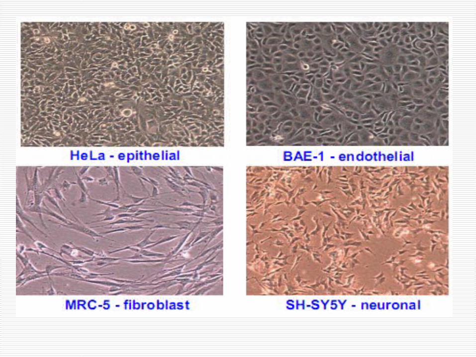
The form taken by a cell line reflects the tissue from which it was derived
from blood tend to grow in suspension from solid tissue (lungs, kidney) tend to grow
as monolayer's. Attached cell lines can be classified as
endothelial, epithelial, neuronal or fibroblasts and their morphology reflect the area within the tissue of origin
Anchorage dependent or independent
Cell lines derived from normal tissues are considered as anchorage-dependent grows only on a suitable substrate. ( Epith, CT)
Suspension cells are anchorage-independent e.g. blood cells

Subculture of the Adherent cells
Cells which are anchorage dependent Add enough trypsin/EDTA to cover the
monolayer Incubate the plate at 37 C for 5 min. Tap the vessel from the sides to dislodge
the cells Add complete medium to dissociate and
dislodge the cells with the help of pipette which are remained to be adherent
Add complete medium depends on the subculture requirement either to 75 cm or 175 cm flask

Suspension cells
Easier to passage as no need to detach them
As the suspension cells reach to confluence aseptically remove 1/3rd of medium
Replace with the same amount of pre-warmed medium
Confluence
Once the available substrate surface is covered by cells, growth slows & ceases.

hep 3B - 70% confluency

100% confluence

After 24 h

Confluence
Cells to be kept in healthy & in growing state have to be sub-cultured or passaged, when they reach 80-90% confluence in flask/dishes/plates
Enzyme such as trypsin, dipase, collagenase in combination with EDTA breaks the cellular glue that attached the cells to the surface

Histotypic Culture or Histoculture
The high-density, or “tissue like” culture of one cell type.
Organotypic Culture
The presence of more than one cell type interacting as they might in the organ of origin (or a simulation of such interaction).

Design and Equipment for the Cell Culture Laboratory

Design and Equipment for the Cell Culture Laboratory
1. Laboratory design- in a safe and efficient manner
2. safety cabinets

Laminar- flow hood

Laminar- flow hood
The working environment is protected from dust and contamination by a
Constant, stable flow of filtered air Two types: Horizontal, airflow blow from the side
facing you, parallel to the work surface, and is not circulating
Vertical, air blows down from the top of the cabinet onto the work surface and is drawn through the work surface and either re-circulated or vented.


Laminar- flow hood
- The efficiency of the hood depends on a minimum pressure drop across the filter
- When the filter resistance builds up, the pressure drop increases and the flow rate of air falls.
Below 0.4 m/s (80 ft/min), the stability of the laminar airflow is lost, and sterility can no longer be maintained. The pressure drop can be monitored with a manometer fitted to the cabinet


Laminar- flow hood
Routine maintenance checks of the primary filters are required (every 3-6 months).
They might be removed and discarded or washed in soap and water.
Every 6 months the main high efficiency particulate air (HEPA) filter above the work surface should be checked for airflow and hole

Before use
Ultraviolet lights are used to sterilize the air and exposed work surfaces in laminar flow cabinets between use.
Detergent 70% alcohol

Cell Culture Incubator

It requires a controlled atmosphere with high humidity and super controlled of CO2 tension.
The incubator should be large enough, probably 50-200 liter, have forced air circulation, temperature control and a safety thermostat that cuts out if the incubator overheats.

- It should be stainless steel, and easily cleaned.
• A double cabinet, one above the other, independently regulated, is preferable to one large cabinet.
• Incubators are supplied either with a heated water jacket as a method for distributing the heat evenly around the cabinet or with surface heater elements for heating.

Humid CO2 Incubator
CO2 incubators are more expensive, but there ease of use and superior in the control of CO2 tension and temperature.
A controlled atmosphere is achieved by
using a humidifying tray and controlling the CO2 tension with a CO2-monitoring device, which draws air from the incubator into a sample chamber, determines the concentration of CO2, and injects pure CO2 into the incubator to make up any deficiency.

Air is circulated around the incubator by natural convection or by using a fan to keep both the CO2 level and the temperature uniform.
Dry, heated wall incubators also
encourage less fungal contamination on the walls, as the walls tend to remain dry, even at high relative humidity.

Moist heat denatures proteins by coagulation, caused by H bonds breakage, can happened more quickly in the presence of water
Autoclave: Steam under pressure
Air should be Exhausted
Sterlization Heat
Figure 7.2

Pasteurization reduces spoilage organisms and pathogens and can be safe if refregerated
Equivalent treatments: as the temprature increase the time decreased and the same No of m/o killed 63°C for 30 min High-temperature short-time P 72°C for
15 sec Ultra-high-temperature P: 140°C for <1
sec Thermoduric organisms survive

Dry Heat Sterilization kills by oxidation effect Flaming Incineration Hot-air sterilization temp 170˚C for 2 hrs
Physical Methods of Microbial Control
Hot-air Autoclave
Equivalent treatments
170˚C, 2 hr 121˚C, 15 min

Steam Sterilizer (Autoclave)
A simple bench-top autoclave that generates 100 kPa (1 atm, 15 lb/in.2) may be sufficient, but a larger model with a timer and a choice of presterilization and poststerilization evacuation and temperature recording give more capacity and greater flexibility in use.
Offers the opportunity to comply with good laboratory practice (GLP).

The chamber should also be evacuated after sterilization, to remove steam and promote subsequent drying;
Otherwise the articles will emerge wet, leaving a trace of contamination from the condensate on drying.
To minimize this risk when a ‘‘postvac’’ cycle is not available, always use deionized or reverse-osmosis water to supply the autoclave.

Cell culture contaminants
Two types:1- Chemicals-difficult to detect caused by
endotoxins, plasticizers, metal ions or traces of disinfectants that are invisible
2- Contamination by microorganisms remains a major problem in tissue culture.
Bacteria, mycoplasma, yeast, and fungal spores may be introduced via the operator, the atmosphere, work surfaces, solutions, and many other sources.


Contamination
They competes for nutrients with host cells Secreted acidic or alkaline by-products
ceases the growth of the host cells. Degraded arginine & purine inhibits the
synthesis of histone and nucleic acid They also produces H2O2 which is directly
toxic to cells.

Safety


For best results in tissue culture, we want to work to keep microbial (bacteria, yeast and molds) contamination to a minimum.
Guidelines to follow: Work in a culture hood set-aside for tissue
culture purposes. Most have filtered air that blows across the
surface to keep microbes from settling in the hood.
Turn off the UV/antimicrobial light and turn on the hood 30 minutes prior to entering the hood.

Wear short sleeves or roll your sleeves up. Turn your baseball caps back if you MUST wear them, tie long hair back and remove rings and watches.
Wash hands with soap and water before beginning the procedure and rewash if you touch anything that is not sterile or within the hood.

Spray down your hands, work surface, and anything that will go into the hood with 70% ethanol. Re-wipe at intervals if you are working for a long time in the hood. This will reduce the numbers of bacteria and mold considerably.
Do not breath directly into your cultures, bottles of media, etc. This also means to keep talking to a minimum. No singing or chewing gum.

Work as quickly as you can within limits of your coordination. Also, keep bottles and flasks closed when you are not working with them. Avoid passing your arm or hand over an open bottle.
Use only sterilized pipettes, plates, flasks and bottles in the hood for procedures.

Take special precautions with the sterile pipette. Remove them from the package just before use. Make certain to set up the numbers on the pipette so that they face you.
Never mouth-pipette, use the pipetting aid.