Cell-based Assays - BioCat GmbH · Cell-based Assays The understanding of ... DNA Ladder Assay Kits...
8
BioCat GmbH Technologiepark Tel.: +49 (0) 6221 71415 16 Im Neuenheimer Feld 584 Fax: +49 (0) 6221 71415 29 D-69120 Heidelberg E-Mail: [email protected] www.biocat.com Cell-based Assays The understanding of gene and protein functions and the regulatory mechanisms controlling these, as well as the screening for potential inhibitors or inducers of biological processes, all require the analy- sis of functional cells. Cultured cells constitute a comprehensive unit that enables functional analysis. Screens in cell-based systems offer accurate representation of the real-life cell model and the possibility of a dynamic experiment through in vitro monitoring of the numbers, phenotype or behavior of the live cells. The diversity of biomolecules present in cells and the need to cha- racterize their presence in normal development/diseased states or treated/untreated conditions leads to an increased demand for cor- responding assays. In addition to a broad collection of fluorescent proteins ranging in co- lor from blue to far-red, a wide variety of cell-based assays designed for greater accuracy and increased throughput is provided. They allow for the analysis of apoptosis, cell proliferation and cell damage as well as cell adhesion, migration and invasion. They provide fully quanti- tative results with no manual cell counting required. The majority of the assays is performed in microplate format. Analysis is carried out by colorimetric or fluorometric detection. www.biocat.com/cell-based Apoptosis Caspases & Caspase Inhibitors Cytotoxicity & Proliferation Cell Adhesion Cell Migration & Invasion Fluorescent Proteins In this brochure, new and especially important products are highlighted. For the complete listing as well as for more detailed product and order information, please use the respective web link at the end of each section.
Transcript of Cell-based Assays - BioCat GmbH · Cell-based Assays The understanding of ... DNA Ladder Assay Kits...
BioCat GmbHTechnologiepark Tel.: +49 (0) 6221 71415 16 Im Neuenheimer Feld 584 Fax: +49 (0) 6221 71415 29D-69120 Heidelberg E-Mail: [email protected] www.biocat.com
Cell-based Assays The understanding of gene and protein functions and the regulatory mechanisms controlling these, as well as the screening for potential inhibitors or inducers of biological processes, all require the analy-sis of functional cells. Cultured cells constitute a comprehensive unit that enables functional analysis. Screens in cell-based systems offer accurate representation of the real-life cell model and the possibility of a dynamic experiment through in vitro monitoring of the numbers, phenotype or behavior of the live cells. The diversity of biomolecules present in cells and the need to cha-racterize their presence in normal development/diseased states or treated/untreated conditions leads to an increased demand for cor-responding assays.In addition to a broad collection of fluorescent proteins ranging in co-lor from blue to far-red, a wide variety of cell-based assays designed for greater accuracy and increased throughput is provided. They allow for the analysis of apoptosis, cell proliferation and cell damage as well as cell adhesion, migration and invasion. They provide fully quanti-tative results with no manual cell counting required. The majority of the assays is performed in microplate format. Analysis is carried out by colorimetric or fluorometric detection.
www.biocat.com/cell-based
Apoptosis
Caspases & Caspase Inhibitors
Cytotoxicity & Proliferation
Cell Adhesion
Cell Migration & Invasion
Fluorescent Proteins
In this brochure, new and especially important products are highlighted. For the complete listing as well as for more detailed product and order information, please use the respective web link at the end of each section.
Apoptosis• Analyze different stages of apoptosis• Differentiate between apoptotic and necrotic cells• Select the assay suitable for your sample type Apoptosis, or programmed cell death, plays an important role in many physiological and diseased conditions. In contrast to necrotic cells, apoptotic cells are characterized morphologi-cally by compaction of the nuclear chromatin, shrinkage of the cytoplasm and production of membrane-bound apopto-tic bodies. Biochemically, apoptosis is distinguished by frag-mentation of the genome and cleavage or degradation of several cellular proteins.As no single parameter fully defines cell death in all systems, it is often advantageous to use several different approaches when studying apoptosis. A large number of assays is offered for the detection of apoptosis at the early, middle and late stages of the apoptotic cascade, and for the analysis of apo-ptotic events that occur in different areas of the cell, including the plasma membrane, cytoplasm, mitochondria and nucleus.
Sample Type Product
Live Cells (Suspension or Adherent)
Annexin V Apoptosis Assay KitsCaspGlow Caspase Assay KitsMitoCapture Apoptosis Assay Kit
Cell Lysate (Fresh or Frozen) Caspase (1-12) Fluorometric Assay KitsCaspase (1-10) Colorimetric Assay Kits
Tissue Lysate (Fresh or Frozen) Caspase (1-12) Fluorometric Assay KitsADP, ATP & ADP/ATP Ratio Assay Kits
Tissue Sections (Frozen or Paraffin) Apo-BrdU-IHCActive Caspase-3 Antibody
Cell Area Product
Mitochondrial ChangesMitoCapture Apoptosis Assay KitCytochrome c Releasing Apoptosis Assay Kit
Membrane Changes Annexin V Assay Kits
Nuclear Changes DNA Ladder Assay KitsTUNEL Assay Kits
Cytosol Changes Caspase Assay Kits
Purpose Product
Measure Cytochrome c Release Cytochrome c Releasing Apoptosis Assay Kit
Screen Caspase Inhibitors Caspase Inhibitor Screening Kits
Block Apoptotic Gene Expression siRNA Apoptotic Gene Expression Vectors
Study Apoptosis-related Non-Caspase Proteases
Calpain Assay KitCathepsin Assay Kits
Isolate Apoptotic Cells Apoptotic Cell Isolation Kit
Induce Apoptosis Ready-to-use Apoptosis Inducers
Differentiate betweenApoptosis & Necrosis
Annexin V-FITC Apoptosis KitAnnexin V-EGFP Apoptosis Kit
Autophagy is closely associated with apoptosis. Cells use autophagy to digest damaged or defective molecules, effec-tively eliminating the stressors like misfolded proteins and damaged organelles that can threaten cell health. An ex-tensive collection of autophagy antibodies, including APG8 (MAPLC3), LAMP and APG1, is offered.
www.biocat.com/apoptosis
Caspases & Caspase Inhibitors• Active recombinant caspases and procaspases• Ready-to-use cell-permeable caspase inhibitors• High quality and high activity
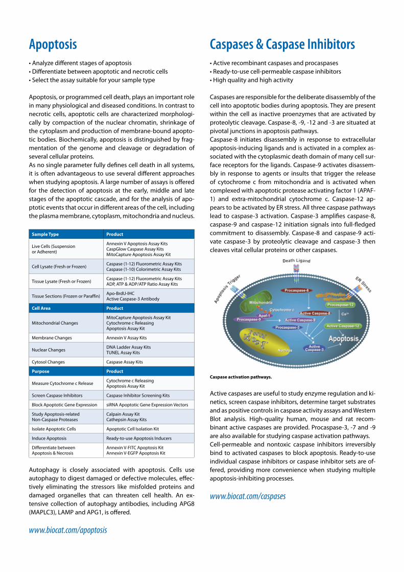
Caspases are responsible for the deliberate disassembly of the cell into apoptotic bodies during apoptosis. They are present within the cell as inactive proenzymes that are activated by proteolytic cleavage. Caspase-8, -9, -12 and -3 are situated at pivotal junctions in apoptosis pathways. Caspase-8 initiates disassembly in response to extracellular apoptosis-inducing ligands and is activated in a complex as-sociated with the cytoplasmic death domain of many cell sur-face receptors for the ligands. Caspase-9 activates disassem-bly in response to agents or insults that trigger the release of cytochrome c from mitochondria and is activated when complexed with apoptotic protease activating factor 1 (APAF-1) and extra-mitochondrial cytochrome c. Caspase-12 ap-pears to be activated by ER stress. All three caspase pathways lead to caspase-3 activation. Caspase-3 amplifies caspase-8, caspase-9 and caspase-12 initiation signals into full-fledged commitment to disassembly. Caspase-8 and caspase-9 acti-vate caspase-3 by proteolytic cleavage and caspase-3 then cleaves vital cellular proteins or other caspases.
Caspase activation pathways.
Active caspases are useful to study enzyme regulation and ki-netics, screen caspase inhibitors, determine target substrates and as positive controls in caspase activity assays and Western Blot analysis. High-quality human, mouse and rat recom-binant active caspases are provided. Procaspase-3, -7 and -9 are also available for studying caspase activation pathways.Cell-permeable and nontoxic caspase inhibitors irreversibly bind to activated caspases to block apoptosis. Ready-to-use individual caspase inhibitors or caspase inhibitor sets are of-fered, providing more convenience when studying multiple apoptosis-inhibiting processes.
www.biocat.com/caspases
Cytotoxicity & ProliferationLDH Cytotoxicity Assay Kit II• Quantify released LDH with high sensitivity• Fast and convenient assay based on enhanced WST• 96-well format, colorimetric detection
Cell death or cytotoxicity is classically evaluated by the quan-tification of plasma membrane damage. Lactate dehydroge-nase (LDH) is a stable enzyme, present in all cell types and rapidly released into the cell culture medium upon damage of the plasma membrane. LDH, therefore, is the most widely used marker in cytotoxicity studies. The LDH Cytotoxicity Assay Kit II developed by BioVision uti-lizes enhanced WST reagent for the fast and highly sensitive detection of LDH released from damaged cells: LDH oxidizes lactate to generate NADH, which then reduces enhanced WST to generate yellow colored formazan. The intensity of the co-lor detected at 450 nm correlates directly with the number of cells lysed. Since enhanced WST is brighter, less amount of culture me-dium is required for the assay, and thus the background from serum and culture medium is significantly reduced. Using the assay, cells can be cultured in regular 10% serum containing medium, no reducing serum or special medium is required for the assay. In addition, as enhanced WST is more stable, the reaction can be read multiple times, and can also be stopped at any time point during the reaction.
Lactate Dehydrogenase
(LDH)
NAD+
NADH
Lactate
Pyruvate Enhanced WST
Formazan
Damaged Cell
Principle of the LDH Cytotoxicity Assay Kit II.
The Quick Cell Proliferation Assay Kit II (colorimetric) is de-signed to determine the number of living cells. It is based on the cleavage of the tetrazolium salt WST (enhanced version) to formazan by cellular mitochondrial dehydrogenase. The amount of dye generated by the activity of dehydrogenase is directly proportional to the number of living cells.
www.biocat.com/cytotoxicity
Cell AdhesionCytoSelect Cell Adhesion Assay (ECM Array)• Quantify cell adhesion without manual cell counting• Analyze cell interactions with 5 ECM proteins• Colorimetric or fluorometric detection
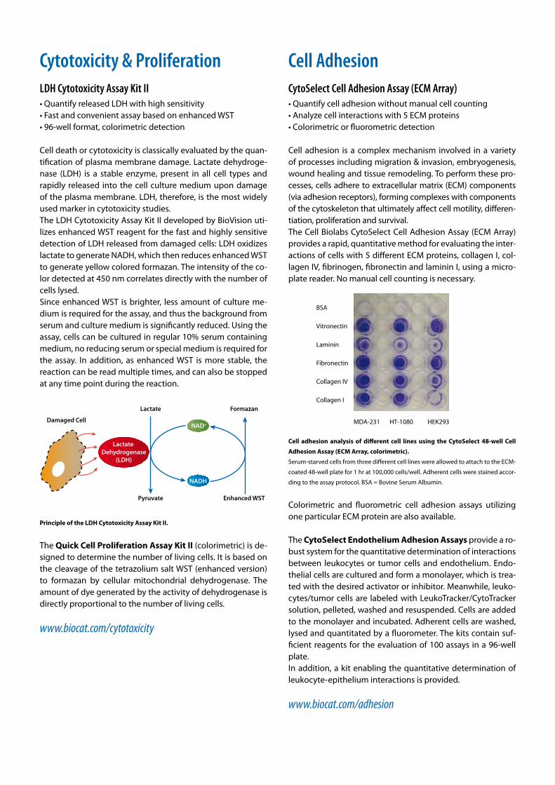
Cell adhesion is a complex mechanism involved in a variety of processes including migration & invasion, embryogenesis, wound healing and tissue remodeling. To perform these pro-cesses, cells adhere to extracellular matrix (ECM) components (via adhesion receptors), forming complexes with components of the cytoskeleton that ultimately affect cell motility, differen-tiation, proliferation and survival. The Cell Biolabs CytoSelect Cell Adhesion Assay (ECM Array) provides a rapid, quantitative method for evaluating the inter-actions of cells with 5 different ECM proteins, collagen I, col-lagen IV, fibrinogen, fibronectin and laminin I, using a micro-plate reader. No manual cell counting is necessary.
MDA-231 HT-1080 HEK293
BSA
Vitronectin
Laminin
Fibronectin
Collagen IV
Collagen I
Cell adhesion analysis of different cell lines using the CytoSelect 48-well Cell
Adhesion Assay (ECM Array, colorimetric).
Serum-starved cells from three different cell lines were allowed to attach to the ECM-
coated 48-well plate for 1 hr at 100,000 cells/well. Adherent cells were stained accor-
ding to the assay protocol. BSA = Bovine Serum Albumin.
Colorimetric and fluorometric cell adhesion assays utilizing one particular ECM protein are also available.
The CytoSelect Endothelium Adhesion Assays provide a ro-bust system for the quantitative determination of interactions between leukocytes or tumor cells and endothelium. Endo-thelial cells are cultured and form a monolayer, which is trea-ted with the desired activator or inhibitor. Meanwhile, leuko-cytes/tumor cells are labeled with LeukoTracker/CytoTracker solution, pelleted, washed and resuspended. Cells are added to the monolayer and incubated. Adherent cells are washed, lysed and quantitated by a fluorometer. The kits contain suf-ficient reagents for the evaluation of 100 assays in a 96-well plate.In addition, a kit enabling the quantitative determination of leukocyte-epithelium interactions is provided.
www.biocat.com/adhesion
Cell Migration & InvasionCell migration is a highly integrated, multistep process that orchestrates embryonic morphogenesis, tissue repair and re-generation. It plays a pivotal role in the disease progression of cancer, mental retardation, atherosclerosis and arthritis. The initial response of a cell to a migration-promoting agent is to polarize and extend protrusions in the direction of the attractant; these protrusions can consist of large, broad lamel-lipodia or spike-like filopodia. In either case, these protrusions are driven by actin polymerization and can be stabilized by extracellular matrix (ECM) adhesion or cell-cell interactions (via transmembrane receptors). There are various types of cell migration including chemotaxis, haptotaxis and transmigra-tion.Cell invasion is related to, and encompasses, cell migration, except that cells do more than migrate. Invasive cells move through the extracellular matrix into neighboring tissues in a process that involves ECM degradation and proteolysis. Meta-static cells produce many proteolytic enzymes (e.g. lysosomal hydrolysates, collagenases, plasminogen activators) while the expression of certain cell surface protease receptors is also in-creased. The ability of malignant tumor cells to invade normal surrounding tissue contributes in large part to the significant morbidity and mortality of cancers.Cell migration assays developed by Cell Biolabs are offered in two formats: Boyden Chamber Assays and Gap Closure Assays.
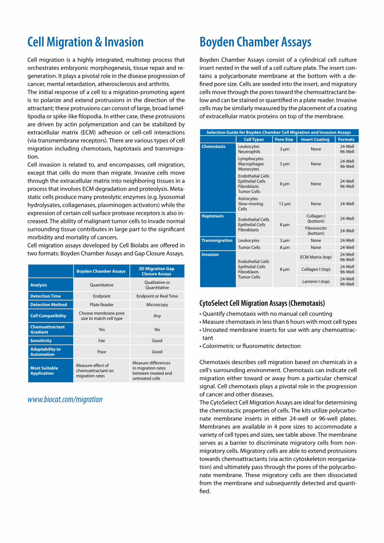
Boyden Chamber Assays 2D Migration Gap Closure Assays
Analysis Quantitative Qualitative or Quantitative
Detection Time Endpoint Endpoint or Real Time
Detection Method Plate Reader Microscopy
Cell Compatibility Choose membrane pore size to match cell type Any
Chemoattractant Gradient Yes No
Sensitivity Fair Good
Adaptability to Automation Poor Good
Most Suitable Application
Measure effect of chemoattractant on migration rates
Measure differences in migration rates between treated and untreated cells
www.biocat.com/migration
Boyden Chamber AssaysBoyden Chamber Assays consist of a cylindrical cell culture insert nested in the well of a cell culture plate. The insert con-tains a polycarbonate membrane at the bottom with a de-fined pore size. Cells are seeded into the insert, and migratory cells move through the pores toward the chemoattractant be-low and can be stained or quantified in a plate reader. Invasive cells may be similarly measured by the placement of a coating of extracellular matrix proteins on top of the membrane.
Selection Guide for Boyden Chamber Cell Migration and Invasion Assays
Cell Types Pore Size Insert Coating Formats
Chemotaxis LeukocytesNeutrophils 3 µm None 24-Well
96-Well
LymphocytesMacrophagesMonocytes
5 µm None 24-Well96-Well
Endothelial CellsEpithelial CellsFibroblastsTumor Cells
8 µm None 24-Well96-Well
AstrocytesSlow-moving Cells
12 µm None 24-Well
HaptotaxisEndothelial CellsEpithelial Cells Fibroblasts
8 µm
Collagen I (bottom) 24-Well
Fibronectin (bottom) 24-Well
Transmigration Leukocytes 3 µm None 24-Well
Tumor Cells 8 µm None 24-Well
Invasion
Endothelial CellsEpithelial CellsFibroblastsTumor Cells
8 µm
ECM Matrix (top) 24-Well96-Well
Collagen I (top) 24-Well96-Well
Laminin I (top) 24-Well96-Well
CytoSelect Cell Migration Assays (Chemotaxis)• Quantify chemotaxis with no manual cell counting• Measure chemotaxis in less than 6 hours with most cell types• Uncoated membrane inserts for use with any chemoattrac-
tant• Colorimetric or fluorometric detection
Chemotaxis describes cell migration based on chemicals in a cell‘s surrounding environment. Chemotaxis can indicate cell migration either toward or away from a particular chemical signal. Cell chemotaxis plays a pivotal role in the progression of cancer and other diseases. The CytoSelect Cell Migration Assays are ideal for determining the chemotactic properties of cells. The kits utilize polycarbo-nate membrane inserts in either 24-well or 96-well plates. Membranes are available in 4 pore sizes to accommodate a variety of cell types and sizes, see table above. The membrane serves as a barrier to discriminate migratory cells from non-migratory cells. Migratory cells are able to extend protrusions towards chemoattractants (via actin cytoskeleton reorganiza-tion) and ultimately pass through the pores of the polycarbo-nate membrane. These migratory cells are then dissociated from the membrane and subsequently detected and quanti-fied.
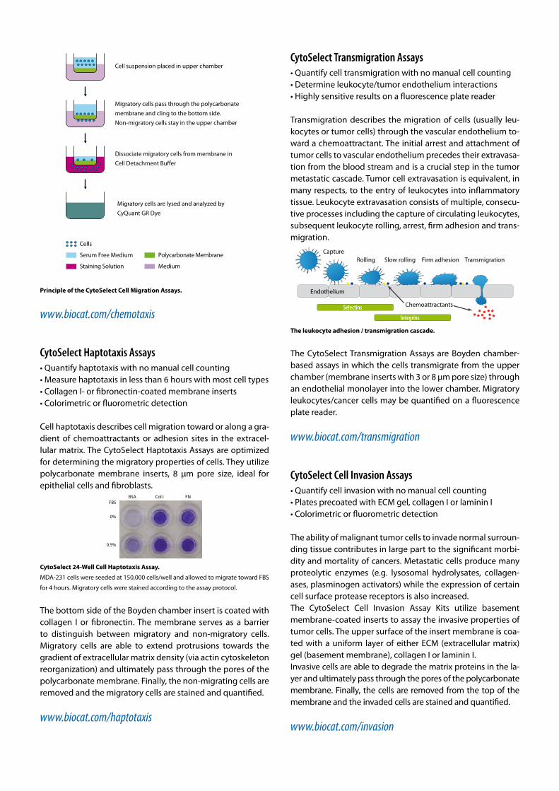
Cell suspension placed in upper chamber
Migratory cells pass through the polycarbonate membrane and cling to the bottom side. Non-migratory cells stay in the upper chamber
Dissociate migratory cells from membrane inCell Detachment Bu�er
Migratory cells are lysed and analyzed byCyQuant GR Dye
Staining Solution
Polycarbonate MembraneSerum Free Medium
Cells
Medium
Principle of the CytoSelect Cell Migration Assays.
www.biocat.com/chemotaxis
CytoSelect Haptotaxis Assays• Quantify haptotaxis with no manual cell counting• Measure haptotaxis in less than 6 hours with most cell types• Collagen I- or fibronectin-coated membrane inserts• Colorimetric or fluorometric detection
Cell haptotaxis describes cell migration toward or along a gra-dient of chemoattractants or adhesion sites in the extracel-lular matrix. The CytoSelect Haptotaxis Assays are optimized for determining the migratory properties of cells. They utilize polycarbonate membrane inserts, 8 μm pore size, ideal for epithelial cells and fibroblasts.
BSA Col I FNFBS
0%
0.5%
CytoSelect 24-Well Cell Haptotaxis Assay.
MDA-231 cells were seeded at 150,000 cells/well and allowed to migrate toward FBS
for 4 hours. Migratory cells were stained according to the assay protocol.
The bottom side of the Boyden chamber insert is coated with collagen I or fibronectin. The membrane serves as a barrier to distinguish between migratory and non-migratory cells. Migratory cells are able to extend protrusions towards the gradient of extracellular matrix density (via actin cytoskeleton reorganization) and ultimately pass through the pores of the polycarbonate membrane. Finally, the non-migrating cells are removed and the migratory cells are stained and quantified.
www.biocat.com/haptotaxis
CytoSelect Transmigration Assays• Quantify cell transmigration with no manual cell counting• Determine leukocyte/tumor endothelium interactions• Highly sensitive results on a fluorescence plate reader
Transmigration describes the migration of cells (usually leu-kocytes or tumor cells) through the vascular endothelium to-ward a chemoattractant. The initial arrest and attachment of tumor cells to vascular endothelium precedes their extravasa-tion from the blood stream and is a crucial step in the tumor metastatic cascade. Tumor cell extravasation is equivalent, in many respects, to the entry of leukocytes into inflammatory tissue. Leukocyte extravasation consists of multiple, consecu-tive processes including the capture of circulating leukocytes,subsequent leukocyte rolling, arrest, firm adhesion and trans-migration.
CaptureRolling Slow rolling Firm adhesion Transmigration
Selectins
Integrins
Chemoattractants
Endothelium
The leukocyte adhesion / transmigration cascade.
The CytoSelect Transmigration Assays are Boyden chamber-based assays in which the cells transmigrate from the upper chamber (membrane inserts with 3 or 8 µm pore size) through an endothelial monolayer into the lower chamber. Migratory leukocytes/cancer cells may be quantified on a fluorescence plate reader.
www.biocat.com/transmigration
CytoSelect Cell Invasion Assays• Quantify cell invasion with no manual cell counting• Plates precoated with ECM gel, collagen I or laminin I• Colorimetric or fluorometric detection
The ability of malignant tumor cells to invade normal surroun-ding tissue contributes in large part to the significant morbi-dity and mortality of cancers. Metastatic cells produce many proteolytic enzymes (e.g. lysosomal hydrolysates, collagen-ases, plasminogen activators) while the expression of certain cell surface protease receptors is also increased.The CytoSelect Cell Invasion Assay Kits utilize basement membrane-coated inserts to assay the invasive properties of tumor cells. The upper surface of the insert membrane is coa-ted with a uniform layer of either ECM (extracellular matrix) gel (basement membrane), collagen I or laminin I. Invasive cells are able to degrade the matrix proteins in the la-yer and ultimately pass through the pores of the polycarbonate membrane. Finally, the cells are removed from the top of the membrane and the invaded cells are stained and quantified.
www.biocat.com/invasion
Gap Closure AssaysGap Closure Assays create a defined area across which migra-tory cells can move. Migration can be monitored in real-time by microscopy. These assays include the new Radius techno-logy which uses a biocompatible hydrogel to create a circular area across which cells can migrate, and the Wound Healing Assay which is a more consistent alternative to the traditional scratch assay.
CytoSelect Wound Healing Assays• Measure cell migration, cell proliferation and wound closure• More consistent compared to homemade scratch assays• Inert inserts leave no residues that could impede proliferation
or migration of cells
Wounded tissue initiates a complex and structured series of events in order to repair the damaged region. These events may include increased vascularization by angiogenic factors, an increase in cell proliferation and extracellular matrix depo-sition, and infiltration by inflammatory immune cells as part of the process to destroy necrotic tissue. The wound healing process begins as cells polarize toward the wound, initiate protrusion, migrate and close the wound area. Traditionally scratch assays have been used to study cell mi-gration, cell proliferation and wound healing. However, these assays lack a consistently defined wound gap and can result in high inter-sample variation.
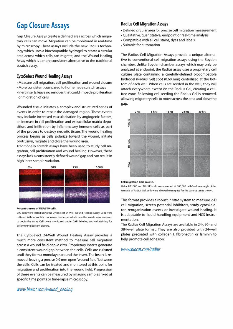
0% 50% 75% 100%
Percent closure of MEF/STO cells.
STO cells were tested using the CytoSelect 24-Well Wound Healing Assay. Cells were
cultured 24 hours until a monolayer formed, at which time the inserts were removed
to begin the assay. Cells were monitored under DAPI labeling and cell staining for
determining percent closure.
The CytoSelect 24-Well Wound Healing Assay provides a much more consistent method to measure cell migration across a wound field gap in vitro. Proprietary inserts generate a consistent wound gap between the cells. Cells are cultured until they form a monolayer around the insert. The insert is re-moved, leaving a precise 0.9 mm open “wound field” between the cells. Cells can be treated and monitored at this point for migration and proliferation into the wound field. Progression of these events can be measured by imaging samples fixed at specific time points or time-lapse microscopy.
www.biocat.com/wound_healing
Radius Cell Migration Assays• Defined circular area for precise cell migration measurement• Qualitative, quantitative, endpoint or real-time analysis• Compatible with all cell stains, dyes and labels• Suitable for automation
The Radius Cell Migration Assays provide a unique alterna-tive to conventional cell migration assays using the Boyden chamber. Unlike Boyden chamber assays which may only be analyzed at endpoint, the Radius assay uses a proprietary cell culture plate containing a carefully-defined biocompatible hydrogel (Radius Gel) spot (0.68 mm) centralized at the bot-tom of each well. When cells are seeded in the well, they will attach everywhere except on the Radius Gel, creating a cell-free zone. Following cell seeding the Radius Gel is removed, allowing migratory cells to move across the area and close the gap.
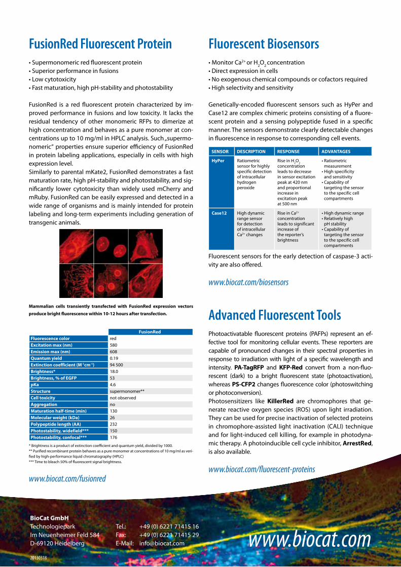
HeL
aH
T108
0N
IH3T
3
0 hrs 5 hrs 18 hrs 24 hrs 30 hrs
Cell migration time course.
HeLa, HT1080 and NIH3T3 cells were seeded at 100,000 cells/well overnight. After
removal of Radius Gel, cells were allowed to migrate for the various times shown.
This format provides a robust in vitro system to measure 2-D cell migration, screen potential inhibitors, study cytoskele-ton reorganization events or investigate wound healing. It is adaptable to liquid handling equipment and HCS instru-mentation.The Radius Cell Migration Assays are available in 24-, 96- and 384-well plate format. They are also provided with 24-well plates precoated with collagen I, fibronectin or laminin to help promote cell adhesion.
www.biocat.com/radius

Fluorescent ProteinsTurboFPs • Superbright fluorescence• Very fast maturation• Optimal for cell labeling
TurboFPs are fluorescent proteins of different colors characte-rized by superbright fluorescence and superior fast maturation. These proteins are recommended for applications requiring fast appearance of bright fluorescence, including cell and organelle labeling or tracking promoter activity. The far-red marker TurboFP635 is ideal for whole body imaging applica-tions.
DsRed-Express and TurboFP635 expression in transgenic Xenopus laevis.
Transgenic 2.5 months living animals expressing DsRed-Express or TurboFP635 un-
der the control of cardiac actin promoter are shown from the dorsal side. TurboFP635
(on the right) is excellently visible in the whole body, while DsRed-Express (on the
left) can be hardly visualized.
The comprehensive fluorescent protein offering developed by Evrogen comprises expression/source vectors, antibodies, recombinant proteins and stable cell lines.
www.biocat.com/fluorescent_proteins
TagFPs• Bright monomeric proteins• Excellent performance in fusions• Wide color palette
TagFPs comprise monomeric fluorescent proteins that are optimized for protein localization, protein interaction studies and stable expression in long term cultures. They can also be used for other cell labeling applications. TagFPs demonstrate successful performance in fusions with cellular proteins and can be expressed in various heterolo-gous systems. Ranging in color from blue to far-red, they pro-vide unique possibilities for multicolor labeling of subcellular structures.
A B C D
Multicolor labeling of subcellular structures in transiently transfected mamma-
lian cells using TagFPs.
(A) TagBFP-histone H2B fusion (blue), TagGFP-actin fusion (green), mitochondria-
targeted PhiYFP (yellow), Golgi-targeted TagRFP (orange), mKate2-zyxin fusion (red).
(B) TagCFP-actin fusion (cyan), mitochondria-targeted PhiYFP (yellow) and mKate2-
clathrin fusion (red) in HeLa cells. (C) TagRFP-cytokeratin 14 fusion (red) and mito-
chondria-targeted TagGFP2 (green) in REF3 cells with Hoechst staining (blue). (D)
TagGFP2-actin fusion (green) and mKate2-zyxin fusion (red) in REF52 cells.
TURBO FLUORESCENT PROTEINS (TurboFPs)TurboGFP TurboYFP TurboRFP TurboFP602 TurboFP635 TurboFP650
Fluorescence color green yellow red (orange) true-red far-red near-infraredExcitation max (nm) 482 525 553 574 588 592Emission max (nm) 502 538 574 602 635 650
Quantum yield 0.53 0.53 0.67 0.35 0.34 0.24Extinction coefficient (M-1cm-1) 70 000 105 000 92 000 74 400 65 000 65 000Brightness* 37.1 55.7 61.6 26.0 22.1 15.6Brightness, % of EGFP 112 169 187 79 67 47
pKa 5.2 5.9 4.4 4.7 5.5 5.7Structure dimer dimer dimer dimer dimer dimerCell toxicity not observed at high
concentrations**not observed not observed not observed not observed
Aggregation no at high concentrations**
no no no no
Maturation rate at 37°C super fast super fast super fast fast super fast super fastMolecular weight (kDa) 26 26 26 26 26 26
TAG FLUORESCENT PROTEINS (TagFPs)TagBFP TagCFP TagGFP2 TagYFP TagRFP mKate2
Fluorescence color blue cyan green yellow red (orange) far-redExcitation max (nm) 402 458 483 508 555 588Emission max (nm) 457 480 506 524 584 633Quantum yield 0.63 0.57 0.6 0.62 0.48 0.40Extinction coefficient (M-1cm-1) 52 000 37 000 56 500 50 000 100 000 62 500Brightness* 32.8 21.1 33.9 31 48.0 25.0Brightness, % of EGFP 99 64 105 94 148 74pKa 2.7 4.7 5.0 5.5 3.8 5.4Structure monomer monomer monomer monomer monomer monomerCell toxicity not observed not observed not observed not observed not observed not observedAggregation no no no no no noMaturation rate at 37°C fast fast fast fast fast fastMolecular weight (kDa) 26 27 27 27 27 26
*Brightness is a product of extinction coefficient and quantum yield, divided by 1000.**For stable cell line generation and organelle labeling we recommend that you use PhiYFP.
FusionRed Fluorescent Protein• Supermonomeric red fluorescent protein• Superior performance in fusions• Low cytotoxicity • Fast maturation, high pH-stability and photostability FusionRed is a red fluorescent protein characterized by im-proved performance in fusions and low toxicity. It lacks the residual tendency of other monomeric RFPs to dimerize at high concentration and behaves as a pure monomer at con-centrations up to 10 mg/ml in HPLC analysis. Such „supermo-nomeric“ properties ensure superior efficiency of FusionRed in protein labeling applications, especially in cells with high expression level.Similarly to parental mKate2, FusionRed demonstrates a fast maturation rate, high pH-stability and photostability, and sig-nificantly lower cytotoxicity than widely used mCherry and mRuby. FusionRed can be easily expressed and detected in a wide range of organisms and is mainly intended for protein labeling and long-term experiments including generation of transgenic animals.
Mammalian cells transiently transfected with FusionRed expression vectors
produce bright fluorescence within 10-12 hours after transfection.
FusionRedFluorescence color redExcitation max (nm) 580Emission max (nm) 608Quantum yield 0.19Extinction coefficient (M-1cm-1) 94 500Brightness* 18.0Brightness, % of EGFP 53pKa 4.6Structure supermonomer**Cell toxicity not observedAggregation noMaturation half-time (min) 130Molecular weight (kDa) 26Polypeptide length (AA) 232Photostability, widefield*** 150Photostability, confocal*** 176
* Brightness is a product of extinction coefficient and quantum yield, divided by 1000.** Purified recombinant protein behaves as a pure monomer at concentrations of 10 mg/ml as veri-fied by high-performance liquid chromatography (HPLC)*** Time to bleach 50% of fluorescent signal brightness.
www.biocat.com/fusionred
Fluorescent Biosensors• Monitor Ca2+ or H2O2 concentration• Direct expression in cells• No exogenous chemical compounds or cofactors required• High selectivity and sensitivity
Genetically-encoded fluorescent sensors such as HyPer and Case12 are complex chimeric proteins consisting of a fluore-scent protein and a sensing polypeptide fused in a specific manner. The sensors demonstrate clearly detectable changes in fluorescence in response to corresponding cell events.
SENSOR DESCRIPTION RESPONSE ADVANTAGES
HyPer Ratiometric sensor for highly specific detection of intracellular hydrogen peroxide
Rise in H2O2 concentration leads to decrease in sensor excitation peak at 420 nm and proportional increase in excitation peak at 500 nm
• Ratiometric measurement
• High specificity and sensitivity
• Capability of targeting the sensor to the specific cell compartments
Case12 High dynamic range sensor for detection of intracellular Ca2+ changes
Rise in Ca2+ concentration leads to significant increase of the reporter’s brightness
• High dynamic range • Relatively high
pH stability• Capability of
targeting the sensor to the specific cell compartments
Fluorescent sensors for the early detection of caspase-3 acti-vity are also offered.
www.biocat.com/biosensors
Advanced Fluorescent ToolsPhotoactivatable fluorescent proteins (PAFPs) represent an ef-fective tool for monitoring cellular events. These reporters are capable of pronounced changes in their spectral properties in response to irradiation with light of a specific wavelength and intensity. PA-TagRFP and KFP-Red convert from a non-fluo-rescent (dark) to a bright fluorescent state (photoactivation), whereas PS-CFP2 changes fluorescence color (photoswitching or photoconversion). Photosensitizers like KillerRed are chromophores that ge-nerate reactive oxygen species (ROS) upon light irradiation. They can be used for precise inactivation of selected proteins in chromophore-assisted light inactivation (CALI) technique and for light-induced cell killing, for example in photodyna-mic therapy. A photoinducible cell cycle inhibitor, ArrestRed, is also available.
www.biocat.com/fluorescent-proteins
www.biocat.comBioCat GmbHTechnologiepark Tel.: +49 (0) 6221 71415 16 Im Neuenheimer Feld 584 Fax: +49 (0) 6221 71415 29D-69120 Heidelberg E-Mail: [email protected]
20130318