Celiac Disease: Epidemiology, Pathogenesis, Diagnosis, and...
16
Celiac Disease: Epidemiology, Pathogenesis, Diagnosis, and Nutritional Management Detlef Schuppan, MD, PhD, Melinda D. Dennis, MS, RD, LDN, Ciaran P. Kelly, MD ABSTRACT Celiac disease (CD) is an inflammatory small intestinal disorder that can lead to severe villous atrophy, malabsorption, and malignancy. It is triggered by the gluten proteins of wheat, barley, and rye. All patients express the antigen-presenting molecules human leukocyte antigen-DQ2 (HLA-DQ2) and/or HLA-DQ8, which bind gluten peptides and thus activate de- structive intestinal T cells. Patients with untreated CD have circulating IgA autoantibodies to the enzyme tissue transglu- taminase (tTG), a component of endomysium. Testing for se- rum IgA tTG has a high predictive value. Therapy of CD is a lifelong gluten-free diet. Counseling by an expert dietitian and association with a celiac support group are important in help- ing the patient embark on a healthy gluten-free diet. Current research focuses on non-dietary therapies and treatment of refractory (diet-unresponsive) CD. Nutr Clin Care. 2005;8: 54–69 KEY WORDS: autoantibody, autoantigen, celiac disease, cytokine, DQ2, DQ8, gene dose, gluten, gliadin, human leuko- cyte antigen, HLA, interferon, matrix metalloproteinase, MMP, pathogenesis, transforming growth factor-beta, TGF-beta, tu- mor necrosis factor-alpha, TNF-alpha, T cell, therapy, tissue transglutaminase © 2005 International Life Sciences Institute HISTORY AND EPIDEMIOLOGY Classical celiac disease (CD) is a small intestinal in- flammatory disease that is characterized by global malabsorption of nutrients, minerals, and vitamins. Histologically, it is characterized by a predominant T-cell infiltration in the epithelium and the lamina propria of the intestine, which can lead to complete destruction of the villi, through which nutrient re- sorption occurs. 1-5 CD was fatal in 12% of affected children in a retrospective study in 1939, 6 and its cause remained elusive until the Dutch pediatrician Willem Dicke recognized an association between the consumption of wheat products and the relapsing diarrhea of CD. During periods of food shortage in the second World War, the illness lessened in his patients when bread was replaced by non-cereal-containing foods. After the war, Dicke and van de Kamer 7 con- firmed this observation by performing controlled ex- periments exposing children with CD to defined diets followed by determination of fecal weight and fecal fat as a measure of malabsorption. These studies clearly demonstrated that wheat, barley, and rye trig- gered CD and that the condition could be reversed after their exclusion from the diet. 7 The major toxicity was found in the alcohol-soluble gliadin fraction of wheat gluten. 8 The celiac lesion of mucosal inflammation, crypt hyperplasia, and villous atrophy in the proximal small intestine, although already observed by Rudolph Ben- necke from Marburg, Germany in 1910, was first de- scribed in detail in 1954, 9 and its classification was refined by Marsh. 10 With the advent of serum tests such as immunoglobulin A (IgA) antibodies to gliadin, Dr. Schuppan, Ms. Dennis, and Dr. Kelly are with the Division of Gastro- enterology, Beth Israel Deaconess Medical Center, Harvard Medical School, Boston, Massachusetts. Address for correspondence: Dr. Detlef Schuppan, Division of Gastro- enterology and Hepatology, Beth Israel Deaconess Medical Center, Harvard Medical School, 330 Brookline Ave., Boston, MA 02215; Phone: 617-667-8377; Fax: 617-667-2767; E-mail: [email protected]. edu. Nutrition in Clinical Care, Volume 8, Number 2, 2005 54 – 69
Transcript of Celiac Disease: Epidemiology, Pathogenesis, Diagnosis, and...

Celiac Disease: Epidemiology, Pathogenesis,Diagnosis, and Nutritional Management
Detlef Schuppan, MD, PhD, Melinda D. Dennis, MS, RD, LDN,Ciaran P. Kelly, MD
� ABSTRACTCeliac disease (CD) is an inflammatory small intestinal disorderthat can lead to severe villous atrophy, malabsorption, andmalignancy. It is triggered by the gluten proteins of wheat,barley, and rye. All patients express the antigen-presentingmolecules human leukocyte antigen-DQ2 (HLA-DQ2) and/orHLA-DQ8, which bind gluten peptides and thus activate de-structive intestinal T cells. Patients with untreated CD havecirculating IgA autoantibodies to the enzyme tissue transglu-taminase (tTG), a component of endomysium. Testing for se-rum IgA tTG has a high predictive value. Therapy of CD is alifelong gluten-free diet. Counseling by an expert dietitian andassociation with a celiac support group are important in help-ing the patient embark on a healthy gluten-free diet. Currentresearch focuses on non-dietary therapies and treatment ofrefractory (diet-unresponsive) CD. Nutr Clin Care. 2005;8:54–69 �
KEY WORDS: autoantibody, autoantigen, celiac disease,cytokine, DQ2, DQ8, gene dose, gluten, gliadin, human leuko-cyte antigen, HLA, interferon, matrix metalloproteinase, MMP,pathogenesis, transforming growth factor-beta, TGF-beta, tu-mor necrosis factor-alpha, TNF-alpha, T cell, therapy, tissuetransglutaminase© 2005 International Life Sciences Institute
HISTORY AND EPIDEMIOLOGY
Classical celiac disease (CD) is a small intestinal in-flammatory disease that is characterized by global
malabsorption of nutrients, minerals, and vitamins.Histologically, it is characterized by a predominantT-cell infiltration in the epithelium and the laminapropria of the intestine, which can lead to completedestruction of the villi, through which nutrient re-sorption occurs.1-5 CD was fatal in 12% of affectedchildren in a retrospective study in 1939,6 and itscause remained elusive until the Dutch pediatricianWillem Dicke recognized an association between theconsumption of wheat products and the relapsingdiarrhea of CD. During periods of food shortage in thesecond World War, the illness lessened in his patientswhen bread was replaced by non-cereal-containingfoods. After the war, Dicke and van de Kamer7 con-firmed this observation by performing controlled ex-periments exposing children with CD to defined dietsfollowed by determination of fecal weight and fecalfat as a measure of malabsorption. These studiesclearly demonstrated that wheat, barley, and rye trig-gered CD and that the condition could be reversedafter their exclusion from the diet.7 The major toxicitywas found in the alcohol-soluble gliadin fraction ofwheat gluten.8
The celiac lesion of mucosal inflammation, crypthyperplasia, and villous atrophy in the proximal smallintestine, although already observed by Rudolph Ben-necke from Marburg, Germany in 1910, was first de-scribed in detail in 1954,9 and its classification wasrefined by Marsh.10 With the advent of serum testssuch as immunoglobulin A (IgA) antibodies to gliadin,
Dr. Schuppan, Ms. Dennis, and Dr. Kelly are with the Division of Gastro-enterology, Beth Israel Deaconess Medical Center, Harvard MedicalSchool, Boston, Massachusetts.Address for correspondence: Dr. Detlef Schuppan, Division of Gastro-enterology and Hepatology, Beth Israel Deaconess Medical Center,Harvard Medical School, 330 Brookline Ave., Boston, MA 02215; Phone:617-667-8377; Fax: 617-667-2767; E-mail: [email protected]. Nutrition in Clinical Care, Volume 8, Number 2, 2005 54–69
and especially to an autoantigen present in reticulinand endomysium (the connective tissue of soft tissuesand around smooth muscle, respectively), CD couldbe more easily differentiated from other malabsorp-tive disorders that cause villous atrophy, such as trop-ical (microbial) sprue and bacterial overgrowth.11
More importantly, population studies using the auto-antibody tests as screening tools with subsequentsmall intestinal biopsy for confirmation revealed ahitherto unexpectedly high prevalence of CD in thewestern world, in northern Africa, the near East, andthe Middle East, ranging from about 1:80 to 1:150.12-14
In these studies, the vast majority (�80%) of screen-ing-detected individuals presented with clinically si-lent or atypical forms of the disease. If and to whatextent screening-detected asymptomatic or minimallysymptomatic individuals may develop clinically overtCD, secondary autoimmune diseases, or even malig-nancy (see below) when continuing on a gluten-con-taining diet is still unclear.
GLUTEN: THE CAUSATIVE AGENT IN CD
Gluten comprises the storage proteins of wheat.These proteins can be separated into the ethanol-insoluble glutenins, which are responsible for thedesired baking properties of this cereal, and the alco-hol-soluble gliadins. Each wheat variant produces anestimated 40 to 50 gliadins, which are structurallyrelated proteins containing 250 to 500 amino acids,and a lower number of high- and low-molecular-weight glutenins, with 650 to 800 and 270 to 320amino acids, respectively. Both gliadins and gluteninsdisplay a high content of the amino acids glutamine(32%–56 %) and proline (15%–30%) and, due to theircysteine content, glutenins can form complex ho-mopolymers and heteropolymers with gliadins.15,16
Storage proteins similar to gliadins (generally termedprolamines) have been found in rye (secalins) andbarley (hordeins), while the avenins of oats and espe-cially the zeins of rice are more distantly related (Ta-ble 1). On the basis of their electrophoretic propertiesand primary structure, the gliadins are subdivided intothe classes of �-, �-, and �-gliadins, which can befurther resolved into distinct proteins such as �1-11,�1-6, and �1-5. Although the pro-inflammatory effectof all gliadin fractions has been shown in vitro,17,18
most experimental and human studies have focusedon �-gliadins, and in particular on the pro-inflamma-tory peptides in its amino-terminal region.19
THE GENETIC COMPONENT ANDASSOCIATED AUTOIMMUNITY
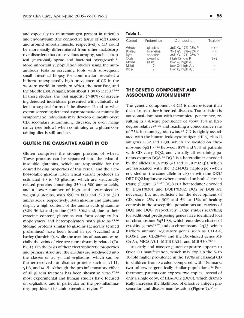
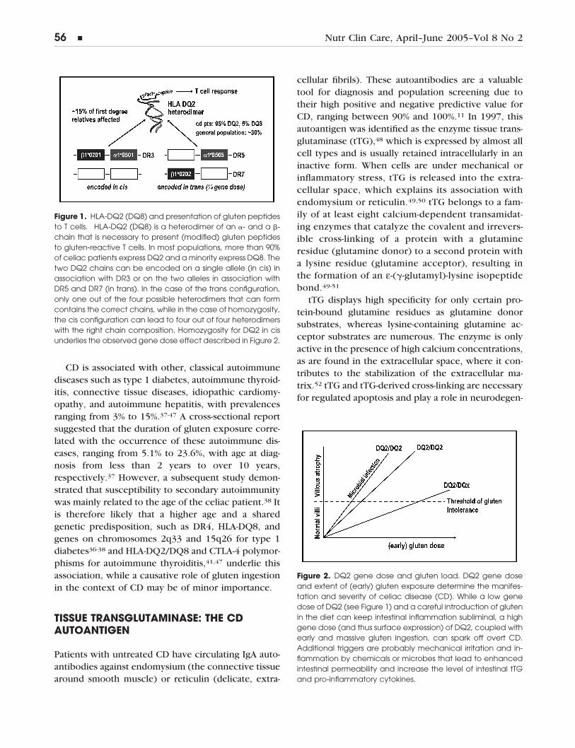
The genetic component of CD is more evident thanthat of most other inherited diseases. Transmission isautosomal dominant with incomplete penetrance, re-sulting in a disease prevalence of about 15% in first-degree relatives20-22 and reaching a concordance rateof 75% in monozygotic twins.23 CD is tightly associ-ated with the human leukocyte antigen (HLA) class IIantigens DQ2 and DQ8, which are located on chro-mosome 6p21.21-22 Between 85% and 95% of patientswith CD carry DQ2, and virtually all remaining pa-tients express DQ8.24 DQ2 is a heterodimer encodedby the alleles DQA1*05 (�) and DQB1*02 (�), whichare associated with the DR3-DQ2 haplotype (whenencoded on the same allele in cis) or with the DR5/DR7-DQ2 haplotype (when encoded on both alleles intrans) (Figure 1).21-22 DQ8 is a heterodimer encodedby DQA1*0301 and DQB1*0302. DQ2 or DQ8 arenecessary but not sufficient for the development ofCD, since 25% to 30% and 5% to 15% of healthycontrols in the susceptible populations are carriers ofDQ2 and DQ8, respectively. Large studies searchingfor additional predisposing genes have identified locion chromosome 5q31-33, which encodes a cluster ofcytokine genes25-27, and on chromosome 2q33, whichharbors immune regulatory genes such as CTLA-4,ICOS-1, and CD2828,29 and the DR3-linked genes MI-CA-A4, MICA-A5.1, MICB-CA24, and MIB-350.30,31
An early and massive gluten exposure appears tofavor CD manifestation, which may explain the 5- to10-fold higher prevalence in the 1970s of classical CDin children from Sweden compared with Denmark,two otherwise genetically similar populations.32 Fur-thermore, patients can express two copies, instead ofonly a single copy, of HLA-DQ2 (DQ8), which dramat-ically increases the likelihood of effective antigen pre-sentation and disease manifestation (Figure 2).33-36
Table 1.
Cereal Prolamines Composition “Toxicity”
Wheat gliadins 36% Q, 17%–23% P ���Barley hordeins 36% Q, 17%–23% P ��Rye secalins 36% Q, 17%–23% P ��Oats avenins high Q, low P (�)Maize zeins low Q, high A,L �Millet ? low Q, high A,L �Rice ? low Q, high A,L �
Nutr Clin Care, April–June 2005–Vol 8 No 2 � 55
CD is associated with other, classical autoimmunediseases such as type 1 diabetes, autoimmune thyroid-itis, connective tissue diseases, idiopathic cardiomy-opathy, and autoimmune hepatitis, with prevalencesranging from 3% to 15%.37-47 A cross-sectional reportsuggested that the duration of gluten exposure corre-lated with the occurrence of these autoimmune dis-eases, ranging from 5.1% to 23.6%, with age at diag-nosis from less than 2 years to over 10 years,respectively.37 However, a subsequent study demon-strated that susceptibility to secondary autoimmunitywas mainly related to the age of the celiac patient.38 Itis therefore likely that a higher age and a sharedgenetic predisposition, such as DR4, HLA-DQ8, andgenes on chromosomes 2q33 and 15q26 for type 1diabetes36-38 and HLA-DQ2/DQ8 and CTLA-4 polymor-phisms for autoimmune thyroiditis,41,47 underlie thisassociation, while a causative role of gluten ingestionin the context of CD may be of minor importance.
TISSUE TRANSGLUTAMINASE: THE CDAUTOANTIGEN
Patients with untreated CD have circulating IgA auto-antibodies against endomysium (the connective tissuearound smooth muscle) or reticulin (delicate, extra-
cellular fibrils). These autoantibodies are a valuabletool for diagnosis and population screening due totheir high positive and negative predictive value forCD, ranging between 90% and 100%.11 In 1997, thisautoantigen was identified as the enzyme tissue trans-glutaminase (tTG),48 which is expressed by almost allcell types and is usually retained intracellularly in aninactive form. When cells are under mechanical orinflammatory stress, tTG is released into the extra-cellular space, which explains its association withendomysium or reticulin.49,50 tTG belongs to a fam-ily of at least eight calcium-dependent transamidat-ing enzymes that catalyze the covalent and irrevers-ible cross-linking of a protein with a glutamineresidue (glutamine donor) to a second protein witha lysine residue (glutamine acceptor), resulting inthe formation of an ε-(�-glutamyl)-lysine isopeptidebond.49-51
tTG displays high specificity for only certain pro-tein-bound glutamine residues as glutamine donorsubstrates, whereas lysine-containing glutamine ac-ceptor substrates are numerous. The enzyme is onlyactive in the presence of high calcium concentrations,as are found in the extracellular space, where it con-tributes to the stabilization of the extracellular ma-trix.52 tTG and tTG-derived cross-linking are necessaryfor regulated apoptosis and play a role in neurodegen-
Figure 2. DQ2 gene dose and gluten load. DQ2 gene doseand extent of (early) gluten exposure determine the manifes-tation and severity of celiac disease (CD). While a low genedose of DQ2 (see Figure 1) and a careful introduction of glutenin the diet can keep intestinal inflammation subliminal, a highgene dose (and thus surface expression) of DQ2, coupled withearly and massive gluten ingestion, can spark off overt CD.Additional triggers are probably mechanical irritation and in-flammation by chemicals or microbes that lead to enhancedintestinal permeability and increase the level of intestinal tTGand pro-inflammatory cytokines.
Figure 1. HLA-DQ2 (DQ8) and presentation of gluten peptidesto T cells. HLA-DQ2 (DQ8) is a heterodimer of an �- and a �-chain that is necessary to present (modified) gluten peptidesto gluten-reactive T cells. In most populations, more than 90%of celiac patients express DQ2 and a minority express DQ8. Thetwo DQ2 chains can be encoded on a single allele (in cis) inassociation with DR3 or on the two alleles in association withDR5 and DR7 (in trans). In the case of the trans configuration,only one out of the four possible heterodimers that can formcontains the correct chains, while in the case of homozygosity,the cis configuration can lead to four out of four heterodimerswith the right chain composition. Homozygosity for DQ2 in cisunderlies the observed gene dose effect described in Figure 2.
56 � Nutr Clin Care, April–June 2005–Vol 8 No 2
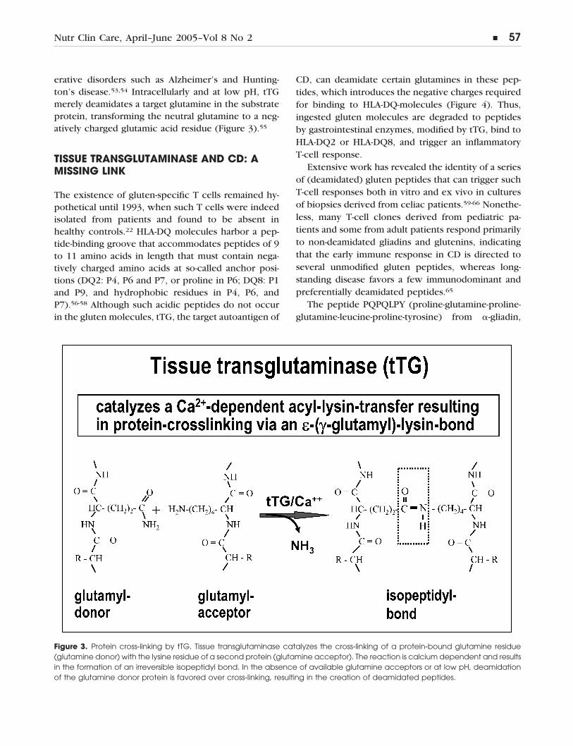
erative disorders such as Alzheimer’s and Hunting-ton’s disease.53,54 Intracellularly and at low pH, tTGmerely deamidates a target glutamine in the substrateprotein, transforming the neutral glutamine to a neg-atively charged glutamic acid residue (Figure 3).55
TISSUE TRANSGLUTAMINASE AND CD: AMISSING LINK
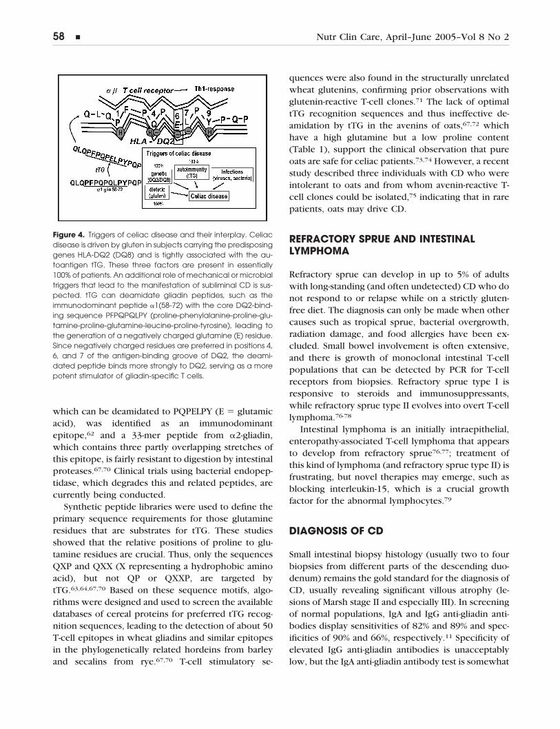
The existence of gluten-specific T cells remained hy-pothetical until 1993, when such T cells were indeedisolated from patients and found to be absent inhealthy controls.22 HLA-DQ molecules harbor a pep-tide-binding groove that accommodates peptides of 9to 11 amino acids in length that must contain nega-tively charged amino acids at so-called anchor posi-tions (DQ2: P4, P6 and P7, or proline in P6; DQ8: P1and P9, and hydrophobic residues in P4, P6, andP7).56-58 Although such acidic peptides do not occurin the gluten molecules, tTG, the target autoantigen of
CD, can deamidate certain glutamines in these pep-tides, which introduces the negative charges requiredfor binding to HLA-DQ-molecules (Figure 4). Thus,ingested gluten molecules are degraded to peptidesby gastrointestinal enzymes, modified by tTG, bind toHLA-DQ2 or HLA-DQ8, and trigger an inflammatoryT-cell response.
Extensive work has revealed the identity of a seriesof (deamidated) gluten peptides that can trigger suchT-cell responses both in vitro and ex vivo in culturesof biopsies derived from celiac patients.59-66 Nonethe-less, many T-cell clones derived from pediatric pa-tients and some from adult patients respond primarilyto non-deamidated gliadins and glutenins, indicatingthat the early immune response in CD is directed toseveral unmodified gluten peptides, whereas long-standing disease favors a few immunodominant andpreferentially deamidated peptides.65
The peptide PQPQLPY (proline-glutamine-proline-glutamine-leucine-proline-tyrosine) from �-gliadin,
Figure 3. Protein cross-linking by tTG. Tissue transglutaminase catalyzes the cross-linking of a protein-bound glutamine residue(glutamine donor) with the lysine residue of a second protein (glutamine acceptor). The reaction is calcium dependent and resultsin the formation of an irreversible isopeptidyl bond. In the absence of available glutamine acceptors or at low pH, deamidationof the glutamine donor protein is favored over cross-linking, resulting in the creation of deamidated peptides.
Nutr Clin Care, April–June 2005–Vol 8 No 2 � 57
which can be deamidated to PQPELPY (E � glutamicacid), was identified as an immunodominantepitope,62 and a 33-mer peptide from �2-gliadin,which contains three partly overlapping stretches ofthis epitope, is fairly resistant to digestion by intestinalproteases.67,70 Clinical trials using bacterial endopep-tidase, which degrades this and related peptides, arecurrently being conducted.
Synthetic peptide libraries were used to define theprimary sequence requirements for those glutamineresidues that are substrates for tTG. These studiesshowed that the relative positions of proline to glu-tamine residues are crucial. Thus, only the sequencesQXP and QXX (X representing a hydrophobic aminoacid), but not QP or QXXP, are targeted bytTG.63,64,67,70 Based on these sequence motifs, algo-rithms were designed and used to screen the availabledatabases of cereal proteins for preferred tTG recog-nition sequences, leading to the detection of about 50T-cell epitopes in wheat gliadins and similar epitopesin the phylogenetically related hordeins from barleyand secalins from rye.67,70 T-cell stimulatory se-
quences were also found in the structurally unrelatedwheat glutenins, confirming prior observations withglutenin-reactive T-cell clones.71 The lack of optimaltTG recognition sequences and thus ineffective de-amidation by tTG in the avenins of oats,67,72 whichhave a high glutamine but a low proline content(Table 1), support the clinical observation that pureoats are safe for celiac patients.73,74 However, a recentstudy described three individuals with CD who wereintolerant to oats and from whom avenin-reactive T-cell clones could be isolated,75 indicating that in rarepatients, oats may drive CD.
REFRACTORY SPRUE AND INTESTINALLYMPHOMA
Refractory sprue can develop in up to 5% of adultswith long-standing (and often undetected) CD who donot respond to or relapse while on a strictly gluten-free diet. The diagnosis can only be made when othercauses such as tropical sprue, bacterial overgrowth,radiation damage, and food allergies have been ex-cluded. Small bowel involvement is often extensive,and there is growth of monoclonal intestinal T-cellpopulations that can be detected by PCR for T-cellreceptors from biopsies. Refractory sprue type I isresponsive to steroids and immunosuppressants,while refractory sprue type II evolves into overt T-celllymphoma.76-78
Intestinal lymphoma is an initially intraepithelial,enteropathy-associated T-cell lymphoma that appearsto develop from refractory sprue76,77; treatment ofthis kind of lymphoma (and refractory sprue type II) isfrustrating, but novel therapies may emerge, such asblocking interleukin-15, which is a crucial growthfactor for the abnormal lymphocytes.79
DIAGNOSIS OF CD
Small intestinal biopsy histology (usually two to fourbiopsies from different parts of the descending duo-denum) remains the gold standard for the diagnosis ofCD, usually revealing significant villous atrophy (le-sions of Marsh stage II and especially III). In screeningof normal populations, IgA and IgG anti-gliadin anti-bodies display sensitivities of 82% and 89% and spec-ificities of 90% and 66%, respectively.11 Specificity ofelevated IgG anti-gliadin antibodies is unacceptablylow, but the IgA anti-gliadin antibody test is somewhat
Figure 4. Triggers of celiac disease and their interplay. Celiacdisease is driven by gluten in subjects carrying the predisposinggenes HLA-DQ2 (DQ8) and is tightly associated with the au-toantigen tTG. These three factors are present in essentially100% of patients. An additional role of mechanical or microbialtriggers that lead to the manifestation of subliminal CD is sus-pected. tTG can deamidate gliadin peptides, such as theimmunodominant peptide �1(58-72) with the core DQ2-bind-ing sequence PFPQPQLPY (proline-phenylalanine-proline-glu-tamine-proline-glutamine-leucine-proline-tyrosine), leading tothe generation of a negatively charged glutamine (E) residue.Since negatively charged residues are preferred in positions 4,6, and 7 of the antigen-binding groove of DQ2, the deami-dated peptide binds more strongly to DQ2, serving as a morepotent stimulator of gliadin-specific T cells.
58 � Nutr Clin Care, April–June 2005–Vol 8 No 2
better. However, for IgA anti-gliadin antibodies, therewill still be 10 times as many positives (up to 10% ofthe adult population) as there are biopsy-proven pa-tients with CD, especially in the age group above 50years.80 Only long-term positivity of IgA anti-gliadinantibodies indicates the presence of CD with higherspecificity.81 IgA autoantibodies to endomysium,which are detected by indirect immunofluorescenceon sections of esophagus, umbilical cord, or liver, area sensitive and the most specific serological test toscreen for CD. The sensitivity reported from refer-ence laboratories in Europe is 93% to 87% and speci-ficity is 99% to 99.7%.11 However, lower sensitivitieshave been observed in the United States,5 apparentlyas a result of poor standardization. In a CD referencecenter in the Netherlands,82 IgA anti-endomysial anti-bodies were only associated with more severe intes-tinal lesions (Marsh IIIc � IIIb � IIIa), but were notfound in patients with mild lesions (Marsh II). Further-more, it remains difficult to determine the absolutesensitivities and specificities in a population screen,since duodenal biopsy, the supposed gold standard, isnot feasible in all subjects tested.
With the identification of tTG as the target CD au-toantigen recognized by endomysial autoantibodies,48
the development of ELISA tests, which are easy to per-form and observer independent, has become possible.This autoantigen has a particular link to CD pathogene-sis, as described above. For population screening, theIgA autoantibodies to tTG serve as an excellent tool topredict CD with a sensitivity and specificity of 94% and97%, respectively, when optimized assays based on hu-man recombinant tTG are used. The original assay ver-sions were based on guinea pig tTG extracted from liverand should not be used because, despite similar sensi-tivity, their specificity is lower (93%) due to contamina-tion and thus cross-reactivity with liver antigens. In con-trast to endomysial autoantibody testing, which largelydepends on the expertise of the laboratory, it is thequality of the commercially available human recombi-nant tTG assays that determine diagnostic accuracy,making selection of a good assay supplier essential.11
NON-DIETARY THERAPIES
Due to tremendous recent advances, CD is consideredthe best-understood HLA-linked disorder. The diseaseis driven by a defined external (nutritional) trigger,namely gluten proteins from wheat, rye, and barley. It
develops on the background of a genetic associationwith HLA-DQ2/DQ8 and involves tTG as an autoanti-gen that plays a key role in disease pathogenesis,namely antigenic potentiation of gluten peptides. Inaddition, the amount of gluten ingested, the genedose of HLA-DQ2 (DQ8), and the local expression oftTG appear to be important determinants of CD man-ifestation and severity. A sizable number of immuno-dominant gluten peptides have been identified, partof them resisting degradation by intestinal peptidasesand thus reaching the intestinal mucosa intact, wherereactive T cells are recruited. Th1 cytokines releasedfrom these activated, gluten-specific T cells or fromintraepithelial lymphocytes cause the typical mucosaldamage of CD.
Effective therapy of CD requires adherence to astrictly gluten-free diet, which is a burden for mostpatients. Therefore, alternative treatments are be-ing explored.83 These include degradation of immu-nodominant gliadin peptides that resist intestinalproteases by use of exogenous bacterial prolyl en-dopeptidases66,69; inhibition of intestinal tTG activ-ity by specific inhibitors84; inhibition of CD-specificT-cell stimulation by peptides that bind to HLA-DQ2or -DQ8 but not to the gluten peptide-specific T cellreceptors85; and modulation of the intestinal cyto-kine milieu, e.g. by immunomodulatory cytokinesor by cytokine antagonists.83 Apart from benefitingpatients with CD, such novel therapies may providea template for the treatment of other autoimmunediseases.
NUTRITIONAL THERAPY
Nutritional therapy for CD is a lifelong gluten-freediet. Early referral to, and counseling by, an expertdietitian is enormously important in helping the pa-tient to make the challenging adjustments needed toembark on a gluten-free lifestyle. The patient’s partic-ipation in an established celiac support group thatprovides updated product information, resources, andsocial and emotional support is also extremely help-ful.
The National Institutes of Health (NIH) held a land-mark consensus conference in June 2004, culminatingin concise guidelines regarding the diagnosis andmanagement of CD, as well as recommendations forfuture research.86 According to the consensus confer-ence statement, the six key elements in the manage-
Nutr Clin Care, April–June 2005–Vol 8 No 2 � 59
ment of CD are: 1) consultation with a skilled dieti-tian, 2) education about the disease, 3) lifelongadherence to the gluten-free diet, 4) identification andtreatment of nutritional deficiencies, 5) access to asupport group, and 6) continuous long-term follow-upby a multidisciplinary team.
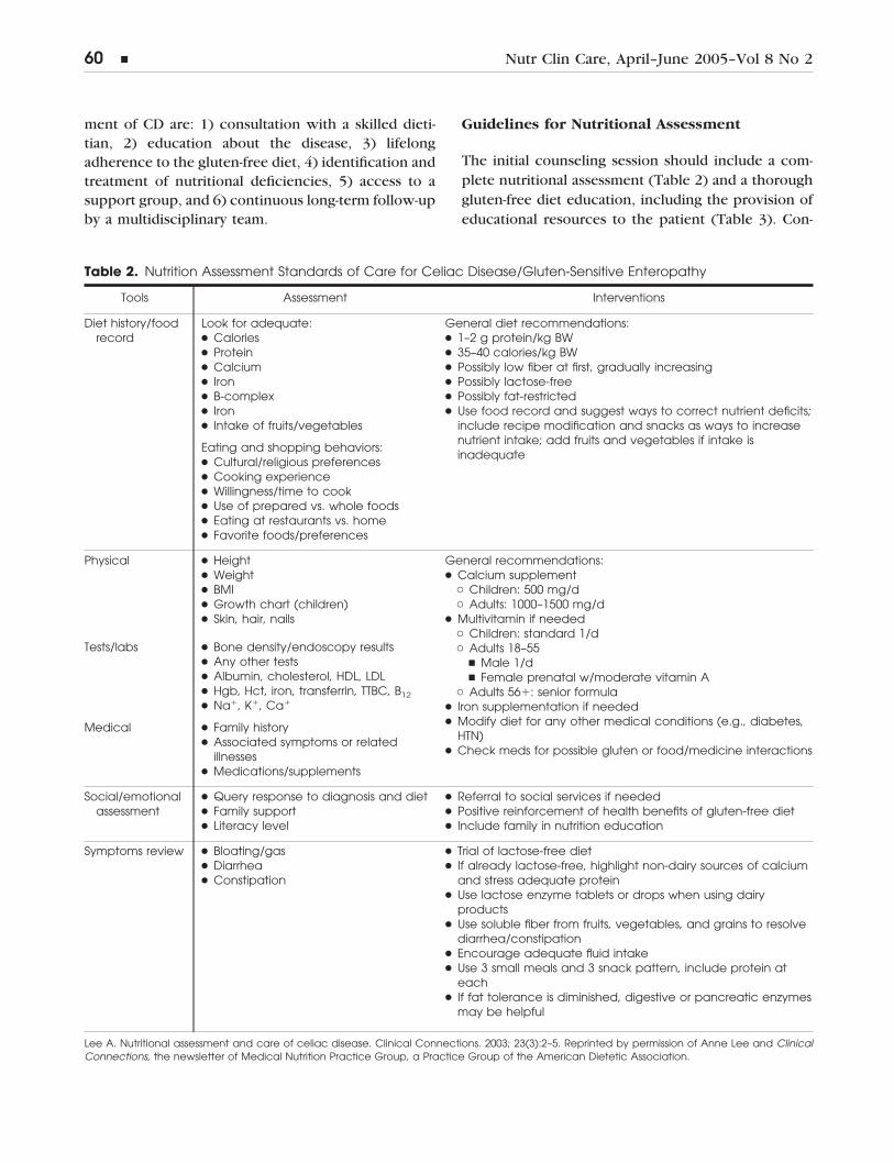
Guidelines for Nutritional Assessment
The initial counseling session should include a com-plete nutritional assessment (Table 2) and a thoroughgluten-free diet education, including the provision ofeducational resources to the patient (Table 3). Con-
Table 2. Nutrition Assessment Standards of Care for Celiac Disease/Gluten-Sensitive Enteropathy
Tools Assessment Interventions
Diet history/foodrecord
Look for adequate:● Calories● Protein● Calcium● Iron● B-complex● Iron● Intake of fruits/vegetables
Eating and shopping behaviors:● Cultural/religious preferences● Cooking experience● Willingness/time to cook● Use of prepared vs. whole foods● Eating at restaurants vs. home● Favorite foods/preferences
General diet recommendations:● 1–2 g protein/kg BW● 35–40 calories/kg BW● Possibly low fiber at first, gradually increasing● Possibly lactose-free● Possibly fat-restricted● Use food record and suggest ways to correct nutrient deficits;
include recipe modification and snacks as ways to increasenutrient intake; add fruits and vegetables if intake isinadequate
Physical ● Height● Weight● BMI● Growth chart (children)● Skin, hair, nails
General recommendations:● Calcium supplement
� Children: 500 mg/d� Adults: 1000–1500 mg/d
● Multivitamin if needed� Children: standard 1/d� Adults 18–55
� Male 1/d� Female prenatal w/moderate vitamin A
� Adults 56�: senior formula● Iron supplementation if needed● Modify diet for any other medical conditions (e.g., diabetes,
HTN)● Check meds for possible gluten or food/medicine interactions
Tests/labs ● Bone density/endoscopy results● Any other tests● Albumin, cholesterol, HDL, LDL● Hgb, Hct, iron, transferrin, TTBC, B12
● Na�, K�, Ca�
Medical ● Family history● Associated symptoms or related
illnesses● Medications/supplements
Social/emotionalassessment
● Query response to diagnosis and diet● Family support● Literacy level
● Referral to social services if needed● Positive reinforcement of health benefits of gluten-free diet● Include family in nutrition education
Symptoms review ● Bloating/gas● Diarrhea● Constipation
● Trial of lactose-free diet● If already lactose-free, highlight non-dairy sources of calcium
and stress adequate protein● Use lactose enzyme tablets or drops when using dairy
products● Use soluble fiber from fruits, vegetables, and grains to resolve
diarrhea/constipation● Encourage adequate fluid intake● Use 3 small meals and 3 snack pattern, include protein at
each● If fat tolerance is diminished, digestive or pancreatic enzymes
may be helpful
Lee A. Nutritional assessment and care of celiac disease. Clinical Connections. 2003; 23(3):2–5. Reprinted by permission of Anne Lee and ClinicalConnections, the newsletter of Medical Nutrition Practice Group, a Practice Group of the American Dietetic Association.
60 � Nutr Clin Care, April–June 2005–Vol 8 No 2
sideration of the patient’s cultural preferences, eatingand shopping behaviors, and social/emotional mind-set is also critical for effective education.87 Finally,
incorporation of additional dietary adaptations may beneeded for co-existing medical conditions such asdiabetes, iron deficiency, or lactose intolerance.
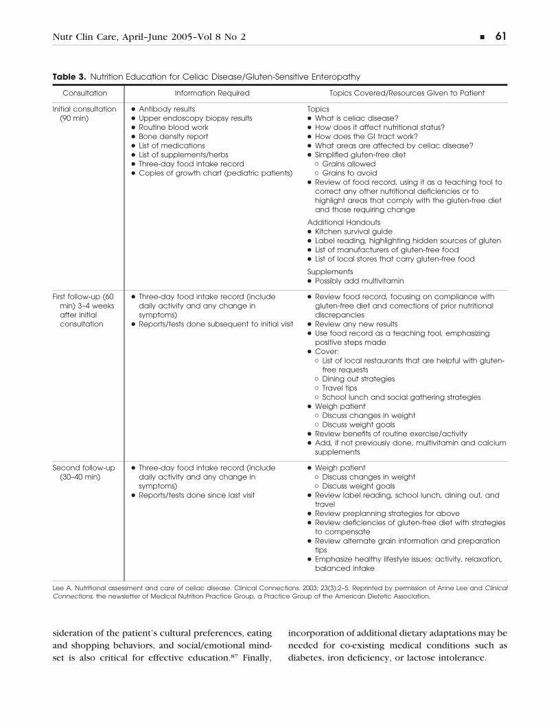
Table 3. Nutrition Education for Celiac Disease/Gluten-Sensitive Enteropathy
Consultation Information Required Topics Covered/Resources Given to Patient
Initial consultation(90 min)
● Antibody results● Upper endoscopy biopsy results● Routine blood work● Bone density report● List of medications● List of supplements/herbs● Three-day food intake record● Copies of growth chart (pediatric patients)
Topics● What is celiac disease?● How does it affect nutritional status?● How does the GI tract work?● What areas are affected by celiac disease?● Simplified gluten-free diet
� Grains allowed� Grains to avoid
● Review of food record, using it as a teaching tool tocorrect any other nutritional deficiencies or tohighlight areas that comply with the gluten-free dietand those requiring change
Additional Handouts● Kitchen survival guide● Label reading, highlighting hidden sources of gluten● List of manufacturers of gluten-free food● List of local stores that carry gluten-free food
Supplements● Possibly add multivitamin
First follow-up (60min) 3–4 weeksafter initialconsultation
● Three-day food intake record (includedaily activity and any change insymptoms)
● Reports/tests done subsequent to initial visit
● Review food record, focusing on compliance withgluten-free diet and corrections of prior nutritionaldiscrepancies
● Review any new results● Use food record as a teaching tool, emphasizing
positive steps made● Cover:
� List of local restaurants that are helpful with gluten-free requests
� Dining out strategies� Travel tips� School lunch and social gathering strategies
● Weigh patient� Discuss changes in weight� Discuss weight goals
● Review benefits of routine exercise/activity● Add, if not previously done, multivitamin and calcium
supplements
Second follow-up(30–40 min)
● Three-day food intake record (includedaily activity and any change insymptoms)
● Reports/tests done since last visit
● Weigh patient� Discuss changes in weight� Discuss weight goals
● Review label reading, school lunch, dining out, andtravel
● Review preplanning strategies for above● Review deficiencies of gluten-free diet with strategies
to compensate● Review alternate grain information and preparation
tips● Emphasize healthy lifestyle issues: activity, relaxation,
balanced intake
Lee A. Nutritional assessment and care of celiac disease. Clinical Connections. 2003; 23(3):2–5. Reprinted by permission of Anne Lee and ClinicalConnections, the newsletter of Medical Nutrition Practice Group, a Practice Group of the American Dietetic Association.
Nutr Clin Care, April–June 2005–Vol 8 No 2 � 61
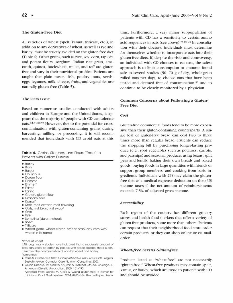
The Gluten-Free Diet
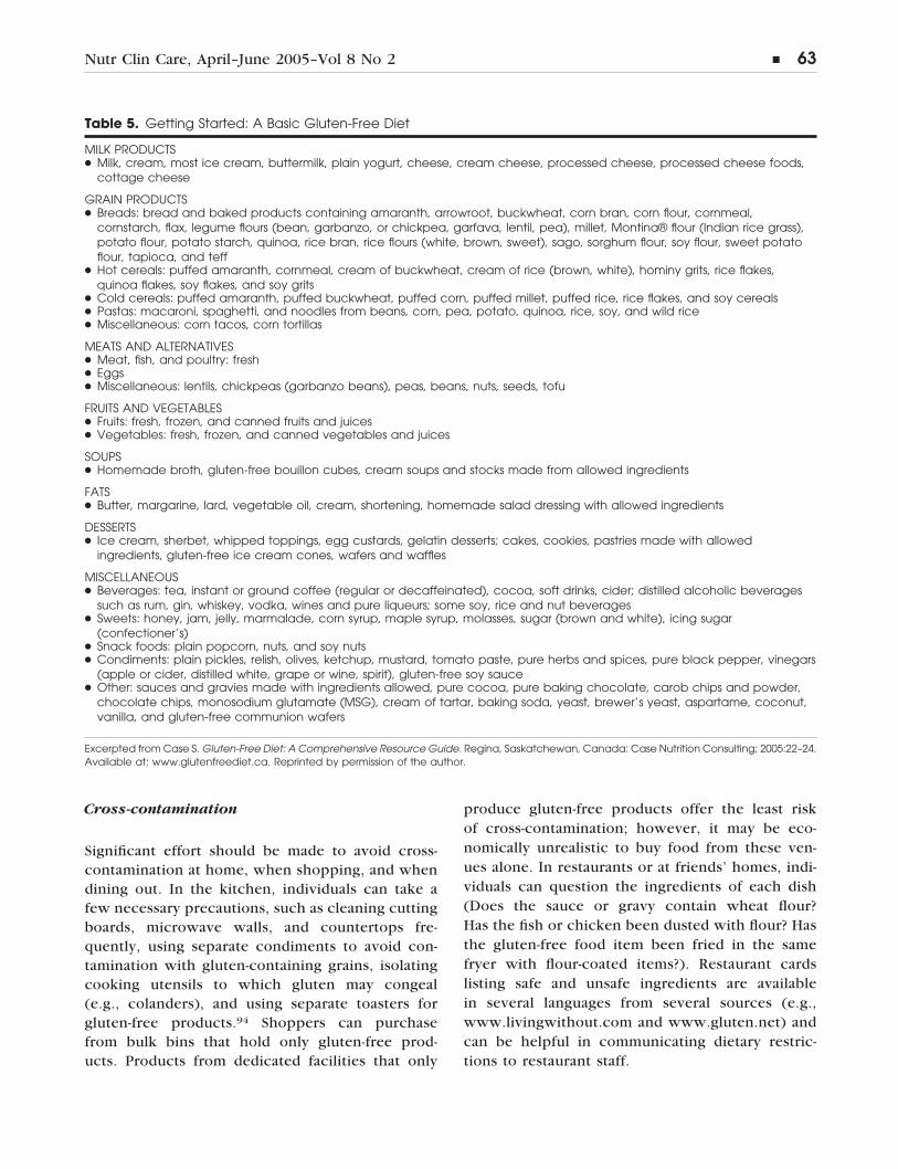
All varieties of wheat (spelt, kamut, triticale, etc.), inaddition to any derivatives of wheat, as well as rye andbarley, must be strictly avoided on the gluten-free diet(Table 4). Other grains, such as rice, soy, corn, tapiocaand potato flours, sorghum, Indian rice grass, ama-ranth, quinoa, buckwheat, millet, and teff are glutenfree and vary in their nutritional profiles. Patients aretaught that plain meats, fish, poultry, nuts, seeds,eggs, legumes, milk, cheese, fruits, and vegetables arenaturally gluten free (Table 5).
The Oats Issue
Based on numerous studies conducted with adultsand children in Europe and the United States, it ap-pears that the majority of people with CD can tolerateoats.72,73,88-93 However, due to the potential for cross-contamination with gluten-containing grains duringharvesting, milling, or processing, it is still recom-mended that individuals with CD avoid oats at this
time. Furthermore, a very minor subpopulation ofpatients with CD has a sensitivity to certain aminoacid sequences in oats (see above).75,88-93 In consulta-tion with their doctors, individuals must determinefor themselves whether to incorporate oats into theirgluten-free diets. If, despite the risks and controversy,an individual with CD chooses to eat oats, the safestapproach is to limit consumption to amounts foundsafe in several studies (50–70 g of dry, whole-grainrolled oats per day), to choose oats that have beentested and deemed free of contamination,93 and tocontinue to be closely monitored by a physician.
Common Concerns about Following a Gluten-Free Diet
Cost
Gluten-free commercial foods tend to be more expen-sive than their gluten-containing counterparts. A sin-gle loaf of gluten-free bread can cost two to threetimes more than regular bread. Patients can reducethe shopping bill by purchasing longer-lasting pro-duce (e.g., root vegetables such as potatoes, carrots,and parsnips) and seasonal produce; using beans, splitpeas and lentils; baking their own breads and bakedgoods; buying foods in large quantities with friends orsupport group members; and cooking from basic in-gredients. Individuals with CD may claim the gluten-free diet as a medical expense deduction on their USincome taxes if the net amount of reimbursementsexceeds 7.5% of adjusted gross income.
Accessibility
Each region of the country has different grocerystores and health food markets that offer a variety ofgluten-free products, some more than others. Patientscan request that their neighborhood food store ordercertain products, or they can shop online or via mail-order.
Wheat-free versus Gluten-free
Products listed as “wheat-free” are not necessarily“gluten-free.” Wheat-free products may contain spelt,kamut, or barley, which are toxic to patients with CDand should be avoided.
Table 4. Grains, Starches, and Flours “Toxic” toPatients with Celiac Disease
● Barley● Bran● Bulgur● Couscous● Durum flour● Einkorn*● Emmer*● Farro*● Farina● Gluten, gluten flour● Graham flour● Kamut*● Malt, malt extract, malt flavoring● Oats, oat bran, oat syrup†
● Orzo● Rye● Semolina (durum wheat)● Spelt● Triticale● Wheat germ, wheat starch, wheat bran, any item with
wheat in its name
*Types of wheat†Although many studies have indicated that a moderate amount ofoats can safely be eaten by people with celiac disease, there is con-cern over the contamination of oats by wheat and barley.References:● Case S. Gluten-Free Diet: A Comprehensive Resource Guide. Regina,
Saskatchewan, Canada: Case Nutrition Consulting; 2003.● Celiac Disease. In: Manual of Clinical Dietetics. 6th ed. Chicago, IL:
American Dietetic Association; 2000: 181–190.Adapted from: Dennis M, Case S. Going gluten-free: a primer forclinicians. Pract Gastroenterol. 2004;28:86–104. Used with permission.
62 � Nutr Clin Care, April–June 2005–Vol 8 No 2
Cross-contamination
Significant effort should be made to avoid cross-contamination at home, when shopping, and whendining out. In the kitchen, individuals can take afew necessary precautions, such as cleaning cuttingboards, microwave walls, and countertops fre-quently, using separate condiments to avoid con-tamination with gluten-containing grains, isolatingcooking utensils to which gluten may congeal(e.g., colanders), and using separate toasters forgluten-free products.94 Shoppers can purchasefrom bulk bins that hold only gluten-free prod-ucts. Products from dedicated facilities that only
produce gluten-free products offer the least riskof cross-contamination; however, it may be eco-nomically unrealistic to buy food from these ven-ues alone. In restaurants or at friends’ homes, indi-viduals can question the ingredients of each dish(Does the sauce or gravy contain wheat flour?Has the fish or chicken been dusted with flour? Hasthe gluten-free food item been fried in the samefryer with flour-coated items?). Restaurant cardslisting safe and unsafe ingredients are availablein several languages from several sources (e.g.,www.livingwithout.com and www.gluten.net) andcan be helpful in communicating dietary restric-tions to restaurant staff.
Table 5. Getting Started: A Basic Gluten-Free Diet
MILK PRODUCTS● Milk, cream, most ice cream, buttermilk, plain yogurt, cheese, cream cheese, processed cheese, processed cheese foods,
cottage cheese
GRAIN PRODUCTS● Breads: bread and baked products containing amaranth, arrowroot, buckwheat, corn bran, corn flour, cornmeal,
cornstarch, flax, legume flours (bean, garbanzo, or chickpea, garfava, lentil, pea), millet, Montina® flour (Indian rice grass),potato flour, potato starch, quinoa, rice bran, rice flours (white, brown, sweet), sago, sorghum flour, soy flour, sweet potatoflour, tapioca, and teff
● Hot cereals: puffed amaranth, cornmeal, cream of buckwheat, cream of rice (brown, white), hominy grits, rice flakes,quinoa flakes, soy flakes, and soy grits
● Cold cereals: puffed amaranth, puffed buckwheat, puffed corn, puffed millet, puffed rice, rice flakes, and soy cereals● Pastas: macaroni, spaghetti, and noodles from beans, corn, pea, potato, quinoa, rice, soy, and wild rice● Miscellaneous: corn tacos, corn tortillas
MEATS AND ALTERNATIVES● Meat, fish, and poultry: fresh● Eggs● Miscellaneous: lentils, chickpeas (garbanzo beans), peas, beans, nuts, seeds, tofu
FRUITS AND VEGETABLES● Fruits: fresh, frozen, and canned fruits and juices● Vegetables: fresh, frozen, and canned vegetables and juices
SOUPS● Homemade broth, gluten-free bouillon cubes, cream soups and stocks made from allowed ingredients
FATS● Butter, margarine, lard, vegetable oil, cream, shortening, homemade salad dressing with allowed ingredients
DESSERTS● Ice cream, sherbet, whipped toppings, egg custards, gelatin desserts; cakes, cookies, pastries made with allowed
ingredients, gluten-free ice cream cones, wafers and waffles
MISCELLANEOUS● Beverages: tea, instant or ground coffee (regular or decaffeinated), cocoa, soft drinks, cider; distilled alcoholic beverages
such as rum, gin, whiskey, vodka, wines and pure liqueurs; some soy, rice and nut beverages● Sweets: honey, jam, jelly, marmalade, corn syrup, maple syrup, molasses, sugar (brown and white), icing sugar
(confectioner’s)● Snack foods: plain popcorn, nuts, and soy nuts● Condiments: plain pickles, relish, olives, ketchup, mustard, tomato paste, pure herbs and spices, pure black pepper, vinegars
(apple or cider, distilled white, grape or wine, spirit), gluten-free soy sauce● Other: sauces and gravies made with ingredients allowed, pure cocoa, pure baking chocolate, carob chips and powder,
chocolate chips, monosodium glutamate (MSG), cream of tartar, baking soda, yeast, brewer’s yeast, aspartame, coconut,vanilla, and gluten-free communion wafers
Excerpted from Case S. Gluten-Free Diet: A Comprehensive Resource Guide. Regina, Saskatchewan, Canada: Case Nutrition Consulting; 2005:22–24.Available at: www.glutenfreediet.ca. Reprinted by permission of the author.
Nutr Clin Care, April–June 2005–Vol 8 No 2 � 63
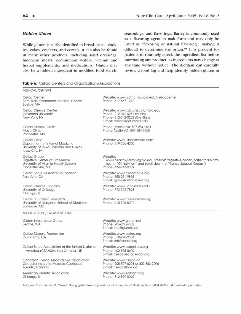
Hidden Gluten
While gluten is easily identified in bread, pasta, cook-ies, cakes, crackers, and cereals, it can also be foundin many other products, including salad dressings,luncheon meats, communion wafers, vitamin andherbal supplements, and medications. Gluten mayalso be a hidden ingredient in modified food starch,
seasonings, and flavorings. Barley is commonly usedas a flavoring agent in malt form and may only belisted as “flavoring or natural flavoring,” making itdifficult to determine the origin.95 It is prudent forpatients to routinely check the ingredient list beforepurchasing any product, as ingredients may change atany time without notice. The dietitian can carefullyreview a food log and help identify hidden gluten in
Table 6. Celiac Centers and Organizations/Associations
MEDICAL CENTERS
Celiac Center Website: www.bidmc.harvard.edu/celiaccenterBeth Israel Deaconess Medical Center Phone: 617-667-1272Boston, MA
Celiac Disease Center Website: www.cdcc.hs.columbia.eduColumbia University Phone: 212-342-0251 (Nurse)New York, NY Phone: 212-342-0252 (Dietitian)
E-mail: [email protected]
Celiac Disease Clinic Phone (clinicians): 507-284-2631Mayo Clinic Phone (patients): 507-284-5255Rochester, MN
Celiac Clinic Website: www.uihealthcare.comDepartment of Internal Medicine Phone: 319-356-4060University of Iowa Hospitals and ClinicsIowa City, IA
Celiac Group Website:Digestive Center of Excellence www.healthsystem.virginia.edu/internet/digestive-health/patientcare.cfmUniversity of Virginia Health System (go to “GI Nutrition” and scroll down to “Celiac Support Group”)Charlottesville, VA Phone: 434-243-9309
Celiac Sprue Research Foundation Website: www.celiacsprue.orgPalo Alto, CA Phone: 650-251-9865
E-mail: [email protected]
Celiac Disease Program Website: www.uchospitals.eduUniversity of Chicago Phone: 773-702-7593Chicago, IL
Center for Celiac Research Website: www.celiaccenter.orgUniversity of Maryland School of Medicine Phone: 410-706-8021Baltimore, MD
ASSOCIATIONS/ORGANIZATIONS
Gluten Intolerance Group Website: www.gluten.netSeattle, WA Phone: 206-246-6652
E-mail: [email protected]
Celiac Disease Foundation Website: www.celiac.orgStudio City, CA Phone: 818-990-2354
E-mail: [email protected]
Celiac Sprue Association of the United States ofAmerica (CSA/USA, Inc) Omaha, NE
Website: www.csaceliacs.orgPhone: 402-558-0600E-mail: [email protected]
Canadian Celiac Association/L’association Website: www.celiac.caCanadienne de la Maladie Coeliaque Phone: 905-507-6208 or 800-363-7296Toronto, Canada E-mail: [email protected]
American Dietetic Association Website: www.eatright.orgChicago, IL Phone: 312-899-0040
Adapted from: Dennis M, Case S. Going gluten-free: a primer for clinicians. Pract Gastroenterol. 2004;28:86–104. Used with permission.
64 � Nutr Clin Care, April–June 2005–Vol 8 No 2
medications, supplements, meals eaten in and awayfrom home, body care products that enter the mouthor nose, and other potential exposure to gluten.
Intentional Gluten Ingestion
Temptations to eat gluten may loom large for anyonewith CD; for those patients who are asymptomatic, thebenefits of a strict, lifelong diet may be even moredifficult to accept. These patients should be educated onthe consequences of long-term exposure to gluten andthen encouraged to comply, fortified with a variety ofhelpful resources such as books, magazines, and news-letters. They should also be given assistance in findingconvenient gluten-free foods and should join a supportgroup for emotional and social assistance. Table 6 listsUS medical centers and North-American organizationsthat specialize in CD, with resources for both patientsand clinicians.
IMPACT OF CD ON NUTRITIONAL STATUS
Malabsorption is common in CD due to a decrease inthe absorptive mucosal area and the enterocyte brushborder digestive enzymes. Studies show a 4% inci-dence of anemia in newly diagnosed patients with CDin the United States.96 According to Annibale et al.,97
recovery from anemia in adult patients can occur on agluten-free diet alone. Research by Hallert et al.98
indicates that even those CD patients who have beencarefully treated with a gluten-free diet for severalyears have higher total plasma homocysteine levelsand lower mean daily intakes of folate and vitamin B12
than control subjects, suggesting that vitamin defi-ciencies may occur even with a gluten-free diet; there-fore, vitamin status should be reviewed annually in alladult CD patients.
Immediately after diagnosis, gluten-free vitaminsand minerals in therapeutic doses may be required tocorrect iron, folate,4 or any other vitamin or mineraldeficiency while the mucosa heals. Depending onindividual factors, including age, laboratory test re-sults, compliance with the gluten-free diet, and over-all eating habits, a standard gluten-free multivitaminand mineral supplement is likely to be indicated.94,99
The need for iron supplementation should be evalu-ated on an individual basis. When prescribing medi-cation or supplements for a patient with CD, thephysician should indicate “as ordered, if gluten-free,
or provide a gluten-free equivalent” on the prescrip-tion.96
A well-balanced, gluten-free diet can provide ade-quate amounts of most nutrients. Prepared gluten-freecereals and commercial grain products, however, of-ten tend to have reduced quantities of B vitamins,iron, and fiber compared with products containinggluten.100-102 Patients should be encouraged to in-clude in their diet foods rich in folate (e.g., dark leafygreens, citrus fruit, dried beans and legumes, and flaxseeds) and vitamin B12 (e.g., meat, milk, fish, andpoultry), as well as foods rich in heme iron (leanmeats, poultry, and seafood) and non-heme iron (nuts,seeds, and legumes, taken with a vitamin C-rich foodto increase iron absorption). Amaranth, buckwheat,and quinoa are good sources of iron, fiber, and someof the B vitamins. Enriched, gluten-free commercialproducts should regularly be selected over refinedones.103
Bone Disease
Adequate calcium and vitamin D intake should beencouraged through dietary sources and supple-mentation, particularly for those patients with os-teopenic bone disease.12 Calcium-rich foods in-clude milk, gluten-free yogurt, cheese, sardines orcanned salmon with bones, calcium-fortified bever-ages such as orange or apple juice, and enriched,gluten-free soy, almond, or rice milk. Good sourcesof vitamin D, necessary for calcium absorption,include vitamin D-fortified milk, fatty fish and fishoils, liver, egg yolk, some gluten-free enriched bev-erages, and exposure to sunshine during latespring, summer, and early fall. Supplementationwith calcium and vitamin D may be necessary87
depending on age, gender, menopausal status, andother conditions. Weight-bearing exercise is alsovery important for bone health.
Lactose Intolerance
Bloating, gas, and diarrhea may be suggestive of lac-tose intolerance, a common problem in individualswith CD. If these symptoms occur, patients shouldavoid or limit lactose consumption for one or moremonths while lactase enzyme production recovers.101
Nutr Clin Care, April–June 2005–Vol 8 No 2 � 65
Gluten-free lactase enzyme supplements,* lactose-re-duced or lactose-free products such as Lactaid® milk,aged cheese, and gluten-free yogurt with live, activecultures are usually well tolerated. Alternatives tocommon dairy products include enriched dairy-free/gluten-free beverages such as soy, almond, or ricemilk.
Constipation and Diarrhea
Constipation can best be managed by gradually in-creasing fluids and dietary fiber (vegetables, fruitswith edible seeds such as berries or kiwi, bean flours,and whole, gluten-free grains and seeds94). Patientspresenting with diarrhea may benefit from a tempo-rary lactose-free or lactose-reduced diet with adequatefluid intake.94 Gluten-free fiber supplements may behelpful for both diarrhea and constipation.
Nutritional Follow-up
After the educational sessions have been completed,follow-up should be scheduled on an individual basisand should include monitoring the patient’s nutri-tional status based on laboratory tests, diet and activ-ity history, and weight changes. Newly presentingissues, such as increased weight gain and elevatedlipid levels from improved absorption, may need to beaddressed.87 Ongoing support, education, and atten-tion to changing nutritional needs are critical factorsin the patient’s successful adaptation to this new dietand lifestyle.
REFERENCES
1. Trier JS. Celiac sprue. N Engl J Med. 1991;325:1709–1719.
2. Schuppan D. Current concepts of celiac diseasepathogenesis. Gastroenterology. 2000; 119:234–242.
3. Ciclitira PJ, King AL, Fraser JS. AGA technicalreview on celiac sprue. American GastroenterologicalAssociation. Gastroenterology. 2001;120:1526–1540.
4. Farrell RJ, Kelly CP. Celiac sprue. N Engl J Med.2002;346:180–188.
5. Green PH, Jabri B. Coeliac disease. Lancet. 2003;362:383–391.
6. Hardwick C. Prognosis in coeliac disease. ArchDis Child. 1939;14:279.
7. Dicke WK, Weijers HA, van de Kamer JH. Coeliacdisease. II. The presence in wheat of a factor having adeleterious effect in cases of coeliac disease. Acta Paedi-atr. 1953;42:34–42.
8. Van de Kamer JH, Weijers HA, Dicke WK. Coeliacdisease IV. An investigation into the injurious constitu-ents of wheat in connection with their action on patientswith coeliac disease. Acta Paediatr. 1953;42:223–231.
9. Paulley JW. Observation on the aetiology of idio-pathic steatorrhoea; jejunal and lymph-node biopsies. BrMed J. 1954;4900:1318–1321.
10. Marsh MN. Gluten, major histocompatibility com-plex, and the small intestine: A molecular and immuno-biologic approach to the spectrum of gluten sensitivity(‘celiac sprue’). Gastroenterology. 1992;102:330–354.
11. Wong RC, Steele RH, Reeves GE, Wilson RJ, PinkA, Adelstein S. Antibody and genetic testing in coeliacdisease. Pathology. 2003;35:285–304.
12. Catassi C, Ratsch IM, Fabiani E, et al. Coeliacdisease in the year 2000: exploring the iceberg. Lancet.1994;343:200–203.
13. Fasano A, Catassi C. Current approaches to diag-nosis and treatment of celiac disease: an evolving spec-trum. Gastroenterology. 2001;120:636–651.
14. Maki M, Mustalahti K, Kokkonen J, et al. Preva-lence of celiac disease among children in Finland. N EnglJ Med. 2003;348:2517–2524.
15. Wieser H. The precipitating factor in coeliac dis-ease. Baillieres Clin Gastroenterol. 1995;9:191–207.
16. Shewry PR, Halford NG, Belton PS, Tatham AS.The structure and properties of gluten: an elastic proteinfrom wheat grain. Philos Trans R Soc Lond B Biol Sci.2002;357:133–142.
17. Howdle PD, Ciclitira PJ, Simpson FG, LosowskyMS. Are all gliadins toxic in coeliac disease? An in vitrostudy of alpha, beta, gamma, and omega gliadins. ScandJ Gastroenterol. 1984;19:41–47.
18. Ciclitira PJ, Evans DJ, Fagg NL, Lennox ES, Dowl-ing RH. Clinical testing of gliadin fractions in coeliacpatients. Clin Sci (Lond). 1984;66:357–364.
19. Sturgess R, Day P, Ellis HJ, et al. Wheat peptidechallenge in coeliac disease. Lancet. 1994;343:758–761.
20. Howell MD, Austin RK, Kelleher D, Nepom GT,Kagnoff MF. An HLA-D region restriction length poly-morphism associated with celiac disease. J Exp Med.1986;164:333–338.
21. Sollid LM, Markussen G, Ek J, Gjerde H, Vartdal F,Thorsby. Evidence for a primary association of celiacdisease to a particular HLA-DQ �/� heterodimer. J ExpMed. 1989;169:345–350.
22. Sollid LM, Thorsby E. HLA susceptibility genes in
* According to the manufacturer, Lactaid® Original Strength caplets,Lactaid® Ultra caplets, and Lactaid® Ultra chewables are consideredto have no detectable levels of gluten at the writing of this article.
66 � Nutr Clin Care, April–June 2005–Vol 8 No 2
celiac disease: genetic mapping and role in pathogene-sis. Gastroenterology. 1993;105:910–922.
23. Greco L, Romino R, Coto I, et al. The first largepopulation based twin study of coeliac disease. Gut.2002;50:624–628.
24. Karell K, Louka AS, Moodie SJ, et al.; EuropeanGenetics Cluster on Celiac Disease. HLA types in celiacdisease patients not carrying the DQA1*05–DQB1*02(DQ2) heterodimer: results from the European GeneticsCluster on Celiac Disease. Hum Immunol. 2003;64:469–477.
25. Greco L, Babron MC, Corazza GR, et al. Existenceof a genetic risk factor on chromosome 5q in Italiancoeliac disease families. Ann Hum Genet. 2001;65:35–41.
26. Babron MC, Nilsson S, Adamovic S, et al.; Euro-pean Genetics Cluster on Coeliac Disease. Meta andpooled analysis of European coeliac disease data. Eur JHum Genet. 2003;11:828–834.
27. Percopo S, Babron MC, Whalen M, et al. Satura-tion of the 5q31-q33 candidate region for coeliac disease.Ann Hum Genet. 2003;67:265–268.
28. Popat S, Hearle N, Hogberg L, et al. Variation inthe CTLA4/CD28 gene region confers an increased riskof celiac disease. Ann Hum Genet. 2002;66(part 2):125–137.
29. Holopainen P, Naluai AT, Moodie S, et al.; Mem-bers of the European Genetics Cluster on Coeliac Dis-ease. Candidate gene region 2q33 in European familieswith coeliac disease. Tissue Antigens. 2004;63:212–222.
30. Bilbao JR, Martin-Pagola A, Vitoria JC, Zubillaga P,Ortiz L, Castano L. HLA-DRB1 and MHC class 1 chain-related A haplotypes in Basque families with celiac dis-ease. Tissue Antigens. 2002;60:71–76.
31. Bolognesi E, Karell K, Percopo S, et al. Additionalfactor in some HLA DR3/DQ2 haplotypes confers a four-fold increased genetic risk of celiac disease. Tissue Anti-gens 2003;61:308–316.
32. Weile B, Cavell B, Nivenius K, Krasilnikoff PA.Striking differences in the incidence of childhood celiacdisease between Denmark and Sweden: a plausible ex-planation. J Pediatr Gastroenterol Nutr. 1995;21:64–68.
33. Petronzelli F, Multari G, Ferrante P, et al. Differentdose effect of HLA-DQ alpha beta heterodimers in insu-lin-dependent diabetes mellitus and celiac disease sus-ceptibility. Hum Immunol. 1993;36:156–162.
34. Congia M, Cucca F, Frau F, et al. A gene dosageeffect of the DQA1*0501/DQB1*0201 allelic combina-tion influences the clinical heterogeneity of celiac dis-ease. Hum Immunol. 1994;40:138–142.
35. Zubillaga P, Vidales MC, Zubillaga I, OrmaecheaV, Garcia-Urkia N, Vitoria JC. HLA-DQA1 and HLA-DQB1genetic markers and clinical presentation in celiac dis-ease. J Pediatr Gastroenterol Nutr. 2002;34:548–554.
36. Vader W, Stepniak D, Kooy Y, et al. The HLA-DQ2gene dose effect in celiac disease is directly related to themagnitude and breadth of gluten-specific T cell re-sponses. Proc Natl Acad Sci U S A. 2003;100:12390–12395.
37. Ventura A, Magazzu G, Greco L. Duration of ex-posure to gluten and risk for autoimmune disorders inpatients with celiac disease. SIGEP Study Group for Au-toimmune Disorders in Celiac Disease. Gastroenterol-ogy. 1999;117:297–303.
38. Sategna Guidetti C, Solerio E, Scaglione N, AimoG, Mengozzi G. Duration of gluten exposure in adultcoeliac disease does not correlate with the risk for auto-immune disorders. Gut. 2001;49:502–505.
39. Schuppan D, Ciccocioppo R. Coeliac disease andsecondary autoimmunity. Dig Liver Dis. 2002;34:13–15.
40. Luft LM, Barr SG, Martin LO, Chan EK, Fritzler MJ.Autoantibodies to tissue transglutaminase in Sjogren’ssyndrome and related rheumatic diseases. J Rheumatol.2003;30:2613–2619.
41. King AL, Moodie SJ, Fraser JS, et al. Coeliac dis-ease: investigation of proposed causal variants in theCTLA4 gene region. Eur J Immunogenet. 2003;30:427–432.
42. Naluai AT, Nilsson S, Samuelsson L, et al. TheCTLA4/CD28 gene region on chromosome 2q33 conferssusceptibility to celiac disease in a way possibly distinctfrom that of type 1 diabetes and other chronic inflam-matory disorders. Tissue Antigens. 2000;56:350–355.
43. Susi M, Holopainen P, Mustalahti K, Maki M, Par-tanen J. Candidate gene region 15q26 and genetic sus-ceptibility to coeliac disease in Finnish families. Scand JGastroenterol. 2001;36:372–374.
44. Collin P, Salmi J, Hallstrom O, Reunala T, Paster-nack A. Autoimmune thyroid disorders and coeliac dis-ease. Eur J Endocrinol. 1994;130:137–140.
45. Valentino R, Savastano S, Tommaselli AP, et al.Prevalence of coeliac disease in patients with thyroidautoimmunity. Horm Res. 1999;51:124–127.
46. Volta U, Ravaglia G, Granito A, et al. Coeliacdisease in patients with autoimmune thyroiditis. Diges-tion. 2001;64:61–65.
47. Badenhoop K, Dieterich W, Segni M, et al. HLADQ2 and/or DQ8 is associated with celiac disease-spe-cific autoantibodies to tissue transglutaminase in familieswith thyroid autoimmunity. Am J Gastroenterol. 2001;96:1648–1649.
48. Dieterich W, Ehnis T, Bauer M, et al. Identificationof tissue transglutaminase as the autoantigen of celiacdisease. Nat Med. 1997;3:797–801.
49. Piacentini M, Rodolfo C, Farrace MG, Autuori F.Tissue transglutaminase in animal development. Int JDev Biol. 2000;44:655–662.
50. Aeschlimann D, Thomazy V. Protein crosslinking
Nutr Clin Care, April–June 2005–Vol 8 No 2 � 67
in assembly and remodelling of extracellular matrices:the role of transglutaminases. Connect Tissue Res 2000;41:1–27.
51. Lorand L, Graham RM. Transglutaminases:crosslinking enzymes with pleiotropic functions. NatRev Mol Cell Biol. 2003;4:140–156.
52. Haroon ZA, Hettasch JM, Lai TS, Dewhirst MW,Greenberg CS. Tissue transglutaminase is expressed, ac-tive, and directly involved in rat dermal wound healingand angiogenesis. FASEB J. 1999;13:1787–1795.
53. Lesort M, Tucholski J, Miller ML, Johnson GV.Tissue transglutaminase: a possible role in neurodegen-erative diseases. Prog Neurobiol. 2000;61:439–463.
54. Gentile V, Cooper AJ. Transglutaminases - possi-ble drug targets in human diseases. Curr Drug TargetsCNS Neurol Disord. 2004;3:99–104.
55. Folk JE, Chung SI. Transglutaminases. MethodsEnzymol. 1985;113:358–375.
56. Vartdal F, Johansen BH, Friede T, et al. The pep-tide binding motif of the disease associated HLA-DQ(�1*0501, �1*0201) molecule. Eur J Immunol. 1996;26:2764–2772.
57. van de Wal Y, Kooy YM, Drijfhout JW, Amons R,Koning F. Peptide binding characteristics of the coeliacdisease-associated DQ(�1*0501, �1*0201) molecule. Im-munogenetics. 1996;44:246–253.
58. Godkin A, Friede T, Davenport M, et al. Use ofeluted peptide sequence data to identify the bindingcharacteristics of peptides to the insulin-dependent dia-betes susceptibility allele HLA-DQ8 (DQ 3.2). Int Immu-nol. 1997;9:905–911.
59. Molberg O, McAdam SN, Korner R, et al. Tissuetransglutaminase selectively modifies gliadin peptidesthat are recognized by gut-derived T cells in celiac dis-ease. Nat Med. 1998;4:713–717.
60. van de Wal Y, Kooy Y, van Veelen P, et al. Selec-tive deamidation by tissue transglutaminase strongly en-hances gliadin-specific T cell reactivity. J Immunol. 1998;161:1585–1588.
61. Arentz-Hansen H, Korner R, Molberg O, et al. Theintestinal T cell response to �-gliadin in adult celiacdisease is focused on a single deamidated glutaminetargeted by tissue transglutaminase. J Exp Med. 2000;191:603–612.
62. Anderson RP, Degano P, Godkin AJ, Jewell DP,Hill AV. In vivo antigen challenge in celiac disease iden-tifies a single transglutaminase-modified peptide as thedominant A-gliadin T-cell epitope. Nat Med. 2000;6:337–342.
63. Arentz-Hansen H, McAdam SN, Molberg O, et al.Celiac lesion T cells recognize epitopes that cluster inregions of gliadins rich in proline residues. Gastroenter-ology. 2002;123:803–809.
64. Fleckenstein B, Molberg O, Qiao SW, et al. Gliadin
T cell epitope selection by tissue transglutaminase inceliac disease. J Biol Chem. 2002;277:34109–34116.
65. Vader W, Kooy Y, Van Veelen P, et al. The glutenresponse in children with celiac disease is directed to-ward multiple gliadin and glutenin peptides. Gastroen-terology. 2002;122:1729–1737.
66. Shan L, Molberg O, Parrot I, et al. Structural basisfor gluten intolerance in celiac sprue. Science. 2002;297:2275–2279.
67. Vader WL, de Ru A, van der Wal Y, et al. Speci-ficity of tissue transglutaminase explains cereal toxicityin celiac disease. J Exp Med. 2002;195:643–649.
68. Skovbjerg H, Koch C, Anthonsen D, Sjostrom H.Deamidation and cross-linking of gliadin peptides bytransglutaminases and the relation to celiac disease. Bio-chim Biophys Acta. 2004;1690:220–230.
69. Qiao SW, Bergseng E, Molberg O, et al. Antigenpresentation to celiac lesion-derived T cells of a 33-mergliadin peptide naturally formed by gastrointestinal di-gestion. J Immunol. 2004;173:1757–1762.
70. Vader LW, Stepniak DT, Bunnik EM, et al. Char-acterization of cereal toxicity for celiac disease patientsbased on protein homology in grains. Gastroenterology.2003;125:1105–1113.
71. Molberg O, Solheim Flaete N, Jensen T, et al.Intestinal T-cell responses to high-molecular-weight glu-tenins in celiac disease. Gastroenterology.2003;125:337–344.
72. Janatuinen EK, Pikkarainen PH, Kemppainen TA,et al. A comparison of diets with and without oats inadults with celiac disease. N Engl J Med. 1995;333:1033–1037.
73. Hogberg L, Laurin P, Falth-Magnusson K, et al.Oats to children with newly diagnosed coeliac disease: arandomised double blind study. Gut. 2004;53:649–654.
74. Kilmartin C, Lynch S, Abuzakouk M, Wieser H,Feighery C. Avenin fails to induce a Th1 response incoeliac tissue following in vitro culture. Gut. 2003;52:47–52.
75. Arentz-Hansen H, Fleckenstein B, Molberg O, etal. The molecular basis for oat intolerance in patientswith celiac disease. PLoS Med. 2004;1:e1. Epub 2004 Oct19.
76. Ryan BM, Kelleher D. Refractory celiac disease.Gastroenterology. 2000;119:243–251.
77. Cellier C, Delabesse E, Helmer C, et al. Refractorysprue, coeliac disease, and enteropathy-associated T-celllymphoma. French Coeliac Disease Study Group. Lancet.2000;356:203–238.
78. Wahab PJ, Meijer JW, Goerres MS, Mulder CJ.Coeliac disease: changing views on gluten-sensitive en-teropathy. Scand J Gastroenterol Suppl. 2002;236:60–65.
79. Mention JJ, Ben Ahmed M, Begue B, et al. Inter-
68 � Nutr Clin Care, April–June 2005–Vol 8 No 2
leukin 15: a key to disrupted intraepithelial lymphocytehomeostasis and lymphomagenesis in celiac disease.Gastroenterology. 2003;125:730–745.
80. McMillan SA, Watson RP, McCrum EE, Evans AE.Factors associated with serum antibodies to reticulin,endomysium, and gliadin in an adult population. Gut.1996;39:43–47.
81. Johnston SD, Watson RG, McMillan SA, McMasterD, Evans A. Preliminary results from follow-up of a large-scale population survey of antibodies to gliadin, reticulinand endomysium. Acta Paediatr Suppl. 1996;412:61–64.
82. Rostami K, Kerckhaert J, Tiemessen R, vonBlomberg BM, Meijer JW, Mulder CJ. Sensitivity of anti-endomysium and antigliadin antibodies in untreated ce-liac disease: disappointing in clinical practice. Am J Gas-troenterol. 1999;94:888–894.
83. Schuppan D, Hahn EG. Gluten and the gut-lessonsfor immune regulation. Science. 2002;297:2218–2220.
84. Hausch F, Halttunen T, Maki M, Khosla C. Design,synthesis and evaluation of gluten peptide analogs asselective inhibitors of human tissue transglutaminase.Chem Biol. 2003;10:225–231.
85. Biagi F, Ellis HJ, Parnell ND, et al. A non-toxicanalogue of a coeliac-activating gliadin peptide: a basisfor immunomodulation? Aliment Pharmacol Ther. 1999;13:945–950.
86. NIH Consensus Development Conference on Ce-liac Disease. Baltimore, MD, June 28-30, 2004. Availableonline at: http://consensus.nih.gov/cons/118/118cdc_intro.htm. Accessed May 5, 2005.
87. Lee A. Nutritional assessment and care of celiacdisease. Clinical Connections. Winter 2003;23:2–5.
88. Srinivasan U, Leonard N, Jones E, et al. Absence ofoats toxicity in adult coeliac disease. BMJ. 1996;313:1300–1301.
89. Parnell N, Ellis HJ, Ciclitira P. Absence of toxicityof oats in patients with dermatitis herpetiformis. N EnglJ Med. 1998;338:1470–1471.
90. Janatuinen EK, Kemppainen TA, Pikkarainen PH,et al. Lack of cellular and humoral immunological re-sponses to oats in adults with coeliac disease. Gut. 2000;46:327–331.
91. Hoffenberg EJ, Haas J, Drescher A, et al. A trial of
oats in children with newly diagnosed celiac disease.J Pediatr. 2000;137:361–366.
92. Janatuinen EK, Kemppainen TA, Julkunen RJ, etal. No harm from five-year ingestion of oats in celiacdisease. Gut. 2002;50:332–335.
93. Thompson T. Oats and the gluten-free diet. J AmDiet Assoc. 2003;103:376–379.
94. Dennis M, Case S. Going Gluten-free: A Primerfor Clinicians. Available at: http://www.glutenfreedi-et.ca/Pract%20Gastro%202004.pdf. Accessed May 5,2005.
95. Case S. Gluten-Free Diet: A Comprehensive Re-source Guide. Regina, Saskatchewan, Canada: Case Nu-trition Consulting; 2004: 22–24.
96. Kupper C. Dietary guidelines and implementationfor celiac disease. Gastroenterol. 2005;128(4 suppl 1):S121–S127.
97. Annibale B, Severi C, Chistolini A, et al. Efficacy ofgluten-free diet alone on recovery from iron deficiencyanemia in adult celiac patients. Am J Gastroenterol.2001;96:132–137.
98. Hallert C, Grant C, Grehn S, et al. Evidence ofpoor vitamin status in celiac patients on a gluten-free dietfor 10 years. Aliment Pharmacol Ther. 2002;16:1333–1339.
99. Thompson T, Dennis M, Higgins LA, Lee AR, Shar-rett MK. Gluten-free diet survey: are Americans withcoeliac disease consuming recommended amounts offibre, iron, calcium and grain foods? J Hum Nutr Diet.2005;18:163–169.
100. Thompson T. Thiamin, riboflavin, and niacincontents of the gluten-free diet: is there cause for con-cern? J Am Diet Assoc. 1999;99:858–862.
101. American Dietetic Association. Celiac disease.In: Manual of Clinical Dietetics. 6th ed. Chicago, IL:ADA; 2000: 181–190.
102. Thompson T. Folate, iron, and dietary fiber con-tents of the gluten-free diet. J Am Diet Assoc. 2000;100:1389–1396.
103. Celiac disease (B vitamins, iron, and fiber). In:ADA Nutrition Care Manual. Available by subscriptionat www.nutritioncaremanual.org. Accessed April 1,2005.
Nutr Clin Care, April–June 2005–Vol 8 No 2 � 69