CD4 T CELL MEDIATED TUMOR IMMUNITY FOLLOWING STEM …
150
CD4 + T CELL MEDIATED TUMOR IMMUNITY FOLLOWING TRANSPLANTATION OF TRP-1 TCR GENE MODIFIED HEMATOPOIETIC STEM CELLS Sung Pil Ha Submitted to the faculty of the University Graduate School in partial fulfillment of the requirements for the degree Doctor of Philosophy in the Department of Microbiology and Immunology Indiana University December 2012
Transcript of CD4 T CELL MEDIATED TUMOR IMMUNITY FOLLOWING STEM …

CD4+ T CELL MEDIATED TUMOR IMMUNITY FOLLOWING
TRANSPLANTATION OF TRP-1 TCR GENE MODIFIED HEMATOPOIETIC
STEM CELLS
Sung Pil Ha
Submitted to the faculty of the University Graduate School in partial fulfillment of the requirements
for the degree Doctor of Philosophy
in the Department of Microbiology and Immunology Indiana University
December 2012

ii
Accepted by the Faculty of Indiana University, in partial fulfillment of the requirements for the degree of Doctor of Philosophy.
Christopher E. Touloukian, M.D., Chair
Hal E. Broxmeyer, Ph.D.
Doctoral Committee
Thomas A. Gardner, M.D. June 18, 2010
Maureen A. Harrington, Ph.D.
Johnny He, Ph.D.

iii
DEDICATION
I would like to dedicate this thesis to my family for all their support during my long
journey to finishing medical school.
iv
ACKNOWLEDGEMENTS
I would like to thank Dr. Christopher Touloukian without whom I would
never have begun this journey down this scientific rabbit hole. He has been a
constant proponent of my work and has provided invaluable guidance to me in
my career.
I would also like to thank Dr. Hal Broxmeyer and his lab for their support
not only in providing technical and practical advice, but also for their advice and
encouragement while I pursued my degree.
The other members of my committee Dr. Johnny He, Dr. Maureen
Harrington, and Dr. Thomas Gardner have provided me with valuable insight and
guidance.
I would also like to acknowledge the assistance of Andrew Brandmaier,
Nick Klemen, Garrett Killenbrew, Keri Burns, Lori Sanford and all the other
members past and present of the Touloukian lab.

v
ABSTRACT
Sung Pil Ha
CD4+ T cell mediated tumor immunity following transplantation of TRP-1 TCR
gene modified hematopoietic stem cells
Immunotherapy for cancer has held much promise as a potent modality of
cancer treatment. The ability to selectively destroy diseased cells and leave
healthy cells unharmed has been the goal of cancer immunotherapy for the past
thirty years. However, the full capabilities of cancer immunotherapies have been
elusive. Cancer immunotherapies have been consistently hampered by limited
immune reactivity, a diminishing immune response over time, and a failure to
overcome self-tolerance. Many of these deficiencies have been borne-out by
immunotherapies that have focused on the adoptive transfer of activated or
genetically modified mature CD8+ T cells. The limitations inherent in therapies
involving terminally differentiated mature lymphocytes include limited duration,
lack of involvement of other components of the immune system, and limited
clinical efficacy. We sought to overcome these limitations by altering and
enhancing long-term host immunity by genetically modifying then transplanting
HSCs. To study these questions and test the efficiency of gene transfer, we
cloned a tumor reactive HLA-DR4-restricted CD4+ TCR specific for the
melanocyte differentiation antigen TRP-1, then constructed both a high
expression lentiviral delivery system and a TCR Tg expressing the same TCR

vi
genes. We demonstrate with both mouse and human HSCs durable, high-
efficiency TCR gene transfer, following long-term transplantation. We
demonstrate the induction of spontaneous autoimmune vitiligo and a TCR-
specific TH1 polarized memory effector CD4+ T cell population. Most importantly,
we demonstrate the destruction of subcutaneous melanoma without the aid of
vaccination, immune modulation, or cytokine administration. Overall, these
results demonstrate the creation of a novel translational model of durable
lentiviral gene transfer, the induction of spontaneous CD4+ T cell immunity, the
breaking of self-tolerance, and the induction of anti-tumor immunity.
Christopher E. Touloukian, M.D., Chair

vii
TABLE OF CONTENTS LIST OF TABLES .................................................................................................. x
LIST OF FIGURES ................................................................................................. xi
LIST OF ABBREVIATIONS ................................................................................... xiv
I. INTRODUCTION ................................................................................................. 1
T Cell Development Overview ................................................................................ 1
Hematopoietic Stem Cells ...................................................................................... 1
Thymic Development .............................................................................................. 2
Immunotherapy ....................................................................................................... 3
Discovery of Tumor Infiltrating Lymphocytes .......................................................... 4
Autoimmunity Versus Tolerance ............................................................................. 5
Myelodepletion Enhances Autoimmunity ................................................................ 8
Limitations of CD8+ T Cells in Cancer Immunotherapy ........................................... 9
Importance of CD4+ T Cells in Cancer Immunotherapy .......................................... 11
Isolation of TRP-1 Specific CD4+ TCR ................................................................... 13
Efficiency and Safety of Lentiviral Gene Delivery ................................................... 14
Summary ................................................................................................................ 15
II. RESEARCH GOALS .......................................................................................... 20
III. MATERIALS AND METHODS .......................................................................... 23
Animal and Cell Lines ............................................................................................. 23
Limiting Dilution ...................................................................................................... 24
Detection of Cytokines Using ELISA ...................................................................... 25
Cloning of TRP-1 TCR ............................................................................................ 26

viii
Other Plasmids Used .............................................................................................. 27
Lentiviral Production ............................................................................................... 28
Lentivirus Titering ................................................................................................... 28
Collection of Bone Marrow, Peripheral Blood Cells, and Splenocytes .................... 29
Collection of Human Cord Blood Cells ................................................................... 30
Magnetic Bead Separation ..................................................................................... 31
Transduction Protocol ............................................................................................. 32
Cell Staining and Analysis Using Flow Cytometry .................................................. 32
Bone Marrow Transplantation................................................................................. 34
Immunohistochemistry ............................................................................................ 34
Determination of LV-TCR Gene Expression ........................................................... 35
Lentivirus Integration Analysis ................................................................................ 35
Tumor Treatment Assays ....................................................................................... 37
IV. RESULTS ......................................................................................................... 38
Identification of a TRP-1 Specific TCR ................................................................... 38
Construction of a Bicistronic Lentivirus ................................................................... 38
Efficiency of Transduction ...................................................................................... 39
Transduced Cells Retain Functionality of TCR ....................................................... 40
Development of a TRP-1 TCR Transgenic Mouse ................................................. 41
Transplantation of Lentivirus Gene Modified HSCs ................................................ 42
Lentiviral Transduction of HSCs Leads to Stable Long Term Integration ............... 43
Transduced T Cells Undergo Development in the Thymus .................................... 48
Transduced CD4+ T Cells Retain Specificity and Function ..................................... 49

ix
Development of Autoimmune Vitiligo ...................................................................... 50
Activation Status and Functionality of Transplanted T Cells ................................... 52
Secondary Transplant Experiments and Integration Analysis ................................ 53
Secondary LV-TCR Transplants Reject Subcutaneous Tumors ............................. 55
Gene Modification and Transplantation of Human HSCs ....................................... 56
TRP-1 TCR Confers Tumor Immunity .................................................................... 57
V. DISCUSSION ..................................................................................................... 59
Effects of Lentiviral Transduction on HSC Transplantation .................................... 61
Peripheral Homeostatic Proliferation Leads to Expansion of Selective T
Cell Clones ............................................................................................................. 63
Transplantation of LV-TCR Transduced HSCs Breaks Self-Tolerance .................. 65
Conclusion .............................................................................................................. 68
VI. LIMITATIONS AND FUTURE DIRECTIONS .................................................... 70
Increased Efficiency of TCR Expression ................................................................ 70
Mechanisms of Autoimmune Vitiligo ....................................................................... 71
Skewing the T Cell Repertoire ................................................................................ 76
Tumor Immunity ...................................................................................................... 78
TABLES ................................................................................................................. 80
FIGURES ............................................................................................................... 84
REFERENCE LIST ................................................................................................. 112
CURRICULUM VITAE

x
LIST OF TABLES
Table 1. Comparison of IRES and F2a lentiviral transduction experiments in
JURKAT and human peripheral blood mononuclear cells ...................................... 80
Table 2. Analysis of CD4+ T cell compartment in LV-TCR transduced and
TRP-1 Tg Lin- HSC transplants into either irradiated DR4 or C57BL/6
recipient mice ......................................................................................................... 81
Table 3. INF-γ release as measured by ELISA in splenocytes harvested from
LV-TCR transplants 12 months post transplantation .............................................. 82
Table 4. Summary of development of spontaneous autoimmune vitiligo in all
transplant groups .................................................................................................... 83

xi
LIST OF FIGURES
Figure 1. Central role of CD4+ T cells in tumor immunity ....................................... 84
Figure 2. Cloned α and β TCR subunits TRP-1/DR4 Clone-2 .............................. 85
Figure 3. Schematic diagrams of the TRP-1 TCR lentiviral constructs .................. 86
Figure 4. Comparison of IRES and F2A lentiviral transduction experiments in
human peripheral blood mononuclear cells ............................................................ 87
Figure 5. Representative flow cytometry results for lentiviral transduction of
human CD34+ and murine Lin- cells ....................................................................... 88
Figure 6. Lentiviral constructs efficiently transduce murine Lin- cells and leads
to high expression of transduced gene ................................................................... 89
Figure 7. Peripheral blood mononuclear cells transduced with LV-TCR are
functionally specific ................................................................................................. 90
Figure 8. Positive selection of the TRP-1 TCR following crossbreeding into
DR4 Tg mice ........................................................................................................... 91
Figure 9. Gene modified HSCs successfully engraft and repopulate transplant
host over 6 months ................................................................................................. 92
Figure 10. Representative flow cytometry shows successful engraftment and
emergence of lymphocytes 1 and 4 months post transplantation ........................... 93
Figure 11. Transplantation of LV-TCR and TRP-1 Tg Lin- cells results in high
expression of TRP-1 specific TCR .......................................................................... 94
Figure 12. 12 month analysis of LV-TCR and TRP-1 TCR Tg Lin- HSCs
transplants. Total cells ............................................................................................ 95

xii
Figure 13. 12 month analysis of LV-TCR and TRP-1 TCR Tg Lin- HSCs
transplants. CD3+ cells ........................................................................................... 96
Figure 14. 12 month analysis of LV-TCR and TRP-1 TCR Tg Lin- HSCs
transplants. CD4+ cells ........................................................................................... 97
Figure 15. Analysis of thymocytes in LV-TCR HSC transplanted DR4 Tg
mice ........................................................................................................................ 98
Figure 16. Development of spontaneous autoimmune vitiligo in mice ................... 99
Figure 17. Paraffin embedded immunohistochemistry staining of skin samples
of TRP-1 TCR transplanted mice that developed autoimmune vitiligo .................... 100
Figure 18. Frozen section immunohistochemistry staining of skin samples of
TRP-1 TCR transplanted mice that developed autoimmune vitiligo ........................ 101
Figure 19. Vitiligo is associated with gross distortion of natural skin
architecture, melanocyte destruction, and TCR gene specific CD4+ T cell
infiltration ................................................................................................................ 102
Figure 20. Representative flow cytometry analysis of splenocytes from
transplant experiments ........................................................................................... 103
Figure 21. Summary of activation status of all CD4+ subpopulations .................... 104
Figure 22. CD4+ T cells from secondary transplants exhibit high level gene .......... 105
Figure 23. Integration analysis of secondary transplants exhibit high level
gene specific TCR expression and multi-copy integration ...................................... 106
Figure 24. Six month secondary transplants reject subcutaneous melanoma
tumor injection ........................................................................................................ 107
Figure 25. Paraffin embedded immunohistochemistry staining of tumors

xiii
from LV-TCR secondary transplants demonstrate necrosis .................................. 108
Figure 26. Paraffin embedded immunohistochemistry staining of tumors from
LV-TCR secondary transplants exhibit infiltration by human TCR expressing
CD4+ T cells............................................................................................................ 109
Figure 27. LV-TCR gene-modified human CD34+/CD38- cells from cord blood
functionally engraft and repopulation 6 month old NOD/SCID-2 mice .................... 110
Figure 28. LV-TCR transplants in NOD/SCID-2 mice reject human melanoma
implantation ............................................................................................................ 111

xiv
LIST OF ABBREVIATIONS
ACK ammonium chloride potassium lysing buffer
AIRE autoimmune regulator gene
APC antigen presenting cells
BM bone marrow
BSA bovine serum albumin
CD103 cluster of differentiation 103
CD25 cluster of differentiation 25
CD3 cluster of differentiation 3
CD34 cluster of differentiation 34
CD38 cluster of differentiation 38
CD4 cluster of differentiation 4
CD40 cluster of differentiation 40
CD40L cluster of differentiation 40 ligand
CD44 cluster of differentiation 44
CD45.1 cluster of differentiation 45.1
CD45.2 cluster of differentiation 45.2
CD45RB cluster of differentiation 45RB
CD62L cluster of differentiation 62 ligand
CD8 cluster of differentiation 8
cDNA complementary DNA
CM complete media
CMV cytomegalovirus

xv
CLP common lymphoid precursor cells
cPPT central polypurine tract derived from HIV-polymerase
CTLA-4 cytotoxic T-lymphocyte antigen 4
DC dendritic cells
DMEM Dulbecco’s modified Eagle’s medium
DN double negative
DP double positive
EBV Epstein Barr virus
EDTA ethylene-glycol-tetra-acetic acid
EF1α elongation factor 1α
ELISA enzyme linked immunosorbent assay
ELP early lymphoid precursor cells
F2A Furin self-cleaving sequence
FACS fluorescence activated cell sorter
FBS fetal bovine serum
FITC fluorescein isothiocyanate
gag HIV gag gene
gDNA genomic DNA
GFP green fluorescent protein
GITR glucocorticoid induced TNF receptor
GM-CSF granulocyte macrophage colony stimulating factor
gp100 glycoprotein 100
HA Hemophilus influenza antigen

xvi
HBSS Hank’s buffered salt solution
HEPES 4-(2-hydroxyethyl)-1-piperazineethanesulfonic acid
HIV human immunodeficiency virus
HLA human leukocyte antigen
HP homeostatic proliferation
HSC hematopoietic stem cells
HTLV human T-lymphotropic virus type I
IFN-γ interferon γ
IHC immunohistochemistry
IL-12 interleukin-12
IL-2 interleukin-2
IL-6 interleukin-6
IL-7 interleukin-7
IRES internal ribosomal entry site
KO knockout
LAK lymphokine-activated killer cells
LTR long terminal repeat
LV lentivirus
MART-1 melanoma antigen recognized by T cells
MDA melanocyte differentiation antigen
MHC major histocompatibility complex
MOI multiplicity of infection
mTEC medullary thymic epithelial cells

xvii
NCI National Cancer Institute
NIH National Institute of Health
NOD non-obese diabetic
OA1 ocular albinism protein 1
OKT-3 murine monoclonal anti-CD3 antibody
OVA ovalbumin
PBMC peripheral blood mononuclear cells
PBS phosphate buffered saline
PCR polymerase chain reaction
PE phycoerythrin
PGK phosphoglycerate kinase
pol HIV pol gene
qPCR quantitative real time PCR
RACE rapid amplification of cDNA ends
RBC red blood cells
Rev regulator of virion protein expression
rpm revolutions per minute
RPMI Roswell Park Memorial Institute
RRE Rev response element
SCF stem cell factor
SCID severe combined immunodeficiency
SFM serum free media
SIN self-inactivating

xviii
SP single positive
TCR T cell receptor
TEM T effector memory phenotype
Tg transgenic
Th1 T helper type 1 cells
TIL tumor infiltrating lymphocytes
TNF tumor necrosis factor
TPO thrombopoietin
Treg regulator T cells
TRP-1 tyrosinase related protein 1
TRP-2 tyrosinase related protein 2
WPRE Woodchuck hepatitis virus posttranslational regulatory
element
VSVG vesicular stomatitis virus gene
γ-RV γ retrovirus

I. Introduction
T Cell Development Overview
T lymphocytes, like all cellular components of blood are ultimately derived
from and dependent on constant regeneration from hematopoietic stem cells
(HSCs) found in bone marrow. HSCs are characterized by their multi-lineage
potential and self-renewal capacity. HSCs continuously give rise to lineage-
restricted progenitor cells including myeloid progenitors, erythroid progenitors,
and common lymphoid progenitor cells. Lymphoid progenitors continue their
maturation in secondary lymphoid tissue. Primarily, B cells mature in the bone
marrow and T cells mature in the thymus, thus their respective names. Since the
crux of this thesis, involves the selection and manipulation of the T cell
population, we will focus on T cell development.
Hematopoietic Stem Cells
HSCs are fully capable of self-renewal and differentiation into all the
cellular components of blood. Because of their pluripotent nature, HSCs hold
great therapeutic promise. In particular, manipulation of the genes involved in
HSC development would mean the alteration of all cellular progeny downstream
of these early precursor cells. Thus, much research has gone into identifying
and characterizing these HSCs and their early decedent progenitor cell
populations. HSCs are identified in mice by a distinct surface protein phenotype,
Lin-/Sca-1+/c-kit+/CD44+/CD25-. They further differentiate in the bone marrow to
become lymphoid biased progenitor cells including LSK CD62L cells (Lin-/Sca-
1+/c-kit+/CD62L+), early lymphoid precursors (ELP) (Lin-/Sca-1+/c-
kit+/Flt3+/CD27+), and common lymphoid precursors (CLP) (Lin-/Sca-1-/c-kit-/IL-
1

7Ra+). A common theme amongst these early progenitor cells is the absence of
lineage markers and the expression of c-kit and Sca-1, along with the capacity to
traffic to and efficiently proliferate in the thymus, and their propensity to
differentiate into lymphoid cells (Haddad et al, 2004; Wu, 2006; Hoebeke et al,
2007; Serwold et al, 2008; Awong et al, 2009).
Thymic Development
Lymphoid progenitor cells subsequently traffic to the thymus, a secondary
lymphoid organ devoted to the development, selection, and maturation of T cells.
T cell progenitors originating from bone marrow are released in waves into the
blood where they travel periodically to the thymus (Miller, 1961; Pullen et al,
1989). These immature thymocytes undergo two phases of selection, both
positive and negative. In positive selection, thymocytes recognizing binding
components of MHC molecules are promoted and activated while thymocytes
that do not recognize self MHC molecules are actively deleted (Bevan and Fink,
1978; Zinkernagel et al, 1978; Pullen et al, 1989). In negative selection,
thymocytes that recognize self-antigens are actively deleted thus ensuring
removal of mature lymphocytes capable of inducing autoimmunity (von Boehmer
et al, 1989; McDonald and Lees, 1990). This process of central tolerance is
critical for the prevention of autoimmunity (Schwartz, 1989). Thymic epithelial
cells are believed to play a significant role in the negative selection process. In
particular, the medullary thymic epithelial cells (mTECs) which express the
autoimmune regulator element (AIRE), a regulatory transcription factor which
controls the promiscuous expression of a variety of self-antigens in mTECs
2

allows for the negative selection of self-antigen binding thymocytes. The genes
activated by AIRE include genes for a broad spectrum of protein functional
classes (growth factors, hormones, structural molecules, and tissue specific
proteins that represent essentially every organ) (Gotter et al, 2004; Johnnidis et
al, 2005; Anderson et al, 2005).
Immunotherapy
As evidenced above, researchers have taken great strides in uncovering
the nature, development and selection of the cells of the immune system.
However, this knowledge has not necessarily translated into clinical therapies.
The early attempts to harness the vast therapeutic potential of the immune
system focused primary on global manipulation of the immune response via
application of supraphysiologic levels of cytokines. Initial immunotherapies for
cancer focused on the activation of immune cells by administration of high dose
interleukin-2 (IL-2). Early studies in the 1980s identified cells capable of directly
lysing fresh tumors by incubation of lymphocytes in IL-2 (Yron et al, 1980).
Subsequent adoptive transfer of these lymphokine activated killer (LAK) cells in
conjunction with IL-2 therapy mediated the regression of some advanced
metastatic cancers. Of 55 patients treated with this approach, 21 showed
objective clinical regression, and 5 individuals showed complete regression of
malignancy (Rosenberg, 1985). These early trials showed promise in stimulating
immune response to metastatic melanoma; however, the high doses of IL-2
required for clinical response were intolerable to the majority of patients and
mediated toxic side effects, the most common of which was a capillary
3

permeability leak syndrome that resulted in major fluid retention. Furthermore,
the large number of activated cells necessary to mediate tumor regression was
cumbersome and limited the practicality of treatment on a large scale
(Rosenberg et al, 1985; Rosenberg et al, 1986). Treatment approaches with
cancer vaccines and more advanced adoptive immunotherapies have had
similarly limited outcomes. Due to these limitations, in the past three decades
researchers have sought more efficient approaches to modify the immune
response to cancer.
Discovery of Tumor Infiltrating Lymphocytes
In 1986, Rosenberg, Spiess, and LaFreniere at the National Cancer
Institute of the NIH discovered a subpopulation of lymphocytes that infiltrate into
growing cancers and were 50 to 100 times more potent than LAK cells for use in
adoptive transfer therapies (Rosenberg et al, 1986). These tumor infiltrating
lymphocytes (TIL) could be expanded ex vivo by incubation with IL-2 to sufficient
numbers for effective adoptive transfer. Concomitant TIL adoptive transfer, low
dose IL-2 treatment and immunosuppression with cyclophosphamide proved to
be highly effective in treating hepatic and pulmonary metastases in mice with
colon adenocarcinoma (Rosenberg et al, 1986). Unfortunately, early human
trials of TIL adoptive cell transfer therapy showed only a limited ability to
objectively regress tumor in metastatic cancer patients. However, the isolation of
these tumor infiltrating lymphocytes and subsequent clonal expansion of the
lymphocytes led to the discovery of T cells specific for many melanocyte
differentiation antigens (MDA). CD8+ and CD4+ T cells specific for MDAs have
4

been identified, and the gene encoding the specific TCR has been sequenced.
(Boon and van der Brugges, 1996; Kawakami and Rosenberg, 1997; Jager et al,
1998; Renkvist et al, 2001).
Autoimmunity Versus Tolerance
If tumor antigen specific lymphocytes exist both within the peripheral
circulation and at the tumor site, then the question arises, why is there not a
consistently observed and fully therapeutic immune response? Although, the
exact mechanisms are not clear, the answer lies in the balance between self-
tolerance and autoimmunity. Immune tolerance mechanisms have evolved to
protect the host from autoimmunity. During the normal development of the T cell
repertoire, self-reactive T cells are negatively selected and removed from the
total population of T cells within the medulla of the thymus. Medullary thymic
epithelial cells (mTECs), under the control of the autoimmune regulator (AIRE)
transcription factor, appear to express a range of self-antigens which allows for
the deletion of those T cells that recognize self-antigens, a process known as
negative selection (Gallegos and Bevan, 2004; Gotter et al, 2004; Anderson et al,
2005; Johnnidis et al, 2005). Thus, T cells potentially capable of reacting against
self-antigens undergo apoptosis in the thymus and never enter the peripheral
circulation (Anderson et al 2005). The deletion of these self-reactive T cells is
mediated by both direct and indirect antigen presentation by mTECs and bone
marrow derived antigen presenting cells (APCs) (Gallegos and Bevan, 2004).
mTECs promiscuously express a wide range of self antigens under the control of
AIRE (Magalhaes et al, 2006). Although, direct presentation of self-peptides by
5

mTECs can efficiently remove a subset of T cells, bone marrow derived APCs
extend the range of clonal deletion by cross-presenting antigen captured from
mTECs and removing those T cells. (Gallegos and Bevan, 2005). However, this
thymus dependent central tolerization is clearly not absolute as witnessed by
self-antigen specific T cells identified in autoimmune disorders and within tumors
as TILs and circulating in the peripheral blood of cancer patients (Gallegos and
Bevan, 2006).
Central tolerance alone is insufficient to fully prevent autoimmunity. Aside
from the normal mechanisms for peripheral anergy, evidence has mounted for
the importance of a subset of CD4+ T cells which function as regulatory T (Treg)
cells in the periphery (Javia and Rosenberg, 2002; Baecher-Allen and Anderson,
2006). T cells that were able to actively suppress immune responses were
described as early as the 1970s (Gershon and Kondo, 1971). In the mid-1990s,
these Treg cells, which constitute approximately 10% of CD4+ T cells, were found
to co-express the IL-2 receptor α chain (CD25) (Shevach 2002). Subsequent
isolation and in vitro experiments have shown that Tregs phenotypically express
cytotoxic T-lymphoctye antigen 4 (CTLA-4), CD103, and glucocorticoid induced
TNF receptor (GITR) family related gene at high levels. The transcription factor
Foxp3 appears to act as the master control gene for the development and
function of Tregs (Shevach 2002; Fontenot et al, 2003; Nishimura, et al 2004).
Unlike, typical CD4+ T cells, Tregs do not appear to undergo activation and
proliferation when their TCR recognizes antigen bound to MHC molecules.
Instead, these Treg cells are both hyporesponsive and suppressive of normal
6

CD4+ and CD8+ T cell responses to specific antigens. The exact mechanisms for
suppression remains poorly understood. Direct T cell to T cell interaction
appears to be a necessary requirement for active suppression by Tregs.
However, the function of dendritic cells and other APCs cannot be completely
dismissed (Shevach 2002).
In the context of cancer immunity, tolerance to endogenously expressed
tumor antigens limits the immune response to tumor and allows for unfettered
tumor growth. Tregs isolated from the peripheral blood of metastatic melanoma
patients clearly demonstrate a hypoproliferative response to MDA presentation
and suppressive capabilities (Javia and Rosenberg, 2003). In one study, Treg
cells isolated from MDA peptide vaccinated metastatic melanoma patients were
co-cultured with CD4+CD25- T cells. Treg cells from 11 of 13 patients
suppressed proliferation of the CD4+CD25- T cells by an average of 60% when
stimulated with a combination of anti-CD3 and irradiated allogeneic PBMC
stimulus. The suppression was directly proportional to the number of Treg cells
in the co-culture and could be overcome with the administration of high doses of
IL-2 (Javia and Rosenberg, 2003).
When the mechanisms of self tolerance are broken, autoimmune
responses occur. Autoimmune vitiligo has been observed as a consequence of
immunotherapy for melanoma. This selective autoimmune destruction of
melanocytes is often observed with objective clinical regression of melanoma.
Vitiligo induction has been observed in mice following vaccination with several
MDAs including gp100, TRP-1 and TRP-2 (Hawkins et al, 2000; Overwijk et al,
7

1999; Okamoto et al, 1998). Furthermore, adoptive transfer of gp100 and
tyrosinase specific CD8+ T cells has also been observed to cause autoimmune
vitiligo in mice (Overwijk et al, 2003). Autoimmune vitiligo appears to be
mediated by cytotoxic CD8+ T cells that recognize MDAs which home to the skin
and lead to the destruction of pigmented cells as observed in patients with Vogt-
Koyanagi-Harada disease (Ogg et al, 1998; Sugita et al, 1996). However,
experiments with vaccination of knockout mice revealed a dependence on MHC
Class II molecules as would be expected with a CD4+ T cell mediated response,
but not MHC Class I in the induction of vitiligo. Taken together these results
indicate a central role for CD4+ T cells in the induction of vitiligo (Overwijk et al,
1999).
Myelodepletion Enhances Autoimmunity
The balance between tolerance and autoimmunity can be skewed to favor
autoimmunity with lymphodepletion (North,1982; Javia and Rosenberg, 2002;
Ghiringhelli et al, 2004). Experiments in mouse models showed that adoptive
transfer of tumor specific lymphocytes (North, 1982) and antigen specific
vaccination both evoked a Treg mediated immunosupression (Javia and
Rosenberg, 2002). Permanent regression of tumors could be achieved by
lymphodepletion via administration of cyclophosphamide prior to adoptive
transfer of lymphocytes (North 1982). Accordingly, clinical trials with adoptive
transfer of TILs demonstrated that modulation of the recipient environment by
myelodepletion enhanced clinical responses. In one clinical trial at the NIH, 18 of
35 treated patients with refractory metastatic melanoma showed objective clinical
8

responses to the combination of lymphodepletion via chemotherapy, adoptive
transfer and high dose IL-2 administration, a significantly improved result from
prior adoptive transfer trials without lymphodepletion (Dudley et al, 2005). Two
mechanisms have been proposed to explain the improved results seen with
myelodepletion: 1) the removal of Tregs which otherwise suppress transferred
tumor reactive lymphocytes, 2) the decreased competition by endogenous
lymphocytes for homeostatic regulatory cytokines such as IL-7 and IL-15 (Dudley
et al, 2005).
Limitations of CD8+ T Cells in Cancer Immunotherapy
Given that most tumor cells are MHC Class II negative, the bulk of current
research on immunotherapies for cancer has revolved around CD8+ T
lymphocytes. Effector CD8+ T cells recognize tumor antigen presented by MHC
Class I molecules and directly lyse the offending neoplastic cells. CD8+ T cells
induce cell destruction through the release of inflammatory cytokines such as
tumor necrosis factor-α and IFN-γ, the induction of apoptosis, and cytotoxic
degranulation leading to perforin-mediated lysis. However, the existence of
tumor antigen specific CD8+ T cells in metastatic cancer patients indicate that
CD8+ T cells alone are insufficient to lead to rejection or regression of cancer
(Dudley and Rosenberg, 2003). Researchers have been successful in identifying
a multitude of melanocyte differentiation antigen (MDA) specific MHC class I
restricted CD8+ T cells including CD8+ T cells specific for gp100, TRP-1, TRP-2
and the immunodominant MART-1 (Pardoll 1994; Boon and van der Brugges,
1996; Kawakami and Rosenberg, 1997; Renkvist et al, 2001). Vaccinations with
9

these peptide antigens succeeded in routinely generating tumor reactive CD8+ T
lymphocytes in patients, but only lead to sporadic regression of tumor (Dudley
and Rosenberg, 2003).
Adoptive transfer of autologous T cells derived from patients with
metastatic melanoma has proven to be effective in objective tumor regression in
a limited number of patients. Subsequent experiments on human patients with
refractory metastatic melanoma have shown some clinical response to adoptive
transfer of TILs. Tumor reactive T cell subpopulations were selected from TILs
by screening for cytokine secretion. TILs were presented with lysates of
autologous and established tumor cell lines. T cell subpopulations with high
recognition of tumor antigens as measured by cytokine secretion were isolated.
TIL cultures that exhibited specific tumor recognition were expanded for
treatment using a rapid expansion protocol with irradiated allogeneic feeder cells,
anti-CD3 antibody, and IL-2. (Dudley et al, 2002; Dudley and Rosenberg, 2003)
In one clinical trial, adoptive transfer of TILs in combination with administration of
high dose IL-2, resulted in the objective regression of tumor in 51% of patients
(18 of 35 patients) (Dudley et al, 2005).
One limitation of adoptive transfer of TILs is the need to find preexisting
tumor reactive colonies. Researchers have addressed this concern by transfer of
genetically modified CD8+ T lymphocytes. CD8+ T cells were conferred tumor
recognition by transduction with a retrovirus encoding a T cell receptor specific
for a MART-1 epitope. Adoptive transfer of these gene modified CD8+ T cells in
15 patients resulted in tumor regression in 2 patients who maintained high levels
10

of circulating gene modified CD8+ T cells for at least one year post infusion
(Morgan et al, 2006).
A second limitation of adoptive cell transfer therapies is the lack of a
permanent change in the T cell repertoire. Persistence of antigen specific T cells
following adoptive transfer is necessary for effective clinical regression of tumor
(Zhou et al, 2004; Robbins et al, 2004; Zhou et al, 2005). Telomere length has
been positively correlated with persistence of reactive T cells following adoptive
transfer (Zhou et al, 2005; Shen et al, 2007). TILs proliferate extensively
following adoptive transfer, but fail to induce substantial telomerase activity, and
undergo rapid decreases in telomere length and quickly become replicatively
senescent. Those clonotypes with the shortest telomeres at the time of transfer
become driven to senescence, while only those with longer telomeres are able to
persist and mediate antitumor effects (Shen et al, 2007).
Although the adoptive transfer of CD8+ T cells shows promise, clinical
responses have been modest. We believe that the limited success of CD8+ T
cells is due in part to the lack of a CD4+ T cell component to the immune
response. Successful immunotherapies for cancer will almost certainly require a
CD4+ T cell response.
Importance of CD4+ T Cells in Cancer Immunotherapy
CD4+ T lymphocytes play a central role in conducting the initiation and
maintenance of the adaptive immune response (Figure 1). It has been
demonstrated that CD4+ T lymphocytes help the activation and expansion of
CD8+ T cells. In particular the release of cytokines such as IL-2, IL-12, and IFN-γ
11

by Th1 helper cells aid in the activation and expansion of CD8+ cytotoxic
lymphocytes. Furthermore, CD4+ T cells appear to be necessary at the time of
priming of CD8+ T cells for the effective establishment of CD8+ T cell memory
cells and for long term maintenance of antigen-activated CD8+ T cells (Gerloni
and Zanetti, 2005; Williams et al, 2006).
Studies on the role of the CD4+ T cells in antitumor immunity have shown
the importance of cytokines such as IL-2, IL-12, and IFN-γ to promote activation
and expansion of CD8+ cytotoxic T lymphocytes (Gerloni and Zanetti, 2005;
Kennedy and Celis, 2008). Further evidence indicates that effective presentation
of tumor antigens to CD8+ T cells may require the activation of antigen
presenting cells (APC) by CD4+ T cells. Tumor antigen recognition by CD4+ T
cells and subsequent interactions between CD40 on CD4+ T cells and the CD40
ligand on APCs activates the APC to present antigen and costimulates the
priming of CD8+ T cells. In this model, the activation of CD8+ T cells is achieved
through the cross-priming of APCs by CD4+ T cells (Pardoll and Topalian, 1998;
Bennettet al, 1998; Toes et al, 1998; Schoenberger et al, 1998).
Cell based vaccine models of antitumor immunity indicates that CD4+ T
cells can mediate a CD8+ T cell independent pathway for tumor resistance. In
particular, there is evidence that CD4+ T cells can activate and recruit
macrophages and eosinophils to tumor sites. Activation of macrophages and
eosinophils can produce reactive oxygen intermediates that can directly destroy
tumor cells (Hock et al, 1991; Hung et al, 1998; Cohen et al, 2000). Vaccination
of mice with a recombinant vaccinia virus encoding the murine TRP-1 antigen led
12

to autoimmune vitiligo and subsequent rejection of B16 tumor challenge.
Vaccination of CD4+ T cell depleted mice did not develop vitiligo and failed to
reject the tumor challenge. In contrast, β2m knockout mice which have severely
limited numbers of CD8+ T lymphocytes developed autoimmune vitiligo and
resistance of B16 tumor challenge at a statistically similar rate as normal C57BL6
mice (Overwijk et al, 1999). In a separate study, vaccination and tumor
challenge in nitric oxide synthase knockout mice showed a failure to reject a
tumor challenge (Pardoll and Topalian, 1998). Taken together these
experiments indicate a CD8+ T cell independent pathway for activated CD4+ T
cells in tumor resistance, perhaps through the recruitment and activation of
macrophages and eosinophils.
Given the pivotal position that CD4+ T lymphocytes occupy in the adaptive
immune response, it is clear that activation of tumor antigen specific CD4+ T cells
is necessary for effective long term immune responses to tumor.
Isolation of TRP-1 Specific CD4+ TCR
Researchers in our lab had previously identified an MHC Class II HLA-
DR4 restricted CD4+ T cell population capable of reacting against the naturally
occurring TRP-1 protein (Touloukian et al, 2002). Human lymphocytes specific
and reactive to a 21-residue epitope of TRP-1 (positions 277-297) were isolated
from the peripheral blood of a metastatic melanoma patient by limiting dilution. In
brief, peripheral blood mononuclear cells (PBMC) were collected by pheresis and
cultured in 96 well plates at a concentration of 0.3 cells/well. The cultures were
subsequently stimulated with allogeneic PBMC (5x10⁴ cells/well), IL-2 (50
13

CU/ml), and murine monoclonal anti-CD3 antibody (OKT-3) (30 ng/ml). The cells
were then screened for TRP-1 antigen reactivity by ELISA for cytokine release
upon presentation with irradiated APCs loaded with TRP-1 peptide. Positive
clones were then expanded by further stimulation with allogeneic PBMC, IL-2,
and OKT-3. The selected clones were further screened for cytokine release by
ELISA to TRP-1 peptide loaded EBV transformed B cells, tumor lysate, and
whole tumor. By this strategy a single clonal population of CD4+ T cells reactive
to TRP-1 was identified. The isolated clonal population of CD4+ T cells showed a
dosage dependent release of IFN-γ to increasing concentration of peptide.
Furthermore, the CD4+ T cells showed a specific response only to TRP-1 epitope
presented in the context of the MHC Class II HLA-DR4 molecule.
Efficiency and Safety of Lentiviral Gene Delivery
Over the past decade γ-retroviruses (γ-RVs) have been used widely in
both animal models and human clinical trials to introduce gene products into
peripheral T cells, dendritic cells (DCs), and HSCs (Schnell et al, 2000;
Cavazanna-Calvo et al, 2000; Yang et al, 2002; Moeller et al, 2005; Morris et al,
2005; Morgan et al, 2006). Despite their widespread use, γ-RVs appear to have
limited intrinsic value because of several factors, including requirement of a long-
term in vitro preactivation period, limited transgene expression in early progenitor
cells, attenuation of gene expression after progenitor differentiation and/or long-
term culture and engraftment, and ongoing fears regarding biosafety (Miller et al,
1990; Roe et al, 1993; Gothot et al, 1998; Glimm et al, 2000; Klug et al, 2000;
Baum, 2007). To achieve higher levels of gene transfer, greater infectivity in
14

metabolically inactive target cells, and improved biosafety profile, recent methods
have shifted to lentiviral (LV) technology (Woods et al, 2002; Bartosch and
Cosset, 2004). Unlike native HIV or HTLV, current generation lentiviral vectors
lack the accessory components necessary for viral self-replication and have been
modified to contain a chimeric Rous sarcoma virus/HIV 5’ LTR enhancer and a
heterologous internal promoter (e.g. CMV, EF1α, PGK) (Woods et al, 2002). The
use of a stronger promoter has been shown to eliminate the need for the HIV Tat
protein (Sastry et al, 1996). Lentivectors have also been made self-inactivating
(SIN) by deleting the majority of the U3 region in the 3’ LTR thereby inhibiting
viral RNA production in target cells (Zufferey et al, 1998; Dull et al, 1998).
Current lentiviral vectors have also been successfully used to deliver complex
gene products into non-dividing (mitotically quiescent, G0/G1 phase) HSCs. Most
importantly, there is increasing evidence of enhanced persistence and biosafety
of both genomically integrated and non-integrated vector DNA, inducing
theoretically lower rates of insertional mutagenesis (Montini et al, 2006; Yanez-
Munoz et al, 2006). For these reasons, we chose a lentiviral construct as our
gene delivery vector.
Summary
As outlined above, T cell selection is a two step mechanism involving both
positive and negative selection. Lymphocytic precursor cells migrate from the
bone marrow to the thymus. T cells mature in the thymus which provides
structural support for the sequence of steps necessary for T cell selection (Miller,
1961). Immature T cells are located in the cortex of the thymus and progress
15

through a series of alterations which can be traced by the differing expression of
surface antigens (CD3, 4, 8, and the TCR) using surface marker specific
antibodies. The vast majority of thymocytes undergo apoptosis unless
successfully selected by positive selection (McDonald and Lees, 1990).
Positive selection is mediated by thymic cortical epithelial cells that select
only those T cells with TCRs that recognize self-MHC molecules (Bevan and
Fink, 1978; Zinkernagel et al, 1978; Pullen et al, 1989). Furthermore, positive
selection promotes the survival of only those T cells with proper matching of
MHC molecules and co-receptor. Thus, T cells with TCRs that recognize MHC
class I molecules and express the CD8 co-receptor go on to mature into cytotoxic
T cells, while those binding MHC class II molecules express CD4 and mature into
cytokine releasing T cells (Bevan and Fink, 1978; Zinkernagel et al, 1978;
McDonald and Lees, 1990; McDonald and Lees, 1990). The overall effect of
positive selection is the survival of only those T cells with TCRs that bind self-
MHC molecules and express the corresponding co-receptors. The surviving
maturing T cells then progress deeper into the thymic medulla where negative
selection occurs.
Negative selection is mediated for the large part by medullary thymic
epithelial cells expressing AIRE and bone marrow derived dendritic cells located
in the corticomedullary junction of the thymus and macrophages scattered
throughout the medulla (Gallegos and Bevan, 2004; Gotter et al, 2004; Anderson
et al, 2005; Johnnidis et al, 2005). T cells that have been positively selected are
presented with a wide array of self-peptides bound to MHC molecules by these
16

antigen presenting cells. Those T cells that recognize self-antigen are lead to
undergo apoptosis, thus removing any T cells that recognize self-antigens
(McDonald and Lees, 1990; von Boehmer et al, 1989; Schwartz, 1989; Anderson
et al, 2005). The remaining T cells are released from the thymus into the
periphery and remain small inactive mature T cells until activated by binding to
non-self-antigens bound to MHC molecules. The overall effect of positive and
negative selection is two-fold. Positive selection results in the peripheral
circulation of only those T cells that recognizes self-MHC molecules with
expression of the appropriate co-receptor leading to MHC restriction (Bevan and
Fink, 1978; Zinkernagel et al, 1978; von Boehmer et al, 1989; Nikolic-Zulgic and
Bevan; 1990). Negative selection results in the removal of T cells that recognize
self-antigen bound to self-MHC molecules leading to self-tolerance. (von
Boehmer et al, 1989; Schwartz, 1989; McDonald and Lees, 1990; Gotter et al,
2004; Johnnidis et al, 2005; Anderson et al, 2005)
The central tolerance mechanisms of positive and negative selection are
not absolute as evidenced by the existence of T cells specific for self antigens in
melanoma patients (Knuth et al, 1992; Anichini et al, 1993; Houghton, 1994;
Topalian et al, 1994; Cox et al, 1994; Bowne et al, 1999; Bouneaud et al, 2000).
Experiments with high doses of IL-2 have shown the existence of lymphocytes
capable of recognizing tumor cells. These tumor-infiltrating lymphocytes have
been successfully cultivated in the presence of high concentrations of IL-2. TILs
are an oligoclonal expansion of T cells with specificity for tumor antigens that
surround and invade the tumor. In melanomas a series of melanocyte
17

differentiation antigens (MDA) have been identified that are specifically targeted
by T cells. These MDAs which in large part are associated with the membrane of
melanosomes are glycoproteins. Many potential melanosome antigens exist, yet
only a few seem to be recognized by the immune system. Furthermore,
evidence indicates that MDAs such as MART-1, OA1 and TRP-1 are capable of
reversing tolerance to self-antigens and inducing the proliferation of specific T
cells reactive to self-antigens leading to autoimmune vitiligo (Okamoto et al,
1998; Overwijk et al, 1999; Hawkins et al, 2000; Garbelli et al, 2005).
T cells capable of recognizing melanoma antigens have clearly been
demonstrated by the presence of MDA recognizing T cells. TIL isolated from
melanoma patients have yielded T cells with specificity for several MDAs
including MART-1, gp100, tyrosinase, TRP-1 and TRP-2 (Boon and van der
Brugges, 1996; Kawakami and Rosenberg, 1997; Kawakami, Dudley et al, 2001).
Early immunotherapies for melanoma have taken advantage of these tumor
specific T cell populations either by intrinsic expansion with administration of
cytokines, vaccinations or by ex vivo expansion and adoptive transfer. However,
clinical efficacy has proven elusive. Current therapeutic approaches have
focused primarily on CD8+ cytotoxic T cells, which have limited durability and
capability to recruit the other components of the immune response. In particular,
CD4+ T cells play a central role in a robust immune response including induction
and maintenance of CD8+ T cells, activation of APCs, upregulation of MHC
molecules, and in limited cases the direct lysis of tumor cells (Hock et al, 1991;
Hung et al, 1998; Cohen et al, 2000; Pardoll and Topalian, 1998; Bennettet et al,
18

1998; Toes et al, 1998; Schoenberger et al, 1998; Gerloni and Zanetti, 2005;
Williams et al, 2006; Kennedy and Celis, 2008).
The promise of cancer immunotherapy is the permanent modification of
the immune system to recognize tumor antigens. A powerful tool in achieving
this goal has been the development of gene therapy by viral vector delivery
systems. Recent clinical applications have shifted to lentiviral technology
because of their safer integration profile and greater infectivity in mitotically
quiescent cells. Implementing a lentiviral construct, I was able to create a gene
delivery system capable of efficiently and permanently alter the T cell repertoire
to express a highly avid and function T cell receptor capable of recognizing and
destroying melanoma tumor cells in recipient mice.
19

II. Research Goals
Immunotherapeutic approaches to treat melanoma have become
increasingly sophisticated with attempts to manipulate the immune response via
high dose IL-2, adoptive transfer, and gene therapy. Although, many of these
approaches show promise, clinical response appears modest, with adoptive
transfer of melanoma antigen specific CD8+ T cells showing the most impressive
results. CD8+ cytotoxic lymphocyte therapies are limited by lack of support from
the remaining cellular components of the immune system such as dendritic cells,
memory cells, B cells, and CD4+ T cells. Increasing evidence indicates that
CD4+ T helper cells play a crucial function as a facilitator in the anti-tumor
response, particularly through the induction and maintenance of CD8+ T cell
immunity (Ossendorp et al, 1998; Overwijk et al, 1999; Langlade-Demoyen et al,
2003; Wang et al, 2004: Sun Et al, 2004; Williams et al, 2006). Yet despite their
important role there have been limited efforts to exploit CD4+ T cells to enhance
the immune response to cancer.
Based on the synthesis of the current research given above, the
overarching goal of this research was to gain a better understanding of lentiviral
transduction on hematopoietic stem cell transplantation, development and
selection, and potential anti-tumor immunity of CD4+ T cells specific for
melanocyte differentiating antigens. In particular, I was interested in the kinetics
of bone marrow transplantation in terms of engraftment, reconstitution and long-
term viability and function of gene modified CD4+ T cells specific for a naturally
occurring self-antigen, TRP-1. Furthermore, I was seeking to better understand
20

the role of central and peripheral selection in the proliferation and expansion of a
tumor antigen/self-antigen specific CD4+ T cells in both the non-transplant and
post-transplant setting. Finally, we hoped to discover anti-tumor capacities of
this novel TRP-1 specific T cell.
To address this goal, I first identified, characterized, and isolated a TRP-1
peptide specific TCR. I then created a lentiviral delivery system optimized to
transfer the TRP-1 TCR gene to hematopoietic stem cells. In parallel, I created a
novel TRP-1 TCR Tg mice expressing a chimeric TCR with our TRP-1 TCR
variable region and a murine constant region. I then conducted a series of
primary and secondary bone marrow transplant experiments. By analyzing the
kinetics, expression, viability, and function of the transduced HSC population and
its derivative cells I sought to identify the modalities for cancer immunotherapy
that address some of the shortcomings of current clinical approaches.
The overall strategy consisted of identification and isolation of a TRP-1
specific TCR from a human peripheral blood mononuclear cell (PBMC) colony,
construction of a lentiviral construct encoding the TRP-1 TCR, subsequent
transduction of HSCs and bone marrow transplantation of gene modified
hematopoietic stem cells into myelodepleted recipients. I sought to address
several basic questions regarding engraftment and development of gene
modified HSCs in bone marrow transplants and to further investigate the anti-
tumor capacity of lentivirus transferred CD4+ T cells. Initially, I investigated the
kinetics of engraftment and development of lentivirus transduced HSCs in bone
marrow transplants. Secondly, I investigated the role of MHC molecules in
21

central and peripheral selection of a lentivirally delivered self-antigen specific
TRP-1 TCR in the post transplant setting. Finally, I sought to determine the
functional capacity of TCR gene transferred CD4+ T cells to mediate
autoimmunity and anti-tumor immunity.
22

III. Materials and Methods
Animal and Cell Lines
TRP-1 TCR Tg mice were generated in collaboration with the Restifo Lab at the
National Cancer Institute at the NHI (Bethesda, MD). The α and β genomic
variable domains of the human TRP-1 TCR were PCR amplified from the same
full length sequence (described below) and TA-cloned into pCR4TOPO
(Invitrogen) validated by sequencing, sub-cloned into TCR cassette vectors
provided by Dr. D. Mathis (Harvard Medical School, Boston, MA (Kouskoff et al,
1995)). The TRP-1 TCR cassette vectors were then sent to the Restifo Lab and
co-injected into fertilized C57BL/6 embryos yielding 9 TCR transgenic founder
lines. The founder mice were shipped to the Indiana University LARC animal
facilities where the founder mice were screened by PCR for the presence of the
transgene, and then crossed with themselves and with DR4 Tg mice to generate
experimental colonies. Progeny were screened for allelic copies of the TCR and
DR4 genes by PCR, or by flow cytometry using a PE-labeled DR4 UltimerTM
containing the TRP-1277-297 epitope (ProImmune Ltd, Oxford, UK). Murine class
II-deficient, DR4-IE transgenic mice (DR4 Tg) (Ito et al, 1996), fully backcrossed
onto a C57BL/6, were purchased from Taconic Farms. B6.SJLPtprcaPepcb/BoyJ
(or Boy-J) and NOD/SCID/IL2rγKO (NOD/SCID-2) mice were purchased from
Jackson Labs. All mice were 6-10 weeks old for experiments, and housed and
bred at the IU LARC barrier facility under an established study. Murine tumor
lines B16 (ATCC, Manassas, VA), MC-38 (ATCC); human melanoma lines 624
and 1102 Mel, human EBV-B cell line 1088 EBV-B (provided by S.A. Rosenberg,
23

Surgery Branch, NCI/NIH), and human lymphoid cells line Sup-T1 (ATCC) were
maintained in complete media (CM) (Touloukian et al, 2000). The HLA-DRB1*
genotypes of human tumor lines used in the following experiments included 624
Mel (0401,0701); 1102 Mel (0401,1502), and 1088 EBV-B (0301,0401),
(Toulokian et al, 2001). Human melanoma line 1102 Mel was transduced with
the retrovirus pLXSN (Clontech, Mountain View, CA) expressing either GFP or
TRP-1 (provided by P. Robbins, Surgery Branch, NCI/NIH). Murine tumors B16
and MC-38 were transduced with a lentivirus expressing DRA and DRB1*040.
DR4 expressing tumors were periodically enriched by flow sorting to maintain
expression at >80%.
Limiting Dilution
T cell clones specific for melanocyte differentiating antigens were isolated
using limiting dilution. Human peripheral blood mononuclear cells harvested
from HLA-DR4+ donors were harvested by pheresis and donated by the
Rosenberg Lab (NCI/NIH). PBMCs were plated in 96 well U-bottom culture
plates at a concentration of 0.3 cells/well co-cultured with DR4+ 1088 EBV B cells
pulsed with target peptides and cultured for 7 days. The T cell clones were
subsequently stimulated with allogeneic PBMCs from at least three different
donors (5x10⁴ cells/well) in human culture media supplemented with IL-2 (50
CU/ml) and OKT3 (30 ng/ml). Cells were cultured from 7-14 days and screened
for growth and antigen reactivity via ELISA for IL-2 and IFN-γ. Selected clones
were expanded using a previously described rapid expansion protocol in T25
culture flasks with allogeneic PBMC (2.5x10⁷ cells/flask supplemented with IL-2
24

50 CU/ml, and OKT3 30 ng/ml). Clones expanded by rapid expansion were
screened for target antigen specificity by ELISA with co-cultures for peptide,
tumor lysates and whole tumor.
Detection of Cytokines Using ELISA
Purified peptides (HA306-318; PKYVKQNTLKLAT and human TRP-1277-297;
ISPNSVFSQWRVVCDSLEDYD) were purchased from BioSource (Invitrogen,
Carlsbad, CA. Monoclonal antibodies (mAbs) were used to block T cell
interactions (at 50 µg/ml) and included: L243 (against HLA-DR; IgG2a;
eBioscience, San Diego, CA), W6/32 (against HLA-A, B, C; IgG2a; eBioscience),
L3T4 (against human CD4; IgG1; eBioscience), and HIT8a (against human CD8;
IgG1; eBioscience). Isolated T cells were washed and plated in triplicate on
round bottom 96 well plates at a concentration of 105 cells/well. T cells were
stimulated with either medium alone or co-cultured with DR4+ 1088 EBV B cells
alone, 1088 EBV B cells pulsed with human TRP-1 (amino acids 277-297),
murine TRP-1 (amino acids 277-297) and 2H hTRP-1 (amino acids 266-477)
proteins, a control OVA recombinant peptide all at a concentration of 50µg/ml,
murine (B16-F10) and human (1088 and SK23 Mel) melanoma lysates, along
with control 1088 EBV-B cell lysate, and DR4- B cells loaded with 1088 Mel
lysate. To up-regulate MHC class II, 624 Mel was pre-treated with IFN-γ x 48 hr
at 200 U/ml). All targets (105) and T cells (105) were cocultured in duplicates in
U-bottom 96-well plates for 24 h. Culture supernatants were assayed for IFN-
γ and IL-2, using commercially available ELISA kits (BD Bioscience, San Jose,
CA). In brief, 96 well immunosorbent plates were coated with IL-2 and IFN-γ
25
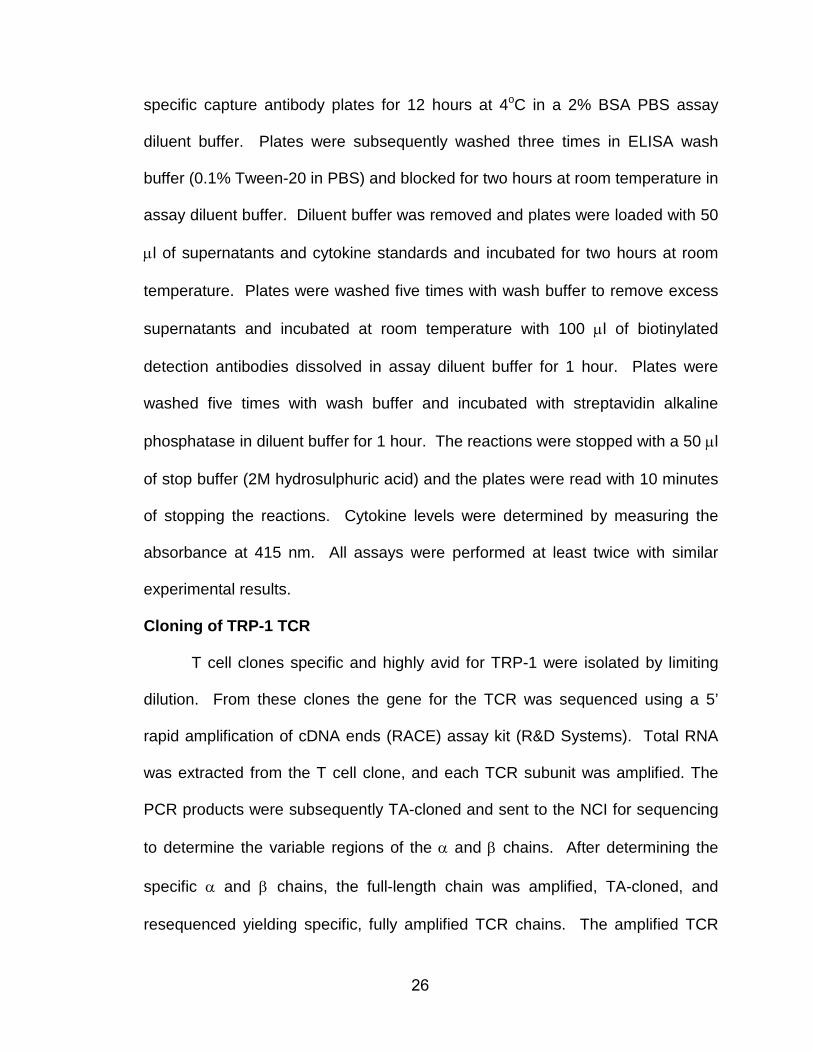
specific capture antibody plates for 12 hours at 4oC in a 2% BSA PBS assay
diluent buffer. Plates were subsequently washed three times in ELISA wash
buffer (0.1% Tween-20 in PBS) and blocked for two hours at room temperature in
assay diluent buffer. Diluent buffer was removed and plates were loaded with 50
µl of supernatants and cytokine standards and incubated for two hours at room
temperature. Plates were washed five times with wash buffer to remove excess
supernatants and incubated at room temperature with 100 µl of biotinylated
detection antibodies dissolved in assay diluent buffer for 1 hour. Plates were
washed five times with wash buffer and incubated with streptavidin alkaline
phosphatase in diluent buffer for 1 hour. The reactions were stopped with a 50 µl
of stop buffer (2M hydrosulphuric acid) and the plates were read with 10 minutes
of stopping the reactions. Cytokine levels were determined by measuring the
absorbance at 415 nm. All assays were performed at least twice with similar
experimental results.
Cloning of TRP-1 TCR
T cell clones specific and highly avid for TRP-1 were isolated by limiting
dilution. From these clones the gene for the TCR was sequenced using a 5’
rapid amplification of cDNA ends (RACE) assay kit (R&D Systems). Total RNA
was extracted from the T cell clone, and each TCR subunit was amplified. The
PCR products were subsequently TA-cloned and sent to the NCI for sequencing
to determine the variable regions of the α and β chains. After determining the
specific α and β chains, the full-length chain was amplified, TA-cloned, and
resequenced yielding specific, fully amplified TCR chains. The amplified TCR
26

chains were sent back to our laboratory wherethe genes were TA cloned using a
TOPO TA cloning kit from Invitrogen. The gene for the α and β chains were
subsequently PCR cloned into an Invitrogen Gateway entry plasmid (pENTR)
separated by either an internal ribosomal entry site (IRES) or a Furin self-
cleaving sequence (F2A). The TCR alpha and beta chains were re-amplified
with an optimal Kozak initiation sequence (Kozak, 2002) and restriction sites
using Platinum-Pfx polymerase (Invitrogen). To generate the IRES vector, the
α-chain was cloned into an IRES-containing derivative of pENTR1a (Invitrogen)
from DraI to a SpeI site 5’ to IRES. The β-chain was cloned 3’ from an NcoI site
at the 13th ATG of IRES to XbaI. Both chains were then sequenced and LR-
clonased, into a modified Gateway (Invitrogen) destination lentiviral plasmid
(pRRL-Dest-Wsin) (Dull et al, 1998). These modifications included a central
polypurine tract derived from HIV-pol(cPPT) sequence inserted between the REV
response element (RRE), the CMV promoter, and the Woodchuck hepatitis virus
posttranslational regulatory element (WPRE) element proximal to the 3’ SIN-LTR.
To generate the F2A vector, Furin-2A peptide sequence was exchanged through
a series of intermediates for the IRES element, transferred into the pENTR
plasmid, then LR-cloned from the pENTR plasmid into pRRL-Dest-Wsin.
Similarly constructed vectors which contain GFP (LV-GFP) and (LV-DR4) were
also prepared.
Other Plasmids Used
Lentivirus component plasmids and a lentivirus encoding for the green
fluorescent protein (pCSCGW-GFP) were donated by the Cornetta Lab and
27

included packaging plasmid pMDLgpRRE (containing the gag-pol accessory
genes), rev expressing plasmid pRSV-rev, and envelope plasmid pMDG-VSVG.
Once again employing the Invitrogen Gateway System, our collaborators in the
Cornetta Lab were able to construct a bicistronic lentiviral plasmid encoding for
the α and β chains of the human HLA-DRA-DRB1*0401 MHC class II molecule
(pCSCGW-DR4 IRES) using the same cloning methods described above.
Lentiviral Production
LV-GFP and LV-TRP1-IRES or LV-TRP1-F2A lentiviral supernatants were
prepared by transient transfection of 293T cells in T-150 flasks with vector DNA
(26.4 μg), packaging plasmid pMDLgpRRE (containing the gag-pol accessory
genes) (13.2 μg), rev expressing plasmid pRSV-rev (6.6 μg), and envelope
plasmid pMDG-VSVG (9.2 μg) using a Promega ProFection Mammalian
Transfection System-Calcium Phosphate Kit. Culture medium was replaced 16-
18h after transfection with GIbco OptiPro SFM. Viral supernatants were then
harvested at 24 and 48 hours after media exchange, centrifuged at 3500 rpm,
filtered through 0.45mm cellulose acetate filter, and concentrated with a
CentriconPlus-70 device (Millipore) at 3500 rpm x 30 minutes. The concentrated
viral supernatants were aliquoted and frozen at -80oC before calculation of titer.
Lentivirus Titering
To determine the titer of frozen viral supernatants, 293 cells were plated at
105 cells/well in 6 well plates, and serial dilutions (10-3, 10-4, 10-5) of concentrated
vector supernatants were prepared in DMEM media and incubated with cells for
4 hours with polybrene at 8μg/ml (5% CO2, 37oC). Cells were maintained in
28

culture for 72 hours, trypsinized and aliquoted, stained with a PE conjugated
TCR β-chain antibody, and analyzed by flow cytometry. Titers were calculated
using the following formula: titer = (F x Co/V) x D. F is the frequency of GFP-
positive or TCR-β chain positive cells determined by flow cytometry; Co is the
total number of target cells infected; V is the volume of the inoculum; D is the
virus dilution factor.
Collection of Bone Marrow, Peripheral Blood Cells, and Splenocytes
Mice were euthanized by cervical dislocation and bone marrow cells were
harvested by bilateral surgical resection of the tibia and femur. Collected tibia
and femur specimens were collected in RPMI complete media (5% FBS in RPMI
1640 supplemented with 1% L-glutamine, 1% non-essential amino acids, 1%
sodium pyruvate, 1% penicillin/streptomycin) to minimize cell death. Harvested
tibia and femurs were thoroughly removed of excess muscle and connective
tissue. Bone marrow was then washed out of the bones by insertion of a 27
gauge syringe needle and flushing with RPMI complete media from both proximal
and distal ends of the bones. The crude bone marrow cell collection was then
collected in 50 ml conical tubes and centrifuged at 1200 rpm and excess media
discarded. Erythrocytes were lysed using an RBC lysing buffer (Qiagen, Inc.) for
10 minutes at room temperature. The cells were then filtered through a 75 mm
nylon filter to remove clumps and ensure a single cell solution. The cells were
centrifuged at 1200 rpm for 5 minutes and washed twice with appropriate media
or buffer.
29

Peripheral blood cells were harvested by tail vein bleeds. Mice were
inserted into specially designed metal mouse holders which immobilized the mice
while allowing access to the tails. Using surgical scalpels, superficial incisions
were made as distally as possible to nick the tail veins. The tail veins were then
bled by external manipulation and peripheral blood collected in heparin treated
microcapillary tubes. Collected peripheral blood was subsequently transferred to
1ml of 2% heparin solution in flow cytometry tubes to prevent clotting. Cells were
then centrifuged and excess heparin discarded. Peripheral blood cells were
treated were RBC lysis buffer and filtered as described for bone marrow cells.
Spleens were collected by surgical resection and homogenized by mechanical
mastication with a sterile glass slide and a blunt instrument. Crudely
disaggregated splenocytes were then treated with RBC lysis buffer, filtered to
yield a single cell suspension, and washed as previously described.
Collection of Human Cord Blood Cells
Cord blood cells were collected by fractionation using Ficoll gradient.
Cord blood was graciously donated by the Broxmeyer Lab. Only cord blood less
than 24 hours old were used in these experiments. The cord blood was mixed
with an equal volume of EDTA Wash Buffer (2mM EDTA in PBS) solution to
prevent clotting and lyse RBCs. This cord blood mixture was carefully layered
over 20 ml of Ficoll plaque in 20 ml aliquots in a 50 ml conical tube. The tubes
were then centrifuged at 1500 rpm for 30 minutes and the white cell layer formed
between the Ficoll and aqueous layer was collected. Collected cells were
washed with 40 ml of EDTA Wash Buffer to remove excess Ficoll plaque and
30

centrifuged at 2000 rpm for 20 minutes. Excess buffer was discarded and the
remaining cord blood cells washed twice with appropriate media or buffer.
Magnetic Bead Separation
Murine bone marrow cells and human cord blood cells collected as
previously described were treated with antibody labeled magnetic microbeads to
extract murine Lin- cells and human CD34+ cells, respectively, using Miltenyi
Biotec magnetic microbead separation kits (Miltenyi Biotec). Cells were washed
with MACS buffer (2% bovine serum albumin in PBS) and labeled with an
appropriate amount of specific Biotin-Antibody Cocktail for 10 minutes at 4oC.
The cells were than incubated with an anti-biotin microbead solution for an
additional 20 minutes at 4oC. The cells were washed with MACS buffer,
centrifuged at 1200 rpm and excess buffer discarded. Samples were then
suspended in MACS buffer and run through a MACS column (LS column) and
MACS separator (Miltenyi Biotec). Collected cells were centrifuged, washed with
MACS buffer and counted. A small aliquot was set aside for antibody staining
and analysis by flow cytometry to check the efficiency of separation and purity of
the collected samples. The remaining cells were cultured in the appropriated
pre-stimulating media for 12 to 36 hours prior to lentiviral transduction. Murine
Lin- cells were prestimulated for 12 to 36 hrs in a murine prestimulating media
(Gibco StemPro 34 SFM Media, 1% Penicillin/Streptomycin, 1% L-Glutamine,
100 ng/ml SCF, 10 ng/ml TPO, 50 ng/ml Flt-3 Ligand). Human CD34+ cells were
prestimulated for 12 to 36 hrs in a human prestimulating media (Gibco StemPro
34 SFM Media, 1% Penicillin/Streptomycin, 1% L-Glutamine, 100ng/ml SCF, 50
31

ng/ml TPO, 100 ng/ml IL-6). Pre-stimulated cell populations were then
transduced with lentivirus.
Transduction Protocol
Cells were collected and plated at 5x10⁵ cells per well in 24 well flat
bottom culture plates and incubated overnight in appropriate media. Cells well
centrifuged and excess media removed. Cells were then spinoculated with
concentrated lentiviral supernatant in appropriate media supplemented with 8
µg/ml of polybrene at 1200 rpm for at least 2 hours at 25oC. Spinoculated cells
were immediately incubated for a further 2 hours at 37oC. Following this 4 hour
spinoculation/incubation period the cells were centrifuged at 1200 rpm for 5
minutes and excess media was removed by aspiration. All cells were transduced
at multiplicity of infections ranging from 10:1 to 100:1 depending on the
experiment. Following remove of the viral supernatant/media fresh media
appropriate for the cell type was added to the wells. The transduced cells were
incubated at 37oC for 72 hours. Transduced cells were then either antibody
stained for analysis by flow cytometry or prepared for bone marrow
transplantation.
Cell Staining and Analysis Using Flow Cytometry
Isolated cells were placed in flow cytometry tubes and washed twice with
FACS buffer (2% fetal bovine serum in PBS). Following centrifugation at 1,200
rpm for 5 minutes at 4oC, wash buffer was discarded and 1µl/tube of anti-mouse
FcIII/II receptor antibody (Fc Block; BD Biosciences) was added to each tube and
incubated for at least 15 minutes at 4oC. When staining with the TRP-1 Ultimer
32

(ProImmune, Inc.), Ultimer was added (5 to 10 µl/tube depending on cell count),
vortexed, and incubated in darkness at room temperature for 4 hours. The cells
were then washed twice with FACS buffer and excess wash buffer discarded.
For antibody staining, specific flourochrome conjugated antibody was added (1 to
10 µl depending on the antibody), vortexed, and incubated in darkness at 4oC for
at least 30 minutes. The cells were then washed twice with FACS buffer and
excess buffer discarded. If the analysis was to occur on the same day as
collection and staining, samples were kept at 4oC in darkness until flow
cytometric analysis. If the analysis was to occur the following day or later, the
cells were fixed by addition of a 2% formaldehyde in PBS fixation buffer (200
µl/tube) and storage at 4oC in darkness. Samples were analyzed by flow
cytometry on FACScan, FACS Calibur, or LSR II machines and data was
analyzed using BD CellQuest (BD Biosciences). To determine gene specific T
cell frequency a combination of antibodies against CD45 (FITC, eBioscience),
CD45.1 (FITC, eBioscience, CD3 (PE Cy5, BD Bioscience), CD4 (APC, BD
Bioscience), CD8 (Pacific Blue, eBioscience, and the DR4 UltimerTM containing
either the TRP-1277-297 epitope (TRP1-Ultimer) or the control gp10044-59 epitope
(both PE, ProImmune) were used. To determine T cell activation status, a
combination of antibodies against CD4 (APC), TRP1-Ultimer (PE), CD44 (PE
Cy5, eBioscience), CD45RB (FITC, eBioscience), and CD62L (Pacific Blue,
eBioscience) were used. To determine the frequency of post-transplant human
mononuclear cells following transplantation into NOD/SCID-2 mice, peripheral
blood samples were stained with mCD45.2 (FITC, eBioscience), hCD45 (PE
33

Cy5, BD Bioscience), hCD4 (APC, BD Bioscience), hCD8 (Pacific Blue,
eBioscience), and the TRP1-Ultimer (PE). Statistical significance in gene
expression between groups was determined using a Student T-test.
Bone Marrow Transplantation
Transduced cells were incubated for three days and subsequently
collected, washed in PBS, and counted. Recipient mice were placed on
doxycycline feeds four days prior to transplantation and lethally irradiated (950
rads) one day prior to transplantation. Each mouse was administered 5x10⁴ of
either the LV-TCR transduced or TRP-1 TCR Tg Lin- bone marrow cells along
with 5x10⁵ either HLA-DR4 Tg or C57BL/6 competitor unfractionated bone
marrow cells via tail vein injection. The mice were carefully placed in
immobilizing containers and using a 27 gauge needle, a syringe preloaded with
the proper concentration of both the HSCs and the competitor rescue whole bone
marrow cells was threaded into the lateral dorsal tail vein.
Immunohistochemistry
Skin or tumor samples were both fixed in formalin and embedded in
paraffin or snap frozen in liquid nitrogen. The fixed or frozen samples were sent
to the Indiana University Pathology Laboratories where standard H&E staining
was done on all specimens. For skin samples, blocks were stained with S100
(rabbit polyclonal 1:1000, Dako, Glostrup, Denmark; Envision Rabbit Polymer
DAB), CD3 (rabbit polyclonal 1:200, Dako; Envision Rabbit Polymer DAB), or
human- TCR-β (mouse IgG1 1:50, Santa Cruz Biotechnology, Santa Cruz, CA;
Envision Rabbit Polymer DAB). Skin sample were also frozen in liquid nitrogen
34

and stained with antibodies to CD4 (RM4-5 1:500, Serotec, Oxford, UK; Donkey
anti-Rat DAB) and CD8 (169.4 1:200, Serotec; Donkey anti-Rat DAB). For tumor
samples, blocks were stained with CD3 (Envision Rabbit Polymer NovaRed), or
human- TCR-β (Envision Rabbit Polymer NovaRed).
Determination of LV-TCR Gene Expression
Total RNA was isolated from 1x10⁵ BM cells obtained from 6 month
secondary transplant recipients (both LV-TCR and Control) and converted to
cDNA. A qPCR analysis using ABI One-Step Real-Time PCR System (Applied
Biosystems - ABS, Foster City, CA) was then performed using a custom
designed α-variable-chain probe (ABS) specific for the LV-TRP-1 vector.
Relative TCR gene expression (RQ) was based on the amplification ratio of LV-
TCR/Control Txp using β-actin as an endogenous control. The PCR reactions
were set up with TaqMan PCR Core Reagents (ABS) using about 100 ng of
cDNA (50 µl total reaction volume), and amplified at 95°C for 10 minutes plus 40
cycles of 95°C for 15 seconds and 60°C for 1 minute.
Lentivirus Integration Analysis
Genomic DNA (gDNA) was prepared (Qiagen, Valencia, CA) from 1x10⁵
BM cells obtained from 6 month secondary transplant recipients (both LV-TCR
and Control). Concentration and purity of the gDNA was determined by
spectrometry. All samples showed a 260/280 ratio of 1.7 or greater indicating a
high purity. Sample concentrations varied from 0.400 to 0.0500 mg/ml. The
gDNA from transplant recipients were diluted to a concentration of 250 ng/ml.
The gDNA samples were then sent to the Cornetta Labe where the samples
35

were then individually digested with Tsp5091 (New England BioLabs Ipswich,
MA) at 65°C for 2 hours. Following digestion, gDNA fragments were purified
using a MinElute Reaction Cleanup Kit (Qiagen). In short, 150 uL of Elution
Reaction Cleanup (ERC) Buffer was added to each sample and transferred to
spin columns. Columns were centrifuged for 1 minute at >10,000 x G and the
pass-through was discarded. 750 mL of Buffer PE was added to each column
and spun for 1 minute at > 10,000 x G. Pass-through was discarded and the
column was dried by spinning for 2 min. at 10,000 x G. The purified DNA
samples were eluted from the columns with 12 ml of EB buffer. Primers specific
for the LTR region of the lentiviral plasmid were biotinylated with native Pfu DNA
polymerase (Invitrogen). The primer extension products were subsequently
purified using the ERC kit. Microbeads with covalently attached streptavidin
(Dynabeads M-280, Invitrogen) were washed and 40 ml of the bead slurry was
added to each biotinylated primer. Beads were subsequently washed and blunt
end adaptors were ligated with T4 DNA ligase. Integration sites were then
identified by nested PCR: master mix consisting of 2 x Extensor HiFi PCR mix
(Invitrogen) and gDNA fragments were aliquoted into 0.2 mL PCR reaction tubes.
1 ml of the microbead-primer bead slurry was added and PCR amplified (94°C x
2 min, 95°C x 15 sec, 60°C x 30 sec, 68°C x 2 min for 30 cycles; 68°C x 10 min,
4°C x hold). After the 1st round of nested PCR a 1:50 dilution was done from
each PCR reaction tube and a 2nd round of nested PCR was performed. Each
PCR sample was analyzed by agarose gel electrophoresis.
36

Tumor Treatment Assays
Six month old secondary DR4 Tg transplant recipients involving an
experimental arm (LV-TCR-F2a) and a control arm were subcutaneously
implanted with B16-DR4 melanoma cells (5x10⁵ cell/200 ml HBSS) at 5 mice per
group (2 groups). Perpendicular tumor diameters were blindly recorded every 3
days. Mice were sacrificed at 20 days post-implantation and tumors were
excised for IHC. The statistical significance between groups was based on tumor
size (using a Student T-test). Experiments were performed twice in a blinded,
randomized fashion with similar results.
37

IV. Results
Identification of a TRP-1 Specific TCR
Using a clonal population of CD4+ T cells specific for the TRP-1277-297
epitope (Touloukian, et al 2002), the gene sequence of the individual α and β
subunits of the TCR were identified from these lymphocytes via a 5’ RACE
strategy at the NCI (Invitrogen GeneRacer Kit). In short, total RNA was extracted
from the T cell clone, and each TCR subunit was amplified. The PCR products
were subsequently TA-cloned and sequenced to determine the variable regions
of the α and β chains. After determining the specific α and β chains, the full-
length chain was amplified, TA-cloned, and resequenced, yielding specific, fully
amplified chains: TCR-α: Vα13-2*01, J22*01; TCR-β: V5.4*01, J1-1*01, D1*01.
The VDJ regions were analyzed using the V-QUEST search engine from the
IMGT web site (http://imgt.cines.fr/IMGT_vquest/vquest?livret=0&Option=
humanTcR). (Figure 2)
Construction of a Bicistronic Lentivirus
Next, I constructed a bicistronic lentivirus containing the genes for the α
and β subunits. I chose to implement a bicistronic lentivirus to deliver the α and
β chains of the TCR because the co-expression and successful pairing of the two
chains is essential for the proper expression of the TCR. Previous experiments
have shown efficient transduction of hematopoietic stem cells with multicistronic
viral systems (Cui et al, 2003; Yang et al, 2002; Yang and Baltimore, 2005;
Morgan et al, 2006). I ultimately chose to implement a lentiviral vector because
of their reported advantage at transducing mitotically quiescent cells such as
38

HSCs and clinically safer genetic integration profile (Schroder et al, 2002; Wu et
al, 2003). Two approaches were used to link the two target genes together. I
constructed both an internal ribosomal entry site (IRES) and Furin self cleavage
protein (F2A) bicistronic lentiviruses. Schematic diagrams for our lentiviral
constructs are shown in Figure 3.
Efficiency of Transduction
To evaluate the efficiency and functionality of TCR gene transfer, I tested
the newly generated LV-TCRs (LV-TCR-IRES or LV-TCRF2A) on human PBMCs
prestimulated with IL-7. I used a TRP-1277–297–specific PE-labeled pentamer
(Ultimer; TRP-1 Ultimer) to measure specific cell surface expression, to avoid
confusing true expression levels of the introduced α/β heterodimer with levels
that result from mispairings with endogenous T cell α and β chains. As shown in
Table 1, I observed statistically significant differences in TCR gene expression
using the LV-TCR-F2A construct at increasing viral titers (37.5% vs. 56.4%; P =
0.0008 at MOI 100). Both the IRES and F2A constructs efficiently transduced
JRT3 cells and PBMCs. However, transduction with the target TCR proved to be
more efficient using the F2A construct in both JRT3 cells (p=0.022 at an MOI of
10:1) and PBMCs (p=0.0008 at MOI of 100:1) particularly at higher MOIs (Figure
4). Thus, the remainder of the experiments focused on the F2A construct.
I next tested the efficiency of the lentiviral construct to transduce both
murine and human HSCs. Murine HSCs were harvested from bone marrow and
enriched by depletion of lineage positive cells using a Miltenyi Biotec Lineage
Depletion Microbead Kit. Murine HSCs were prestimulated for 12 to 36 hrs in a
39

prestimulating media (Gibco StemPro 34 SFM Media, 1%
Penicillin/Streptomycin, 1% L-Glutamine, 100 ng/ml SCF, 10 ng/ml TPO, 50
ng/ml Flt-3 Ligand). Human HSCs were harvested from human cord blood.
Following fractionation the cells were enriched using a Miltenyi Biotec CD34+
Microbead Kit. Human HSCs were prestimulated for 12 hrs in a prestimulating
media (Gibco StemPro 34 SFM Media, 1% Penicillin/Streptomycin, 1% L-
Glutamine, 100ng/ml SCF, 50 ng/ml TPO, 100 ng/ml IL-6). Following
prestimulation HSCs were spinoculated with lentivirus at MOIs from 10:1 to 30:1
with 8 µg/ml of polybrene at 1200 rpms at 25oC for 2 hrs. The cells were then
incubated for an additional 2 hrs at 370 C. Following incubation, the media was
changed for fresh prestimulating media and incubated for 72 hrs.
Results as measured by flow cytometry using a peptide-antibody complex
(TRP-1 Ultimer provided by ProImmune Inc.) showed approximately 10-50%
transduction efficiency in human CD34+ cells and approximately 10-20% in
murine Lin- cells at MOIs of 15:1 to 30:1 (Figure 5). I conducted multiple repeat
experiments with Lin- HSC enriched populations with lentivirus encoding green
fluorescent protein (LV-GFP) as a control group (Figure 6). Lin- cell transduction
with the LV-TRP-1 TCR showed high expression at 28.7% ± 3.0 as measured by
flow cytometry. Overall, the results demonstrate a high level of expression of the
complete TRP-1 TCR in transduced Lin- cells.
Transduced Cells Retain Functionality of TCR
To evaluate the in vitro functionality of the LV-TCR-F2A lentiviral
construct, we transduced IL-7 prestimulated PBMCs at an MOI of 15. After
40

overnight incubation, transduced PBMCs were co-cultured with DR4+ 1088 EVB
B cells loaded with a panel of control and experimental peptides. Transduced
cells were found to recognize the specific TRP-1277-297 peptide, melanoma tumor
lysates (B16), and TRP-1 positive tumor lines (B16-DR4, 624 Mel pretreated with
IFN-γ, and 1102 Mel stably transduced with TRP-1) only in the context of the
proper MHC class II HLA-DR4 restriction element as measured by specific IFN-γ
production on ELISA (Figure 7). No increase in IFN-γ was seen with the control
peptide HA306-318, control lysate (TRP-1 negative 1102 melanoma tumor
lysate), or control intact tumor (1102 melanoma tumor cells transduced with a
vaccina virus encoding GFP). Moreover, interactions with peptide pulsed targets
were found to be both DR-restricted (blocked by L243) and CD4-specific
(blocked by L3T4), but were not effected by control class I and CD8 specific
antibodies.
Development of a TRP-1 TCR Transgenic Mouse
To help evaluate the efficiency and effects of lentiviral transduction on
HSC development our collaborators in the Restifo Lab created a transgenic
mouse strain which constitutively expressed the same variable α and β genetic
components as the LV-TCR (TRP-1 TCR Tg). The variable regions of the α and
β chain of the TRP-1 TCR were cloned into TCR cassette vectors which encoded
the murine constant regions of the α and β chains. Our collaborator Dr. Nicholas
Restifo at the National Cancer Institute, injected the TCR cassette vector
encoding this chimeric TRP-1 specific TCR into the embryos of fertilized C57BL/6
mice. The founder mice were screened by PCR for the transgene, specifically
41

the variable region of the α chain. Initial characterization of the naïve TRP-1
TCR Tg mice by flow cytometry showed very limited presence of TRP-1 TCR
specific lymphocytes in the peripheral blood or spleen using the TRP-1 Ultimer.
The low or absent levels of TRP-1 specific T cells in the periphery of TRP-1 Tg
mice is not surprising given the lack of expression of the proper MHC Class II
restriction element in this transgenic mice. In the native TRP-1 Tg mice levels of
Ultimer positivity was near that of negative controls and only 6.4% ± 1.0 of
peripheral blood CD4+ T cells were specific for the TRP-1 Ultimer (Figure 8). I
subsequently bred the TRP-1 TCR Tg mice with the MHC Class II HLA-DR4 Tg
mice to obtain the proper restriction element. In this double transgenic mouse
which expresses the proper MHC restriction element, peripheral blood antibody
and Ultimer staining revealed that 79% ± 8.3 of all circulating CD4+ T cells are
specific for the TRP-1 Ultimer (Figure 8). Mice crossed onto a HLA-DR4
background strongly expressed specific TCR (CD4+/Ultimer+, 79% ± 8.3), while
those on a C57BL/6 background expressed low levels only (CD4+/Ultimer+, 6.4%
± 1.0; P < 0.0001). These results strongly suggested the necessity of aproper
restriction element (HLA-DR4) in positive T cell selection. To control for
experimental specificity and background (< 1%), I used a Ultimer specific for a
gp10044-59 DR4-restricted epitope. This multimeric antibody control was used for
all subsequent flow cytometry analysis.
Transplantation of Lentivirus Gene Modified HSCs
To better understand the cellular dynamics and kinetics of lentivirally
transduced HSCs to home, engraft, and reconstitute the immune system
42
following transplantation I began a series of transplantation experiments. I
initially sought to understand the basic mechanics of transplantation with
lentivirally manipulated HSCs by conducting a pilot lentiviral transplant
experiment. Using a lentiviral construct encoding a gene for green fluorescent
protein (LV-GFP), I transduced Lin- HSCs from Boy/J donor mice. 2.5x10⁴
transduced Lin- cells were transplanted per mice along with 5x10⁵ competitor
rescue bone marrow from unmanipulated C57BL/6 mice into 30 C57BL/6
recipients. Five mice were subsequently euthanized and the peripheral blood,
splenocytes, thymus, and bone marrow were analyzed by flow cytometry for
CD3, CD4, CD8, CD45.1, CD45.2, and GFP expression (Figure 9).
Transplanted, transduced Lin- cells maintain GFP expression in the peripheral
blood (CD45.1+/GFP+), developing into a high percentage (over 20% by 12
weeks post transplant) of mature T cells (TCR+/CD3+) and CD4+ T cells. Gene
modified cells are also present and remain stable within spleen, thymus, and
bone marrow for up to 6 months post transplant. Furthermore, transduced HSCs
expressing GFP appear to undergo nor thymic development progressing from
double positive thymocytes to single positive mature thymocytes.
Lentiviral Transduction of HSCs Leads to Stable Long Term Integration
Several lentiviral protocols have already been described for gene transfer
into transformed cells, lymphocytes, DCs, and HSCs (Cui et al, 2003; Cavalieri et
al, 2003; Lizee et al, 2004; He et al, 2005; Mostoslavsky et al, 2005). Although
different heterologous promoters (PGK, CAG, EF1α, and CMV), vectors (both
mono and bicistronic), pseudotyped envelopes, and inoculation protocols have
43

been used, the vast majority involve the use of cytokine prestimulation (Miyoshi,
2004). Because HSCs are relatively resistant to even high concentrations of
lentiviral particles (Sutton et al, 1998) and susceptible to cell death following viral
manipulation, I based the transduction protocol on both an overnight
prestimulation with SCF, TPO and Flt-3L, a spin/rest inoculation at an MOI of 15,
followed by an overnight recovery phase in cytokine media prior to
transplantation (Barrette et al, 2000; Mostoslavsky et al, 2005).
To quantify the engraftment potential, gene transfer efficiency, and long-
term stability of transduced HSC, I employed a standard CD45.1/CD45.2
competitive isocongenic repopulation (Tao et al, 2007). I initiated a series of
transplant experiments into DR4 Tg and C57BL/6 mice to determine the
engraftment efficiency of unmanipulated TRP-1 Tg HSCs and LV-TCR
transduced Lin- HSCs. To assess the impact of lentiviral transduction on the
engraftment and development of HSCs we established a CD45.1/CD45.2
competitive transplant experiment with either TCR Tg or LV-TCR transduced Lin-
BoyJ (CD45.1) HSCs and DR4 Tg (CD45.2) competitor bone marrow cells. Bone
marrow was harvested from the femur and tibia bilaterally and treated with ACK
lysing buffer to remove RBCs. The cells were subsequently disaggregated into a
single cell solution and filtered through a 100µm nylon filter. The bone marrow
cells were then depleted of lineage positive cells by using a Miltenyi Biotec
Murine Lineage Depletion Microbead Kit. BoyJ HSCs were prestimulated
overnight in a prestimulation cytokine media, and transduced the following
morning with a concentrated LV-TCR supranatant at an MOI of 30 to 1.
44

Transduced cells were incubated for three days and subsequently collected,
washed in PBS, and counted. Ten DR4 Tg recipient mice and five C57BL/6
recipient mice were placed on doxycycline feeds four days prior to
transplantation and lethally irradiated (950 rads) one day prior to transplantation.
Each mouse was administered 5x10⁴ of either the LV-TCR transduced or TRP-1
TCR Tg Lin- bone marrow cells along with 5x10⁵ DR4 Tg competitor
unfractionated bone marrow cells via tail vein injection. Five DR4 Tg recipient
mice received the LV-TCR transduced HSCs (LV-TCR/DR4). Five DR4 Tg
recipients were given HSCs from the TRP-1 TCR Tg donors (TCR Tg/DR4). Five
C57BL/6 mice were transplanted with LV-TCR transduced HSCs (LV-TCR/BL6).
A control transplant group of 5 DR4 Tg mice were established which were
administered bone marrow cells from a C57BL/6 mouse. Peripheral blood was
obtained at 1, 4, 6, and 9 month time points and stained for murine CD3, CD4,
and CD8 along with the TRP-1 Ultimer complex. By 4 months, immune
reconstitution appears robust with successful repopulation of both CD8+ and
CD4+ T cells. Figure 10 shows a representative flow cytometry analysis, which
schematically outlines our analysis. Each mouse was analyzed, by gating initially
around CD3+CD4+ cells. The Ultimer positivity within this population of gated
cells was then determined. As can be seen in this example, a high percentage of
circulating lymphocytes in both experimental transplant groups were positive for
the specific TCR with 43.3% and 43.8% of mature CD3+/CD4+ lymphocytes
exhibiting specificity for the TRP-1 Ultimer in the LV-TCR and TRP-1 Tg mice,
respectively.
45
I further analyzed the entire pool of circulating lymphocytes in the
peripheral blood, to measure the percentage of all lymphocytes that were specific
for the TRP-1 Ultimer (Figure 11). At 1 month post-transplant, TRP-1 specific
mature CD3+CD4+ T cells constituted less than 5% of all lymphocytes in all the
transplant groups. However, by 4 months post transplant, differences in
CD3+CD4+ Ultimer+ populations were evident in the LV-TCR HSCs transplanted
into DR4 Tg recipients (LV-TCR/DR4) versus those transplanted into the
C57BL/6 recipient mice (LV-TCR/BL6) (15.8% ± 2 versus 8.2% ± 2, p<0.05).
Statistically similar to the LV-TCR/DR4 group, 11.6% ± 2.5 of mature CD3+CD4+
lymphocytes from the TRP-1 TCR Tg HSCs transplanted into DR4 Tg recipients
(TCR Tg/DR4) were specific for the TRP-1 TCR.
As seen on Table 2, by 6 months post-transplant this difference in TRP-1
TCR positivity between the LV-TCR/DR4 and LV-TCR/BL6 groups had narrowed
and was no longer statistically significant (33% ± 4.6 vs 38% ± 7.5; P > 0.05). All
groups showed continued increases in TRP-1 TCR positive T cells in the mature
T cell population. TRP-1 TCR positivity in the peripheral mature CD3+CD4+ T
cell populations was 33% ± 4.6 in the LV-TCR/DR4 transplants, 25% ± 4.7% in
the LV-TCR/BL6 transplants, and 38% ± 7.5 in the TRP-1 Tg/DR4 transplants.
I subsequently performed two additional repeat competitive transplantation
experiments with TRP-1 TCR lentivirus transduced HSCs and DR4 Tg recipient
host mice (5 mice in each group) and performed similar peripheral blood and
splenocytes analyses at various time points up to 12 months. My results
demonstrate that the unmanipulated TRP-1 Tg HSC transplants on average had
46

a higher level of TRP-1 Ultimer positivity, particularly after the 4 month time point
in all cellular compartments analyzed as compared to the lentivirally transduced
TCR transplants. However, the LV-TCR transduced transplants demonstrated
robust high level expression of the TRP-1 TCR up to 12 months post transplant.
I also learned some valuable information regarding the timing of the emergence
of lymphocytes into the periphery and the kinetics of reconstitution of the
lymphocyte population in our TRP-1 TCR model. Lymphocytes expressing the
TRP-1 TCR first emerge into the periphery around 4-6 weeks post transplant and
steadily increase. I analyzed several different compartments within the
peripheral mononuclear cell population including the total peripheral blood
mononuclear cells, all CD3+ cells, and finally the mature CD3+CD4+ T cell
population. As a percentage of all cells in the peripheral blood, TRP-1 TCR
positive cells steadily increased from about 2.5% at 2 months to 5% in 4 months
for both the LV-TCR and the TRP-1 Tg transplants. However, by 12 months, the
TRP-1 Tg transplants were significantly higher in the TRP-1 Ultimer positivity
than the LV TCR transplants (8% ± 1.7 versus 22.6% ± 1.5; P = 0.001). (Figure
12). When analyzing only the CD3+ lymphocyte population, a similar pattern of
emergence and steady expansion is seen in both the LV-TCR and TRP-1 Tg
groups. However, at all time points the TRP-1 Tg group exhibited significantly
higher levels of TRP-1 TCR expression peaking at 12 months, 17.8% ± 2.3
versus 36.7% ± 2.4; P = 0.006 (Figure 13). Finally, I analyzed the mature
CD3+CD4+ T cell population (Figure 14). In this population, TRP-1 TCR
expression peaked around 6 months at 75.4% for the TRP-1 Tg transplant and
47

48.1% in the LV-TCR transplants, and slightly decreased at 12 months. Even at
12 months post transplants, both groups maintained a high level of TRP-1 TCR
express (65.2% ± 6.3 in the TRP-1 TCR group and 26.1% ± 2.4 in the LV-TCR
group; P = 0.004).
Transduced T Cells Undergo Development in the Thymus
The vast majority of early lymphoid progenitors travel to the thymus and
undergo a complex multi-step process of differentiation, proliferation, and
repertoire selection (Walker and Abbas, 2002; Ohashi, 2003; Heinzel et al,
2007). Although much of this process occurs early in mammalian life, there also
appears to be increasing clinical and experimental evidence that thymopoetic
activity undergoes substantial re-amplification (or “thymic rebound”) well into late
adult life (Hakim et al, 2005). These findings are particularly true in patients
successfully treated for HIV or following chemotherapy-induced lymphopenia and
subsequent transplantation (Sfikakis et al, 2005). Given the results that both an
appropriate restriction element (HLA-DR4) and co-receptor expression (CD4, not
CD8) are critical for TCR expression, I sought to determine both the cellular
density and compositional makeup of individual thymic subpopulations in 12
month old post-transplant recipients (approximately 14 months old overall).
The thymus of lentivirus transplanted mice were collected and analyzed.
The thymii were surgically removed, mechanically homogenized, filtered and
stained with TRP-1 Ultimer and antibodies against CD3, CD4, CD8 and CD45.
Despite the old age of the mice, a high percentage of TCR specific gene
expression was observed in double negative (DN), double positive (DP), and
48

single positive (SP) populations, these findings appear consistent with continued
HSC production and thymic replenishment of gene-specific T cell progenitors.
Analysis by flow cytometry revealed TRP-1 TCR expression at all stages of
thymocyte development from the immature DN to DP and SP state (Figure 15).
At 12 months 36% of DN thymocytes, 45% of DP thymocytes, 18% of CD8+
thymocytes, and 13% of CD4+ thymocytes expressed the TRP-1 TCR (Figure
16). Also observed was a steep and significant drop off (DP 45% ± 7.3; SP CD4
13% ± 3.1; P = 0.002) in TCR gene expression as thymocytes transitioned from
DP to SP, results consistent with partial central elimination of auto-reactive TRP-
1277-297-specific DR4-restricted specific T cells. Overall these findings appear
in agreement with existing models of self-tolerance (Bouneaud et al, 2000;
Gallegos and Bevan, 2004; Anderson et al, 2005), as well as those unique to
TRP-1 in which higher avidity T cells are generated in mice genetically deficient
in TRP-1 (Brandmaier et al, 2009).
Transduced CD4+ T Cells Retain Specificity and Function
Splenocytes harvested from LV-TCR tranplants were tested ex vivo for
specificity and function by ELISA measuring IFN-γ release. Splenocytes were
fractionated for CD4+ T cells by magnetic microbead separation as previousy
described then co-cultured overnight with DR4+ 1088 EBV B cells preloaded with
a panel of TRP-1 specific and non-specific peptides and tumor lysates (Table 3).
CD4+ T cell populations showed dramatic release to cytokine in response to the
TRP-1 peptide in a dose dependent manner. Furthermore, these T cells
49

recognized 624 melanoma tumor lysate, but not 1102 melanoma lysate which
does not express high levels of TRP-1.
Development of Autoimmune Vitiligo
A result of the LV-TCR transduced HSC transplantation has been the
development of spontaneous autoimmune vitiligo (Figure 17). Between 8-16
weeks post transplantation, LV-TCR mice were consistently found to develop
progressive vitiligo (Table 4). This pattern of depigmented skin bordered by
areas with no apparent signs of pigment loss resembled vitiligo development in
other models of TRP-1 autoreactivity (Overwijk et al, 1999; Bowne et al, 1999;
Leitner et al, 2003; Muranksi et al, 2008), as well those observed in patients that
have responded to IL-2 treatment (Rosenberg and White, 1996), or those treated
with myeloablative chemotherapy and the adoptive transfer of TIL (Dudley et al,
2002). A high proportion of transplant recipients regardless of DR4 background
developed vitiligo when transplanted with either the LV-TCR HSC (16 of 19 mice)
or the TRP-1 TCR Tg HSC (26 of 37 mice) and the DR4 Tg competitor bone
marrow regardless of recipient background, while none of the control transplants
including those with the LV-GFP and went on to develop autoimmune vitiligo.
The emergence of spontaneous vitiligo in the C57BL/6 recipient mice indicates
that thymic expression of HLA-DR4 and positive selection in the thymus is not
necessary for autoimmune vitiligo. However, the lack of vitiligo in all mice
receiving C57BL/6 competitor bone marrow indicates the necessity for a
peripheral antigen presenting cell expressing the proper MHC restriction element.
Similar vitiligo development was also observed in mice transplanted with HSCs
50

derived the TCR Tg, but none in the endogenous, non-transplanted TCR Tg
(despite the higher gene-specific frequency), nor in those lacking expression of
HLA DR4. These results suggested that myelodepletion and transplantation
combined with specific immune reconstitution (either from LV gene-modified or
Tg HSCs in combination with HLA-DR4 expression) provided the appropriate
conditions for the development of autoimmune vitiligo.
To address the pathogenesis of autoimmune vitiligo, I analyzed both
paraffin embedded and liquid nitrogen frozen skin samples from LV-TCR and
control transplants by immunohistochemistry (IHC). A small patch of the skin
sample was fixed in paraffin and stained for immunohistology using markers for
CD3 and S-100 (a marker specific for neurocrest cells such as melanocytes).
Sub-dermal hair follicle centered melanocytes were nearly completed obliterated
with effacement of the normal architecture based on staining with H&E.
Furthermore, affected skin samples demonstrated a dense increase in CD3+ T
cell lymphocytic infiltration in the vitiliginous dermal space surrounding the hair
follicles but not in the control mice (Figure 18). Frozen sections of the vitiligo
afflicted skin patches stained for CD4 and CD8 revealed that the lymphocytic
infiltrate consisted of both CD8+ and CD4+ T cells (Figure 19). To determine if
TCR gene-specific T cells were invading the sub-dermal compartment, sections
were stained with an exclusively human TCR β-chain specific antibody (Figure
20). As observed, staining was both identical in pattern and location with the
CD3 staining and distribution. To verify antibody specificity, human tonsil and
51

mouse spleen samples were similarly analyzed and found to be appropriately
positive and negative.
Activation Status and Functionality of Transplanted T Cells
To further understand the pathogenesis behind post-transplant vitiligo, I
investigated the activation status of TCR-specific CD4+ T cells. I had already
observed that non-transplanted endogenous TCR Tg mice failed to develop
vitiligo. Based on these early observations, I hypothesized that post-transplant
specific T cells would exhibit surface bound phenotypic features consistent with
spontaneous immune activation (Dutton et al, 1998; Klebanoff et al, 2005; Yang
et al, 2006; Stockinger et al, 2006). To capture the phenotype of the TRP-1 TCR
expressing CD4+ T cells, I collected splenocytes from 6 month old transplanted
mice and separated CD4+ T cells using a no touch murine CD4+ magnetic
Microbead separation kit (Miltenyi Biotec). The CD4+ T cells were then stained
with the TRP-1 Ultimer and antibodies for CD4, CD44, CD45RB, and CD62L and
analyzed by flow cytometry. Untreated, unmanipulated age matched DR4 Tg
mice splenocytes were used as a control group. In the native DR4 Tg control
group, the CD4+ T cell population was CD44low, CD45RBhigh, and CD62Lhigh
indicative of a naïve phenotype. By comparison, the control transplant group
exhibited a more generalized effector phenotype with CD44high, CD45RBhigh,
CD62Lint activation profile. In contrast, the activation profile of CD4+/Ultimer+ T
cells from LV-TCR transplants was CD44high, CD45RBlow, and CD62Llow
indicative of a T effector memory phenotype (TEM). Importantly, the
CD4+/Ultimer- subpopulation was similar in its expression profile to that of the
52

control transplant population (CD44high, CD45RBhigh, CD62Lint), suggesting the
divergence of two distinct T cell populations within the same host, the clonal-like
TEM CD4+/Ultimer+ vs. the polyclonal effector CD4+/Ultimer-, independent of the
effects of transplantation (Figure 21).
Given the spontaneous TEM-status of CD4+/Ultimer+ cells, I then
addressed whether or not T cells from LV-TCR transplants, would immediately
react to TRP-1 specific targets. To test this question, splenocytes from 6 month
old transplants (both LV-TCR and control) were harvested then stimulated ex
vivo for 24 hours with peptide or tumor. As seen Figure 22, cells obtained from
the LV-TCR group responded to TRP-1 specific targets, secreting IFN-γ in
response to TRP-1277-297 at titering peptide concentrations and to B16-DR4
tumor, but not to control targets HA306-318 or to control tumor MC38-DR4 tumor.
These results appear consistent with a specific and high avidity (recognition was
observed at 1 nM) TH1-polarized T cell population.
Secondary Transplant Experiments and Integration Analysis
Following the successful primary transplantation of LV-TCR transduced
HSCs, I performed three secondary transplant experiments using the bone
marrow from the primary transplants. Bone marrow was harvested from the
donor mice at the time of euthanization, in the LV-TCR transplant experiments as
previously described. 5x10⁵ whole bone marrow cells were non-competitively
transplanted per mouse into DR4 Tg mice (5 mice per group). Since these mice
had previously received DR4 Tg competitor rescue bone marrow, a second
competitive reconstitution was deemed unnecessary. All mice were followed and
53
analyzed at 6 months post transplant for CD3, CD4, CD8, CD45 and TRP-1 TCR
expression (Figure 23). As shown, the levels of CD4+ T cells expressing the
TRP-1 TCR closely matched that of the primary transplants and remained stable
for the duration of the analysis. TCR specific cells were identified at a level of
47.5% ± 8.3 (CD45+/CD3+/Ultimer+) within the global CD3 compartment and at a
level of 53.1% ± 10.7 within the CD4 sub-compartment (CD45+/CD3+/CD4+/
Ultimer+). These results were notable for the higher expression levels observed
within both populations compared with the 12 month expression levels observed
in the parental primary transplant group (12 m primary vs. 6 m secondary, P =
0.02).
Having now demonstrated stable and high level TCR gene expression 18
months after a primary transplantation, I sought to further understand the genetic
stability of the 6 month old secondary transplant recipients. Total RNA and
gDNA was prepared from 1x10⁵ BM cells (from both LV-TCR and control
transplants) and analyzed by both qPCR for relative TCR gene expression and
by standard PCR using a vector specific probe to determine the number of
lentiviral integrants. As seen in Figure 24A, using a probe specific for the
variable chain of the humanized α subunit, TCR gene expression from LV-TCR
transplants was found to be more than 1000-times greater than the background
signal produced in control transplants but 43-fold less than that observed in the
TCR-Tg group (P = 0.01 for LV-TCR vs. TCR-Tg). These results were consistent
with differences in protein level TCR expression. Using a probe specific for the
LV vector backbone, 4-6 integration sites and internal control fragment were
54
identified by nested PCR in 3 separate LV-TCR transplant samples, compared
with control transplant (0 integrants) (Figure 24B). Both results clearly
demonstrate long-term post transplantation genetic integration and stability.
Secondary LV-TCR Transplants Reject Subcutaneous Tumors
Immune reconstitutions after BM transplantation, dendritic cell vaccination,
or following the adoptive transfer of antigen-specific lymphocytes have been
shown to enhance anti-tumor immune responses (Borrello et al, 2000; Gattinoni
et al, 2005; Moyer et al, 2006). Given the high level TCR gene expression,
spontaneous autoimmunity, TEM activation, and in vitro anti-tumor activity, I asked
if long-term secondary transplant recipients would induce protective tumor
immunity. To address this question, transplant recipients (both LV-TCR and
control, 5 mice per group) were subcutaneously injected with B16-DR4 tumor
cells. Because of the preexisting “activated” TEM status of the transplanted
animals no additional cytokines, immune modulators (such as α-CTLA-4) or
vaccinations were added to enhance the anti-tumor effect. Beginning three days
after injection, and every 3 days thereafter, the product of the perpendicular
tumor diameters were recorded. As seen in Figure 25, secondary LV-TCR
transplants significantly rejected tumor growth compared with control transplants
over a 20 day time period. Tumor size between d12-d20 in the LV-TCR arm was
also non-significant (P = 0.09) suggesting cessation of tumor growth.
Based on IHC staining of paraffin-embedded specimens, tumors from LV-
TCR transplants but not controls were found almost entirely necrotic and
associated with hemorrhagic infiltrates (Figure 26). Tumors were also found to
55

contain dense trafficking populations of CD3+ and human TCR-β+ (gene-specific)
tumor-infiltrating lymphocytes (Figure 26), suggesting a similar infiltrative pattern
as that observed within the autoreactive vitiligo skin specimens. The nova-red
substrate was used instead of DAB to more effectively differentiate between
necrotic tumor/pigment and the cellular infiltrate.
Gene Modification and Transplantation of Human HSCs
Running in parallel with the murine HSC transplant studies I conducted a
series of transplant experiments to elucidate the characteristics and kinetics of
human CD34+ HSCs in non-obese diabetic/severe combined immunodeficient IL-
2 receptor γ chain knock out (NOD/SCID-2) mice. CD34+ HSCs were extracted
from fresh cord blood by fractionation as described in Materials and Methods.
In brief, fresh cord blood (less than 24 hours old) was treated with an equal
volume of 2mM EDTA in PBS solution. 20ml aliquots of the cord
blood/EDTA/PBS mixture were carefully layered on top of 20 ml of Ficoll plaque
in 50 ml conical tubes. The cord blood was centrifuged at 1500 rpm for 30
minutes to fractionate the lymphocyte layer. The cell layer was collected and
washed with 30 mls of the 2mM EDTA/PBS solution and centrifuged at 2000 rpm
for 20 mins at 25oC. The supernatant was discarded leaving behind the
mononuclear cells. The CD34+ cells were enriched for using a Miltenyi Biotec
CD34+ Microbead magnetic bead separation kit. Following selection of CD34+
cells, the CD34+ HSCs were incubated overnight at 5x10⁵ cells per well in 6 well
plates with human prestimulation media (StemPro SFM34 Media, 1% L-
glutamine, 1% penicillin/ streptomycin, 10ng/ml SCF, 100ng/ml TPO, 100ng/ml
56

IL-6). The following morning the cells were transduced using our established
transduction protocol with the TRP-1 TCR lentivirus. Three days post-
transduction, the cells were collected and a sample of the transduced CD34+
cells was analyzed by flow cytometry using a CD34 antibody and the TRP-1
Ultimer. After confirmation of successful transduction, the transduced CD34+
cells were transplanted into previously irradiated (350 rads) NOD/SCID-2 mice at
a concentration of 2.5x10⁴ cells/mice along with 2.5x10⁵ DR4Tg BM cells.
Peripheral blood lymphocytes were analyzed by flow cytometry with antibodies
for human CD45, CD3, CD4, CD8, HLA-DR, murine CD45.2 and the TRP-1
Ultimer at set intervals up to 6 months post transplant.
Human lymphocytes were isolated by magnetic microbead separation for
CD45 (Miltenyi Biotec) and analyzed for TRP-1 TCR expression. At 3 months
post transplant, I initially observed a higher than normal number of double
positive immature T lymphocytes in the peripheral blood (Figure 27). These
double positive lymphocytes exhibited high levels of TRP-1 TCR expression
(78%). By 4 months post transplant, the number of double positive T cells had
drastically decreased to 2% of all CD4+ T cells (Figure 28). At 4 months, 10.3%
of all mature single positive CD3+CD4+ T cells expressed TRP-1 TCR as
measured by Ultimer staining and flow cytometry.
TRP-1 TCR Confers Tumor Immunity
Five of the NOD/SCID-2 mice transplanted with LV-TCR modified HSCs
were enlisted in a tumor rejection assay. A human melanoma line that had
previously been transduced with a retrovirus encoding the gene for the MHC
57

class II transcription factor CIITA, 624 Mel CIITA was injected subcutaneously
into the transplanted mice and 5 control NOD/SCID-2 mice. The engraftment
and growth of the melanoma was carefully monitored and measured for 24 days
post injection (Figure 29). As observed, none of the LV-TCR transplants
developed measurable tumors. However, all of the control transplants allowed
for the engraftment and growth of the melanoma. Tumors first became apparent
in the control transplants at day 9 and continued to steadily grow up to 24 days
post transplant.
58

V. Discussion
While most gene-therapy strategies have focused on the introduction of
defective genes or antigens or used fluorescent gene products to track long-term
development, few have attempted to introduce TCR genes into HSCs (Dudley et
al, 2000). In those examples that do exist, TCR genes introduced with RVs have
targeted only experimental antigens (OVA) (Morgan et al, 2003; Zhao et al,
2005). Herein, I report a new translational model of TCR-based immunotherapy
directly applicable to patients with metastatic melanoma. I demonstrate both
spontaneous autoimmunity and the destruction of subcutaneous melanoma that
occur following long-term transplantation with gene-modified HSCs. My studies,
based on the use of a high expression lentiviral system to deliver to HSCs a
potent class II-restricted CD4+ TCR, were assisted by neither the use of
vaccines, cytokines, or immune modulators to either induce a population of
autoreactive memory T cells or to achieve its antitumor effects.
To design a model encompassing features that would address both self-
tolerance and tumor immunity and simultaneously serve as a potential source of
preclinical information, I utilized a previously identified tumor-reactive, self-
reactive, TRP-1-specific, DR4-restricted T cell clone. I then engineered both a
high expression LV and a TCR Tg expressing the same TCR genes. As
mentioned earlier, I focused on current generation SIN lentivectors, not only
because of their enhanced capacity to stably infect metabolically dormant HSCs,
but because of their improved biosafety profile compared with standard γ-RVs
(Sinn et al, 2005; Levine et al, 2006). These experimental observations have
59

been further supported by clinical trials either ongoing or those in preparation for
treatment of patients with HIV, β-thalassemia, Parkinson’s disease, and Wiskott-
Aldrich syndrome (Hargrove et al, 2008; Isacson and Kordower, 2008; Galy et al,
2008; D’Costa et al, 2009). With a functional LV-TCR and TCR Tg in hand, we
demonstrate engraftment, thymic trafficking and differentiation, long-term TCR
gene expression in both murine and humanized transplants, spontaneous post-
transplant autoimmunity, and hemorrhagic necrosis of subcutaneous melanoma.
Although experimental answers obtained from well-constructed transgenic
models are largely precise and elucidating, they are also fraught with the
potential for over-simplicity and a failure to translate into beneficial human
medical care (Rivera and Tessarollo, 2008). In this model, I used the TCR Tg
solely to gauge the efficiency of gene-transfer. The information gleaned from the
TCR Tg was, therefore, designed to be in service to the greater translational
value served by the LV-TCR.
Using these building blocks I propose a novel melanoma based murine
model to examine the role of CD4+ T cells in cancer immunology using a lentiviral
delivery system to modify hematopoietic stem cells. The model involves four key
components: 1) isolation of a T cell receptor reactive to a specific melanocyte
differentiation antigen, tyrosinase related protein 1 (TRP-1); 2) development of a
lentiviral construct for the delivery of the gene encoding the TRP-1 TCR; 3)
creation of a constitutively TRP-1 TCR expressing transgenic mouse (TRP-1
TCR Tg) to study gene transfer efficiency; 4) transplantation of gene modified
HSCs.
60

Effects of Lentiviral Transduction on HSC Transplantation
Through the transplantation experiments I was able to elucidate the
kinetics of transplantation following manipulation of HSC with lentiviral
constructs. Lentiviral transduction, although a traumatic and toxic event appears
to have little or no deleterious effects on the ability of HSCs to home, engraft and
reconstitute a lethally irradiated recipient. The initial experiments with lentivirus
encoding GFP allowed me to approximate the kinetics of transplantation and
effectively set the stage for the larger, more ambitious LV-TRP-1 TCR
experiments. The general kinetics of transplantation of gene modified HSCs
were consistent across all of the transplantation experiments. Early peripheral
blood mononuclear cells began emerging from the bone marrow at around 6-8
weeks post transplant (Figure 8, 9 and 11). Total cell number increases
dramatically from 2 months to 4 months, then levels off until it reaches a steady
state around 6 months post transplant. These time points are mirrored in both
the lentivirus transduced transplants and the unmanipulated TRP-1 Tg
transplants (Figure 11) indicating that lentiviral transduction did little harm to the
capacity of the HSCs to home, engraft and differentiated into blood component
cells.
A permanently reconfigured immune system of durable high-frequency
lymphocytes has long been a goal of immunologists and clinicians in pursuit of
treatments for viral pathogens and cancer (Parish, 2003). Concerns persist,
however, regarding the feasibility of delivering such a treatment within a narrow
enough time-frame. Most clinical therapies and translational models have
61

focused on fully actualized therapies that deliver their effect with little delay, such
as vaccines (peptide, virus, DCs, heat shock proteins, gene-modified tumor
cells), cytokine infusions (IL-2, IL-7, IL-12, GM-CSF), immune modulators
(myeloablative chemotherapy, anti-CTLA-4) or adoptive immunotherapies (TIL,
cloned T cells, gene-modified T cells) (Parish, 2003; Rosenberg, 2004).
Limitations and delays in immune reconstitution are a well described physiologic
consequence of autologous HSC transfer, and the kinetics of individual
mononuclear populations have been shown to favor the early return of
granulocytes and B cells, with CD4+ T cells often being the last subpopulation to
return to normal levels (Talmadge et al, 1997; Laurenti et al, 2004). In this
model, such observations were also found to be true. To improve T cell
recovery, IL-7 administration has been successfully used in non-human primates
(Storek et al, 2003) and patients in with cancer (Rosenberg et al, 2006).
Nevertheless, the Achilles heel of such a potential treatment in patients pertains
to the relative late emergence of the CD4+ T cell subpopulation. Despite these
concerns, the timing of maximum (or even normal) T cell recovery may not be the
most meaningful physiologic end point of gene-modified HSC transfer. Although
gene-specific T cells peaked 4-6 months after transplantation, low levels were
detected after 6-8 weeks and chronologically correlated with the development of
vitiligo (occurring between 8-12 weeks). These findings suggested that the
magnitude of the TCR-specific frequency was less an important indicator of
functionality than their qualitative behavior or the amplitude of their emergence
into the periphery. Such observations have been supported by other models in
62

which small numbers of T cell clones differing only by activation status or T
helper phenotype more effectively induce autoimmunity when compared with
control populations (Lanzavscchia and Sallusto, 2002; Minang et al, 2010).
Given the lack of information pertaining to the kinetics of engraftment and
development of lentivirus transduced HSCs, I was able to address several critical
questions regarding the use of lentivirally transduced HSCs in bone marrow
transplantation. These experiments indicate that the time from transplantation,
engraftment, and emergence from the bone marrow and thymus to the periphery
for lymphocytes is around 6-8 weeks. Furthermore, lymphocyte reconstitution
happens briskly between 2-6 months then stabilizes after the 6 month time point
and decreases slightly, but remains relatively stable for the duration of the
experiments up to 12 months in almost all transplanted groups.
Peripheral Homeostatic Proliferation Leads to Expansion of Selective T Cell
Clones
Homeostatic proliferation (HP) represents a naturally occurring multi-log
expansion of lymphocytes in response to depletion or ablation followed by either
BM reconstitution or adoptive transfer (Mackall et al, 1997; King et al, 2004).
Immune responses that occur during HP appear to favor the dramatic expansion
of T cells and lead to the precipitation of autoimmune events (King et al, 2004;
Kruprica et al, 2006). These observations have been shown associated with a
number of factors, including the increased availability of homeostatic cytokines
(IL-7, IL-15), the depletion or inhibition or Tregs, generalized host inflammation,
and the availability of self-ligands (Mackall et al, 1997; Fry et al, 2001; Boyman et
63

al, 2007). IL-7 availability, in particular, has also been found to be a critical factor
in T helper and thymocyte survival along with the promoting of HP of both naïve
and memory T cell subpopulations (Fry et al, 2001).
There are a number of possible explanations for the HP of TCR-specific
CD4+ T cells. Even though the importance of IL-7 has been clearly stated, I
found that IL-7 receptor expression by flow cytometry on thymic DPs and SP
CD4s as well as peripheral blood and splenic CD4s to be statistically no different
within either the Ultimer+ and Ultimer- population. On the other hand, although
generalized antigen may be limiting to the vast majority of T cells, TRP-1 loaded
DCs may be uniquely available to the CD4+/Ultimer+ subpopulation. Unlike most
antigens, a fragment of the TRP-1 protein (the highest expressed of all
melanocyte differentiation antigens) containing the 277-297 epitope has been
shown to be secreted into the peripheral circulation (Xu et al, 1997). In theory,
circulating TRP-1 could be captured by immature APCs (either in the skin or
within peripheral lymphoid organs), that in turn become activated during the
course of systemic injury from lethal irradiation (Hill et al, 1997; Zhang et al,
2002). Expansion of CD4+ T cells has been tied to not only the relative load of
antigen, but to the prolongation of that antigen load well after initial exposure
(Obst et al, 2005; Jelley-Gibbs et al, 2005). With all this being said, the
magnitude of the burst size may be a simple function of mass effect given the
high frequency of gene specific HSCs and T cell progenitors introduced at the
time of primary transplantation. Lin– HSCs examined by flow cytometry 3 days
64

after transduction had TCR-specific levels that ranged between 35-70% (data not
shown).
Other explanations may also account for both the magnitude of the burst
size and the unique TEM status of the CD4+/Ultimer+ subpopulation. In general,
two competing models of memory T cell development have been proposed. The
first involving a separate lineage from effectors, and the second proposing that T
cells pass initially through an effector stage (Hu et al, 2001; Opferman et al,
1999). As a consequence of either scenario, T cell clones can then give rise to
multiple effector and memory T cell populations, by means of asymmetrical cell
division. After antigen exposure, memory cell expansion is classically followed
by contraction, and stable persistence. Although I showed activation status at 6
months post primary transplantation (similar profiles were found at all time
points), I found a sharp increase in the total CD4+/Ultimer+ T cell population
between the 12 month primary and 6 months secondary transplants, consistent
with both memory decay and re-expansion. I also found a splitting of two distinct
T cell populations within the same host (the clonal-like TEM CD4+/Ultimer+ vs. the
polyclonal effector CD4+/Ultimer-) irrespective of the effects of transplantation. In
the end, the CD4+/Ultimer+ T cell population may have had a competitive survival
advantage and propensity for memory differentiation based on the relative high
availability of TRP-1.
Transplantation of LV-TCR Transduced HSCs Breaks Self-Tolerance
The proposed TRP-1 TCR model allows for the novel study of a self-
antigen specific TCR in the post-transplant setting. Although researchers in the
65

past have successfully transduced HSCs using viral systems none have explicitly
characterized the fate of HSCs modified to express an endogenous self-antigen
specific TCR. Dr. David Baltimore and colleagues transduced HSCs to express
an OVA specific CD4+ TCR. However, because of the highly immunogenic non-
self nature of the antigen chosen it left unclear the effects of thymic selection on
a normally occurring self antigen specific TCR such as TRP-1 (Yang and
Baltimore, 2005). As noted in the Results, engraftment and development of
HSCs transduced with LV-TCR appeared robust up to 12 months post
transplantation with only slightly reduced numbers of circulating TCR specific T
cells in transplants without the expression of the proper MHC Class II restriction
element in the thymus (Figure 9). Initially, the LV-TCR/BL6 transplant group
lagged behind the LV-TCR/DR4 transplants in terms of CD4+ T cell expression of
TRP-1 TCR. The lag in TRP-1 TCR positive T cells in the LV-TCR/BL6 group is
not altogether surprising given the lack of expression of HLA-DR4 in the thymic
epithelium of C57BL/6 mice. As we saw previously (Figure 7), in the non-
transplanted native setting, TRP-1 TCR Tg mice on a C57BL/6 background
exhibit little to no T cells in the peripheral with TRP-1 TCR expression. However,
in the post-transplant setting a far higher number of TRP-1 specific T cells are
found in the periphery. By 6 months, there is statistically no difference in TRP-1
TCR expression in the DR4 Tg and C57BL/6 recipients. Most likely, the initial
depression in TRP-1 specific T cells in the C57BL6 transplants is due to central
deletion. The lack of the proper HLA-DR4 restriction molecule in these recipient
mice in conjunction with the expression of a self-antigen specific TCR leads to
66

the apoptosis and deletion of the T cell. However, some TRP-1 TCR expressing
T cells do escape deletion and appear in the periphery as detected by flow
cytometry. Considering that the sheer number of lymphocytes expressing the
TRP-1 TCR has been artificially increased many fold higher than any naturally
occurring TCR by gene modification, it is easy to imagine that the normal thymic
deletion mechanisms might become overwhelmed. Once, these self-antigen
specific T cells escape into the periphery, they would be confronted with a
microenvironment rich in homeostatic cytokines due to irradiation of the recipient
mice and subsequent elimination of competing lymphocytes. Further, these self-
antigen specific T cells would encounter antigen presenting cells loaded with the
TRP-1 antigen. In such a favorable stimulation- rich environment, the TRP-1 T
cells could readily expand to fill the empty niche. This homeostatic and antigen
driven peripheral expansion would readily explain the transplantation kinetics I
have observed in the transplant experiments.
In TRP-1 model, vitiligo developed in C57BL/6 recipients (along with a
corresponding similar level of TCR specific cells Table 3), not just in DR4 Tg
recipients. These findings underscore a broader range of effects by the gene-
specific CD4+ T cell population, regardless of the relative magnitude of circulating
cells present. The polarized TH1 production of IFN-γ may have produced multiple
effects, including direct melanocyte cytotoxicity, up-regulation of MHC molecules
that enhance target recognition, and an enhancement of APC activation leading
to further amplification of the T helper response. Such functions have been well
associated with the unique, but underutilized properties of CD4+ T cells. While
67

long-term cellular persistence was an initial objective of this model, the amplitude
of gene-specific T cell expansion (between 8-24 weeks) and TEM activation were
clearly dramatic, and we propose associated with both autoimmunity and
antitumor activity.
These transplant experiments hint that in the myelodepleted irradiated
state engraftment and selection of TRP-1 specific CD4+ T cells does occur as
indicated by the observance of spontaneous autoimmune vitiligo in these HLA-
DR4 negative recipient mice. I thus, hypothesize that although central selection
in the thymus may be a significant factor in maximizing the selection of TRP-1
TCR specific CD4+ T cells, mechanisms of selection and homeostatic
proliferation exist that either bypass or overwhelm the normal selection of T cells
in the thymus.
Conclusion
These results highlight several important findings regarding reconstitution
of the hematopoietic cells in the post transplant setting: 1) the expression of the
proper MHC Class II restriction element, HLA-DR4, in the thymic epithelium may
be necessary for the maximal central selection of T cells. However, peripheral
homeostatic expansion of peripheral lymphocytes in the context of HLA-DR4 can
compensate for central deletion, 2) the LV-TCR transduced HSCs competed,
engrafted, and developed comparably to the unmanipulated TCR Tg HSCs
indicating that lentiviral transduction did not adversely affect the capacity and
function of the HSCs, 3) the lentiviral gene delivery system successfully led to a
high level of circulating TEM CD4+ lymphocytes expressing the TRP-1 TCR.
68

The most clinically significant evidence I present is the near total
liquefaction of tumor following subcutaneous implantation in long-term secondary
transplants. These results demonstrate not only potent antitumor activity 18
months after initial transplantation, but the dense cellular trafficking of LV-TCR
specific T cells. While many adoptive immunotherapies have either maximized
their therapeutic effect or been totally dependent upon the addition of cytokine
administration, vaccination, myelodepletion (lethal irradiation or RAG KOs) or the
use of immune modulators (α-CTLA-4) I elected to demonstrate this effect
without such assistance (Overwijk et al, 2003; Quezada et al, 2010). I propose
that these freestanding antitumor effects were likely secondary to both the HP
and the spontaneous TEM activation of TCR-specific cells. In conclusion, I
suggest that this novel translational model of immunotherapy, utilizing the unique
properties of HSC transplantation, LV technology, and CD4+ T cells, has the
opportunity to bring new medical care to patients with metastatic melanoma.
69

VI. Limitations and Future Directions
Increased Efficiency of TCR Expression
A major potential limitation is the inefficient expression of our bicistronic
TCR gene in vivo. I have shown in preliminary optimization studies that the F2A
construct is superior to the IRES construct in expression of the TCR; however,
the successful transcription and translation of such a long single protein chain
might be limiting and reduce the expression of both gene products. Moreover, a
single general promoter such as CMV might not be sufficient to drive the
expression of high levels of our bicistronic gene in developing lymphoid cells.
Along with inefficient expression of both chains of the TRP-1 TCR, mismatched
pairing of the α and β chain to endogenously expressed α and β chains may
further reduce the functional efficiency of transduced lymphoid cells to recognize
TRP-1.
To address these concerns I have investigated the possibility of
constructing a dual promoter lentiviral construct, with each chain of the TCR
under the control of two separate promoters. Although, the implementation of the
natural lymphocyte specific CD2 promoter would be favorable, the CD2 promoter
has never been shown to be a strong enough promoter to drive the expression of
a TCR taken out of its natural context. As such, I have focused my attention on
two potent, general promoters, EF1α and CMV. The use of independent dual
internal promoters in a bicistronic lentiviral construct has shown increased
expression of the transgenes in CD34+ HSCs as compared to a single promoter
construct. In particular, the dual promoter bicistronic constructs appear to be
70

advantageous in the expression of the downstream cistron (Yu et al, 2003; Ben-
Dor et al, 2006). Furthermore, I am seeking to increase the efficiency of
transduction by implementation of several other lentiviral envelope proteins such
as the feline endogenous virus RD114 glycoprotein and the murine leukemia
virus glycoprotein. One or both of these alternative envelope proteins might
prove to be either more effective or less toxic in transducing HSCs.
Mechanisms of Autoimmune Vitiligo
These transplantation experiments with donor HSCs from both TRP-1 Tg
mice and TRP-1 TCR lentivirus transduced HSCs have demonstrated consistent
spontaneous autoimmune vitiligo. Transplantation of GFP lentivirus transduced
HSCs into irradiated mice did not develop vitiligo in prior experiments indicating
that this phenomenon is an antigen specific event associated with the TRP-1
TCR. My data indicates that the myelodepletion of recipient mice coupled with
the homeostatic proliferation of highly self-reactive TRP-1 TCR CD4+ T cells in
the post transplant setting breaks the normal tolerance mechanisms leading to
autoimmunity. Furthermore, no other autoimmune responses were observed in
TRP-1 TCR transduced HSC transplanted mice. The autoimmunity appears to
be a melanocyte specific response to the transplantation of TRP-1 TCR
expressing HSCs. The breaking of self-tolerance is a significant step in priming
the immune system to promote tumor regression, and numerous studies have
demonstrated autoimmune vitiligo as a side effect of melanoma immunotherapy
in both mice and humans (Garbelli et al, 2005; Okamoto et al, 1998; Overwijk et
al 1999). The cellular mechanism of autoimmune vitiligo in melanoma immunity
71

is unknown. Although, melanocyte destruction might ultimately depend on lysis
by CD8+ effector T cells, Ihypothesize that the breaking of tolerance and
development of vitiligo is a TRP-1 antigen specific CD4+ T cell dependent
response. The elucidation of the cellular mechanisms of autoimmune vitiligo is a
future direction of the project. I will establish a series of transplants with
lentivirally transduced HSCs. Both C57BL/6 and DR4 Tg mice will be
transplanted with TRP-1 TCR lentivirus transduced HSCs. Recipient mice
developing autoimmune vitiligo will be analyzed at set intervals (onset of vitiligo,
2, 4, 6, 8, 12 wks post onset of vitiligo). Lymphocytes harvested from peripheral
blood, spleen, bone marrow, and thymus will be characterized for phenotypic
markers (CD45.1, CD45.2, CD3, CD4, CD8, CD25, Foxp3, human αβ TCR, and
TRP-1 Ultimer) by antibody staining and flow cytometry. As described
previously, the epidermis of these mice will be fixed in formalin and sent for
histologic staining for CD3, S-100, IL-2 and IL-17 to observe the lymphocytic
infiltrations typically found in vitiligo. We will seek to characterize the cytokine
profile of the lymphocytes by co-culture and subsequent ELISA and ELISPOT for
IFN-γ, IL-2, and IL-17 as previously described.
Lymphocytes harvested from the spleen of recipients developing vitiligo
will be analyzed for activation status. CD4+ T cells will be selected by using a
Miltenyi Biotec CD4 Microbead Kit. The CD4+ T cells will then be stained with
antibodies for activation status markers CD44, CD45RB, CD62L, CD69, and
CCR7 and phenotype markers CD4 and the TRP-1 Ultimer. Analysis by flow
cytometry should allow us to capture the activation status of the CD4+ T cells in
72

general and the TRP-1 TCR+ T cells in specific. I have previously run this
activation status experiment on naïve BoyJ and DR4 Tg mice splenocytes and
will use the activation status marker profile from these prior experiments as the
naïve control group for comparison to the vitiligo transplant mice. In vitro
experiments to test for melanocyte recognition and destruction will be established
by directly placing splenocytes to an established HLA-DR expressing melanocyte
culture with or without administration of various cytokines such as IL-2 and IL-17.
After characterization of the lymphocytes and timing of the development of
vitiligo in the transplanted mice we will establish two transplant groups (6 DR4 Tg
mice per group) which will undergo either CD4+ or CD8+ T cell depletion by
specific anti-CD4 and anti-CD8 antibodies from hybridoma cell lines (ATCC,
Manassas, VA, USA). Following irradiation and transplantation of either TRP-1
TCR Tg HSCs or TRP-1 TCR lentivirus transduced HSCs, recipient mice will
receive a total of six intraperitoneal injections of either rat anti-mouse CD4 (clone
GK1.5) or rat anti-mouse CD8 (clone 2.43) monoclonal antibodies in a PBS
solution. Normal rat IgG will be used as a control group (Miyazaki et al, 2005).
The exact timing of the injections will be set to occur prior to and during the peak
occurrence of vitiligo as observed in the previous transplant group. The
transplanted mice will be carefully monitored for the development of vitiligo and
lymphocytes collected from the peripheral blood will be analyzed by flow
cytometry and antibody staining for CD45.1, CD45.2, CD3, CD4, CD8, and TRP-
1 Ultimer on weeks 4, 6, 8, 10, 12, 16, 20 and 24.
73

Spontaneous autoimmune vitiligo is a significant development in our
mouse melanoma model, and an understanding of the cellular mechanisms
involved in the development of vitiligo in the transplanted mice could have
potential implications for the ultimate goal of treating melanoma by
transplantation of HSCs. I fully expect that many of the lentivirus transduced
HSC transplant recipients will develop vitiligo as in the previous experiments. In
the irradiated lymphodepleted mice, I suspect that the high expression of the
TRP-1 TCR in the transplanted HSCs will overcome the normal tolerance
mechanisms allowing for the selection and proliferation of these TRP-1 self-
peptide specific T cells. The exact timing of the development of vitiligo and
kinetics of lymphocyte development and activation will allow me to properly time
the injections for CD4+ and CD8+ T cell depletion in vivo. In the earlier
transplants, low levels of lymphocytes became detectable around 4-6 weeks. The
development of vitiligo will lag behind the reconstitution of the bone marrow
following irradiation and transplantation. My data indicates vitiligo to occur
between 6-10 weeks post transplant
Due to the limitations of formalin fixed paraffin embedded
immunohistology, our analysis of lymphocytes in the skin has been limited to
staining for CD3, S-100, and the cytokines IFN-γ, IL-2 and IL-17. I am currently
working to circumvent this limitation by developing a method to directly analyze
the lymphocytes in the skin by flow cytometry. The preliminary experimental
approach involves the harvesting of lymphocytes from the skin of vitiliginous mice
by disruption and enzymatic digestion by dispase and collagenase similar to
74

protocols used in the extraction of thymocytes from whole thymus used by other
researchers. In brief the procedure involves the harvesting of the entire
epidermis from the mice, and subsequent disruption using a surgical scalpel.
Following disruption, the tissue undergoes a series of digestions with
collagenase and dispase. The cellular mixture is then carefully layered over
Ficoll plaque and centrifuged at 1200 rpm for 20 minutes. The cellular layer is
than subsequently collected for analysis. Lymphocyte yields from this approach
have been limited to approximately 50,000 cells per mice, which has been
sufficient to perform simple flow cytometric analysis. However, the results have
been inconclusive. I will continue to refine this technique to increase the yield
and better characterize the phenotypes of the lymphocytic infiltrates
demonstrated in the skin of the vitiligo mice.
The CD4+ and CD8+ T cell depletion experiments will allow for a better
understanding of the roles that these two cells play in the development of
autoimmune vitiligo. Most likely, the CD8+ T cells act as the effector T cells that
directly destroy the melanocytes and will be found to be necessary for the
development of vitiligo. However, I suspect that the TRP-1 specific CD4+ T cells
are equally necessary for the development of vitiligo, either by release of IL-17
and subsequent recruitment of inflammatory cells, or perhaps by activation of
dendritic cells and subsequent increased presentation of antigen to naïve CD8+ T
cells as in the cross presentation model discussed earlier. Furthermore, it is
possible that these TRP-1 specific CD4+ T cells might directly lyse melanocytes.
75

Skewing the T Cell Repertoire
Ther overall strategy of this thesis was to conduct a series of lentivirus
transduced HSC bone marrow transplants as described above. In future
experiments I will seek to manipulate the reconstitution of the T cell repertoire by
early vaccination of the transplanted mice with a recombinant vaccina virus
encoding a gene for the human TRP-1 peptide (rVV-hTRP-1). The optimal
immunization schedule is unknown, therefore, I will implement immunization at
two different time points (8 wks and 12 wks post transplant). To measure the
kinetics and effects of immunization I will analyze the transplanted mice at
various time points (2, 4, 6, 8, 12, 16, 20, and 24 wks) and in various organs
(thymus, spleen, inguinal lymph nodes, peripheral blood, and bone marrow). As
previously, I will concurrently transplant TRP-1 Tg Lin- cells into DR4 Tg recipient
mice to use as a comparison group to the lentivirus transduced transplant group.
I will analyze the lymphocytes from the various organs by flow cytometry
implementing fluorescent antibodies to CD45.1, CD45.2, CD4, CD8, CD3, human
αβ TCR, and TRP-1 Ultimer. In addition I will seek to capture the activation
status of the immune response by flow cytometry of various markers for naïve T
cells (TRP-1 Ultimer, CD45.1+, CD4+, CD25low/int, CD44low, CD45RBhigh,
CD62Lhigh, and CD69low), effector T cells (TRP-1 Ultimer, CD4+, CD25high,
CD44high, CD45RBhigh , CD62Lint, CD69high), and memory T cells (TRP-1 DR4
Ultimer, CD4+, CD25low, CD44high, CD45RBlow, CD62Llow, CD69low).
I will seek to capture the functional capacity of the TRP-1 TCR CD4+ T
cells to specifically recognize TRP-1 peptide in the context of HLA DR4 MHC
76

Class II molecule by ex vivo co-culture and ELISA and ELISPOT analysis for
release of cytokines to peptide loaded APCs, tumor lysates, and whole tumors.
Co-cultures will be established following the protocols previously described.
ELISA and ELISPOT will be conducted by standard established protocols.
I will further seek to manipulate the post transplant host environment to
favor the repopulation of the T cell repertoire by administration of the homeostatic
proliferation promoting cytokine, IL-7. Increasing evidence indicates that post-
transplant administration of IL-7 increases T cell regeneration (Mackall et al,
2001). I will establish TRP-1 TCR lentivirus transduced HSC transplants as
described above and test the effects of IL-7 on T cell survival and proliferation.
IL-7 will be administered via intraperitoneal injection (10µg/injection) twice a day
for seven consecutive days and started immediately after the first immunization,
or not given at all (control arm). To control for non-specific mimicking of TCR
signaling through IL-7, I will also incorporate a control immunogen using the
MDA, gp100 (gene-gun gp100 plasmid or DC’s transduced with an adenovirus
encoding gp100). Fourteen days after the 3rd immunization with/without IL-7,
animals will be sacrificed and tissues harvested (lymph nodes and spleen).
Samples will then be assayed by flow cytometry for changes in frequency,
absolute numbers, and activation status as previously described. As before,
changes in T cell function will be assessed using ELISPOT and ELISA assays for
specific responses to TRP-1 peptide, tumor lysates, and whole tumor cells to
measure changes in T cell avidity.
77

Tumor Immunity
While this study has established the protective effects of LV-TCR
transduced HSC transplantation to reject subcutaneous tumor, I have not tested
the potential to treat established tumors. I will test the capacity of the TRP-1
TCR CD4+ T cells to mediate tumor immunity by a series of adoptive transfer
experiments into recipient mice with previously established melanoma tumors.
Subcutaneous tumors will be established in DR4 Tg mice by the injection of
1x10⁴ B16 tumor cells suspended in 100 µl of HBSS into the right and left flanks
of 5 DR4 Tg mice 14 days prior to adoptive transfer. Lymphocytes harvested
from the spleen of TRP-1 TCR lentivirus transduced HSCs transplanted mice will
be adoptively transferred at 1.5x10⁷ splenocytes per mice via tail vein injection.
Tumor volume will be assessed twice weekly using calipers to measure tumor
growth or regression. Tumor area will be calculated as the product of width and
length.
I will further seek to elucidate the cellular mechanisms of any observed
tumor regression by selective adoptive transfer of either CD4+ or CD8+ T cells
from the splenocytes of TRP-1 TCR transduced HSC transplant mice. CD4+ and
CD8+ T cells will be selected using a Miltenyi Biotec CD4+ and CD8+ Microbead
Selection Kit and adoptively transferred into previously established melanoma
injected in DR4 Tg recipient mice as described above. Similarly, I will deplete
individual T cell populations starting 3 days post transfer using antibodies specific
for CD4+, CD8+, and CD25+ T cells. Adoptive transfer experiments will be
performed as described above with splenocytes from TRP-1 TCR lentivirus
78

transduced HSC transplant recipient mice. Anti-CD4, anti-CD8, and anti-CD25
antibodies from hybridoma cell lines will be administered as intraperitoneal
injections (0.1 mg/mouse) every 3 days post adoptive transfer until the day of
analysis. Tumor width and length will be measured to assess tumor regression.
Active, specific, persistent tumor immunity is the ultimate goal. As
observed with the development of autoimmune vitiligo in the transplanted mice,
the TRP-1 TCR is fully capable of recognizing TRP-1 peptide and actively
mediating the destruction of TRP-1 expressing melanocytes in vivo. The cellular
mechanism and the role of CD4+ and CD8+ T cells in this antitumor response are
unclear. However, I hope to elucidate some of the basic roles played by these T
cell populations with the T cell population depletion and specific CD4+ and CD8+
T cell adoptive transfer experiments. Furthermore, I seek to better understand
the post transplantation host conditions that will promote the survival and
proliferation of our gene modified HSCs by immunization and administration of
IL-7.
Building on prior experiments investigating the basic effects of lentiviral
transduction on HSC transplantation and elucidating the mechanisms of
autoimmune vitiligo, I hope to address essential questions regarding enhancing
tumor immunity by antigen presentation and transplantation of gene modified
HSCs. Ultimately, I hope that these findings can be the basis for a clinical trial
with human patients with metastatic melanoma.
79

Table 1. Comparison of IRES and F2A lentiviral transduction experiments in Jurkat (JRT3) and human peripheral blood mononuclear cells (PBMC). A lentiviral construct encoding the green flourescent protein (GFP) was used as a control group to test the efficiency of the transduction protocol. Both the IRES and F2A constructs efficiently transduced JRT3 and PBMCs, however, the F2A construct proved to be more efficient, particularly in PBMCs at high multiplicities of infection (MOI) (p=0.012 at MOI of 30:1, p=0008 at MOI of 100:1).
80

Table 2. Analysis of CD4+ T cell compartment in LV-TCR transduced and TCR-Tg Lin- HSC transplants into either irradiated DR4 or C57BL/6 recipient mice (5 mice per group). Peripheral blood mononuclear cells were collected and stained with fluorescent antibodies for murine CD3, CD4, CD8, CD45.1 and the TRP-1 pentamer at 6 months post transplantation. Antibody binding to surface markers was measured by flow cytometry as previously described. At 6 months transplantation there was no statistical difference between the LV-TCR transduced and TRP-1 Tg transplant groups (33%+/-4.6 vs 38%+/-7.5). The LV-TCR transplants into C57BL/6 recipients also exhibited Ultimer speci-ficity at 25%+/-4.7. LV-GFP was used as a transduction and transplant control group. In the LV-TCR/DR4 transplant group, 64%+/-4.1 of CD45.1+/CD3+/CD4+ T cells were specific for the TRP-1 Ultimer.
81

Table 3. IFN- release as measured by ELISA in spleno-cytes harvested from LV-TCR transplants 12 months post transplantation. Cell media (CM), negative control peptide (HA), TRP-1277-297 epitope at increasing concentrations (TRP1), control 1102 melanoma cells (1102 mel), and 624 melanoma whole tumor cells pretreated with IFN (624 Mel + IFN).
82

Table 4. Summary of development of spontaneous autoimmune vitiligo in all transplant groups. The development of vitiligo as measured by external phe-notypic changes in coat color was recorded for all transplant groups. As shown in the table the development of vitiligo was dependent on the presence of the TRP-1 TCR (via lentiviral transduction or the TCR transgenic) and the proper MHC class II DR4 element (via the competitor BM).
83

Figure 1. Central role of CD4+ T cells in tumor immunity. CD4+ T cells are in-volved in: 1. The induction and maintenance of CD8+ T cells. 2. The activation of antigen presenting cells (APC) 3. the upregulation of MHC molecules. 4. Enhanced antigen processing. 5. B-cell isotype switching. 6. Direct cytolysis of tumor cells.
84
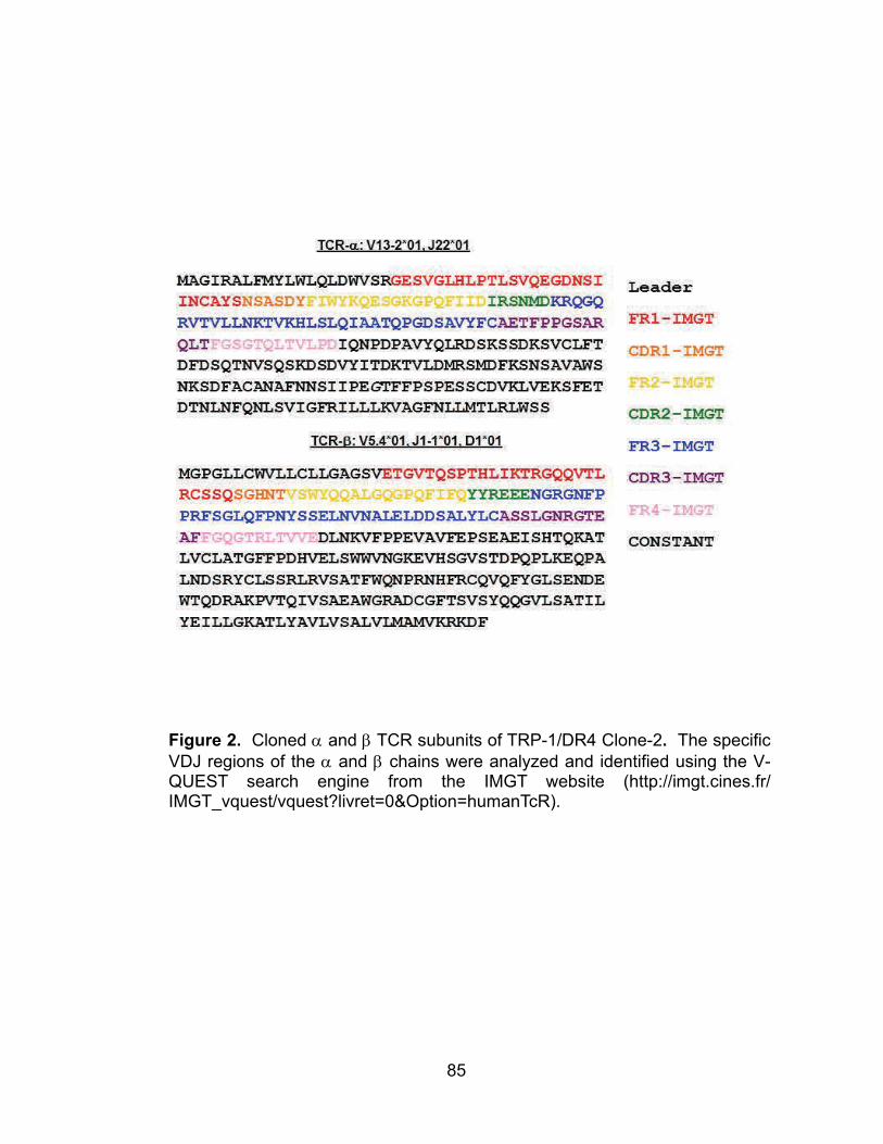
Figure 2. Cloned and TCR subunits of TRP-1/DR4 Clone-2. The specific VDJ regions of the and chains were analyzed and identified using the V-QUEST search engine from the IMGT website (http://imgt.cines.fr/IMGT_vquest/vquest?livret=0&Option=humanTcR).
85

Figure 3. Schematic diagrams of the TRP-1 TCR lentiviral constructs. A. Schematic of lentiviral constructs implementing either an internal ribosomal entry site (IRES) or Furin self-cleavage site (2A) between genes encoding the a and b chains of the TRP-1 epitope specific TCR. B. Detailed diagrams of the TRP-1 TCR lentiviral constructs. The non-specific general promoter of the cytomeglovirus (CMV) was chosen to drive the expression of the downstream a and b chains.
86

Figure 4. Comparison of IRES and F2A lentiviral transduction experiments in human peripheral blood mononuclear cells (PBMC). Both the IRES and F2A constructs efficiently transduced PBMCs, however, the F2A construct proved to be more efficient, particularly in PBMCs at high multiplicities of infection (MOI) (p=0.012 at MOI of 30:1, p=0008 at MOI of 100:1).
87

Figure 5. Representative flow cytometry results for lentiviral transduc-tion of human CD34+ and murine Lin- cells with the TRP-1 TCR F2A len-tiviral construct. A gp100 specific pentamer was used as a negative control to gauge the background non-specific binding of pentamer com-plexes, shown in row 1. A lentivirus encoding the green fluorescent pro-tein (GFP) was used as a positive control for the transduction protocol as shown row 3. Flow cytometric analysis was performed 72 hours post transduction using a TRP-1 TCR specific Ultimer (Proimmune, Inc.).
88

Figure 6. Lentiviral constructs efficiently transduce murine Lin- cells and leads to high expression of transduced gene (28.7%+3) A lentivirus encoding the green fluorescent protein (GFP) was used as a positive control for the transduction protocol (56.2%+8) Flow cytometric analysis was performed 72 hours post transduction using a TRP-1 TCR specific Ultimer (Proimmune, Inc.). A gp100 specific Ultimer was used as a negative control to gauge the background non-specific binding of pentamer complexes. The results repre-sent five separate transduction experiments with at least 3 wells of independ-ent cells in each group.
89

Figure 7. Peripheral blood mononuclear cells transduced with LV-TRP1-F2A (LV-TCR) are functionally specific. Specific IFN- production detected by ELI-SA to the specific peptide (TRP-1277-297), lysate (B16) and intact tumors (B16-DR4, 624 Mel pretreated with IFN-, and 1102 Mel stably transduced with TRP-1), but not to the control peptide (HA306-318), control lysate (MC38) or control tumors (wild type B16, MC38-DR4, and 1102 Mel stably transduced with GFP). Interactions with TRP-1277-297 pulsed targets were blocked by L243 (HLA-DR) and L3T4 (CD4), but not by control W6/32 (class I) or HIT8a (CD8) antibodies. All peptides were pulsed onto DR4+ 1088 EBV-B cells at 50 M x 3 hrs.
90

Figure 8. Positive selection of the TRP-1 TCR following crossbreeding into DR4 Tg mice. Peripheral blood cells were stained for CD3, CD4 and the TRP-1 Pentamer in both TRP-1 TCR Tg mice and TRP-1 TCR/DR4 Tg mice. Each group contains at least 5 mice that were individually bled, stained and ana-lyzed by flow cytometry. In the TRP-1 TCR Tg mice, levels of pentamer posi-tivity was near that of negative controls and ranged from 2-7% in CD4 T cells (Lighter shaded bars). Once crossed on to the HLA-DR4 Tg mice, nearly 80% of CD4 T cells in the peripheral blood were specific for the TRP-1 Ultimer (Darker shaded bars). Within the CD4 cells, 79% ± 8.3% versus 6.4% ± 1.0%; P < 0.0001. Data (mean ± SEM with P value; n = 5–7 per group)
91

Figure 9. Gene modified HSCs successfully engraft and repopulate transplant host over 6 months. CD45.1/CD45.2 competitive repopulation involving co-transplantation of GFP transduced Lin- BoyJ (CD45.1) HSCs and unmanipulat-ed congenic bone marrow cells from a C57BL/6 mouse (CD45.2) transplanted into irradiated DR4 Tg recipient mice. A. Transplanted, transduced Lin- cells maintain GFP expression in the peripheral blood (CD45.1+/GFP+) , developing into a high percentage of mature T cells (TCR+/CD3+) and CD4+ T cells. B. Gene modified cells area also present and remain stable in within the spleen. C. Developing double positive (CD4+/CD8+/GFP+) thymocytes carrying GFP are also identified and maintained in the thymus. D. Gene modified donor cells (CD45.1+/GFP+) are also stable in the bone marrow. Percentages repre-sent 5 mice in each group, for each time point.
92

Figure 10. Representative flow sample shows successful engraftment and emergence of lymphocytes 1 and 4 months post transplantation with either LV-TCR transduced Lin- HSCs (LV-TCR/DR4) or TCR-Tg Lin- HSCs (TCR-Tg/DR4) into irradiated DR4 recipient mice. At 1 month post transplant, few CD4 staining cells are seen in the peripheral blood (1.1% in the LV-TCR/DR4 and 3.6% in the TCR-Tg/DR4). By 4 months post transplantation lymphocytes (both CD4+ and CD8+) have reconstituted the peripheral blood. Of the mature CD3+/CD4+ T cells 43.3% and 43.8% stain positive for the TRP-1 Ultimer in the LV-TCR/DR4 and TCR-Tg/DR4 groups, respectively. Data are representa-tive of flow cytometry studies done on 4 separate transplant experiments.
93

Figure 11. Transplantation of LV-TCR and TRP-1 TCR Tg Lin- HSCs results in high expression of TRP-1 specific TCR. As previously described, competi-tive bone marrow transplants were established with LV-TCR transduced and TRP-1 Tg lineage depleted BM cells and transplanted into irradiated recipient DR4 Tg and C57BL/6 mice. Peripheral blood was obtained at 1 and 4 months and stained with PE labeled TRP-1 pentamer, APC labeled anti-CD4 antibody, and PE-CY5 labeled anti-CD3 antibody. At 1 month post transplantation (grey bars), TRP-1 pentamer specific lymphocytes constituted less than 5% of the total circulating CD3+/CD4+ lymphocytes in all transplant groups. At 4 months post transplantation (blue bars), the LV-TCR in DR4 Tg recipients (LV-TCR/DR4) had the highest percentage of circulating TCR specific CD3+/CD4+ T cells at 15.8%+/-2, followed closely by the TCR-Tg in DR4 Tg recipients (TCR-Tg/DR4) at 11.6%+/-2.5. The LV-TCR in C57BL/6 group only expressed the specific TCR in 8.2%+/-2 of CD3+/CD4+ cells, statistically less than the LV-TCR/DR4 group (p<0.05). A control group of LV-GFP transduced HSCs in DR4 Tg recipients (LV-GFP/DR4) only expressed GFP in 5.5%+/-1 of CD3+/CD4+ T cells at 4 months.
94

Figure 12. 12 month analysis of LV-TCR and TRP-1 TCR Tg Lin- HSCs trans-planted into DR4 Tg mice (n=10 for each group). Percent TRP-1 Ultimer posi-tive as a function of total number of peripheral mononuclear cells. Results show high expression of TRP-1 specific TCR in both groups with TRP-1 TCR Tg transplants expressing higher numbers after 4 months. At 12 months, 8% ± 1.7% versus 22.6% ± 1.5%; P = 0.001. As previously described, competitive bone marrow transplants were established with LV-TCR transduced and TRP-1 Tg lineage depleted BM cells and transplanted into irradiated recipient DR4 Tg mice. Peripheral blood was obtained at 1,2,4,6, and 12 months and stained with PE labeled TRP-1 Ultimer, FITC labeled anti-CD8 antibody, APC labeled anti-CD4 antibody, and PE-CY5 labeled anti-CD3 antibody.
95

Figure 13. 12 month analysis of LV-TCR and TRP-1 TCR Tg Lin- HSCs trans-planted into DR4 Tg mice (n=10 for each group). Percent TRP-1 Ultimer posi-tive as a function of CD3+ peripheral mononuclear cells. Results show high expression of TRP-1 specific TCR in both groups, but TRP-1 TCR Tg trans-plants expressed higher percentages at all time points. At 12 months, 17.8% ± 2.3% versus 36.7% ± 2.4%; P = 0.006. As previously described, competitive bone marrow transplants were established with LV-TCR transduced and TRP-1 Tg lineage depleted BM cells and transplanted into irradiated recipient DR4 Tg mice. Peripheral blood was obtained at 1,2,4,6, and 12 months and stained with PE labeled TRP-1 Ultimer, FITC labeled anti-CD8 antibody, APC labeled anti-CD4 antibody, and PE-CY5 labeled anti-CD3 antibody.
96

Figure 14. 12 month analysis of LV-TCR and TRP-1 TCR Tg Lin- HSCs trans-planted into DR4 Tg mice (n=10 for each group). Percent TRP-1 Ultimer posi-tive as a function of CD4+ peripheral mononuclear cells. Results show high expression of TRP-1 specific TCR in both groups, but TRP-1 TCR Tg trans-plants expressed higher percentages at all time points. At 12 months, 26.1% ± 2.4% versus 65.2% ± 6.3%; P = 0.004. As previously described, competitive bone marrow transplants were established with LV-TCR transduced and TRP-1 Tg lineage depleted BM cells and transplanted into irradiated recipient DR4 Tg mice. Peripheral blood was obtained at 1,2,4,6, and 12 months and stained with PE labeled TRP-1 Ultimer, FITC labeled anti-CD8 antibody, APC labeled anti-CD4 antibody, and PE-CY5 labeled anti-CD3 antibody.
97

Figure 15. Analysis of thymocytes in LV-TCR HSC transplanted DR4 Tg mice. At 12 months the thymus from LV-TCR transplants were harvested and analyzed by flow cytometry. A. This representative flow sample exhibits our analysis technique. Thymocytes were stained for TRP-1 Ultimer, CD3, CD4, and CD8. Each compartment of the maturing thymocyte population (double negative, double positive, and CD4 and CD8 single positive) were analyzed for TRP-1 Ultimer positivity. B. Summary of TCR gene expression in DN, DP, SP CD8, SP CD4 subpopulations. 36% of DN thymocytes, 45% of DP thymo-cytes, 15% of CD8+ thymocytes, and 13% of CD4+ thymocytes expressed the TRP-1 TCR. As expected, a large percentage of cells are deleted from the double positive stage to the mature single positive stage of central thymic se-lection. DP 45% ± 7.3% versus SP CD4 13.3% ± 3.1%; P = 0.002. Data (mean ± SEM with P value; n = 4–6 per group).
98

Figure 16. Development of spontaneous autoimmune vitiligo in mice trans-planted with LV-TCR Lin- bone marrow cells. Depigmentation of the hair folli-cles in the vitiliginous mouse (right) exhibits an irregular, spotted pattern. A control transplant mouse (left) is shown for comparison.
99

Figure 17. Skin samples of LV TRP-1 TCR BM transplanted mice that devel-oped spontaneous autoimmune vitiligo. The samples were fixed in paraffin and stained for S-100 (a marker for neurocrest derived cells such as melano-cytes) and mouse CD3. The top two panels show the staining pattern in the skin of a normal immune replete control mouse. Melanocytes typically reside around the hair follicles as highlighted by the dotted circles (Slide1), and few lymphocytes are found in normal skin (Slide 2). In the vitiliginous mice, there is a clear loss of melanocytes around the hair follicles in affected skin areas as indicated by the arrow in Slide 3. Unlike normal skin in vitiliginous skin, a profuse lymphocytic infiltration particularly around the hair follicles is observed (Slide 4).
100

Figure 18. Skin samples of LV TRP-1 TCR BM transplanted mice that devel-oped spontaneous autoimmune vitiligo. The left two panels show paraffin em-bedded staining for S-100 and CD3 as previously described. The right two panels show frozen section samples of vitiliginous skin stained with antibodies specific for murine CD4 and CD8, respectively.
101

Figure 19. Vitiligo is associated with gross distortion of the natural skin archi-tecture, melanocyte destruction, and TCR gene specific CD4+ T cell infiltration. Immunohistochemistry staining at 6 month post-transplantation of paraffin-embedded skin sample from LV-TCR transplant with antibody specific for hu-man-TCR-.
102

Figure 20. Representative flow cytometry analysis of splenocytes from transplant experiments. Transplant recipients exhibit a TEM phenotype and react spontaneously to tumor ex vivo. Peripheral blood samples at 6 months post-transplantation were obtained and stained with antibodies against CD4, TRP1-Ulimter, CD44, CD45RB, and CD62L. LV-TCR trans-plant CD4+/Ultimer+ cells exhibit a memory-effector (TEM) phenotype (CD45RBlow, CD62Llow, CD44high), while the polyclonal CD4+/Ultimer- and CD4+ control population exhibit a more generalized effector profile (CD45RBhigh, CD62Lint, CD44high), contrasting with naïve CD4+/Ultimer+ cells (CD45RBhigh, CD62Lhigh, CD44low) from endogenous non-transplanted TCR Tg.
103

Figure 21. Summary of activation status of all CD4+ subpopulations. Trans-plant recipients exhibit a TEM phenotype and react spontaneously to tumor ex vivo. Peripheral blood samples at 6 months post-transplantation were obtained and stained with antibodies against CD4, TRP1-Ulimter, CD44, CD45RB, and CD62L. LV-TCR transplant CD4+/Ultimer+ cells exhibit a memory-effector (TEM) phenotype (CD45RBlow, CD62Llow, CD44high), while the polyclonal CD4+/Ultimer- and CD4+ control population exhibit a more generalized effector pro-file (CD45RBhigh, CD62Lint, CD44high), contrasting with naïve CD4+/Ultimer+ cells (CD45RBhigh, CD62Lhigh, CD44low) from endogenous non-transplanted TCR Tg. Data (mean ± SEM; n = 5 per group).
104

Figure 22. CD4+ T cells from secondary transplant recipients exhibit high level gene specific TCR expression. Peripheral blood from 6 month secondary transplants were stained with antibodies against CD4, CD8, CD3 and the TRP-1 pentamer and analyzed by flow cytometry. Total, CD3, and CD4 popula-tions: 7.1% ± 4.5% within the total cellular compartment (CD45+Ultimer+), 47.5% ± 8.3% within the global CD3 compartment (CD45+CD3+Ultimer+), and 53.1% ± 10.7% within the CD4 subcompartment (CD45+CD3+CD4+Ultimer+). Data (mean ± SEM;n = 5 per group)
105

Figure 23. CD4+ T cells from secondary transplant recipients exhibit high level gene specific TCR expression and multi-copy integration of the LV-TCR gene product. A. Relative TCR gene expression is >1000 times greater than control transplants (P=0.01). Total RNA was prepared from BM obtained from 6 month old secondary LV-TCR and Control transplants and analyzed by qPCR for rel-ative TCR gene expression using an -chain specific probe. Relative gene ex-pression (RQ = LV-TCR/Control) is displayed. B. Multicopy integration of the LV-TCR gene product is found in secondary transplant recipients. Using a vec-tor backbone specific probe, 4-6 integrants were identified in three separate LV-TCR transplant samples compared with Control.
106

Figure 24. Six month secondary TCR transplant recipients reject subcutane-ous melanoma. LV-TCR and control transplants (5 mice per group) were sub-cutaneously injected with 5x10⁵ B16-DR4 tumor cells. Three days after injec-tion, tumor size was recorded. Data are displayed as mean ± SEM with P val-ue; n = 5 per group. Statistical significance between groups was based on tu-mor size using a Student T-test. Experiments were performed in a blinded, randomized fashion, and executed independently 3 times.
107

Figure 25. Tumors from LV-TCR secondary transplants are necrotic. Im-munohistochemistry staining of paraffin-embedded tumors resected from 6 month transplants. A. Resected tumor cells were stained with antibody spe-cific for murine CD3. Panel 1 shows B16-DR4 tumors from control transplant. Panel 2 shows tumor from LV-TCR transplant. B. H&E staining of tumor re-sections from control transplant (Panel 1) and LV-TCR (Panel 2) shows ne-crotic changes in LV-TCR transplant.
108

Figure 26. Paraffin-embedded tumor resection from LV-TCR secondary trans-plant with immonhistochemistry staining specific for human TCR- chain ex-pression. Tumors from LV-TCR secondary transplants exhibit lymphocytic in-filitration expressing human TCR-
109

Figure 27. LV-TCR gene-modified human CD34+/CD38- HPCs obtained from cord blood functionally engraft and repopulate 6 month old NOD-SCID-IL2r KO transplant recipients. A, Flow cytometry analysis of peripheral blood demonstrates high level TCR gene expression: hCD45/mCD45.2/CD4/CD8/Ultimer staining. B, Six month transplant summary of TCR gene expression within the hCD45+/CD4+/Ultimer+ subpopulation. Data are displayed as mean + SEM; n = 5 per group.
110

Figure 28. LV-TCR transplants in NOD-SCID-IL2r KO recipients reject hu-man melanoma implantation. 6 month old control and LV-TCR transplants (5 mice in each group) were injected subcutaneously with 5x10⁵ human 624 melanoma cells at Day 0. Tumor size was measured every 3 days for 24 days. No tumor was visibly observable in any of the LV-TCR transplants through 24 days post-tumor implantation, while all 5 mice in the control trans-plants developed visibly observable tumors between 9 and 12 days.
111

REFERENCE LIST
1. Anderson, M.S., Venanzi, E.S., Chen, Z., Berzins, S. P., Benoist, C. and
Mathis., D. 2005. The cellular mechanism of Aire control of T cell
tolerance. Immunity. 23:227-239.
2. Anichini, A., Maccall, C., Motarini, R., Salvi, S., Mazzocchi, A., Squarcina,
P., Herlyn, M. and Parmiani, G. 1993. Melanoma cells and normal
melanocytes share antigens recognized by HLA-A2-restricted cytotoxic
T cell clones from melanoma patients. J. Exp. Med. 177:989-998.
3. Awong, G., Herer, E., Surh, C., Dick, J., La Motte-Mohs, R. and Zuniga-
Pflucker., J.C. 2009. Characterization in vitro and engraftment
potential in vivo of human progenitor T cells generated from
hematopoietic stem cells. Blood. 114:972-982.
4. Baecher-Allan, C. and Anderson, D.E. 2006. Immune regulation in tumor-
bearing hosts. Curr. Opin. Immunol. 18:214-219.
5. Barrette, S., Douglas, J.L., Seidel, N.E., Bodine, D.M. 2000. Lentivirus-
based vectors transduce mouse hematopoietic stem cells with similar
efficiency to moloney murine leukemia virus-based vectors. Blood.
96:3385-3391.
6. Bartosch, B. and Cosset, F.L. 2004. Strategies for retargeted gene delivery
using vectors derived from lentiviruses. Curr. Gene Ther. 4:427-443.
7. Baum, C. 2007. Insertional mutagenesis in gene therapy and stem cell
biology. Curr. Opin. Hematol. 14:337-342.
8. Ben-Dor, I., Itsykson, P., Goldenberg, D., Galun, E. and Reubinoff, B.E.
2006. Lentiviral vectors harboring a dual-gene system allow high and
homogeneous transgene expression in selected polyclonal human
embryonic stem cells. Mol. Ther. 14:255-267.
9. Bennett, S.R., Carbone, F., Karamalis, F., Flavell, R.A., Miller, J.F. and
Heath, W.R. 1998. Help for cytotoxic T cell responses is mediated by
CD40 signaling. Nature. 393:478-480.
112
10. Bevan, M. and Fink, P. 1978. The influence of thymus H-2 antigens on the
specificity of maturing killer and helper cells. Immunol. Rev. 42:3-19.
11. Boon, T. and van der Brugges, P. 1996. Human tumor antigens recognized
by T lymphocytes. J. Exp. Med. 183:725-729.
12. Borrello, I., Sotomayor, E.M., Rattis, F., Cooke, S.K., Gu, L. and Levitsky,
H.I. 2000. Sustaining the graft-versus-tumor effect through post
transplant immunization with granulocyte-macrophage colony-
stimulating factor (GM-CSF) producing tumor vaccines. Blood.
95:3011-3019.
13. Bouneaud, C., Kourilsky, P. and Bousso, P. 2000. Impact of negative
selection on the T cell repertoire reactive to a self-peptide: a large
fraction of T cell clones escapes clonal deletion. Immunity. 13:829-840.
14. Bowne, W.B., Srinivasan, R., Wolchok, J.D., Hawkins, W.G., Blachere, N.E.,
Dyall, R., Lewis, J.J. and Houghton, A.N. 1999. Coupling and
uncoupling of tumor immunity and autoimmunity. J. Exp. Med.
190:1717-1722.
15. Boyman, O., Purton, J.F., Surh, C.D. and Sprent, J. 2007. Cytokines and T
cell homeostasis. Curr. Opin. Immunol. 19:320-326.
16. Brandmaier, A.G., Leitner, W.W., Ha, S.P., Sidney, J., Restifo, N.P. and
Touloukian, C.E. 2009. High-avidity autoreactive CD4+ T cells induce
host CTL, overcome T(regs) and mediate tumor destruction. J.
Immunother. 32:677-688.
17. Cavalieri, S., Cazzaniga, S., Geuna, M., Magnani, Z., Bordignon, C.,
Naldini, L. and Bonini, C. 2003. Human T lymphocytes transduced by
lentiviral vectors in the absence of TCR activation maintain an intact
immune competence. Blood 102:497-505.
18. Cavazzana-Calvo, M., Hacein-Bey, S., de Saint, B.G., Gross, F., Yvon, E.,
Nusbaum, P., Selz, F., Hue, C., Certain, S. and Casanova, J.L. 2000.
Gene therapy of human severe combined immunodeficiency (SCID)-
X1 disease. Science 288:669-672.
113
19. Cohen, P.A., Peng, L., Plantz, G.E., Kim, J.A., Wang, D.E. and Shu, S.
2000. CD4+ T cells in adoptive immunotherapy and the indirect
mechanism of tumor rejection. Crit. Rev. Immunol. 20:17-56.
20. Cox, A.L., Skipper, J., Chen, Y., Henderson, R.A., Darrow, T.L.,
Shabanowitz, J., Engelhard, V.H., Hunt, D.F. and Slingluff, C.L.S.
1994. Identification of a peptide recognized by five melanoma-specific
human cytotoxic T cell lines. Science. 264:716-719.
21. Cui,Y., Kelleher, E., Straley, E., Fuchs, E., Gorski, K., Levitsky, H., Borrello,
I., Civin, C.I., Schoenberger, S.P., Cheng, L., Pardoll, D.M. and
Whartenby, K.A. 2003. Immunotherapy of established tumors using
bone marrow transplantation with antigen gene--modified
hematopoietic stem cells. Nat. Med. 9:952-958.
22. D'Costa, J., Mansfield, S.G. and Humeau, L.M. 2009. Lentiviral vectors in
clinical trials: Current status. Curr. Opin. Mol. Ther. 11:554-564.
23. Dudley, M.E. and Rosenberg, S.A. 2003. Adoptive-cell-transfer therapy for
the treatment of patients with cancer. Nat. Rev. Cancer 3:666-675.
24. Dudley, M.E., Wunderlich, J.R., Yang, J.C., Sherry, R.M., Topalian, S.L.,
Restifo, N.P., Royal, R.E., Kammula, U., White, D.E., Mavroukakis,
S.A., Rogers, L.J., Gracia, G.J., Jones, S.A., Mangiameli, D.P.,
Pelletier, M.M., Gea-Banacloche, J., Robinson, M.R., Berman, D.M.,
Filie, A.C., Abati, A. and Rosenberg, S.A. 2005. Adoptive cell transfer
therapy following non-myeloablative but lymphodepleting
chemotherapy for the treatment of patients with refractory metastatic
melanoma. J. Clin. Oncol. 23:2346-2357.
25. Dudley, M.E., Wunderlich, J.R., Robbins, P.F., Yang, J.C., Hwu, P.,
Schwartzentruber, D.J., Topalian, S.L., Sherry, R., Restifo, N.P.,
Hubicki, A.M., Robinson, M.R., Raffeld, M., Duray, P., Seipp, C.A.,
Rogers-Freezer, L., Morton, K., Mavroukakis, S.A., White, D.A. and
Rosenberg, S.A. 2002. Cancer regression and autoimmunity in
patients after clonal repopulation with antitumor lymphocytes. Science.
298:850-854.
114

26. Dudley, M.E., Wunderlich, J., Nishimura, M.I., Yu, D., Yang, J.C., Topalian,
S.L., Schwartzentruber, D.J., Hwu, P., Marincola, F.M., Sherry, R.,
Leitman, S.F. and Rosenberg, S.A. 2001. Adoptive transfer of cloned
melanoma-reactive T lymphocytes for the treatment of patients with
metastatic melanoma. J. Immunother. 24:363-373.
27. Dudley, M.E., Ngo, L.T., Westwood, J., Wunderlich, J.R. and Rosenberg,
S.A. T cell clones from melanoma patients immunized against an
anchor-modified gp100 peptide display discordant effector phenotypes.
Cancer J. 2000. Mar-Apr.; 6:69-77.
28. Dull, T., Zufferey, R., Kelly, M., Mandel, R.J., Nguyen, M., Trono, D. and
Naldini, L. 1998. A third-generation lentivirus vector with a conditional
packaging system. J. Virol. 72:8463-8471.
29. Dutton, R.W., Bradley, L.M., and Swain,S.L. 1998. T cell memory. Annu.
Rev. Immunol. 16:201-223.
30. Fontenot, J.D., Gavin, M.A. and Rudensky, A.Y. 2003. Foxp3 programs the
development and function of CD4+CD25+ regulatory T cells. Nat.
Immunol. 4:330-336.
31. Fry, T.J., Connick, E., Falloon, J., Lederman, M., Liewehr, D.J., Spritzler, J.,
Steinberg, S.M., Wood, L.V., Yarchoan, R., Zuckerman, J., Landay, A.
and Mackall, C.L. 2001. A potential role for interleukin-7 in T cell
homeostasis. Blood. 97:2983-2990.
32. Gallegos, A.M. and Bevan, M.J. 2004. Central tolerance to tissue-specific
antigens mediated by direct and indirect antigen presentation. J. Exp.
Med. 200:1039-1049.
33. Gallegos, A.M. and Bevan, M.J. 2006. Central tolerance: good but
imperfect. Immunol. Rev. 209:290-6.:290-296.
34. Galy ,A., Roncarolo, M.G., and Thrasher, A.J. 2008. Development of
lentiviral gene therapy for Wiskott Aldrich syndrome. Expert. Opin. Biol.
Ther. 8:181-190.
115

35. Garbelli, S., Mantovani, S., Palermo, B. and Giachino, C. 2005. Melanocyte-
specific, cytotoxic T cell responses in vitiligo: the effective variant of
melanoma immunity? Pigment Cell Res. 18:234-242.
36. Gattinoni, L., Finkelstein, S.E., Klebanoff, C.A., Antony, P.A., Palmer, D.C.,
Spiess, P.J., Hwang, L.N., Yu, Z., Wrzesinski, C., Heimann, D.M. et al
2005. Removal of homeostatic cytokine sinks by lymphodepletion
enhances the efficacy of adoptively transferred tumor-specific CD8+ T
cells. J. Exp. Med. 202:907-912.
37. Gerloni, M. and Zanetti, M. 2005. CD4 T cells in tumor immunity. Springer
Semin. Immunopathol. 27:37-48.
38. Gershon, R. K. and Kondo, K. 1971. Infectious immunological tolerance.
Immunology 21:903-914.
39. Gershon, R.K., Cohen, P.,Hencin, R., Liebhaber, S.A. 1972. Suppressor T
cells. J. Immunol. 108:586-590.
40. Ghiringhelli, F., Larmonier, N., Schmitt, E., Parcellier, A., Cathelin, D.,
Garrido, C., Chauffert, B., Solary, E., Bonnotte, B. and Martin, F. 2004.
CD4+CD25+ regulatory T cells suppress tumor immunity but are
sensitive to cyclophosphamide which allows immunotherapy of
established tumors to be curative. Eur. J. Immunol. 34:336-344.
41. Glimm, H., Oh, I.H. and Eaves, C.J. 2000. Human hematopoietic stem cells
stimulated to proliferate in vitro lose engraftment potential during their
S/G(2)/M transit and do not reenter G(0). Blood. 96:4185-4193.
42. Gothot, A., van der Loo, J.C., Clapp, D.W., and Srour, E.F. 1998. Cell cycle-
related changes in repopulating capacity of human mobilized
peripheral blood CD34(+) cells in non-obese diabetic/severe combined
immune-deficient mice. Blood. 92:2641-2649.
43. Gotter, J., Brors, B., Hergenhahn, M. and Kyewski, B. 2004. Medullary
epithelial cells of the human thymus express a highly diverse selection
of tissue-specific genes colocalized in chromosomal slusters. J. Exp.
Med. 199:155-166.
116

44. Haddad R., Guardiola P., Izac B., Thibault C., Radich J. and Delezoide,
A.L. 2004. Molecular characterization of early human T/NK and B-
lymphoid progenitor cells in umbilical cord blood. Blood. 104:3918-
3926.
45. Hakim, F.T., Memon, S.A., Cepeda, R., Jones, E.C., Chow, C.K., Kasten-
Sportes, C., Odom, J., Vance, B.A., Christensen, B.L. and Mackall,
C.L. 2005. Age dependent incidence, time course, and consequences
of thymic renewal in adults. J. Clin. Invest. 115:930-939.
46. Hargrove, P.W., Kepes, S., Hanawa, H., Obenauer, J.C., Pei, D., Cheng, C.,
Gray, J.T., Neale, G. and Persons, D.A. 2008. Globin lentiviral vector
insertions can perturb the expression of endogenous genes in beta-
thalassemic hematopoietic cells. Mol. Ther. 16:525-533.
47. Hawkins, W.G., Gold, J.S., Dyall, R., Wolchok, J.D., Hoos, A., Bowne, W.B.,
Srinivasan, R., Houghton, A.N. and Lewis, J.J. 2000. Immunization
with DNA coding for gp100 results in CD4 T cell independent antitumor
immunity. Surgery. 128:273-280.
48. He, Y., Zhang, J., Mi, Z., Robbins, P. and Falo, L.D. 2005. Immunization
with lentiviral vector-transduced dendritic cells induces strong and
long-lasting T cell responses and therapeutic immunity. J. Immunol.
174:3808-3817.
49. Heinzel, K., Benz, C., Martins, V.C., Haidl, I.D. and Bleul, C.C. 2007. Bone
marrow derived hemopoietic precursors commit to the T cell lineage
only after arrival in the thymic microenvironment. J. Immunol. 178:858-
868.
50. Hill, G., Crawford, J.M., Cooke, K.R., Brinson, Y.S., Pan, L. and Ferrara,J.L.
1997. Total body irradiation and acute graft-versus-host disease: the
role of gastrointestinal damage and inflammatory cytokines. Blood.
90:3204-3213.
51. Hock, H., Dorsch, M., Diamantstein, T. and Blankenstein, T. 1991.
Interleukin-7 induces CD4+ T cell-dependent tumor rejection. J. Exp.
Med. 174:1291-1298.
117

52. Hoebeke I., De Smedt, M., Stolz, F., Pike-Overzet, K., Staal, J.T., Plum, J.
and Leclercq, G. 2007. T-, B- and NK-lymphoid, but not myeloid cells
arise from human CD34+ CD38- CD7+ common lymphoid progenitors
expressing lymphoid-specific genes. Leukemia. 21:311-319.
53. Houghton, A.N. 1994. Cancer antigens: immune recognition of self and
altered self. J. Exp. Med. 180:1-4.
54. Hu, H., Huston, G., Duso, D., Lepak, N., Roman, E. and Swain, S.L. 2001.
CD4(+) T cell effectors can become memory cells with high efficiency
and without further division. Nat. Immunol. 2:705-710.
55. Hung, K., Hayashi, R., Lafand-Walker, A., Lowenstein, C., Pardoll, D.,
Levitsky, D. 1998. The central role of CD4+ T cells in the antitumor
immune resonse. J. Exp. Med. 188:2357-2368.
56. Isacson, O. and Kordower, J.H. 2008. Future of cell and gene therapies for
Parkinson's disease. Ann. Neurol. 64 Suppl 2:S122-S138.
57. Ito, K., Bian, H.J., Molina, M., Han, J., Magram, J., Saar, E., Belunis, C.,
Bolin, D.R., Arceo, R. and Campbell, R. 1996. HLA-DR4-IE chimeric
class II transgenic, murine class II-deficient mice are susceptible to
experimental allergic encephalomyelitis. J. Exp. Med. 183:2635-2644.
58. Jager, E., Chen, Y.T., Drijfhout, J.W., Karbach, J., Ringhoffer, M., Jager, D.
Arand, M., Wada, H., Noguchi, Y., Stockert, E., Old, L.J. and Knuth, A.
1998. Simultaneous humoral and cellular immune response against
cancer-testis antigen NY-ESO-1: definition of human histocompatibility
leukocyte antigen (HLA)-A2-binding peptide epitopes. J. Exp. Med.
187:265-270.
59. Javia, L.R. and Rosenberg, S.A. 2003. CD4+CD25+ suppressor
lymphocytes in the circulation of patients immunized against
melanoma antigens. J. Immunother. 26:85-93.
60. Jelley-Gibbs, D.M., Brown, D.M., Dibble, J.P., Haynes, L., Eaton, S.M. and
Swain, S.L. 2005. Unexpected prolonged presentation of influenza
antigens promotes CD4 T cell memory generation. J. Exp. Med.
202:697-706.
118

61. Johnnidis, J.B., Venanzi, E.S., Taxman, D.J., Ting, J.P., Benoist, C. and
Mathis. D.J. 2005. Chromosomal clustering of genes controlled by the
aire transcription factor. Proc. Natl. Acad. Sci. 102:7233-7238.
62. Kawakami, Y. and Rosenberg, S.A.. 1997. Human tumor antigens
recognized by T cells. Immunol. Res. 16:313-339.
63. Kennedy, R. and Celis, E. 2008. Multiple roles for CD4+ T cells in anti-tumor
immune responses. Immunol. Rev. 222:129-144.
64. King ,C., Ilic ,A., Koelsch, K. and Sarvetnick, N. 2004. Homeostatic
expansion of T cells during immune insufficiency generates
autoimmunity. Cell. 117:265-277.
65. Klebanoff, C.A., Gattinoni, L., Torabi-Parizi, P., Kerstann, K., Cardones,
A.R., Finkelstein, S.E., Palmer, D.C., Antony, P.A., Hwang, S.T. and
Rosenberg, S.A. 2005. Central memory self/tumor-reactive CD8+ T
cells confer superior antitumor immunity compared with effector
memory T cells. Proc. Natl. Acad. Sci. U. S. A 102:9571-9576.
66. Klug, C.A., Cheshier, S. and Weissman, I.L. 2000. Inactivation of a GFP
retrovirus occurs at multiple levels in long-term repopulating stem cells
and their differentiated progeny. Blood. 96:894-901.
67. Knuth, A., Wolfel, T. and Meyer Zum Buschenfelde, K.H. 1992. T cell
responses to human malignant tumors. Cancer Surveys. 13:39-52.
68. Kozak, M. 2002. Pushing the limits of the scanning mechanism for initiation
of translation. Gene 299:1-34.
69. Kouskoff, V., Signorelli, K., Benoist, C. and Mathis, D. 1995. Cassette
vectors directing expression of T cell receptor genes in transgenic
mice. J. Immunol. Methods. 180:273-280.
70. Krupica, T., Fry, T.J. and Mackall, C.L. 2006. Autoimmunity during
lymphopenia: a two-hit model. Clin. Immunol. 120:121-128.
71. Langlade-Demoyen, P., Garcia-Pons, F., Castiglioni, P., Garcia, Z.,
Cardinaud, S., Xiong, S., Gerloni, M. and Zanetti, M. 2003. Role of T
cell help and endoplasmic reticulum targeting in protective CTL
response against influenza virus. Eur. J. Immunol. 33:720-728.
119

72. Leitner, W.W., Hwang, L.N., deVeer, M.J., Zhou, A., Silverman, R.H.,
Williams, B.R., Dubensky, T.W., Ying, H. and Restifo, N.P. 2003. Alpha
virus-based DNA vaccine breaks immunological tolerance by activating
innate antiviral pathways. Nat. Med. 9:33-39.
73. Levine, B.L., Humeau, L.M., Boyer, J., MacGregor, R.R., Rebello, T., Lu, X.,
Binder, G.K., Slepushkin, V., Lemiale, F. and Mascola, J.R. 2006.
Gene transfer in humans using a conditionally replicating lentiviral
vector. Proc. Natl. Acad. Sci. U.S.A. 103:17372-17377.
74. Lizee, G., Gonzales, M.I. and Topalian, S.L. 2004. Lentivirus vector-
mediated expression of tumor-associated epitopes by human antigen
presenting cells. Hum. Gene Ther. 15:393-404.
75. Mackall, C.L., Hakim, F.T. and Gress, R.E. 1997. Restoration of T cell
homeostasis after T cell depletion. Semin. Immunol. 9:339-346.
76. Magalhaes, D.A., Silveira, E.L., Junta, C.M., Sandrin-Garcia, P., Fachin,
A.L., Donadi, E.A., Sakamoto-Hojo, E.T. and Passos, G.A. 2006.
Promiscuous gene expression in the thymus: the root of central
tolerance. Clin. Dev. Immunol. 13:81-99.
77. McDonald, H.R. and Lees, R.K. 1990. Programmed death of autoreactive
thymocytes. Nature. 343:642-644.
78. Miller, J. 1961. Immunological function of the thymus. Lancet. 2:748-749.
79. Miller, D.G., Adam, M.A. and Miller, A.D. 1990. Gene transfer by retrovirus
vectors occurs only in cells that are actively replicating at the time of
infection. Mol. Cell Biol. 10:4239-4242.
80. Miyazaki, M., Nakatsura, T., Yokomine, K., Senju, S., Monji, M., Hosaka, S.,
Komori, H., Yoshitake, Y., Motomura, Y., Minohara, M., Kubo, T.,
Ishihara, K., Hatayama, T., Ogawa, M. and Nishimura, Y. 2005. DNA
vaccination of HSP105 leads to tumor rejection of colorectal cancer
and melanoma in mice through activation of both CD4 T cells and CD8
T cells. Cancer Sci. 96:695-705.
81. Miyoshi, H. 2004. Gene delivery to hematopoietic stem cells using lentiviral
vectors. Methods Mol. Biol. 246:429-438.
120

82. Moeller, M., Haynes, N.M., Kershaw, M.H., Jackson, J.T., Teng, M.W.,
Street, S.E., Cerutti, L., Jane, S.M., Trapani, J.A. and Smyth, M.J.
2005. Adoptive transfer of gene-engineered CD4+ helper T cells
induces potent primary and secondary tumor rejection. Blood.
106:2995-3003.
83. Montini, E., Cesana, D., Schmidt, M., Sanvito, F., Ponzoni, M.,
Bartholomae, C., Sergi, S.L., Benedicenti, F., Ambrosi, A. and Di, S.C.
2006. Hematopoietic stem cell gene transfer in a tumor-prone mouse
model uncovers low genotoxicity of lentiviral vector integration. Nat.
Biotechnol. 24:687-696.
84. Morgan, R.A., Dudley, M., Wunderlich, J., Hughes, M., Yang, J., Sherry, R.,
Royal, R., Topalian, S., Kammula, U., Restifo, N., Zheng, Z., Nahvi, A.,
de Vries, C.R., Rogers-Freezer, L., Mavroukakis, S. and Rosenberg, S.
2006. Cancer regression in patients after transfer of genetically
engineered lymphocytes. Science. 314:126-129.
85. Morgan, R.A., Dudley, M.E., Yu, Y.Y., Zheng, Z., Robbins, P.F., Theoret,
M.R., Wunderlich, J.R., Hughes, M.S., Restifo, N.P. and Rosenberg,
S.A. 2003. High efficiency TCR gene transfer into primary human
lymphocytes affords avid recognition of melanoma tumor antigen
glycoprotein 100 and does not alter the recognition of autologous
melanoma antigens. J. Immunol. 171:3287-3295.
86. Morris, E.C., Tsallios, A., Bendle, G.M., Xue, S.A. and Stauss, H.J. 2005. A
critical role of T cell antigen receptor-transduced MHC class I-restricted
helper T cells in tumor protection. Proc. Natl. Acad. Sci. U. S. A
102:7934-7939.
87. Mostoslavsky, G., Kotton, D.N., Fabian, A.J., Gray, J.T., Lee, J.S. and
Mulligan, R.C. 2005. Efficiency of transduction of highly purified murine
hematopoietic stem cells by lentiviral and oncoretroviral vectors under
conditions of minimal in vitro manipulation. Mol. Ther. 11:932-940.
121

88. Moyer, J.S., Maine, G, and Mule, J.J. 2006. Early vaccination with tumor-
lysate pulsed dendritic cells after allogeneic bone marrow
transplantation has antitumor effects. Biol. Blood Marrow Transplant.
12:1010-1019.
89. Muranski, P., Boni, A., Antony, P.A., Cassard, L., Irvine, K.R., Kaiser, A.,
Paulos, C.M., Palmer, D.C., Touloukian, C.E. and Ptak, K. 2008.
Tumor-specific Th17-polarized cells eradicate large established
melanoma. Blood. 112:362-373.
90. Nikolic-Zugic, J. and Bevan, M.J. 1990. Role of self peptides in positively
selecting the T cell repertoire. Nature. 344:65-67.
91. Nishimura, E., Sakihama, T., Setoguchi, R.,Tanaka, K. and Sakaguchi, S.
2004. Induction of antigen-specific immunologic tolerance by in vivo
and in vitro antigen-specific expansion of naturally arising
Foxp3+CD25+CD4+ regulatory T cells. Int. Immunol. 16:1189-1201.
92. North, R.J. 1982. Cyclophosphamide-facilitated adoptive immunotherapy of
an established tumor depends on elimination of tumor-induced
suppressor T cells. J. Exp. Med. 155:1063-1074.
93. Obst, R., van Santen, H.M., Mathis, D. and Benoist, C. 2005. Antigen
persistence is required throughout the expansion phase of a CD4(+) T
cell response. J. Exp. Med. 201:1555-1565.
94. Ogg, G.S., Rod, D.P., Romero, P., Chen, J.L. and Cerundolo, V. 1998. High
frequency of skin-homing melanocyte-specific cytotoxic T lymphocytes
in autoimmune vitiligo. J. Exp. Med. 188:1203-1208.
95. Ohashi, P.S. 2003. Negative selection and autoimmunity. Curr. Opin.
Immunol. 15:668-676.
96. Okamoto, T., Irie, R.F., Fuji, S., Huang, S.K., Nizze, A.J., Morton, D.L. and
Hoon, D.S. 1998. Anti-tyrosinase-related protein-2 immune response in
vitiligo patients and melanoma patients receiving active-specific
immunotherapy. J. Invest Dermatol. 111:1034-1039.
122

97. Opferman, J.T., Ober, B.T. and Ashton-Rickardt, P.G. 1999. Linear
differentiation of cytotoxic effectors into memory T lymphocytes.
Science. 283:1745-1748.
98. Ossendorp, F., Mengede, E., Camps, M., Filius, R. and Melief, C.J.. 1998.
Specific T helper cell requirement for optimal induction of cytotoxic T
lymphocytes against major histocompatibility complex class II negative
tumors. J. Exp. Med. 187:693-702.
99. Overwijk, W.W., Lee, D.S., Surman, D., Irvine, K., Touloukian, CE., Chan,
C.C., Carroll, M.W., Moss, B., Rosenberg, S.A. and Restifo, N.P. 1999.
Vaccination with a recombinant vaccinia virus encoding a self antigen
induces autoimmune vitiligo and tumor cell destruction in mice:
requirement for CD4(+) T lymphocytes. Proc. Natl. Acad. Sci. U. S. A.
96:2982-2987.
100. Overwijk, W.W., Theoret, M.R., Finkelstein, S.E., Surman, D.R., de Jong,
L.A., Vyth-Dreese, F.A., Dellemijn, T.A., Antony, P.A., Spiess, P.J.,
Palmer, D.C., Heimann, D.M., Klebanoff, C.A., Yu, Z., Hwang, L.N.,
Feigenbaum, L., Kruisbeek, A.M., Rosenberg, S.A. and Restifo, N.P.
2003. Tumor regression and autoimmunity after reversal of a
functionally tolerant state of self-reactive CD8+ T cells. J. Exp. Med.
198:569-580.
101. Pardoll, D.M. 1994. Tumor antigens: A new look for the 1990s. Nature.
369:357.
102. Pardoll, D.M. and Topalian, S.L. 1998. The role of CD4+ T cell responses in
antitumor immunity. Curr. Opin. Immunol. 10:588-594.
103. Parish, C.R. 2003. Cancer immunotherapy: the past, the present and the
future. Immunol. Cell Biol. 81:106-113.
104. Pullen, A.M., Kappler, J.W., Marrack, P. 1989. Tolerance to self antigens
shapes the T cell repertoire. Immunol. Rev. 107:125-139.
123

105. Quezada, S.A., Simpson, T.R., Peggs, K.S., Merghoub, T., Vider, J., Fan,
X., Blasberg, R., Yagita, H., Muranski, P. and Antony, P.A. 2010.
Tumor-reactive CD4+ T cells develop cytotoxic activity and eradicate
large established melanoma after transfer into lymphopenic hosts. J.
Exp. Med. 207:637-650.
106. Renkvist, N., Castelli, C., Robbins, P.F. and Parmiani, G. 2001. A listing of
human tumor antigens recognized by T cells. Cancer Immunol.
Immunother. 50:3-15.
107. Rivera, J. and Tessarollo, L. 2008. Genetic background and the dilemma of
translating mouse studies to humans. Immunity. 28:1-4.
108. Robbins, P.F., Dudley, M.E., Wunderlich, J., El-Gamil, M., Li, Y.F., Zhou, J.,
Huang, J., Powell, D.J. and Rosenberg, S.A. 2004. Cutting edge:
Persistence of transferred lymphocyte clonotypes correlates with
cancer regression inpatients receiving cell transfer therapy. J.
Immunol. 173:7125-7130.
109. Roe, T., Reynolds, T.C., Yu, G. and Brown, P.O. 1993. Integration of murine
leukemia virus DNA depends on mitosis. EMBO J. 12:2099-2108.
110. Rosenberg, S.A., Lotze, M.T., Muul, L.M., Leitman, S., Chang, A.E.,
Ettinghausen, S.E., Matory, Y.L., Skibber, J.M. and Vetto, J.T. 1985.
Observations on the systemic administration of autologous
lymphokine-activated killer cells and recombinant interleukin-2 to
patients with metastatic cancer. N. Engl. J. Med. 313:1485-1492.
111. Rosenberg, S.A., Spiess, P. and Lafreniere, R. 1986. A new approach to the
adoptive immunotherapy of cancer with tumor-infiltrating lymphocytes.
Science. 233:1318-1321.
112. Rosenberg, S.A. and White, D.E. 1996. Vitiligo in patients with melanoma:
normal tissue antigens can be targets for cancer immunotherapy. J.
Immunother. Emphasis. Tumor Immunol. 19:81-84.
113. Rosenberg, S.A. 2004. Development of effective immunotherapy for the
treatment of patients with cancer. J. Am. Coll. Surg. 198:685-696.
124

114. Rosenberg, S.A., Sportes, C., Ahmadzadeh, M., Fry, T.J., Ngo, L.T.,
Schwarz, S.L., Stetler-Stevenson, M., Morton, K.E., Mavroukakis ,S.A.
and Morre, M. 2006. IL-7 administration to humans leads to expansion
of CD8+ and CD4+ cells but a relative decrease of CD4+ T-regulatory
cells. J. Immunother. 29:313-319.
115. Sastry, K.J., Marin, M.C., Nehete, P.N., McConnell, K., el-Naggar, A.K. and
McDonnell, T.J. 1996. Expression of human immunodeficiency virus
type I tat results in down-regulation of bcl-2 and induction of apoptosis
in hematopoietic cells. Oncogene. 13:487-493.
116. Schnell, S., Young, J.W., Houghton, A. and Sadelain, M. 2000. Retrovirally
transduced mouse dendritic cells require CD4+ T cell help to elicit
antitumor immunity: implications for the clinical use of dendritic cells. J.
Immunol.164:1243-1250.
117. Schoenberger, S.P., Toes, R.E., van der Voort, E.I., Offringa. R., Melief,
C.J. 1998. T cell help for cytotoxic T lymphocytes is mediated by
CD40-CD40L interactions. Nature. 393:480-483.
118. Schroder, A.R.W., Shinn, P., Chen, H., Berry, C., Ecker, J. and Bushman, F.
2002. HIV-1 Integration in the Human Genome Favors Active Genes
and Local Hotspots. Cell. 110:521-529.
119. Schwartz, R.H. 1989. Acquisition of immunologic self tolerance. Cell.
57:1073-1081.
120. Serwold, T., Ehrlich, L. and Weissman, I. 2008. Reductive isolation from
bone marrow and blood implicates common lymphoid progenitors as
the major source of thymopoiesis. Blood. 113:807-815.
121. Sfikakis, P.P., Gourgoulis, G.M., Moulopoulos, L.A., Kouvatseas, G.,
Theofilopoulos, A.N., and Dimopoulos, M.A. 2005. Age-related thymic
activity in adults following chemotherapy-induced lymphopenia. Eur. J.
Clin. Invest 35:380-387.
125

122. Shen, X., Zhou, J., Hathcock, K.S., Robbins, P., Powell, D.J., Rosenberg,
S.A. and Hodes, R.J. 2007. Persistence of tumor infiltrating
lymphocytes in adoptive immunotherapy correlates with telomere
length. J. Immunother. 30:123-129.
123. Shevach, E.M. 2002. CD4+ CD25+ suppressor T cells: more questions than
answers. Nat. Rev. Immunol. 2:389-400.
124. Sinn, P.L., Sauter, S.L. and McCray, P.B., Jr. 2005. Gene therapy progress
and prospects: development of improved lentiviral and retroviral
vectors design, biosafety, and production. Gene Ther. 12:1089-1098.
125. Stockinger, B., Bourgeois, C. and Kassiotis, G. 2006. CD4+ memory T cells:
functional differentiation and homeostasis. Immunol. Rev. 211:39-48.
126. Storek, J., Gillespy, T., Lu, H., Joseph, A., Dawson, M.A., Gough, M.,
Morris, J., Hackman, R.C., Horn, P.A. and Sale, G.E. 2003. Interleukin-
7 improves CD4 T cell reconstitution after autologous CD34 cell
transplantation in monkeys. Blood. 101:4209-4218.
127. Sugita, S., Sagawa, K., Mochizuki, M., Shichijo, S. and Itoh, K. 1996.
Melanocyte lysis by cytotoxic T lymphocytes recognizing the MART-1
melanoma antigen in HLA-A2 patients with Vogt-Koyanagi-Harada
disease. Int. Immunol. 8:799-803.
128. Sun, J.C., Williams, M.A. and Bevan, M.J. 2004. CD4+ T cells are required
for the maintenance, not programming, of memory CD8+ T cells after
acute infection. Nat. Immunol. 5:927-933.
129. Sutton, R.E., Reitsma, M.J., Uchida, N. and Brown, P.O. 1999. Transduction
of human progenitor hematopoietic stem cells by human
immunodeficiency virus type 1-based vectors is cell cycle dependent.
J. Virol. 73:3649-3660.
130. Tan, J.T., Ernst, B., Kieper, W.C., LeRoy, E., Sprent, J. and Surh, C.D.
2002. Interleukin (IL)-15 and IL-7 jointly regulate homeostatic
proliferation of memory phenotype CD8+ cells but are not required for
memory phenotype CD4+ cells. J. Exp. Med. 195:1523-1532.
126

131. Tao, W., Evans, B.G., Yao, J., Cooper, S., Cornetta, K., Ballas, C.B.,
Hangoc, G., and Broxmeyer, H.E. 2007. Enhanced green fluorescent
protein is a nearly ideal long term expression tracer for hematopoietic
stem cells, whereas DsRed-express fluorescent protein is not. Stem
Cells. 25:670-678.
132. Toes, R.E., Schoenberger, S.P., van der Voort, E.I., Offringa, R., Melief,
C.J. 1998. CD40-CD40 ligand interactions and their role in cytotoxic T
Lymphocyte priming and anti-tumor immunity. Sem. Immunol. 10:443-
448.
133. Topalian, S.C., Rivoltini, L., Mancini, M., Markus, N.R., Robbins, P.E.,
Kawakami, Y., Rosenberg, S.A. 1994. Human CD4+ T cells specifically
recognize a shared melanoma-associated antigen encoded by the
tyrosinase gene. Proc. Natl. Acad. Sci. USA. 91:9461-9465.
134. Touloukian, C.E., Leitner, W.W., Topalian, S.L., Li, Y.F., Robbins, P.F.,
Rosenberg, S.A. and Restifo, N.P. 2000. Identification of a MHC class
II-restricted human gp100 epitope using DR4-IE transgenic mice. J.
Immunol. 164(7):3535-42.
135. Touloukian, C.E., Leitner, W.W., Robbins, P.F., Rosenberg, S.A. and
Restifo, N.P. 2001. Mining the melanosome for tumor vaccine targets:
P.polypeptide is a novel tumor-associated antigen. Cancer Res.
61:8100-8104.
136. Von Boehmer, H., The, H.S., Kisielow, P. 1989. The thymus selects the
useful and destroys the harmful. Immunology Today. 10:57-61.
137. Walker, L.S. and Abbas, A.K. 2002. The enemy within: keeping self-reactive
T cells at bay in the periphery. Nat. Rev. Immunol. 2:11-19.
138. Wang, H.Y., Lee, D.A., Peng, G., Guo, Z., Li, Y., Kiniwa, Y., Shevach, E.M.
and Wang, R.F. 2004. Tumor-specific human CD4+ regulatory T cells
and their ligands: implications for immunotherapy. Immunity. 20:107-
118.
127

139. Williams, M.A., Tyznik, A.J. and Bevan, M.J. 2006. Interleukin-2 signals
during priming are required for secondary expansion of CD8+ memory
T cells. Nature. 441:890-893.
140. Williams, M.A., Holmes, B.J., Sun, J.C. and Bevan, M.J. 2006. Developing
and maintaining protective CD8+ memory T cells. Immunol. Rev.
211:146-153.
141. Woods, N.B., Ooka, A. and Karlsson,S. 2002. Development of gene therapy
for hematopoietic stem cells using lentiviral vectors. Leukemia 16:563-
569.
142. Wu, X., Li, Y., Crise, B. and Burgess, S.M. 2003. Transcription start regions
in the human genome are favored targets for MLV integration. Science.
300:1749-1751.
143. Wu, Li. 2006. T lineage progenitors: the earliest steps en route to T
lymphocytes. Current Opinion in Immunology. 18:121-126.
144. Xu, Y., Setaluri, V., Takechi, Y. and Houghton, A.N. 1997. Sorting and
secretion of a melanosome membrane protein, gp75/TRP1. J. Invest
Dermatol. 109:788-795.
145. Yanez-Munoz, R.J., Balaggan, K.S., MacNeil, A., Howe, S.J., Schmidt, M.,
Smith, A.J., Buch, P., MacLaren, R.E., Anderson, P.N. and Barker,
S.E. 2006. Effective gene therapy with nonintegrating lentiviral vectors.
Nat. Med. 12:348-353.
146. Yang, C.P., Sparshott, S.M., Duffy, D., Garside, P. and Bell, E.B. 2006. The
phenotype and survival of antigen-stimulated transgenic CD4 T cells in
vivo: the influence of persisting antigen. Int. Immunol. 18:515-523.
147. Yang, L. and Baltimore, D. 2005. Long-term in vivo provision of antigen-
specific T cell immunity by programming hematopoietic stem cells.
Proc. Natl. Acad. Sci. U.S.A 102:4518-4523.
148. Yang, L., Qin, X.F., Baltimore, D. and Van, P. 2002. Generation of functional
antigen-specific T cells in defined genetic backgrounds by retrovirus-
mediated expression of TCR cDNAs in hematopoietic precursor cells.
Proc. Natl. Acad. Sci. U. S. A 99:6204-6209.
128

149. Yron I., Wood T.A., Spiess P.J. and Rosenberg S.A. 1980. The isolation and
growth of lymphoid cells infiltrating syngeneic solid tumors. J. Immunol.
125:238-245.
150. Yu, X., Zhan, X., D'Costa, J., Tanavde, V.M., Ye, Z., Peng, T., Malehorn,
M.T., Yang, X., Civin, C.I. and Cheng, L. 2003. Lentiviral vectors with
two independent internal promoters transfer high-level expression of
multiple transgenes to human hematopoietic stem-progenitor cells.
Mol. Ther. 7:827-838.
151. Zhao, Y., Zheng, Z., Robbins, P.F., Khong, H.T., Rosenberg, S.A., and
Morgan, R.A. 2005. Primary human lymphocytes transduced with NY-
ESO-1 antigen-specific TCR genes recognize and kill diverse human
tumor cell lines. J. Immunol. 174:4415-4423.
152. Zhou, J., Dudley, M.E., Rosenberg, S.A. and Robbins, P.F. 2004. Selective
growth in vitro and in vivo, of individual T cell clones from tumor-
infiltrating lymphocytes obtained from patients with melanoma. J.
Immunol. 173:7622-7629.
153. Zhou, J., Dudley, M.E., Rosenberg, S.A. and Robbins, P.F. 2005.
Persistence of multiple rumor specific T cell clones is associated with
complete tumor regression in a melanoma patient receiving adoptive
cell transfer therapy. J. Immunotherapy. 28:53-62.
154. Zhou, J., Shen, X., Huang, J., Hodes, R.J., Rosenberg, S.A. and Robbins,
P.F. 2005. Telomere length of transferred lymphocytes correlates with
in vivo persistence and tumor regression in melanoma patients
receiving cell transfer therapy. J. Immunol. 175:7046-7052.
155. Zinkernagel, R.M., Callahan, G.N., Althage, A., Cooper, S., Klein, P.A.,
Klein, S. 1978. On the thymus in the differentiation of “H2 self
recognition” by T cells: Evidence for dual recognition? J. Exp. Med.
147:882-896.
156. Zufferey, R., Dull, T., Mandel, R.J., Bukovsky, A., Quiroz, D., Naldini, L. and
Trono, D. 1998. Self-inactivating lentivirus vector for safe and efficient
in vivo gene delivery. J. Virol. 72:9873-9880.
129

CURRICULUM VITAE Sung Pil Ha EDUCATION
Indiana University, Indianapolis, IN Ph.D. in Microbiology and Immunology 2012 Indiana University School of Medicine, Indianapolis, IN M.D. in Medicine 2010 Yale University, New Haven, CT M.P.H in Health Economics and Management 2003 Yale University, New Haven, CT B.S. in Molecular Biochemistry and Biophysics 2000 West Lafayette High School, West Lafayette, IN Diploma 1996
AWARDS
Roy H. Behnke Scholarship, Indiana University 2003-2007 Dr. Wright Sr. Scholarship, Walther Cancer Institute 2006 National Merit Scholarship, Yale University 1996
PUBLICATIONS AND ABSTRACTS Sung P Ha, Nicholas D Klemen, Garrett H Kinnebrew, Andrew G Brandmaier,
Jon Marsh, Giao Hangoc, Douglas C Palmer, Nicholas P Restifo, Kenneth Cornetta, Hal E Broxmeyer, Christopher E Touloukian. 2010. Transplantation of mouse HSCs genetically modified to express a CD4-restricted TCR results in long-term immunity that destroys tumors and initiates spontaneous autoimmunity. Journal of Clinical Investigation. 102(12):4273-4288.
Andrew G Brandmaier, Wolfgang W Leitner, Sung P Ha, John Sidney,
Nicholas P Restifo, Christopher E Touloukian. 2009. High-avidity autoreactive CD4+ T cells induce host CTL, overcome Tregs and mediate tumor destruction. Journal of Immunotherpay. 32(7):677-688.
Michael C Lipke, Sung P Ha, Christopher D Fischer, Jonas Rydberg, Stephen
M Bonsib, Chandru P Sundaram. 2007. Pathologic characteristics of exophytic renal masses. Journal of Endourology. 21(12):1489-1492.
Sung P Ha, Kenneth Cornetta, Hal E Broxmeyer, Nicholas P Restifo,
Christopher E Touloukian. 2008. CD4+ T cell immunity following gene transfer into hemaopoietic stem cells. 4th Annual Academic Surgical Congress.

WORK EXPERIENCE
University of Wisconsin Hospitals and Clinics, Madison, WI Neurosurgery Resident 2010-2012 Neurosurgery resident. Nicaragua Medical Internship, Nicaragua Intern 2004 Eight week summer internship in various rural and urban hospitals in Nicaragua sponsored through the University of California Medical Schools. Participated in the public health assessment and daily clinic duties of third in urban and rural clinics in Nicaragua. NERA/Mercer Management Consulting, New York, NY Research Associate 2000-2001 Assessed damage analysis for a variety of securities cases ranging from 10b-5 cases to customized long-term studies of restatements and earnings surprise impact on stock price. Neurobiology Laboratory, Yale University, New Haven CT Researcher 1999-2000 Conducted independent electrophysiological studies in memory and learning developing mathematical models of neural networks. Trumbull News, Yale University, New Haven, CT Co-Editor 1998-1999 Co-wrote and edited production of weekly newsletter. Orthopedic Oncology Department, MGH, Boston, MA Summer Intern 1998 Organized and conducted clinical study of Ewing’s Sarcoma collecting and building databases. Observed patient rounds and surgical rotations. Biomedical Engineering, Purdue University, West Lafayette, IN Researcher 1997 Coordinated and conducted experiments in nerve and skin regeneration on extracellular matrices.
MEMBERSHIPS
Congress of Neurological Surgery, Member 2010-2012

Medical School Student Council, Class Vice President 2006-2007 Rock for Riley, Executive Committee Member 2003-2010 Evening of the Arts, Co-Host 2005-2009 Yale Young Authors’ Club, Co-Founder 1999-2000 Korean American Students at Yale, Member 1998-2000 Yale Emergency Medical Trainers Club, Presiding Officer 1998-2000