CCR FOCUS - Clinical Cancer Research...immune cells (i.e., natural killers cells or dendritic cells)...
10
The Intersection of Immune-Directed and Molecularly Targeted Therapy in Advanced Melanoma: Where We Have Been, Are, and Will Be Ryan J. Sullivan 1 , Patricia M. LoRusso 2 , and Keith T. Flaherty 1 Abstract In three years, four drugs have gained regulatory approval for the treatment of metastatic and unresectable melanoma, with at least seven other drugs having recently completed, currently in, or soon to be in phase III clinical testing. This amazing achievement has been made following a remarkable increase of knowledge in molecular biology and immunology that led to the identification of high- valued therapeutic targets and the clinical development of agents that effectively engage and inhibit these targets. The discovery of either effective molecularly targeted therapies or immunotherapies would have led to dramatic improvements to the standard-of-care treatment of melanoma. However, through parallel efforts that have showcased the efficacy of small-molecule BRAF and MAP–ERK kinase (MEK) inhibitors, as well as the immune checkpoint inhibitors, namely ipilimumab and the anti-PD1/PDL1 antibodies (lambrolizumab, nivolumab, MPDL3280), an opportunity exists to transform the treatment of melanoma specifically and cancer generally by exploring rational combinations of molecularly targeted therapies, immunotherapies, and molecular targeted therapies with immunotherapies. This overview presents the historical context to this therapeutic revolution, reviews the benefits and limitations of current therapies, and provides a look ahead at where the field is headed. Clin Cancer Res; 19(19); 5283–91. Ó2013 AACR. Introduction Melanoma is a deadly disease that is rising in incidence. In 2013, an estimated 76,000 new cases and 9,400 deaths are expected in the United States (1). Still, despite these stark numbers, there is great optimism in the melanoma research and treatment communities due to a number of breakthroughs that are transforming the way this disease is being treated. To highlight the dramatic progress being made to treat patients with melanoma, it is very possible that for the second time in 3 years, the number of ther- apies approved by the U.S. Food and Drug Administration (FDA) for the treatment of advanced melanoma will double in a 12-month period (Fig. 1). These advances have been the result of extraordinary scientific discovery combined with robust clinical and translational efforts. What follows is an overview of the past, current, and near future states of melanoma therapeutics and an introduc- tion to the topics covered in this Clinical Cancer Research Focus section. Immunotherapy and Melanoma Melanoma has long been considered a malignancy that has a complex and unique interaction with the immune system. The first description of immune infiltrates in pri- mary tumors was made decades ago, as was the definition of the prognostic significance of these infiltrates (2, 3). Further interactions between the immune system and melanoma have been posited as the explanation of two fascinating phenomena: (i) the long latency from primary melanoma resection of early-stage disease to the development of wide- spread metastases and (ii) the spontaneous regression of metastatic melanoma in a small number of patients (4, 5). Because of these findings and beliefs, immunotherapy has a long history in the treatment of melanoma starting with injections of immune stimulants (i.e., Bacillus Calmette- Gu erin), moving to treatment with mediators of immune responses (i.e., cytokines) with or without "educated" immune effectors such as primed T lymphocytes (adoptive cell transfer), and more recently, monoclonal antibodies that target critical immune checkpoints and thereby lead to T-lymphocyte (T-cell) activation (6–11). Cytokine therapy In the early days of tumor immunology, it was evident that T-cell activation, in particular cytotoxic T-lymphocyte (CTL) activation, was required (12). Although the under- standing of how T cells become active has evolved over the past four decades, one of the first major discoveries was that Authors' Affiliations: 1 Massachusetts General Hospital Cancer Center, Harvard Medical School, Boston, Massachusetts; and 2 Karmanos Cancer Institute, Wayne State University, Detroit, Michigan Corresponding Author: Keith T. Flaherty, Massachusetts General Hos- pital Cancer Center, 55 Fruit Street, Boston, MA 02114. Phone: 617-724- 4800; Fax: 617-724-6898; E-mail: kfl[email protected] doi: 10.1158/1078-0432.CCR-13-2151 Ó2013 American Association for Cancer Research. CCR FOCUS www.aacrjournals.org 5283 on April 12, 2020. © 2013 American Association for Cancer Research. clincancerres.aacrjournals.org Downloaded from
Transcript of CCR FOCUS - Clinical Cancer Research...immune cells (i.e., natural killers cells or dendritic cells)...
The Intersection of Immune-Directed and MolecularlyTargeted Therapy in Advanced Melanoma: Where We HaveBeen, Are, and Will Be
Ryan J. Sullivan1, Patricia M. LoRusso2, and Keith T. Flaherty1
AbstractIn three years, four drugs have gained regulatory approval for the treatment of metastatic and
unresectable melanoma, with at least seven other drugs having recently completed, currently in, or
soon to be in phase III clinical testing. This amazing achievement has been made following a remarkable
increase of knowledge in molecular biology and immunology that led to the identification of high-
valued therapeutic targets and the clinical development of agents that effectively engage and inhibit
these targets. The discovery of either effective molecularly targeted therapies or immunotherapies would
have led to dramatic improvements to the standard-of-care treatment of melanoma. However, through
parallel efforts that have showcased the efficacy of small-molecule BRAF and MAP–ERK kinase (MEK)
inhibitors, as well as the immune checkpoint inhibitors, namely ipilimumab and the anti-PD1/PDL1
antibodies (lambrolizumab, nivolumab, MPDL3280), an opportunity exists to transform the treatment
of melanoma specifically and cancer generally by exploring rational combinations of molecularly
targeted therapies, immunotherapies, and molecular targeted therapies with immunotherapies. This
overview presents the historical context to this therapeutic revolution, reviews the benefits and
limitations of current therapies, and provides a look ahead at where the field is headed. Clin Cancer
Res; 19(19); 5283–91. �2013 AACR.
IntroductionMelanoma is a deadly disease that is rising in incidence.
In 2013, an estimated 76,000 new cases and 9,400 deathsare expected in the United States (1). Still, despite thesestark numbers, there is great optimism in the melanomaresearch and treatment communities due to a number ofbreakthroughs that are transforming the way this diseaseis being treated. To highlight the dramatic progress beingmade to treat patients with melanoma, it is very possiblethat for the second time in 3 years, the number of ther-apies approved by the U.S. Food and Drug Administration(FDA) for the treatment of advanced melanoma willdouble in a 12-month period (Fig. 1). These advanceshave been the result of extraordinary scientific discoverycombined with robust clinical and translational efforts.What follows is an overview of the past, current, and nearfuture states of melanoma therapeutics and an introduc-tion to the topics covered in this Clinical Cancer ResearchFocus section.
Immunotherapy and MelanomaMelanoma has long been considered a malignancy that
has a complex and unique interaction with the immunesystem. The first description of immune infiltrates in pri-mary tumorswasmade decades ago, as was the definition ofthe prognostic significance of these infiltrates (2, 3). Furtherinteractions between the immune system and melanomahave been posited as the explanation of two fascinatingphenomena: (i) the long latency from primary melanomaresection of early-stage disease to the development of wide-spread metastases and (ii) the spontaneous regression ofmetastatic melanoma in a small number of patients (4, 5).Because of these findings and beliefs, immunotherapy has along history in the treatment of melanoma starting withinjections of immune stimulants (i.e., Bacillus Calmette-Gu�erin), moving to treatment with mediators of immuneresponses (i.e., cytokines) with or without "educated"immune effectors such as primed T lymphocytes (adoptivecell transfer), and more recently, monoclonal antibodiesthat target critical immune checkpoints and thereby lead toT-lymphocyte (T-cell) activation (6–11).
Cytokine therapyIn the early days of tumor immunology, it was evident
that T-cell activation, in particular cytotoxic T-lymphocyte(CTL) activation, was required (12). Although the under-standing of how T cells become active has evolved over thepast four decades, one of the first major discoveries was that
Authors' Affiliations: 1Massachusetts General Hospital Cancer Center,Harvard Medical School, Boston, Massachusetts; and 2Karmanos CancerInstitute, Wayne State University, Detroit, Michigan
Corresponding Author: Keith T. Flaherty, Massachusetts General Hos-pital Cancer Center, 55 Fruit Street, Boston, MA 02114. Phone: 617-724-4800; Fax: 617-724-6898; E-mail: [email protected]
doi: 10.1158/1078-0432.CCR-13-2151
�2013 American Association for Cancer Research.
CCRFOCUS
www.aacrjournals.org 5283
on April 12, 2020. © 2013 American Association for Cancer Research. clincancerres.aacrjournals.org Downloaded from

a number of substances were produced and secreted byimmune cells and could interact with receptors on otherimmune cells as well as tumor cells (13–15). The substancesknown as cytokines were initially grouped as one of twotypes, type 1 associated with CTL activation (so-calledcellular immunity) and type 2, associated with antibodyformation (so-called humoral immunity; ref. 16). Interest-ingly, these two types of cytokines were typically antago-nistic, such that type 1 cytokines would inhibit humoralimmunity and type 2 cytokines would inhibit cellularimmunity. Not surprisingly, a number of type 1 cytokineswere tested as antineoplastic therapies for melanoma,among other malignancies; only IFN-a-2B (IFN2B) andinterleukin-2 (IL-2) showed sufficient benefit to supportregulatory approval for melanoma (17).
High-dose IFN2B is approved for the adjuvant treatmentofpatients with intermediate-to high-risk melanoma (definedas American Joint Committee on Cancer stage IIB, IIC, IIIA,IIIB, and IIIC) basedondata that showed an improvement inrelapse/disease-free survival (RFS) and overall survival (OS;ref. 18). Since this initial report, a number of studies havebeen conducted with high-dose IFN2B showing a consistentimprovement in RFS, yet not necessarily in OS (19). Similardata have been seen with pegylated IFN2B, an agent thatreceived FDA approval in 2011 (20). Although the data withIFN2B led to its FDA approval as an adjuvant therapy forpatients with intermediate-and high-risk melanoma, givenits toxicity profile and underwhelming efficacy, its use in thissetting is more by default due to a lack of more promisingoptions than an endorsement of its effectiveness.
High-dose IL-2 is a highly toxic therapy that leads to acapillary leak syndrome associated with hypotension/shock, massive fluid retention, and renal failure necessitat-ing that it be given in an inpatient, intensive-care levelsetting (8, 21). Its use is associated with a 16% to 23%response rate,with5%to10%ofpatients treated achieving a
durable response that can last for decades (8, 22). Given thehigh toxicity and low response rate, IL-2 is only given in asmall numberof centers, although thepotential for decades-long response is compelling and the reasonwhy this therapyis still considered for highly selected andmotivatedpatients.
Adoptive immunotherapyAnother therapy associated with long-term remissions is
adoptive T-cell therapy (23). This involves the harvesting oftumor-infiltrating lymphocytes (TIL) from metastatictumors, ex vivo expansion, and administration with IL-2following nonablating lymphodepleting chemotherapy(24). In a small series of patients, this has resulted in com-plete remissions in up to 40% of cases, even when otherimmunotherapies have failed (24, 25). This approachremains investigational but is being explored at an increas-ing number of centers. In addition, further manipulationsor genetic modifications of TILs (new culture conditions,altered cytokine secretion) and coadministrationwith otherimmune cells (i.e., natural killers cells or dendritic cells)and/or vaccines are being explored (26–30). Alternatively,tumor reactive T cells for clinical administration are alsobeing engineered from peripheral blood by the introduc-tion of receptors specific for tumor-associated antigens.And, most recently, the potential for molecularly targetedtherapy to augment tumor infiltrates and enhance effectorT-cell function following administration is being explored(reviewed by Kwong and colleagues in this issue; ref. 31).
Immune checkpoint inhibitionOver the past three decades, the complexities of immune
activation, and T-lymphocyte activation specifically, havebeen elucidated. Although cytokines play an important roleindirecting immune effectors, the process of T-cell activationrequires two major signals: (i) T-cell receptor (TCR) recog-nition of antigen in the context of MHC expressed on aprofessional antigen-presenting cell (APC) and (ii) costimu-lation in the form of TCR interactions between the T cell andtheAPC (32–34). This second step of costimulation involvesa number of so-called "checkpoints" that regulate whetherthis process occurs or not (35). The two checkpoints thathave garnered themost attention to date are the CTL antigen4 (CTLA4) and the program death 1 (PD1) molecule.
CTLA4 is a surface protein onT cells that interactswith theAPC membrane-bound costimulatory molecules, B7-1(CD80) and B7-2 (CD86), and functionally competes withthe T-cell costimulatory molecule CD28 (35, 36). After itsidentification as a potent, negative regulator of T-cell acti-vation, it became an attractive target for monoclonal anti-body therapy (37). Ipilimumab is a fully humanizedmono-clonal antibody that binds and inhibits CTLA4 function,thereby releasing a critical brake to T-cell costimulation(10). The preclinical discoveries of CTLA4 and its role inT-cell costimulatory regulation and subsequent clinicaldevelopment of ipilimumab offer an amazing example oftranslational research, as ipilimumab was the first agent tobe proven to prolong OS in patients with metastatic mel-anomaand thefirst agent since IL-2 to achieve FDAapproval
Nivolumab, lambrolizumab
Dabrafenib, trametinib
Vemurafenib, ipilimumab
Dacarbazine
1980 1990 2000 2010
High-dose IL-2
© 2013 American Association for Cancer Research
Figure 1. FDA-approval timeline for metastatic melanoma. Dacarbazine(1976) and high-dose IL-2 (1998) were the only approved agents between1976 and 2011. In 2011, both vemurafenib and ipilimumab wereapproved, thereby doubling the number of approved agents. In 2013,dabrafenib and trametinib were approved, and based on the emergingdata with nivolumab and lambrolizumab, regulatory approval isexpected in the near future, thereby setting up the possibility that thenumber of approved agents will double again within a 12- to 18-monthtime period.
CCRFOCUS
Clin Cancer Res; 19(19) October 1, 2013 Clinical Cancer Research5284
on April 12, 2020. © 2013 American Association for Cancer Research. clincancerres.aacrjournals.org Downloaded from

for this treatment indication (10, 38). Clinical activity ofipilimumab has also been associated with the induction ofserious immune-mediated adverse events,most prominent-ly colitis, implying a broad role for CTLA4 in suppressingautoimmunity. Notably, an agonist CD28 antibody provedto induce a life-threatening cytokine release syndrome andhighlights the delicate balance of immune cell activation/inactivation that must be respected in designing safe andeffective immune checkpoint–targeted therapies (39).A second example of the increased knowledge of immune
checkpoint biology leading to clinical improvement is thedevelopment of antagonists of PD1 and one of its ligands,PDL1. Following chronic T-cell activation, the inhibitoryreceptor PD1 is induced on T cells, and expression of one ofits ligands, PDL1, on tissue-based macrophages and tumorcells can offer protection from immune destruction (40). Asa result, targeting either PD1 or PDL1 offers an opportunityto disable a major mechanism of tumor-mediated immuneevasion. The clinical development of monoclonal antibo-dies that inhibit either PD1 or PDL1 is under way and theresults of early-stage clinical trials of the PD1 antibodies,nivolumab and lambrolizumab, aswell as the PDL1antago-nists, MDX-1107 and MPDL1-3280, are impressive. Tumorresponses (at least 50% appearing to be durable) are seen ina sizable minority of patients, whereas toxicity seems to beless prominent as compared with ipilimumab (11, 41–44).It is not an understatement to say that these agents are themost promising treatments for melanoma that have everbeen developed given the high clinical benefit rate, reason-able tolerability, and potential for being added to otherstandard and experimental agents. This is reviewed ingreater detail by Ott and colleagues in their article onimmunotherapy in this CCR Focus section (45).
Molecular Signaling and MelanomaIn parallel to the amazing developments in the field of
immunotherapy, there has been a remarkable advancementin the understanding of the molecular biology of tumorcells. Perhaps the most profound discovery, as it relates tothe field of targeted therapy development, is the identifica-tion that tumors generally, and melanoma specifically, co-opt and then become dependent upon a small number ofsignal transduction pathways to stimulate cell-cycle pro-gression and angiogenesis, prevent apoptosis, and abrogatehost defense responses. In melanoma, the mitogen-activat-ed protein kinase (MAPK) and phosphoinositide 3-kinase(PI3K) pathways are the two major pathways that mediategrowth and survival signals (Fig. 2; refs. 46, 47). The role ofthe PI3Kpathway is reviewed in detail byKwong andDaviesin this CCR Focus section (48).
Molecular classification of melanomaThe MAPK pathway is almost always overexpressed in
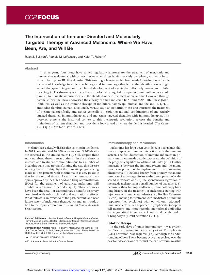
melanoma and is constitutively activated through geneticaberrations, most commonly via specific point mutations,in the greatmajority of the cases (Fig. 3). Themost commonof these genetic aberrations is mutation at the 600 positionof BRAF (V600), present in 40% to 50% of melanomas
(49, 50). Mutations of the N-isoform of RAS are found inanother 15% to 25% of cases and tend to occur at eitherposition 12 or 61 (51). NRAS and BRAF mutations aremutually exclusive (the co-occurrence rate is <<1%), lead tohyperactivation of the MAPK pathway, and are associatedwith a worse prognosis than are melanomas with wild-typeNRASandBRAF (52, 53). A loss-of-functionmutation in thetumor suppressor gene, neurofibromatosis 1 (NF1), wasrecently identified in approximately 10% to 15% of casesand also is associated with abnormal MAPK signaling (54).Whenmutated, NF1 is no longer capable of keeping RAS inits inactive RAS-GDP form and thus leads to constitutiveactivation of RAS and the pathways downstream (55).Genetic mutations or amplifications are seen in other genesleading to upregulation of theMAPKpathway aswell. Theseinclude cKIT mutations, seen in less than 1% of all mela-nomas, although in upwards of 10% to 30% of acral ormucosal melanomas, and mutations in small G-proteinsubunits called GNAQ and GNA11 that are present inmorethan 80%of ocular melanomas, though rarely seen in othermelanoma subtypes (56–58).
In addition to the oncogenicmutations that lead to hyper-activation of the MAPK pathway (BRAF, NRAS, CKIT, NF1,andGNAQ/GAQ11), anumberofother genesmaybealteredin melanoma and serve to complement the primary onco-genic mutations described above. In particular, abnormali-ties of genes involved in cell-cycle regulation, includingcyclin-dependent kinases (CDK), CDK inhibitors [such asP16Ink4a (CDKN2A)], and cyclin D are seen in more than70% of the patients (reviewed in detail by Sheppard andMcArthur in this CCR Focus section; refs. 59, 60). Also, thefunctionof thenegative regulatorof thePI3Kpathway,PTEN,is either lostor impaired inup to30%ofcases (61, 62). This ismost commonly seen in a subset of patients with BRAFmutations, thereby allowing for unregulated signaling ofboth the MAPK pathway, via BRAF mutation, and the PI3Kpathway, through PTEN loss of function (52, 63).
MAPK inhibition and resistanceWith thefirst descriptionof oncogenic BRAFmutations in
melanoma, efforts were made to identify and developclinical inhibitors of both BRAF specifically and the MAPKpathway in general. The initial targeted therapy studies inmelanoma were with agents that are now considered non-specific inhibitors of BRAF, sorafenib and RAF265, andlower potency inhibitors of MAP–ERK kinase (MEK)-1/2,such as selumetinib, PD-325901, andCI-1040 (64–72). It isimportant to note that these studies were open to anypatient with melanoma independent of mutational status.Thus, these trials were doomed for failure as the agents werenot able to inhibit the MAPK pathway sufficiently at toler-able doses, and the patients treated were not preselected toinclude only those most likely to benefit. Mechanisms ofresistance to these agents were impossible to determinegiven the fact that the pathway was suboptimally inhibitedand thus few patients received benefit.
BRAF-directed therapy in BRAF mutants. The first so-called targeted therapy to show substantial efficacy in
Immunotherapy and Molecular Targeted Therapy in Melanoma
www.aacrjournals.org Clin Cancer Res; 19(19) October 1, 2013 5285
on April 12, 2020. © 2013 American Association for Cancer Research. clincancerres.aacrjournals.org Downloaded from

melanoma was vemurafenib (73). In the initial phase Istudy, it was determined early that only patients withoncogenic (i.e, V600) mutations experienced clinical ben-efit and that almost every one of these patients had someevidence of tumor regression with treatment. Furthermore,responses occurred early with improvement of symptomswithin days and near complete 2[18F]fluoro-2-deoxy-D-glu-cose positron emission tomography (FDG-PET) responseswithin 2 weeks from the onset of therapy. A subsequentphase II study confirmed the remarkable response dynamicsand frequency of vemurafenib, and a phase III study con-firmed that vemurafenib conferred a survival advantagecompared with chemotherapy (50, 74). A second, potent,
and specific mutant BRAF inhibitor, dabrafenib, has beenassociated with very similar clinical efficacy as vemurafeniband joined it as the second BRAF inhibitor to achieve FDAapproval (75). A third such BRAF inhibitor, LGX818, hasshown responses at every dose level tested (76).
Although the advantages of BRAF inhibitor therapy arethe rapid onset and high frequency of responses, the dis-advantage is the limited durationof clinical benefit (74, 75).Specifically, BRAF inhibitor treatment is associated with aprogression-free survival (PFS) of only 5 to 7 months as aresult of the development of cellular resistance over thisrelatively short period of time. The mechanisms of thisresistance can be subdivided into those that can be
© 2013 American Association for Cancer Research
HER2
CMET
HGFHER3
Dendriticcell
Nucleus
EGFR
VEGFIGF-1RKIT INSR
RAFMEK
ERK
MART-1
gp100
TRP1/2
CDK4/6
p16INK4A
CDK2Cyclin D Cyclin E
CCR Focus
FOXM1 MYC SMAD3
MEP50/
PRMT5
RB1
E2F POL1
PI3K
PDK1
PTEN
AKT
mTORC2
RAS NF1
RAS
RALGDS
CDC42
Actin
RAL
RALBP1
SHC
PLD
RHO
-GTP
TIAM1
CDC42 RAC1
PLCα
IP3
SOS
GRB2
RIN1
ABL
MEKK
SEK
JNK
DAG
PKCCa2+
FOXO p27
p21GSK3 ΙΚΚα BAD
Caspase-9
Hexokinase GLUT-1PRAS40
PDCD4 S6 eIF4B eIF4E
MDM2
p53
TSC2
TSC1
Rheb
mTOR mTORC1
Raptor
ATG13
Autophagy
P70S6K 4EBP1 HIF-1α
mLST8
AMPK LKB1
Cyclin D1
PD-1
TCR
MHC
(-)
(+)
B7-H1
T cell
Figure 2. Molecular and immunologic signaling in melanoma. Melanoma cells use a diverse set of cell surface receptors and intracellular signalingmolecules to promote growth, cell-cycle progression, cell survival, angiogenesis, and immune evasion. HIF-1a, hypoxia-inducible factor-1a; IGF-1R,insulin-like growth factor receptor.
CCRFOCUS
Clin Cancer Res; 19(19) October 1, 2013 Clinical Cancer Research5286
on April 12, 2020. © 2013 American Association for Cancer Research. clincancerres.aacrjournals.org Downloaded from

predicted on the basis of pretreatment analysis of tumorsand those that clearly were not identified at baseline butrather developed as a result of selective pressure placedupon the tumor cells by BRAF inhibitor treatment.A number of identifiable pretreatment factors have been
described as being associated with either a poorer responseand/or a shorter PFS to BRAF inhibitor treatment. Theseinclude stromal hepatocyte growth factor (HGF) produc-tion, BCL2A1 [an antiapoptotic B-cell leukemia 2 (BCL-2)family member] expression, activation of cyclin D1, andloss of PTEN (77–81). It is interesting that each of theseexamples is associated with critical regulation of growth,survival, or cell-cycle regulation: the activation of the PI3Kpathway (stromal HGF production leads to CMET activa-tion of the pathway; PTEN loss leads to dysregulation of thepathway), resistance to apoptosis (BCL2A1), or cell-cycleprogression (cyclin D1; Fig. 2).Acquired resistance toBRAF inhibitors, definedhere as the
development of cellular resistance by a mechanism notidentified in pretreatment tumors, is associated with reacti-vationof theMAPKpathwayapproximately two thirdsof thetime (82). One of the first described mechanisms of resis-tance (MOR) to BRAF inhibitors was the upregulation ofreceptor tyrosine kinases (RTK) such as insulin-like growthfactor receptor 1 (IGF-R1), platelet-derived growth factor(PDGF), as well as HER3 that can signal through PI3K or theMAPK pathway by activating the C-isoform of RAF (83–86).Although BRAF inhibitors potently inhibit BRAFV600, amutant isoform that signals through a constitutively activekinase in a monomeric form, they paradoxically facilitate
RAFdimerization, thereby leading to activationof theMAPKpathway (87, 88). This so-called "BRAF inhibitor paradox"explains how RTK activation upstream of RAF can reactivatethe MAPK pathway through CRAF homo- or heterodimer-ization and how other MORs to BRAF inhibitor therapyactivate the pathway. For example, concomitantmutationofNRAS and BRAF is seen in more than 20% of the resistancesamples driving MAPK signaling, and an alternative splicevariant of BRAFV600E that can dimerize in the context ofBRAF mutation emerges in 20% to 25% of the resistancesamples; loss of NF1 leading to NRAS activation has alsobeendescribed (86,89, 90). In addition,otherMORs that donot rely on paradoxical activation also are seen and includeincreased expression of BRAFV600, downstream oncogenicmutation of MEK, and alternative MAPK activation (COT)leading to activation of MEK (refs. 91–94; MORs are sum-marized in Fig. 4).
MEK-directed therapy. The clinical development ofmore selective MEK1/2 inhibitors, such as trametinib andMEK162, has led to the proof of principle that MEK is alegitimate target in melanoma, both in BRAF-mutant mel-anoma and, to a lesser degree, in BRAFwild-typemelanoma(harboring NRAS or NF1 mutations; refs. 95, 96). In fact,trametinib has recently been FDA approved for the treat-ment of BRAF-mutant metastatic melanoma based on arandomized phase III study showing that treatment withtremetinib is associatedwith a survival advantage comparedwith conventional chemotherapy [response rate (RR) 22%,PFS 4.8months, 6-monthOS 81% for trametinib vs. RR 8%,PFS 1.5 months, 6-month OS 67% for chemo; ref. 96).MEK162 has also shown clinical efficacy in both BRAF- andNRAS-mutantmelanoma in a phase II study (BRAFmutants:RR 20%, PFS 3.6 months; NRAS mutants: RR 20%, PFS 3.7months; ref. 97). A phase III study is under way to explore
© 2013 American Association for Cancer Research
RTK
CKIT
G-protein coupled
receptor
BRAF
GNA
Q/11
ERK
MEK
PI3K
PTENAKT
RAS
NF1
CCR Focus
Figure 3. Oncogenic mutations in melanoma. Oncogenic mutations andmolecular aberrations are regularly present in the MAPK pathway inmelanoma and include, in order of frequency, BRAF, NRAS, PTEN loss,NF1 loss, CKIT mutation or amplification, and GNAQ/GNA11 (<1% of allmelanoma, >80% of ocular melanoma).
CCR Focus© 2013 American Association for Cancer Research
BRAFBRAF BRAF
NRAS
BRAF
BRAFBRAF MEK
PI3K
COT
CRAF
P
P
MEK
ERK
NF1
ERBB3, PDGFR,
IGFR, FGFR3
1
1B
1A
2
7
4
5
36
Figure 4. Mechanisms of acquired resistance to BRAF inhibitors: 1, RTKactivation that can signal either through CRAF (1A) or the PI3K pathway(1B); 2, concomitant NRAS mutation; 3, emergence of a truncatedBRAFV600E variant from alternative splicing; 4, concomitant MEKmutation; 5, BRAFV600 overexpression; 6, loss of NF1; 7, COT, analternative MAPK activation. ERK, extracellular signal-regulated kinase;FGFR3, fibroblast growth factor receptor 3.
Immunotherapy and Molecular Targeted Therapy in Melanoma
www.aacrjournals.org Clin Cancer Res; 19(19) October 1, 2013 5287
on April 12, 2020. © 2013 American Association for Cancer Research. clincancerres.aacrjournals.org Downloaded from

whether MEK162 is more effective than chemotherapy inNRAS-mutantmelanoma (NCT01763164). Finally, selume-tinib was shown to have modest efficacy in patients withuvealmelanoma. In a randomized, phase II study, treatmentwith selumetinib was associated with a two-fold improve-ment in PFS compared with patients who received chemo-therapy, though notably, OS was not different in the twotreatment groups (98). On the basis of the results of all ofthese studies, it seems clear thatMEK inhibition is associatedwithmodest benefit (20%RR,PFS4–5months) in subsets ofpatientswithmelanoma andwill have a role as a single agentin the treatment of this disease. The MORs of single-agentMEK inhibitor therapy have not been well elucidated.
The future of targeted therapy in melanomaIt is important to acknowledge that targeted therapy in
melanoma remains in its infancy. Only 4 years have passedsince initial clinical data with vemurafenib were presented.During this time, the collective knowledge about bothmechanisms of action and resistance of BRAF and MEKinhibitor therapy has grown nearly exponentially. As thesedata have emerged, so toohave clinical trial ideas using BRAFandMEK inhibitor therapy as the backbone to combinatorialregimens that have been rationally designed from our scien-tific understanding of how these agents change tumor cells.
The first example of this second wave of trials focusing oncombination regimens is a phase I/II combination of dabra-feniband trametinib (99). Itwaspredicted that reactivationofthe MAPK pathway would occur in the setting of BRAFinhibitors (82). Therefore, inhibition of the pathway down-streamofBRAFby targeting eitherMEKor extracellular signal-regulated kinase (ERK) was considered as an approach thatmight lead to further clinical benefit, and perhaps moreremarkably, improvement in severityof toxicity. Interestingly,the sequential administrationof aBRAF inhibitor followedbya MEK inhibitor is ineffective and exposes patients to thepotential toxicities seenwith each single agent, yet concurrenttreatment with both agents is associated with an improve-ment in RR, response depth (i.e., greater maximal response),response duration, and PFS, and a reduction in toxicityseverity (99, 100). This peculiar safety signal is based on thefact that BRAF inhibitors paradoxically activate the MAPKpathway through facilitationofRAFdimerization (87, 88). Asdiscussed above, manyMORs to BRAF inhibitors emerge as aresult of this phenomenon, however, the toxicity of BRAFinhibitors is also likely explained by this. Namely, BRAFinhibitor toxicity is likely a result of upregulation of theMAPK in nonmelanoma cells; the best-described example isthe development of squamous cell carcinomas of the skinsecondary to RAS mutations in skin cells (101). This phe-nomenon is seen at a much lower frequency with the treat-ment of BRAF-mutant melanoma with MEK inhibitors (99).Therefore, in BRAF-mutant cells, BRAF and MEK inhibitorsboth inhibit the pathway leading to augmented inhibitionbut exert differential effects on the MAPK pathway in non-BRAF–mutant cells suchassquamouscells of the skin, leadingto an attenuation of toxicity. There are now two additionalphase I combinations of BRAF plus MEK inhibitors showing
similar improvements in efficacy and abrogation of toxicityseverity (102, 103). Phase III studies of each of these combi-nations are under way (NCT01689519; NCT01597908;NCT01584648) or being planned. It is expected that overthe coming 5 years, triple and quadruple drug regimens willbe studied in the clinic to treat BRAF-mutantmelanomawiththe BRAF and MEK inhibitor combination at the core.
In NRAS-mutant melanoma, it is anticipated that twoevents will occur in the near future that will hopefully leadto the dramatic improvement in how these patients aretreated. First, a phase III study of MEK162 (NCT01763164)has been launched to determine the efficacy of this agentcompared with chemotherapy. If successful, regulatoryapproval would be expected. Second, a number ofcombination regimens are expected on the basis of preclin-ical data showing that a number of agents may augmentMEK inhibitor toxicity in patients with NRAS-mutant mel-anoma (104, 105). Two examples are the combination of aMEK inhibitor with a CDK4/6 inhibitor (NCT01781572)and a combination of a MEK inhibitor with an HDM2antagonist, though many more doublet and triplet combi-nations are expected in the near future.
Grand unification: the intersection of MAPK inhibitionand immunotherapy
When ipilimumab and vemurafenib were approved bythe FDAwithinmonths of eachother in 2011, a great deal ofadvocacy was directed toward the makers of each drug tosupport a combination trial. It turns out that there is actuallya compelling rationale to combine BRAF inhibitors withimmunotherapies that goes beyond the fact that they areboth effective in patients with melanoma. In particular,emerging evidence suggests that oncogenic BRAF is immu-nosuppressive (106, 107). Furthermore, treatment withMAPK inhibitors is associated with enhanced expressionof melanocytic antigens, antigen recognition by T cells, andan influx of CTLs (108–112). These findings offer compel-ling evidence for the development of combined targetedand immune therapies, although based on the earlyattempts at combining BRAF inhibitors with checkpointinhibitors, clinical trials may not be so simple. As anexample, the phase I trial of vemurafenib plus ipilimumabwas closed because of toxicity concerns, namely a high rateof severe hepatic toxicity (113). Still, a number of trials haveopened exploring various BRAF-directed therapies (singleagent, BRAF inhibitor plus MEK inhibitor combinations,etc.) with checkpoint inhibitors and cytokines alike. Thegreat hope is that the ideal combination will be identifiedthat will be associated with a very high rate of durableclinical response and no untoward toxicity.
Conclusions/Future DirectionsFrom the bleakness of the recent past to the great promise
of the near future, the development of melanoma thera-peutics has always relied on a strong connection with hard-core molecular biology and immunology laboratories todrive the clinical progress. This is more important than everas critical issues remain about the ideal sequences and
CCRFOCUS
Clin Cancer Res; 19(19) October 1, 2013 Clinical Cancer Research5288
on April 12, 2020. © 2013 American Association for Cancer Research. clincancerres.aacrjournals.org Downloaded from
combinations of the various agents that have proven pre-clinical and clinical efficacy.
Disclosure of Potential Conflicts of InterestK.T. Flaherty is a consultant/advisory boardmember of GlaxoSmithKline,
Roche/Genentech, and Novartis. No potential conflicts of interest weredisclosed by the other authors.
Authors' ContributionsConception and design: R.J. Sullivan, K.T. FlahertyDevelopment of methodology: R.J. Sullivan
Acquisitionofdata (provided animals, acquired andmanagedpatients,provided facilities, etc.): R.J. SullivanAnalysis and interpretation of data (e.g., statistical analysis, biosta-tistics, computational analysis): R.J. SullivanWriting, review, and/or revision of the manuscript: R.J. Sullivan, P.M.LoRusso, K.T. FlahertyAdministrative, technical, or material support (i.e., reporting or orga-nizing data, constructing databases): R.J. SullivanStudy supervision: P.M. LoRusso
Received August 4, 2013; accepted August 6, 2013; published onlineOctober 2, 2013.
References1. Siegel R,NaishadhamD, JemalA.Cancer statistics, 2013.CACancer
J Clin 2013;63:11–30.2. Clemente CG,MihmMC Jr, Bufalino R, Zurrida S, Collini P, Cascinelli
N. Prognostic value of tumor infiltrating lymphocytes in the verticalgrowth phase of primary cutaneous melanoma. Cancer 1996;77:1303–10.
3. MortonD, Eilber FR,MalmgrenRA,WoodWC. Immunological factorswhich influence response to immunotherapy inmalignant melanoma.Surgery 1970;68:158–63.
4. Tsao H, Cosimi AB, Sober AJ. Ultra-late recurrence (15 years orlonger) of cutaneous melanoma. Cancer 1997;79:2361–70.
5. Baker HW. Spontaneous regression of malignant melanoma. AmSurg 1964;30:825–9.
6. Morton DL, Eilber FR, Holmes EC, Hunt JS, Ketcham AS, SilversteinMJ, et al. BCG immunotherapyofmalignantmelanoma: summaryof aseven-year experience. Ann Surg 1974;180:635–43.
7. Kirkwood JM, Ernstoff MS. Interferons in the treatment of humancancer. J Clin Oncol 1984;2:336–52.
8. Atkins MB, Lotze MT, Dutcher JP, Fisher RI, Weiss G, Margolin K,et al. High-dose recombinant interleukin 2 therapy for patients withmetastatic melanoma: analysis of 270 patients treated between 1985and 1993. J Clin Oncol 1999;17:2105–16.
9. Rosenberg SA, Mule JJ. Immunotherapy of cancer with lymphokine-activated killer cells and recombinant interleukin-2. Surgery 1985;98:437–44.
10. Hodi FS, O'Day SJ, McDermott DF, Weber RW, Sosman JA, HaanenJB, et al. Improved survival with ipilimumab in patients with meta-static melanoma. N Engl J Med 2010;363:711–23.
11. Topalian SL, Hodi FS, Brahmer JR, Gettinger SN, Smith DC, McDer-mott DF, et al. Safety, activity, and immune correlates of anti-PD-1antibody in cancer. N Engl J Med 2012;366:2443–54.
12. Freedman LR, Cerottini JC, Brunner KT. In vivo studies of the role ofcytotoxic T cells in tumor allograft immunity. J Immunol 1972;109:1371–8.
13. Grossberg SE. The interferons and their inducers: molecular andtherapeutic considerations. 3. N Engl J Med 1972;287:122–8.
14. Grossberg SE. The interferons and their inducers: molecular andtherapeutic considerations. 2. N Engl J Med 1972;287:79–85.
15. Grossberg SE. The interferons and their inducers: molecular andtherapeutic considerations. 1. N Engl J Med 1972;287:13–9.
16. Stevens TL, Bossie A, Sanders VM, Fernandez-Botran R, CoffmanRL, Mosmann TR, et al. Regulation of antibody isotype secretion bysubsets of antigen-specific helper T cells. Nature 1988;334:255–8.
17. Sullivan RJ, Atkins MB. Cytokine therapy in melanoma. J CutanPathol 2010;37 Suppl 1:60–7.
18. Kirkwood JM, Strawderman MH, Ernstoff MS, Smith TJ, Borden EC,Blum RH. Interferon alfa-2b adjuvant therapy of high-risk resectedcutaneousmelanoma: the EasternCooperativeOncologyGroupTrialEST 1684. J Clin Oncol 1996;14:7–17.
19. Kirkwood JM, Manola J, Ibrahim J, Sondak V, Ernstoff MS, Rao U. Apooled analysis of eastern cooperative oncology group and inter-group trials of adjuvant high-dose interferon for melanoma. ClinCancer Res 2004;10:1670–7.
20. Eggermont AM, Suciu S, Santinami M, Testori A, Kruit WH, MarsdenJ, et al. Adjuvant therapy with pegylated interferon alfa-2b versus
observation alone in resected stage III melanoma: final results ofEORTC18991, a randomisedphase III trial. Lancet 2008;372:117–26.
21. Atkins MB, Kunkel L, Sznol M, Rosenberg SA. High-dose recombi-nant interleukin-2 therapy in patients with metastatic melanoma:long-term survival update. Cancer J Sci Am 2000;6 Suppl 1:S11–4.
22. Joseph RW, Sullivan RJ, Harrell R, Stemke-Hale K, Panka D, Man-oukian G, et al. Correlation of NRAS mutations with clinical responseto high-dose IL-2 in patientswith advancedmelanoma. J Immunother2012;35:66–72.
23. RosenbergSA, YangJC,SherryRM,KammulaUS,HughesMS,PhanGQ, et al. Durable complete responses in heavily pretreated patientswith metastatic melanoma using T-cell transfer immunotherapy. ClinCancer Res 2011;17:4550–7.
24. Wu R, Forget MA, Chacon J, Bernatchez C, Haymaker C, Chen JQ,et al. Adoptive T-cell therapy using autologous tumor-infiltratinglymphocytes for metastatic melanoma: current status and futureoutlook. Cancer J 2012;18:160–75.
25. Dudley ME, Wunderlich JR, Yang JC, Sherry RM, Topalian SL,Restifo NP, et al. Adoptive cell transfer therapy following non-myeloablative but lymphodepleting chemotherapy for the treat-ment of patients with refractory metastatic melanoma. J Clin Oncol2005;23:2346–57.
26. Dudley ME, Gross CA, Langhan MM, Garcia MR, Sherry RM, YangJC, et al. CD8 þenriched "young" tumor infiltrating lymphocytes canmediate regression of metastatic melanoma. Clin Cancer Res 2010;16:6122–31.
27. Johnson LA, Morgan RA, Dudley ME, Cassard L, Yang JC, HughesMS, et al. Gene therapy with human and mouse T-cell receptorsmediates cancer regression and targets normal tissues expressingcognate antigen. Blood 2009;114:535–46.
28. Zhang L, Kerkar SP, Yu Z, Zheng Z, Yang S, Restifo NP, et al.Improving adoptive T cell therapy by targeting and controlling IL-12 expression to the tumor environment. Mol Ther 2011;19:751–9.
29. ParkhurstMR, Riley JP, DudleyME, Rosenberg SA. Adoptive transferof autologous natural killer cells leads to high levels of circulatingnatural killer cells but does notmediate tumor regression. Clin CancerRes 2011;17:6287–97.
30. Yu J, Tian R, Xiu B, Yan J, Jia R, Zhang L, et al. Antitumor activity of Tcells generated from lymph nodes draining the SEA-expressingmurine B16melanoma and secondarily activated with dendritic cells.Int J Biol Sci 2009;5:135–46.
31. Kwong MM, Neyns B, Yang JC. Adoptive T-cell transfer therapy andoncogene targeted therapy for melanoma: the search for synergy.Clin Cancer Res 2013;19:5292–9.
32. Sondel PM, Bach FH. Recognitive specificity of human cytotoxic Tlymphocytes. I. Antigen-specific inhibition of human cell-mediatedlympholysis. J Exp Med 1975;142:1339–48.
33. Azuma M, Cayabyab M, Buck D, Phillips JH, Lanier LL. CD28interaction with B7 costimulates primary allogeneic proliferativeresponses and cytotoxicity mediated by small, resting T lympho-cytes. J Exp Med 1992;175:353–60.
34. Vonderheide RH, Glennie MJ. Agonistic CD40 antibodies and cancertherapy. Clin Cancer Res 2013;19:1035–43.
35. Melero I, Grimaldi AM, Perez-Gracia JL, Ascierto PA. Clinicaldevelopment of immunostimulatory monoclonal antibodies and
Immunotherapy and Molecular Targeted Therapy in Melanoma
www.aacrjournals.org Clin Cancer Res; 19(19) October 1, 2013 5289
on April 12, 2020. © 2013 American Association for Cancer Research. clincancerres.aacrjournals.org Downloaded from

opportunities for combination. Clin Cancer Res 2013;19:997–1008.
36. Brunet JF, Denizot F, Luciani MF, Roux-Dosseto M, Suzan M, MatteiMG, et al. A new member of the immunoglobulin superfamily–CTLA-4. Nature 1987;328:267–70.
37. Walunas TL, Lenschow DJ, Bakker CY, Linsley PS, Freeman GJ,Green JM, et al. CTLA-4 can function as a negative regulator of T cellactivation. Immunity 1994;1:405–13.
38. Robert C, Thomas L, Bondarenko I, O'Day S, M DJ, Garbe C, et al.Ipilimumab plus dacarbazine for previously untreated metastaticmelanoma. N Engl J Med 2011;364:2517–26.
39. Suntharalingam G, Perry MR, Ward S, Brett SJ, Castello-Cortes A,Brunner MD, et al. Cytokine storm in a phase 1 trial of the anti-CD28 monoclonal antibody TGN1412. N Engl J Med 2006;355:1018–28.
40. Pardoll DM. The blockade of immune checkpoints in cancer immu-notherapy. Nat Rev Cancer 2012;12:252–64.
41. Brahmer JR, Tykodi SS,ChowLQ,HwuWJ, TopalianSL,HwuP, et al.Safety and activity of anti-PD-L1 antibody in patients with advancedcancer. N Engl J Med 2012;366:2455–65.
42. HamidO, Robert C, DaudA, Hodi FS, HwuWJ, KeffordR, et al. Safetyand tumor responses with lambrolizumab (Anti-PD-1) in melanoma.N Engl J Med 2013;369:134–44.
43. Hamid O, Sosman J, Lawrence D, Sullivan RJ, Ibrahim N, Kluger H,et al. Clinical activity, safety, and biomarkers of MPDL3280A, anengineered PD-L1 antibody in patients with locally advanced ormetastaticmelanoma (mM). JClinOncol 31, 2013 (suppl; abstr 9010).
44. Wolchok JD, Kluger H, CallahanMK, PostowMA, Rizvi NA, LesokhinAM, et al. Nivolumab plus ipilimumab in advancedmelanoma. N EnglJ Med 2013;369:122–33.
45. Ott PA, Hodi FS, Robert C. CTLA-4 and PD-1/PD-L1 blockade: newimmunotherapeutic modalities with durable clinical benefit in mela-noma patients. Clin Cancer Res 2013;19:5300–9.
46. Sullivan RJ, Flaherty K. MAP kinase signaling and inhibition inmelanoma. Oncogene 2012;32:2373–9.
47. DaviesMA. The role of the PI3K-AKT pathway inmelanoma. Cancer J2012;18:142–7.
48. Kwong LN, Davies MA. Navigating the therapeutic complexityof PI3K pathway inhibition in melanoma. Clin Cancer Res 2013;19:5310–9.
49. Davies H, Bignell GR, Cox C, Stephens P, Edkins S, Clegg S, et al.Mutations of the BRAF gene in human cancer. Nature 2002;417:949–54.
50. Sosman JA, KimKB, Schuchter L,GonzalezR, Pavlick AC,Weber JS,et al. Survival inBRAFV600-mutant advancedmelanoma treatedwithvemurafenib. N Engl J Med 2012;366:707–14.
51. Omholt K, Platz A, Kanter L, RingborgU, Hansson J. NRAS andBRAFmutations arise early during melanoma pathogenesis and are pre-served throughout tumor progression. Clin Cancer Res 2003;9:6483–8.
52. Tsao H, Goel V, Wu H, Yang G, Haluska FG. Genetic interactionbetween NRAS and BRAFmutations and PTEN/MMAC1 inactivationin melanoma. J Invest Dermatol 2004;122:337–41.
53. Jakob JA, Bassett RL Jr, Ng CS, Curry JL, Joseph RW, Alvarado GC,et al. NRAS mutation status is an independent prognostic factor inmetastatic melanoma. Cancer 2011;118:4014–23.
54. Maertens O, Johnson B, Hollstein P, Frederick DT, Cooper ZA,Messiaen L, et al. Elucidating distinct roles for NF1 in melanomagen-esis. Cancer Discov 2013;3:338–49.
55. Gibney GT, Smalley KS. An unholy alliance: cooperation betweenBRAF and NF1 in melanoma development and BRAF inhibitor resis-tance. Cancer Discov 2013;3:260–3.
56. Curtin JA,BusamK,Pinkel D, BastianBC.Somatic activation ofKIT indistinct subtypes of melanoma. J Clin Oncol 2006;24:4340–6.
57. Van Raamsdonk CD, Bezrookove V, Green G, Bauer J, Gaugler L,O'Brien JM, et al. Frequent somatic mutations of GNAQ in uvealmelanoma and blue naevi. Nature 2009;457:599–602.
58. Van Raamsdonk CD, Griewank KG, CrosbyMB, GarridoMC, VemulaS, Wiesner T, et al. Mutations in GNA11 in uveal melanoma. N Engl JMed 2010;363:2191–9.
59. McArthur GA, Young RJ, Sheppard KE, Mar V, Waldeck K, Fox SB,et al. Clinical significance of genomic alterations of the CDK4-path-way and sensitivity to the CDK4 inhibitor PD 0332991 in melanoma.J Clin Oncol 30, 2012 (Suppl; abstr 8520).
60. Sheppard KE, McArthur GA. The cell-cycle regulator CDK4 anemerging therapeutic target in melanoma. Clin Cancer Res 2013;19:5320–8.
61. Teng DH, Hu R, Lin H, Davis T, Iliev D, Frye C, et al. MMAC1/PTENmutations in primary tumor specimens and tumor cell lines. CancerRes 1997;57:5221–5.
62. Tsao H, Zhang X, Benoit E, Haluska FG. Identification of PTEN/MMAC1 alterations in uncultured melanomas and melanoma celllines. Oncogene 1998;16:3397–402.
63. Tsao H, Zhang X, Fowlkes K, Haluska FG. Relative reciprocity ofNRAS and PTEN/MMAC1 alterations in cutaneous melanoma celllines. Cancer Res 2000;60:1800–4.
64. Hauschild A, Agarwala SS, Trefzer U, Hogg D, Robert C, Hersey P,et al. Results of a phase III, randomized, placebo-controlled study ofsorafenib in combination with carboplatin and paclitaxel as second-line treatment in patients with unresectable stage III or stage IVmelanoma. J Clin Oncol 2009;27:2823–30.
65. Flaherty KT, Lee SJ, Zhao F, Schuchter LM, Flaherty L, Kefford R,et al. Phase III trial of carboplatin and paclitaxel with or with-out sorafenib in metastatic melanoma. J Clin Oncol 2013;31:373–9.
66. Adjei AA, Cohen RB, Franklin W, Morris C, Wilson D, Molina JR, et al.Phase I pharmacokinetic and pharmacodynamic study of the oral,small-molecule mitogen-activated protein kinase kinase 1/2 inhibitorAZD6244 (ARRY-142886) in patients with advanced cancers. J ClinOncol 2008;26:2139–46.
67. Banerji U, Camidge DR, Verheul HM, Agarwal R, Sarker D, Kaye SB,et al. The first-in-human study of the hydrogen sulfate (Hyd-sulfate)capsule of the MEK1/2 inhibitor AZD6244 (ARRY-142886): a phase Iopen-label multicenter trial in patients with advanced cancer. ClinCancer Res 2010;16:1613–23.
68. Delord J, HouedeN, Awada A, TaammaA, Faivre SJ, Besse-HammerT, et al. First-in-human phase I safety, pharmacokinetic (PK), andpharmacodynamic (PD) analysis of the oral MEK-inhibitor AS703026(two regimens [R]) in patients (pts) with advanced solid tumors. J ClinOncol 28:15s, 2010 (suppl; abstr 2504).
69. Lorusso PM, Adjei AA, VarterasianM, Gadgeel S, Reid J, Mitchell DY,et al. Phase I and pharmacodynamic study of the oral MEK inhibitorCI-1040 in patients with advanced malignancies. J Clin Oncol2005;23:5281–93.
70. Boasberg PD, Redfern CH, Daniels GA, Bodkin D, Garrett CR, RicartAD. Pilot study of PD-0325901 in previously treated patients withadvanced melanoma, breast cancer, and colon cancer. Cancer Che-mother Pharmacol 2011;68:547–52.
71. LoRusso PM, Krishnamurthi SS, Rinehart JJ, Nabell LM, Malburg L,Chapman PB, et al. Phase I pharmacokinetic and pharmacodynamicstudy of the oral MAPK/ERK kinase inhibitor PD-0325901 in patientswith advanced cancers. Clin Cancer Res 2010;16:1924–37.
72. Sharfman WH, Hodi FS, Lawrence DP, Flaherty KT, Amaravadi RK,Kim KB, et al. Results from the first-in-human (FIH) phase I study ofthe oral RAF inhibitor RAF265 administered daily to patients withadvanced cutaneous melanoma. J Clin Oncol 29: 2011 (suppl; abstr8508).
73. Flaherty KT, Puzanov I, Kim KB, Ribas A, McArthur GA, Sosman JA,et al. Inhibition of mutated, activated BRAF in metastatic melanoma.N Engl J Med 2010;363:809–19.
74. Chapman PB, Hauschild A, Robert C, Haanen JB, Ascierto P, LarkinJ, et al. Improved survival with vemurafenib in melanoma with BRAFV600E mutation. N Engl J Med 2011;364:2507–16.
75. Hauschild A, Grob JJ, Demidov LV, Jouary T, Gutzmer R, MillwardM,et al. Dabrafenib in BRAF-mutated metastatic melanoma: a multi-centre, open-label, phase 3 randomised controlled trial. Lancet2012;380:358–65.
76. Dummer R, Robert C, Nyakas M, McArthur GA, Kudchadkar RR,Gomez-RocaC, et al. editors. Initial results fromaphase I, open-label,dose escalation study of the oral BRAF inhibitor LGX818 in patients
CCRFOCUS
Clin Cancer Res; 19(19) October 1, 2013 Clinical Cancer Research5290
on April 12, 2020. © 2013 American Association for Cancer Research. clincancerres.aacrjournals.org Downloaded from
with BRAF V600 mutant advanced or metastatic melanoma. J ClinOncol 31, 2013 (suppl; abstr 9028).
77. Haq R, Yokoyama S, Hawryluk EB, Jonsson GB, Frederick DT,McHenry K, et al. BCL2A1 is a lineage-specific antiapoptotic mela-noma oncogene that confers resistance to BRAF inhibition. Proc NatlAcad Sci U S A 2013;110:4321–6.
78. NathansonKL,MartinA, LetreroR,D'AndreaKP,O'DayS, Infante JR,et al. Tumor genetic analyses of patients with metastatic melanomatreated with the BRAF inhibitor GSK2118436 (GSK436). J Clin Oncol29: 2011 (suppl; abstr 8501).
79. Smalley KS, Lioni M, Dalla PalmaM, XiaoM, Desai B, Egyhazi S, et al.Increased cyclin D1 expression can mediate BRAF inhibitor resis-tance in BRAF V600E-mutated melanomas. Mol Cancer Ther 2008;7:2876–83.
80. Straussman R, Morikawa T, Shee K, Barzily-Rokni M, Qian ZR, Du J,et al. Tumour micro-environment elicits innate resistance to RAFinhibitors through HGF secretion. Nature 2012;487:500–4.
81. Wilson TR, Fridlyand J, Yan Y, Penuel E, Burton L, Chan E, et al.Widespread potential for growth-factor-driven resistance to antican-cer kinase inhibitors. Nature 2012;487:505–9.
82. Bollag G, Hirth P, Tsai J, Zhang J, Ibrahim PN, Cho H, et al. Clinicalefficacy of a RAF inhibitor needs broad target blockade in BRAF-mutant melanoma. Nature 2010;467:596–9.
83. Montagut C, Dalmases A, Bellosillo B, CrespoM, Pairet S, IglesiasM,et al. Identification of a mutation in the extracellular domain of theEpidermal Growth Factor Receptor conferring cetuximab resistancein colorectal cancer. Nat Med 2012;18:221–3.
84. MontagutC,SharmaSV, ShiodaT,McDermottU,UlmanM,Ulkus LE,et al. ElevatedCRAF as a potential mechanismof acquired resistanceto BRAF inhibition in melanoma. Cancer Res 2008;68:4853–61.
85. Villanueva J, Vultur A, Lee JT, Somasundaram R, Fukunaga-KalabisM, Cipolla AK, et al. Acquired resistance to BRAF inhibitors mediatedby aRAF kinase switch inmelanoma can be overcome by cotargetingMEK and IGF-1R/PI3K. Cancer Cell 2010;18:683–95.
86. NazarianR,ShiH,WangQ,KongX,KoyaRC, LeeH, et al.Melanomasacquire resistance to B-RAF(V600E) inhibition by RTK or N-RASupregulation. Nature 2010;468:973–7.
87. Heidorn SJ, Milagre C, Whittaker S, Nourry A, Niculescu-Duvas I,Dhomen N, et al. Kinase-dead BRAF and oncogenic RAS cooperateto drive tumor progression through CRAF. Cell 2010;140:209–21.
88. Poulikakos PI, Zhang C, Bollag G, Shokat KM, Rosen N. RAFinhibitors transactivate RAF dimers and ERK signalling in cells withwild-type BRAF. Nature 2010;464:427–30.
89. Whittaker SR, Theurillat JP, Van Allen E, Wagle N, Hsiao J, CowleyGS, et al. A genome-scale RNA interference screen implicates NF1loss in resistance to RAF inhibition. Cancer Discov 2013;3:350–62.
90. PoulikakosPI, PersaudY, JanakiramanM,KongX,NgC,MoriceauG,et al. RAF inhibitor resistance is mediated by dimerization of aber-rantly spliced BRAF(V600E). Nature 2011;480:387–90.
91. Johannessen CM, Boehm JS, Kim SY, Thomas SR, Wardwell L,Johnson LA, et al. COT drives resistance to RAF inhibition throughMAP kinase pathway reactivation. Nature 2010;468:968–72.
92. Shi H,MoriceauG, Kong X, LeeMK, LeeH, KoyaRC, et al. Melanomawhole-exome sequencing identifies (V600E)B-RAF amplification-mediated acquired B-RAF inhibitor resistance. Nat Commun 2012;3:724.
93. Wagle N, Emery C, Berger MF, Davis MJ, Sawyer A, Pochanard P,et al. Dissecting therapeutic resistance toRAF inhibition inmelanomaby tumor genomic profiling. J Clin Oncol 2011;29:3085–96.
94. Wang H, Daouti S, Li WH, Wen Y, Rizzo C, Higgins B, et al. Identi-fication of the MEK1(F129L) activating mutation as a potential mech-anism of acquired resistance to MEK inhibition in human cancerscarrying the B-RafV600E mutation. Cancer Res 2011;71:5535–45.
95. Ascierto PA, Simeone E, Giannarelli D, Grimaldi AM, Romano A,Mozzillo N. Sequencing of BRAF inhibitors and ipilimumab in patientswith metastatic melanoma: a possible algorithm for clinical use.J Transl Med 2012;10:107.
96. Flaherty KT, Robert C, Hersey P, Nathan P, Garbe C, MilhemM, et al.Improved survival with MEK inhibition in BRAF-mutated melanoma.N Engl J Med 2012;367:107-14.
97. Ascierto PA, Schadendorf D, Berking C, Agarwala SS, van HerpenCM, Queirolo P, et al. MEK162 for patients with advanced mel-anoma harbouring NRAS or Val600 BRAF mutations: a non-randomised, open-label phase 2 study. Lancet Oncol 2013;14:249–56.
98. Carvajal RD, Sosman JA, Quevedo F, Milhem M, Joshua AM, Kud-chadkar RR, et al. Phase II study of selumetinib (sel) versus temo-zolomide (TMZ) in gnaq/Gna11 (Gq/11)mutant (mut) uvealmelanoma(UM). J Clin Oncol 31, 2013 (suppl; abstr CRA9003).
99. Flaherty KT, Infante JR, Daud A, Gonzalez R, Kefford RF, Sosman J,et al. Combined BRAF and MEK inhibition in melanoma with BRAFV600 mutations. N Engl J Med 2012;367:1694–703.
100. Kim KB, Kefford R, Pavlick AC, Infante JR, Ribas A, Sosman JA,et al. Phase II study of the MEK1/MEK2 inhibitor Trametinib inpatients with metastatic BRAF-mutant cutaneous melanoma pre-viously treated with or without a BRAF inhibitor. J Clin Oncol 2013;31:482–9.
101. Su F, Viros A, Milagre C, Trunzer K, Bollag G, Spleiss O, et al. RASmutations in cutaneous squamous-cell carcinomas in patients trea-ted with BRAF inhibitors. N Engl J Med 2012;366:207–15.
102. Gonzalez R, Ribas A, DaudA, Pavlick A, Gajewski TF, Puzanov I, et al.Phase IB Study of Vemurafenib in Combination with the MEK inhib-itor, GDC-0973, in Patients (pts) with Unresectable or MetastaticBRAFV600MutatedMelanoma (BRIM7) [abstract]. In: Proceedings ofthe 37th ESMO Congress; 2012 Sep 28–Oct 2; Vienna, Australia.Lugano, Switzerland: ESMO; 2012. Abstract nr 2744.
103. Kefford R, Miller WH Jr, Tan DS-W, Sullivan RJ, Long GV, TaiWMD, et al. Preliminary results from a phase Ib/II, open-label,dose-escalation study of the oral BRAF inhibitor LGX818 in com-bination with the oral MEK1/2 inhibitor MEK162 in BRAF V600-dependent advanced solid tumors.J Clin Oncol 31, 2013 (suppl;abstr 9029).
104. Kwong LN, Costello JC, Liu H, Jiang S, Helms TL, Langsdorf AE, et al.Oncogenic NRAS signaling differentially regulates survival and pro-liferation in melanoma. Nat Med 2012;18:1503–10.
105. Ji Z, Njauw CN, Taylor M, Neel V, Flaherty KT, Tsao H. p53 rescuethrough HDM2 antagonism suppresses melanoma growth andpotentiates MEK inhibition. J Invest Dermatol 2012;132:356–64.
106. Sumimoto H, Imabayashi F, Iwata T, Kawakami Y. The BRAF-MAPKsignaling pathway is essential for cancer-immune evasion in humanmelanoma cells. J Exp Med 2006;203:1651–6.
107. Khalili JS, Liu S, Rodriguez-Cruz TG,WhittingtonM,Wardell S, Liu C,et al. Oncogenic BRAF(V600E) promotes stromal cell-mediatedimmunosuppression via induction of interleukin-1 in melanoma. ClinCancer Res 2012;18:5329–40.
108. Boni A, Cogdill AP, Dang P, Udayakumar D, Njauw CN, Sloss CM,et al. Selective BRAFV600E inhibition enhances T-cell recognition ofmelanoma without affecting lymphocyte function. Cancer Res 2010;70:5213–9.
109. Frederick DT, Piris A, Cogdill AP, Cooper ZA, Lezcano C, FerroneCR, et al. BRAF inhibition is associated with enhanced melanomaantigen expression and a more favorable tumor microenvironmentin patients with metastatic melanoma. Clin Cancer Res 2013;19:1225–31.
110. Donia M, Fagone P, Nicoletti F, Andersen RS, Hogdall E, Straten PT,et al. BRAF inhibition improves tumor recognition by the immunesystem: potential implications for combinatorial therapies againstmelanoma involving adoptive T-cell transfer. Oncoimmunology 2012;1:1476–83.
111. LiuC, PengW, XuC, LouY, ZhangM,Wargo JA, et al. BRAF inhibitionincreases tumor infiltration by T cells and enhances the antitumoractivity of adoptive immunotherapy in mice. Clin Cancer Res2013;19:393–403.
112. Wilmott JS, Long GV, Howle JR, Haydu LE, Sharma RN, Thomp-son JF, et al. Selective BRAF inhibitors induce marked T-cellinfiltration into human metastatic melanoma. Clin Cancer Res2012;18:1386–94.
113. Ribas A, Hodi FS, Callahan M, Konto C, Wolchok J. Hepatotoxicitywith combination of vemurafenib and ipilimumab. N Engl J Med2013;368:1365–6.
Immunotherapy and Molecular Targeted Therapy in Melanoma
www.aacrjournals.org Clin Cancer Res; 19(19) October 1, 2013 5291
on April 12, 2020. © 2013 American Association for Cancer Research. clincancerres.aacrjournals.org Downloaded from

2013;19:5283-5291. Clin Cancer Res Ryan J. Sullivan, Patricia M. LoRusso and Keith T. Flaherty Will BeTherapy in Advanced Melanoma: Where We Have Been, Are, and The Intersection of Immune-Directed and Molecularly Targeted
Updated version
http://clincancerres.aacrjournals.org/content/19/19/5283
Access the most recent version of this article at:
Cited articles
http://clincancerres.aacrjournals.org/content/19/19/5283.full#ref-list-1
This article cites 103 articles, 43 of which you can access for free at:
Citing articles
http://clincancerres.aacrjournals.org/content/19/19/5283.full#related-urls
This article has been cited by 5 HighWire-hosted articles. Access the articles at:
E-mail alerts related to this article or journal.Sign up to receive free email-alerts
Subscriptions
Reprints and
To order reprints of this article or to subscribe to the journal, contact the AACR Publications Department at
Permissions
Rightslink site. Click on "Request Permissions" which will take you to the Copyright Clearance Center's (CCC)
.http://clincancerres.aacrjournals.org/content/19/19/5283To request permission to re-use all or part of this article, use this link
on April 12, 2020. © 2013 American Association for Cancer Research. clincancerres.aacrjournals.org Downloaded from