CCK1- und CCK2- Rezeptoren werden auf pankreatischen...
27
UNIVERSITÄTSKLINIKUM HAMBURG-EPPENDORF Zentrum für Innere Medizin I. Medizinische Klinik und Poliklinik Direktor: Professor Dr. Ansgar W. Lohse CCK1- und CCK2- Rezeptoren werden auf pankreatischen Sternzellen exprimiert und induzieren die Kollagensynthese Dissertation zur Erlangung des Grades eines Doktors der Medizin an der Medizinischen Fakultät der Universität Hamburg. vorgelegt von: Oliver Walter Seiz aus Reutlingen Hamburg 2011
Transcript of CCK1- und CCK2- Rezeptoren werden auf pankreatischen...

UNIVERSITÄTSKLINIKUM HAMBURG-EPPENDORF
Zentrum für Innere MedizinI. Medizinische Klinik und Poliklinik
Direktor: Professor Dr. Ansgar W. Lohse
CCK1- und CCK2- Rezeptoren werden auf pankreatischen Sternzellen exprimiert und induzieren die Kollagensynthese
Dissertation
zur Erlangung des Grades eines Doktors der Medizinan der Medizinischen Fakultät der Universität Hamburg.
vorgelegt von:
Oliver Walter Seiz
aus Reutlingen
Hamburg 2011

Angenommen von der Medizinischen Fakultät der Universität Hamburg am: 25.09.2012
Veröffentlicht mit Genehmigung der Medizinischen Fakultät der Universität Hamburg.
Prüfungsausschuss, der/die Vorsitzende: PD Dr. M. Bläker
Prüfungsausschuss, zweite/r Gutachter/in: PD Dr. M. Bockhorn
Prüfungsausschuss, dritte/r Gutachter/in: PD Dr. A. Pace2
Inhaltsverzeichnis
1. Publikation 4
2. Darstellung der Publikation 14
2.1. Einführung 14
2.1.1.Übersicht zur chronischen Pankreatitis 14
2.1.2.Rolle der pankreatischen Sternzellen an der Entstehung der chronischen Pankreatitis / dem Ablauf der Entzündung 14
2.1.3.Rolle des Cholecystokinin 15
2.1.4.Ziel der Arbeit 16
2.2. Zusammenfassung der Versuche und Ergebnisse 16
2.2.1.Expression von CCK1- und CCK2- Rezeptoren auf Sternzellen 16
2.2.2.Proliferation und Kollagenproduktion in pankreatischen Sternzellen nach CCK-Stimulation 17
2.2.3.Profibrogene Signaltransduktionswege nach CCK-Stimulation 20
2.2.4.Einfluss von Akt, ERK und Src auf Kollagensynthese und Proliferation 20
2.2.5.Einfluss der Rezeptortypen auf Akt Aktivierung und Kollagenproduktion 21
2.2.6.Zusammenfassung 22
2.2.7.Literaturverzeichnis 23
3. Danksagung 25
4. Lebenslauf 26
5. Eidesstattliche Versicherung 27
3

1. Publikation
CCK1 and CCK2 Receptors Are Expressed on PancreaticStellate Cells and Induce Collagen ProductionReceived for publication, March 21, 2010, and in revised form, August 11, 2010 Published, JBC Papers in Press, September 15, 2010, DOI 10.1074/jbc.M110.125534
Marc J. Berna1, Oliver Seiz1, Jan Friso Nast, Daniel Benten, Michael Blaker, Johannes Koch, Ansgar W. Lohse,and Andrea Pace2
From the Universitatsklinikum Eppendorf, Medizinische Klinik I, 20246 Hamburg, Germany
The gastrointestinal hormone cholecystokinin (CCK) caninduce acute pancreatitis in rodents through its action on acinarcells. Treatment with CCK, in combination with other agents,represents the most commonly used model to induce experi-mental chronic pancreatitis. Pancreatic stellate cells (PSC) areresponsible for pancreatic fibrosis and therefore play a predom-inant role in the genesis of chronic pancreatitis. However, it isnot knownwhether PSC express CCK receptors. Using real timePCR techniques, we demonstrate that CCK1 and CCK2 recep-tors are expressed on rat PSC. Interestingly both CCK and gas-trin significantly induced type I collagen synthesis. Moreover,both inhibit proliferation. These effects are comparable withTGF-!-stimulated PSC. Furthermore, the natural agonists CCKand gastrin induce activation of pro-fibrogenic pathways Akt,ERK, and Src.Using specificCCK1 andCCK2 receptor (CCK2R)inhibitors, we found that Akt activation is mainly mediated byCCK2R.Akt activationbyCCKandgastrin could be inhibited bythe PI3K inhibitor wortmannin. Activation of ERK and thedownstream target Elk-1 could be inhibited by the MEK inhibi-tor U0126. These data suggest that CCK and gastrin have directactivating effects on PSC, are able to induce collagen synthesisin these cells, and therefore appear to be important regulators ofpancreatic fibrogenesis. Furthermore, similar to TGF-!, bothCCK and gastrin inhibit proliferation in PSC.
Acute and chronic pancreatitis are responsible for a signifi-cant morbidity and mortality. Although studies have shownthat pancreatic acinar cells activate the mechanisms leading toinflammation and organ destruction in acute pancreatitis (1),another cell type, called pancreatic stellate cells (PSC),3 play apivotal role in the process leading to pancreatic fibrosis andchronic pancreatitis (2). These fibroblast-like cells representonly a minority of cells in the normal pancreas. Growth factors,G-protein-coupled receptors, and toxins activate PSC, leadingto proliferation, migration, and production of extracellularmatrix constituents including collagen, thereby inducing theorgan changes characteristic of chronic pancreatitis (3–8).These cellular effects are mediated by central signal transduc-tion cascades including Akt, ERK, and Src. The gastrointestinalhormone cholecystokinin (CCK) was among the first gastroin-
testinal hormones discovered. CCK binds to two receptors(CCK1R and CCK2R) that are expressed in many tissues,including the exocrine pancreas (9). The stimulation of CCKreceptors by the natural agonists CCK (comparable affinity forCCK1R and CCK2R) and gastrin (1000-fold higher affinity forCCK2R) regulate many physiological and pathophysiologicalprocesses (9). Most notably, CCK induces experimental acutepancreatitis in rodents, a process mediated by the CCK1Rexpressed on pancreatic acinar cells. The pathophysiology ofCCK-induced acute pancreatitis and the underlying signaltransduction pathways have been extensively studied in wholeanimals and isolated acinar cells in the last two decades (10).Repeated administration of CCK, or its agonist cerulein, wasused inmany studies to induce different aspects of chronic pan-creatitis, including activation of PSC and fibrosis. In these stud-ies, it had been assumed that chronic pancreatitis is the result ofrepeated organ inflammation caused by multiple inductions ofacute pancreatitis by CCK. Moreover, some studies found thatpatients suffering from chronic pancreatitis have higher plasmaCCK levels than healthy controls (11–13) and this observationwas the rationale of a clinical study of CCK-antagonist loxiglu-mide in chronic pancreatitis (14). Despite these experimentaland clinical findings pointing to a role of CCK in chronic pan-creatitis, surprisingly, no study has considered the possibility ofa direct effect of CCK or CCK agonists on PSC. Even theexpression of CCK receptors has not been studied on PSC.To systemically analyze the possible direct role of CCK andCCK receptors in the pathophysiology of chronic pancreati-tis, we studied the expression of CCK1R and CCK2R on PSC,as well as the effect of stimulation of these receptors oncollagen production, proliferation, and on pro-fibrogenicsignal transduction pathways.
EXPERIMENTAL PROCEDURES
Materials—Male Sprague-Dawley rats (150–250 g) wereobtained from Charles River Laboratories (Sulzfeld, Germany).Akt Ser(P)473, anti-caspase 3, Src family (Tyr416), ERK (Thr202/Tyr204), U0126, and ELK-pS383were fromCell SignalingTech-nology (Beverly, MA). Anti-goat horseradish peroxidase (HRP)conjugate, anti-actin, anti-p21 Ser146, anti-p21, anti-p27, andanti-rabbit HRP-conjugate antibodies were from Santa CruzBiotechnology (Santa Cruz, CA). Anti-p27 Ser10 was fromAbcam (Cambridge, UK). Collagen antibody was from South-ern Biotech (Birmingham, AL). !-SMA antibody was fromDako (Denmark). Chamber slides and anti-PY20 antibodywerepurchased from BD Biosciences (San Jose, CA). L-364,718 andLY288513 were from Tocris (Ellisville, MO). Wortmannin,
1 Both authors contributed equally to this work.2 To whom correspondence should be addressed. Tel.: 49-40-741056919;
E-mail: [email protected] The abbreviations used are: PSC, pancreatic stellate cell; CCK, cholecystoki-
nin; !-SMA, smooth muscle actin; MTT, 3-(4,5-dimethylthiazol-2-yl)-2,5-di-phenyltetrazolium bromide; DMSO, dimethyl sulfoxide.
THE JOURNAL OF BIOLOGICAL CHEMISTRY VOL. 285, NO. 50, pp. 38905–38914, December 10, 2010© 2010 by The American Society for Biochemistry and Molecular Biology, Inc. Printed in the U.S.A.
DECEMBER 10, 2010 • VOLUME 285 • NUMBER 50 JOURNAL OF BIOLOGICAL CHEMISTRY 38905
at ZENTRUM FUER M
OLEKULARE, on Decem
ber 25, 2010www.jbc.org
Downloaded from
4

TGF-! RI kinase inhibitor VIII, and PI3K" inhibitor II werefrom Calbiochem (Nottingham, UK). Iscove was from Bio-chrome AG (Berlin, Germany). SDS and ammonium sulfatewere from Bio-Rad. Tris, acrylamide (40%), and glycin werefromCarl Roth (Karlsruhe, Germany). Protein ladder was fromFermentas (Burlington, Canada). Agarose G, BCA proteinassay, and SuperSignal West Dura were from Fisher Scientific.Films were from Amersham Biosciences. Donkey anti-goatAlexa Fluor 488, donkey anti-mouse Alexa Fluor 596, rabbitanti-goat Alexa Fluor 596, donkey anti-mouse Alexa Fluor 488,Hanks’ balanced salt solution, FCS, PenStrep, NEAS, Dulbec-co’s PBS, and ETDA were from Invitrogen. Cell culture disheswere from Sarstedt (Numbrecht, Germany). Protease, HEPES,benzamidine, orthovanadate, leupeptin, PMSF, and aprotininwere fromSigma. Protran nitrocellulosemembranes were fromWhatman. DNase and collagenase were from Roche AppliedScience. CCK1R andCCK2R antibodies were fromEverest Bio-tech (Oxfordshire, UK).Animal Care—Animals were cared according to the guide-
lines and under supervision of the local animal welfare board.Tissue Preparation—Pancreatic stellate cells were isolated
using intraductal injection of enzyme solution followed by cellisolation and concentration of stellate cells by gradient centri-fugation as reported previously (15, 16). Briefly, the commonbile ductwas intubated via the papilla using a 24-French syringeand 8 ml of an enzyme mixture (800 mg/liter of collagenase P,400 mg/liter of Protease, and 200 mg/liter of DNase). Pancre-ases were excised injected with enzymes and cut into pieces.The tissue was disrupted by re-suspension in new enzymes andincubation at 37 °C for 10min. Finally, cells were isolated usinga 150-#mmesh. Isolated cells were subjected to Histodenz gra-dient centrifugation as described previously to isolate pancre-atic stellate cells. Stellate cells were cultured for 7 days, thensubjected to stimulants as described.Stellate Cell Lysis—After incubation with stimulants and/or
inhibitors, stellate cells were lysed in lysis buffer (0.5 mM NaF,0.2 mM EDTA, 10% glycerol, 10 mM benzamidine, 0.2 mMsodium orthovanadate). Lysates were centrifuged at 10,000 ! gfor 15 min at 4 °C, and protein concentration was measuredusing the BCA reagent. Equal amounts of samples were ana-lyzed by SDS-PAGE and Western blotting.Immunoprecipitation—After cell lysis, equal amounts of
samples were incubated overnight with agarose G and 4 #g ofthe specific antibody diluted in lysis buffer. Then samples werecentrifuged, washed three times, and analyzed by SDS-PAGEand Western blotting.Western Blotting—Western blotting was performed as de-
scribed previously.Whole cell lysates were subjected to SDS-PAGE using Tris
glycine gels. After electrophoresis, proteins were transferred tonitrocellulose membranes. Membranes were blocked in block-ing buffer (50 mM Tris/HCl, pH 8.0, 2 mM CaCl2, 80 mM NaCl,0.05% Tween 20, 5% nonfat dry milk) at room temperature for1 h. Membranes were incubated overnight with primary anti-body, washed three times in washing buffer (50 mM Tris/HCl,pH 8.0, 2 mM CaCl2, 80 mM NaCl, 0.05% Tween 20) for 5 min,and incubatedwithHRP-conjugated secondary antibody for 1 hat room temperature. Membranes were washed again three
times in washing buffer for 5 min, incubated with chemilumi-nescense detection reagents and finally exposed to film. Theintensity of the protein bands wasmeasured using ImageJ anal-ysis. When reprobing was necessary, membranes were incu-bated in Stripping buffer (Pierce) for 30 min at room tempera-ture, washed twice for 10min in washing buffer, blocked for 1 hin blocking buffer at room temperature, and re-probed asdescribed above.Immunocytochemistry—To detect CCK1R and CCK2R in
isolated PSC, cells were grown on glass chamber slides, fixedwith acetone, blocked in 2% rabbit-PBS, and immuno-stained. The sequence was 1:500 CCK1R and CCK2R, andrabbit anti-goat Alexa Fluor 488 at 1:200. $-SMA was coun-terstained using donkey anti-mouse Alexa Fluor 596. Todetect the effects of CCK and gastrin on PSC, cells weregrown on glass chamber slides. After serum starvation, cellswere stimulated for 24 h with no additions, 1 nM CCK, or1 #M gastrin (stimulants were replaced after 12 h). Then cellswere fixed in acetone and immunostained. Staining se-quence for collagen type I was goat anti-collagen I (1:50) anddonkey anti-goat Alexa Fluor 488 (1:200). Staining for the$-SMA sequence was mouse anti-$-SMA (1:100) and don-key anti-goat Alexa Fluor 594 (1:200). Nuclei were counter-stained with bisbenzimide. Pictures were taken using theIPLab3 software. For the collagen experiments we appliedthe same exposure time and magnification.Immunohistology—To detect CCK1R andCCK2R in rat pan-
creatic tissue, we snap froze pancreatic tissue in liquid nitrogen.5-#m slides were then fixed in acetone, blocked in 2% rabbitPBS, and immunostained. Staining sequence was 1:500 CCK1Rand CCK2R, and 1:200 for $-SMA. Secondary antibodies wererabbit anti-goat Alexa Fluor 596 for the CCK receptors anddonkey anti-mouse Alexa Fluor 488 for $-SMA. Nuclei werecounterstained using bisbenzimide.All micrographs were taken using a Leica DM LB fluores-
cence microscope equipped with a Retiga I300 camera. Weacquired the pictures using the IPLab3 software.ELISA—Indirect ELISA was performed essentially as de-
scribed by Kordes et al. (35). Stellate cells were grown in12-well plates until confluence. After serum starvation, cellswere stimulated for 24 h in serum-free medium containing50 #g/ml of ascorbic acid (Sigma) with no additions, 2 ng/mlof TGF!1 (Peprotech, Hamburg, Germany), 1 nMCCK, and 1#M gastrin. Stimulants were replaced after 12 h. The super-natants were collected and diluted (1:4) in PBS. 50 #l/wellswere used to coat Microtiter plates (Nunc Maxisorp, Lan-genselbold, Germany) overnight at 4 °C. Plates were blockedwith 5% milk powder in PBS/T for 1 h. Plates were washedand incubated with anti-type I collagen for 2 h. Subse-quently, the secondary anti-goat HRP antibody was used.Unbound secondary antibody was washed away and 100#l/well of tetramethylbenzidine substrate (TMB Plus,Kementec, Taestrup, Denmark) were added. Enzymaticreaction was stopped by adding 50 #l/well of 0.2 N H2SO4.Plates were read on a standard plate reader at 540 nm. Puri-fied type I collagen (Serva, Heidelberg, Germany) served asthe control.
CCK1R and CCK2R in Cells and Collagen Production
38906 JOURNAL OF BIOLOGICAL CHEMISTRY VOLUME 285 • NUMBER 50 • DECEMBER 10, 2010
at ZENTRUM FUER M
OLEKULARE, on Decem
ber 25, 2010www.jbc.org
Downloaded from
5

Thymidine Uptake—Rat PSC were isolated and 50,000 cells/well were seeded on 12-well plates. When reaching 60% con-fluence, cellswere serumstarved and then stimulated for 24 h inserum-free medium containing 2 !Ci/ml of [3H]thymidine(PerkinElmer Life Sciences) with no additions, 2 ng/ml of TGF,1 nM CCK, and 1 !M gastrin. After 24 h cells were harvested(Brandel Harvester, Unterfohring, Germany) and [3H]thymi-dine incorporation was measured (Hidex Plate Chameleon,Straubenhardt, Germany).
MTT Assay—Rat PSC were iso-lated and cultured. Cells weretrypsinized and 100,000 cells/wellwere seeded in 6-well plates. Whenreaching subconfluence, cells wereserum-starved and stimulated withno additions, 2 ng/ml of TGF, 1 nMCCK, or 1 !M gastrin for 24 h. Stim-ulating factors were replaced after12 h. Thereafter, 100!g/ml ofMTT(Sigma) were added and cells wereincubated for 2 h. Supernatantswere removed and 250 !l/well ofDMSO (Sigma)was added. Reactionproducts were transferred tomicro-titer plates and read in a standardmicroplate reader at 570 nm.Statistical Analysis—Data are
presented as mean ! S.E. and wereanalyzed using the Student’s t testfor unpaired data using the Prismsoftware (GraphPad). p values "0.05were considered significant.
RESULTS
Rat PSC Express CCK1 and CCK2Receptors—The presence of mRNAfor CCK1 and CCK2 receptors wasassessed by RT-PCR in PSC cul-tured for 7 days. According to themanufacturer of the primers used,specific bands of 118 (CCK1R) and93 bp (CCK2R) were expected. Asshown in Fig. 1A, expression ofCCK1R mRNA was found in PSC(lane 1), but absent in the genomicDNA (lane 2) and negative controlusing water (lane 3). Similarly,CCK2R mRNA was found in PSC(lane 4), whereas the correspondingcontrols using genomic DNA (lane5) or water (lane 6) did not show acorresponding band. We concludethat mRNA for both CCK1R andCCK2R is expressed in rat PSC.Furthermore, the presence of
both receptors was confirmed byimmunohistology (Fig. 1B) and im-munocytochemistry (Fig. 1C).
"-SMA as a tissue-specific marker was used to confirm local-ization on PSC. Snap frozen rat brain sections served as positivecontrol for the antibodies.CCK Causes Collagen Production in PSC—Upon stimulation
by growth factors and severalGprotein-coupled receptors, PSChave been reported to produce extracellular matrix proteinsincluding type I collagen, leading to pancreatic fibrosis, a con-stant feature of chronic pancreatitis. To date TGF-# has beenthemost potent fibrogenic stimulus described in PSC.To assess
FIGURE 1. CCK2 and CCK1 receptors are expressed on rat PSC. A, rat PSC were cultured for 7 days, RNA wasisolated, transcribed, and samples submitted to RT-PCR. Negative controls (genomic DNA and water) wereincluded. Expected product sizes are 93 (CCK2R) and 118 bp (CCK1R). A representative experiment of a total of3 independent experiments is shown. B, rat pancreatic tissue was snap frozen, fixed in acetone, and 5-!msections were immunostained. Shown is a merged image of the CCK1R/CCK2R-stained tissue (red), counter-stained with "-SMA (green). Magnification, #200. C, rat PSC were cultured, trypsinized, and plated on glasschamber slides. Cells were acetone fixed and immunostained for CCK1R/CCK2R and "-SMA. Magnification,#200.
CCK1R and CCK2R in Cells and Collagen Production
DECEMBER 10, 2010 • VOLUME 285 • NUMBER 50 JOURNAL OF BIOLOGICAL CHEMISTRY 38907
at ZENTRUM FUER M
OLEKULARE, on Decem
ber 25, 2010www.jbc.org
Downloaded from
6
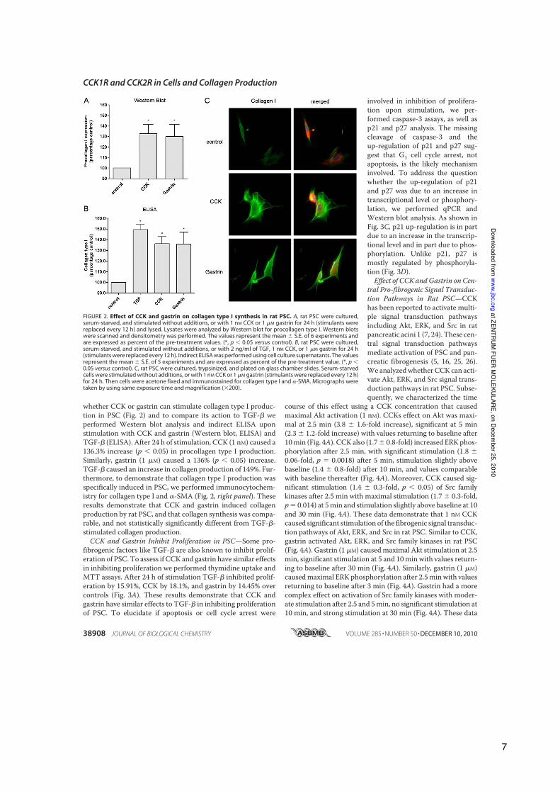
whether CCK or gastrin can stimulate collagen type I produc-tion in PSC (Fig. 2) and to compare its action to TGF-! weperformed Western blot analysis and indirect ELISA uponstimulation with CCK and gastrin (Western blot, ELISA) andTGF-! (ELISA). After 24 h of stimulation, CCK (1 nM) caused a136.3% increase (p ! 0.05) in procollagen type I production.Similarly, gastrin (1 "M) caused a 136% (p ! 0.05) increase.TGF-! caused an increase in collagen production of 149%. Fur-thermore, to demonstrate that collagen type I production wasspecifically induced in PSC, we performed immunocytochem-istry for collagen type I and #-SMA (Fig. 2, right panel). Theseresults demonstrate that CCK and gastrin induced collagenproduction by rat PSC, and that collagen synthesis was compa-rable, and not statistically significantly different from TGF-!-stimulated collagen production.CCK and Gastrin Inhibit Proliferation in PSC—Some pro-
fibrogenic factors like TGF-! are also known to inhibit prolif-eration of PSC. To assess if CCK and gastrin have similar effectsin inhibiting proliferation we performed thymidine uptake andMTT assays. After 24 h of stimulation TGF-! inhibited prolif-eration by 15.91%, CCK by 18.1%, and gastrin by 14.45% overcontrols (Fig. 3A). These results demonstrate that CCK andgastrin have similar effects to TGF-! in inhibiting proliferationof PSC. To elucidate if apoptosis or cell cycle arrest were
involved in inhibition of prolifera-tion upon stimulation, we per-formed caspase-3 assays, as well asp21 and p27 analysis. The missingcleavage of caspase-3 and theup-regulation of p21 and p27 sug-gest that G1 cell cycle arrest, notapoptosis, is the likely mechanisminvolved. To address the questionwhether the up-regulation of p21and p27 was due to an increase intranscriptional level or phosphory-lation, we performed qPCR andWestern blot analysis. As shown inFig. 3C, p21 up-regulation is in partdue to an increase in the transcrip-tional level and in part due to phos-phorylation. Unlike p21, p27 ismostly regulated by phosphoryla-tion (Fig. 3D).Effect of CCK andGastrin on Cen-
tral Pro-fibrogenic Signal Transduc-tion Pathways in Rat PSC—CCKhas been reported to activate multi-ple signal transduction pathwaysincluding Akt, ERK, and Src in ratpancreatic acini 1 (7, 24). These cen-tral signal transduction pathwaysmediate activation of PSC and pan-creatic fibrogenesis (5, 16, 25, 26).We analyzedwhether CCK can acti-vate Akt, ERK, and Src signal trans-duction pathways in rat PSC. Subse-quently, we characterized the time
course of this effect using a CCK concentration that causedmaximal Akt activation (1 nM). CCKs effect on Akt was maxi-mal at 2.5 min (3.8 " 1.6-fold increase), significant at 5 min(2.3 " 1.2-fold increase) with values returning to baseline after10min (Fig. 4A). CCK also (1.7" 0.8-fold) increased ERKphos-phorylation after 2.5 min, with significant stimulation (1.8 "0.06-fold, p # 0.0018) after 5 min, stimulation slightly abovebaseline (1.4 " 0.8-fold) after 10 min, and values comparablewith baseline thereafter (Fig. 4A). Moreover, CCK caused sig-nificant stimulation (1.4 " 0.3-fold, p ! 0.05) of Src familykinases after 2.5 min with maximal stimulation (1.7 " 0.3-fold,p# 0.014) at 5min and stimulation slightly above baseline at 10and 30 min (Fig. 4A). These data demonstrate that 1 nM CCKcaused significant stimulation of the fibrogenic signal transduc-tion pathways of Akt, ERK, and Src in rat PSC. Similar to CCK,gastrin activated Akt, ERK, and Src family kinases in rat PSC(Fig. 4A). Gastrin (1 "M) causedmaximal Akt stimulation at 2.5min, significant stimulation at 5 and 10min with values return-ing to baseline after 30 min (Fig. 4A). Similarly, gastrin (1 "M)causedmaximal ERK phosphorylation after 2.5minwith valuesreturning to baseline after 3 min (Fig. 4A). Gastrin had a morecomplex effect on activation of Src family kinases with moder-ate stimulation after 2.5 and 5min, no significant stimulation at10 min, and strong stimulation at 30 min (Fig. 4A). These data
FIGURE 2. Effect of CCK and gastrin on collagen type I synthesis in rat PSC. A, rat PSC were cultured,serum-starved, and stimulated without additions, or with 1 nM CCK or 1 "M gastrin for 24 h (stimulants werereplaced every 12 h) and lysed. Lysates were analyzed by Western blot for procollagen type I. Western blotswere scanned and densitometry was performed. The values represent the mean " S.E. of 6 experiments andare expressed as percent of the pre-treatment values. (*, p ! 0.05 versus control). B, rat PSC were cultured,serum-starved, and stimulated without additions, or with 2 ng/ml of TGF, 1 nM CCK, or 1 "M gastrin for 24 h(stimulants were replaced every 12 h). Indirect ELISA was performed using cell culture supernatants. The valuesrepresent the mean " S.E. of 5 experiments and are expressed as percent of the pre-treatment value. (*, p !0.05 versus control). C, rat PSC were cultured, trypsinized, and plated on glass chamber slides. Serum-starvedcells were stimulated without additions, or with 1 nM CCK or 1 "M gastrin (stimulants were replaced every 12 h)for 24 h. Then cells were acetone fixed and immunostained for collagen type I and #-SMA. Micrographs weretaken by using same exposure time and magnification ($200).
CCK1R and CCK2R in Cells and Collagen Production
38908 JOURNAL OF BIOLOGICAL CHEMISTRY VOLUME 285 • NUMBER 50 • DECEMBER 10, 2010
at ZENTRUM FUER M
OLEKULARE, on Decem
ber 25, 2010www.jbc.org
Downloaded from
7

demonstrate that, similar to CCK,gastrin stimulated Akt, ERK, andSrc in rat PSC.CCK and Gastrin Modulate Akt
Activation in a Dose-dependentManner in Rat PSC—We haverecently shown that CCK1R in-duced a biphasic modulation of Aktactivity in rat pancreatic acinidepending on the CCK concentra-tion (18). Furthermore, recent pub-lications have shown an importantrole of Akt in PSC activation (6, 19).Phosphorylation of Akt at serine473 has been shown to closely cor-relate with Akt activity in numerousstudies (20, 21). Therefore, weexamined the effect of differentdoses of CCK on Akt Ser473 phos-phorylation in rat PSC (Fig. 4B).Doses as low as 1 pM CCK caused asignificant increase in Akt Ser473phosphorylation. This response wasmaximal at 1 nM (2.36 ! 0.45-foldincrease) and then decreased to bejust slightly above control at 1 !M.These results suggest that CCK hasa complex effect on Akt activationin rat PSC: low doses of CCK inducesignificant Akt activation, whereashigher doses of CCK partiallyreverse this activation. To assess theeffect of CCK2R on Akt activationin rat PSC, we studied the effect ofthe CCK2R-preferring agonist gas-trin on Akt serine 473 phosphoryla-tion (Fig. 4B). Gastrin (0.1 nM)caused a significant (p " 0.01)increase in Akt Ser473 phosphoryla-tion and this effect wasmaximal at 1!M (2.25 ! 0.40-fold). These datasuggest that CCK2R stimulated Aktactivation in rat PSC.CCKs Action on Collagen Produc-
tion, Akt Activation Was Mediatedby Both Receptors but Primarily bythe CCK2R in Rat PSC—AlthoughCCK1R binds CCK with high affin-ity (Kd in the nanomolar range) andgastrin with low affinity (Kd in themicromolar range), the CCK2R hadalmost equal affinity for gastrin andCCK (for review, see Ref. 22). Todetermine whether CCK and gas-trin effects are mediated by CCK1Ror CCK2R, we used L364 andLY288513 as specific CCK1R andCCK2R inhibitors, respectively (for
FIGURE 3. Effect of CCK, Gastrin and TGFbeta1 on proliferation of PSC. A, thymidine uptake: 50,000 cells/wellwere seeded, stimulated for 24 h, and [3H]thymidine incorporation measured. The values represent the mean ! S.E.of 5 experiments and are expressed as percentage of the control. *, p " 0.01 versus control. MTT assay, rat PSC wereisolated and cultured for 7 days. Then 100,000 cells/well were seeded, stimulated, and the MTT assay performed. Thevalues represent the mean ! S.E. of 5 experiments and are expressed as percentage of the control. *, p " 0.01 versuscontrol. B, rat PSC were cultured, serum starved, and stimulated for 24 h with no additions, ow with 2 ng/ml of TGF,1 nM CCK, 1 !M gastrin, 100 !M cisplatin, and then lysed. Lysates were analyzed by Western blot (WB) for p21, p27,and caspase 3. "-Actin served as loading control. Shown is a representative experiment. C, rat PSC were cultured,serum starved, and stimulated for 24 h with no additions, or with 2 ng/ml of TGF, 1 nM CCK, and 1 !M gastrin. Cellswere lysed/harvested and immunoprecipitations (IP) were performed using anti-p21 Ser146. Western blots werethen analyzed for anti-p21 Ser146. RNA from harvested cells was isolated, reverse transcribed, and real time PCR forp21 mRNA was performed. The values indicated represent the mean !S.E. of 4 experiments and are expressed aspercentage of the control. (*, p"0.05 versus control). D, rat PSC were cultured, serum starved, and stimulated for 24 hwith no additionsm or with 2 ng/ml of TGF, 1 nM CCK, and 1 !M gastrin. Cells were lysed/harvested and Western blotswere performed using anti-p27 Ser10. "-Actin served as loading control. RNA from harvested cells was isolated,reverse transcribed, and real time PCR for p27 mRNA was performed. The values indicated represent the mean!S.E.of 4 experiments and are expressed as percentage of the control.
CCK1R and CCK2R in Cells and Collagen Production
DECEMBER 10, 2010 • VOLUME 285 • NUMBER 50 JOURNAL OF BIOLOGICAL CHEMISTRY 38909
at ZENTRUM FUER M
OLEKULARE, on Decem
ber 25, 2010www.jbc.org
Downloaded from
8

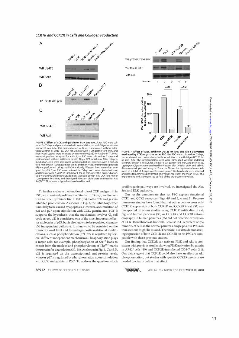
review of CCKR inhibitors, see Ref. 9). The CCK1R inhibitorL364 caused a moderate, but significant (p ! 0.001) inhibi-tion of Akt phosphorylation by 1 nM CCK (Fig. 5A, secondversus fourth lane) and had a similar effect on Akt phosphor-ylation by 1 !M gastrin (Fig. 5A, seventh versus ninth lane).CCK2R inhibitor LY288513 strongly inhibited (p ! 0.0001)Akt phosphorylation caused by 1 pM CCK (Fig. 5A, secondversus third lane) and almost completely suppressed theeffect of 1 !M gastrin on Akt phosphorylation (Fig. 5A, sev-enth versus eighth lane). A combination of both inhibitorsinhibited Akt phosphorylation to a greater and statisticallysignificant extent than the inhibition of one receptor alone,showing clearly that both receptors are involved (Fig. 5A,fifth and tenth lane). The same inhibitors were used to eval-uate CCK1 and -2 receptor inhibition on collagen synthesisand proliferation (Fig. 5B). Similar to the results on Akt acti-vation inhibition of CCK2R leads to a greater decrease ofcollagen synthesis than inhibition of CCK1R; simultaneousinhibition of both receptors reduces collagen productionback to unstimulated levels, whereas proliferation raisesproduction to unstimulated levels. These results clearlyshow that the inhibitory effect of CCK and gastrin is medi-ated through CCK1 and CCK2 receptors.Akt Activation by CCK and Gastrin Are Mediated by PI3K in
Rat PSC—Akt activation by growth factors is generally medi-ated by PI3K in most experimental systems. In rat pancreaticacini, Akt activation by the high affinity state of the CCK1R ismediated by PI3K (18). Therefore, we wanted to study if PI3Kmediates Akt activation by CCK and gastrin in rat PSC. The
highly specific PI3K inhibitor wort-mannin (10!M) caused a significantdecrease of basal Akt Ser473 phos-phorylation in rat PSC (Fig. 6A, sec-ond versus first lane) and almostcompletely suppressed Akt Ser473phosphorylation caused by CCKand gastrin. These data demon-strate that CCK- and gastrin-stimu-lated Akt phosphorylation aremediated by PI3K in rat PSC. PI3Kconsists of two major components:p85, which is the regulatory site andp110, the catalytic domain (27). Tostudy the events involved in CCK-and gastrin-mediated PI3K activa-tion, we inhibited the " and # sub-units of G protein-coupled receptortargeting p110 using the PI3K#inhibitor II. As shown in Fig. 6C noinhibition was seen. On the otherhand, inhibition of the Src pathwayusing the inhibitor PP2 leads to amarked decrease in CCK and gas-trin-stimulated p85 activation (Fig.6B). This result shows that CCK andgastrin activation of the PI3K is reg-ulated by Src-dependent activationof the p85 subunit.
ERK Mediates Activation of Transcription Factor Elk-1 inResponse to Gastrin and CCK in Rat PSC—The transcriptionfactor Elk-1 has been reported to mediate ERK-induced c-fostranscription in response to growth factors, leading to cellu-lar growth and proliferation in some cell systems (28, 29).This pathway has been reported to mediate PSC activationby alcohol and acetaldehyde (30). Therefore, we wanted toinvestigate whether CCK and gastrin cause Elk-1 activationand whether this activation is mediated by ERK. Our exper-iments show that both CCK and gastrin caused a significantincrease in Elk-1 Ser383 phosphorylation, reflecting activa-tion of Elk-1 (Fig. 7). As reported for pancreatic acini (17),the specific MEK inhibitor U0126 inhibited ERK activationinduced by CCK and gastrin (Fig. 7). Moreover, U0126 com-pletely inhibited stimulation of Elk-1 phosphorylationinduced by CCK and gastrin (Fig. 7). These data demonstratethat, similar to findings reported in other cells, in rat PSC,transcription factor Elk-1 is a downstream target of theMEK-ERK pathway activated by CCK and gastrin.Recent studies (31, 32) have shown that beyond its localiza-
tion in the nucleus, transcription factor Elk-1 can be found inthe cytosol. We did not find a cytosolic fraction of Elk-1 in PSCand subsequently no nuclear transition upon stimulation withCCK or gastrin (data not shown).Pathways Involved in CCK and Gastrin-stimulated Collagen
Production and Inhibition of Proliferation—As shown in Fig. 2,CCK and gastrin stimulate collagen production in PSC. Fig. 3shows the inhibitory effect on proliferation of CCK and gastrin.
FIGURE 4. Effect of CCK and gastrin on central fibrogenic signal transduction pathways in rat PSC. A, ratPSC were cultured for 7 days and stimulated without additions (control) with 1 nM CCK (left panels) or with 1 !Mgastrin (right panels) and then lysed. Lysates were analyzed by Western blot (WB) for pAkt, pERK, and pSrc. Blotswere stripped and analyzed for actin. Shown is a representative experiment of a total of 3 experiments. B, ratPSC were cultured for 7 days and treated with CCK (left panel) and gastrin (right panel) for 5 min. Proteins wereanalyzed for phosphorylated Akt by Western blot. Blots were stripped and analyzed for actin. Shown arerepresentative results of a total of 6 experiments.
CCK1R and CCK2R in Cells and Collagen Production
38910 JOURNAL OF BIOLOGICAL CHEMISTRY VOLUME 285 • NUMBER 50 • DECEMBER 10, 2010
at ZENTRUM FUER M
OLEKULARE, on Decem
ber 25, 2010www.jbc.org
Downloaded from
9

Furthermore, we were able to show that CCK and gastrin stim-ulate different signaling pathways. Thereforewe nextwanted toelucidate which pathways are involved in CCK- and gastrin-stimulated collagen production and inhibition of proliferation.As shown in Fig. 8, A (CCK) and B (gastrin), inhibition of Srcwith the specific inhibitor PP2 leads to a statistically significantdecrease in collagen synthesis in PSC, whereas inhibition ofMEK and PI3K shows only a trend to decrease collagen produc-tion without reaching statistical significance. Fig. 8, C (CCK)
and D (gastrin), shows that thesame intracellular mechanisms areinvolved in inhibiting proliferation.
DISCUSSION
Our results clearly demonstratethat both CCK1 and CCK2 recep-tors are expressed in rat PSC. First,using the RT-PCR technique, wefound that mRNA for both CCK1Rand CCK2R is present in rat PSC.For both PCR, the specificity of theprimer-target interaction was con-firmed by the presence of a specificband in agarose-gel electrophoresis(Fig. 1). Furthermore, an interactionwith possible contaminations ofgenomic DNA can be ruled out bythe absence of a specific band incontrols using genomic DNA (Fig.1A). Furthermore, we were able toshow the presence of both receptorsusing immunohistochemistry andimmunocytology, also showing co-localization with !-SMA (Fig. 1, Band C). Most importantly, the datashow that both receptors areinvolved in crucial cellular func-tions such as collagen synthesis andproliferation. Our data demonstratefor the first time that CCK and gas-trin cause a significant increase incollagen I synthesis (Fig. 2). Synthe-sis of type I collagen contributes toorgan fibrosis in the liver and pan-creas. Therefore, our finding thatCCK receptors have a direct effecton PSC by increasing collagen syn-thesis is of particular interest,because it demonstrates for the firsttime that CCK receptors can inducepancreatic fibrosis. CCK serum lev-els are often elevated in patientswith chronic pancreatitis. A directfibrogenic effect of CCK could be animportant mediator of pancreaticfibrosis, suggesting that CCK recep-tors could be an interesting thera-peutic target in patients with
chronic pancreatitis. To date, the cytokine TGF-" had been themost potent stimulator of collagen synthesis in PSC (35). Asshown in Fig. 2, the effect of CCK and gastrin on collagen syn-thesis was comparable with TGF-"-stimulated collagen pro-duction supporting their potential role in development of pan-creatic fibrosis. Although expression of other matrix proteins,such as fibronectin, TIMP-1, and MMP has been described inPSC (36), CCK and gastrin were unable to stimulate the pro-duction of fibronectin and TIMP1.
FIGURE 5. Effect of CCK1R and CCK2R inhibitor on Akt activation, collagen synthesis, and prolifera-tion mediated by CCK or gastrin in rat PSC. A, rat PSC were cultured for 7 days, serum starved, andpreincubated without additions, 1 #M CCK1R inhibitor L364 for 2.5 min, 10 nM CCK2R inhibitor LY288513,or a combination of both for 5 min. Cells were stimulated without additions (control), 1 nM CCK for 5 minor 1 #M gastrin for 5 min and then lysed. Upper panel, lysates were analyzed by Western blot for AktSer(P)473. Blots were stripped and analyzed for actin. Shown is a representative experiment of a total of 6experiments. Lower panel, Western blots (WB) were scanned and densitometry was performed. The valuesrepresent the mean ! S.E. of 6 experiments and are expressed as fold of the maximal stimulation achievedwith CCK or gastrin alone (*, p " 0.05). B, ELISA: rat PSC were cultured for 7 days, serum starved, andpreincubated with 1 #M L364 for 2.5 min, or with 10 nM CCK2R LY288513 for 5 min, or with the combina-tion. Cells were stimulated without additions, or with 1 nM CCK or 1 #M gasrin for 24 h. Stimulants andinhibitors were replaced after 12 h. Indirect ELISA using cell culture supernatants was performed. MTTassay: rat PSC were cultured for 7 days, serum starved, and preincubated with 1 #M L364 for 2.5 min, with10 nM CCK2R LY288513 for 5 min, or with the combination. Cells were stimulated without additions, orwith 1 nM CCK or 1 #M gasrin for 24 h. Stimulants and inhibitors were replaced after 12 h. MTT assays wereperformed. The values represent the mean ! S.E. of 3 experiments and are expressed as percentage of thecontrol. *, p " 0.05 versus control.
CCK1R and CCK2R in Cells and Collagen Production
DECEMBER 10, 2010 • VOLUME 285 • NUMBER 50 JOURNAL OF BIOLOGICAL CHEMISTRY 38911
at ZENTRUM FUER M
OLEKULARE, on Decem
ber 25, 2010www.jbc.org
Downloaded from
10
To further evaluate the functional role of CCK and gastrin inPSC, we examined proliferation. Similar to TGF-!, and in con-trast to other cytokines like PDGF (35), both CCK and gastrininhibited proliferation. As shown in Fig. 3, the inhibitory effectis unlikely to be caused by apoptosis. However, accumulation ofp21 and p27 upon stimulation with CCK, gastrin, and TGF-!supports the hypothesis that the mechanism involves G1 cellcycle arrest. p21 is considered one of the most important effec-tormolecules of p53, but is also known to be regulated viamanyp53 independent pathways. It is known to be regulated on thetranscriptional level and to undergo posttranslational modifi-cations, such as phosphorylation (37). p27 is regulated by sev-eral different independent mechanisms. Phosphorylation playsa major role: for example, phosphorylation of Ser10 leads toexport from the nucleus and phosphorylation of Thr187 marksthe protein for degradation (37, 38). As shown in Fig. 3,C andD,p21 is regulated on the transcriptional and protein levels,whereas p27 is regulated by phosphorylation upon stimulationwith CCK and gastrin in PSC. To address the question which
profibrogenic pathways are involved, we investigated the Akt,Src, and ERK pathways.Our results demonstrate that rat PSC express functional
CCK1 and CCK2 receptors (Figs. 4B and 5, A and B). Becausenumerous studies have found that rat acinar cells express onlyCCK1R, expression of both CCK1R and CCK2R in rat PSC wasunexpected. Previous studies using CCK1R antibodies in rat,pig, and human pancreas (33) or CCK1R and CCK2R autora-diography in human pancreas (35) did not describe expressionof CCK1R on fibroblast-like cells. Because PSC represent only aminority of cells in the normal pancreas, single positive PSCs onthin sectionsmight bemissed. Therefore, our data demonstrat-ing expression of both CCK1R andCCK2R on rat PSC are com-patible with these previous studies.Our finding that CCK2R can activate PI3K and Akt is con-
sistentwith previous studies showing PI3K activation by gastrinin AR42J cells (40) and CCK2R-transfected COS-7 cells (41).Our data suggest that CCK1R could also have an effect on Aktphosphorylation, but studies with specific CCK1R agonists areneeded to clearly define that effect.
FIGURE 6. Effect of CCK and gastrin on PI3K and Akt. A, rat PSC were cul-tured for 7 days and preincubated without additions or with 10 "M wortman-nin for 30 min. After this preincubation, cells were stimulated without addi-tions (control) or with 1 nM CCK for 5 min or with 1 "M gastrin for 5 min, andthen lysed. Lysates were analyzed by Western blot (WB) for Akt Ser(P)473. Blotswere stripped and analyzed for actin. B, rat PSC were cultured for 7 days andpreincubated without additions or with 10 "M PP2 for 60 min. After this pre-incubation, cells were stimulated without additions (control), with 1 nM CCKfor 5 min or with 1 "M gastrin for 5 min, and then lysed. Immunoprecipitation(IP) was performed using anti-PY20 antibody. Western blots were then ana-lyzed for p85. C, rat PSC were cultured for 7 days and preincubated withoutadditions or with 2 "M PI3K# inhibitor II for 60 min. After this preincubation,cells were stimulated without additions (control), or with 1 nM CCK for 5 min or1 "M gastrin for 5 min, and then lysed. Western blots were analyzed for AktSer(P)473. Blots were stripped and analyzed for actin.
FIGURE 7. Effect of MEK inhibitor U0126 on ERK and Elk-1 activationmediated by CCK or gastrin in rat PSC. Rat PSC were cultured for 7 days,serum starved, and preincubated without additions or with 20 "M U0126 for60 min. After this preincubation, cells were stimulated without additions(control), or with 1 nM CCK for 5 min or 1 "M gastrin for 5 min, and then lysed.Upper panel, lysates were analyzed by Western blot (WB) for pERK and pElk-1.Blots were stripped and analyzed for actin. Shown is a representative experi-ment of a total of 3 experiments. Lower panel, Western blots were scannedand densitometry was performed. The values represent the mean ! S.E. of 3experiments and are expressed as fold of the pre-treatment values.
CCK1R and CCK2R in Cells and Collagen Production
38912 JOURNAL OF BIOLOGICAL CHEMISTRY VOLUME 285 • NUMBER 50 • DECEMBER 10, 2010
at ZENTRUM FUER M
OLEKULARE, on Decem
ber 25, 2010www.jbc.org
Downloaded from
11

Our data demonstrate that both CCK and gastrin cause acti-vation of the MEK-ERK pathway in rat PSC, leading to activa-tion of the transcription factor Elk-1. This is compatible withstudies showing activation of ERK by CCK in rat pancreaticacini (10) and with studies showing activation of ERK by CCKand gastrin in AR42J pancreas cancer cells (42). Our findingthat gastrin and CCK induce significant ERK activation is par-ticularly relevant because PSC activation and proliferationcaused by PDGF, one of the strongest known activators of PSC,ismediated by ERK (16, 43).We have shown that bothCCK andgastrin cause reproducible activation of Src family kinases (Fig.4A). This finding is compatible with studies showing activationof Src kinases in rat pancreatic acini by CCK (mediated byCCK1R)(24) and with studies showing activation of Src kinasesby gastrin in AR42J and Panc-1 pancreas cancer cells (44–46)aswell as in othermultiple cells. Activation of Src family kinasesby CCK and gastrin in rat PSC is interesting because Src familykinases activate the JAK2-STAT pathway after PDGF stimula-tion in PSC and thereby could be important regulators of PSCproliferation (26). Furthermore, we were able to show (Fig. 8)
that inhibition of Src reduces colla-gen synthesis and proliferation ina statistically significant manner,pointing to the Src pathway as a piv-otal pathway in CCK- and gastrin-stimulated collagen production andinhibition of proliferation.Chronic pancreatitis is a com-
plex disease and our knowledge ofthe exact pathophysiologic mecha-nisms are still incomplete. Recentstudies suggest that the develop-ment of chronic pancreatitis re-quires a first episode of pancreatitis(sentinel acute pancreatitis event),which, by the release of differentcytokines, triggers activation ofimmune cells and stellate cells,leading to chronic inflammation,fibrosis, and destruction of normalorgan architecture, resulting in lossof organ function (47, 48). In thisprocess, activation of stellate cells iscrucial, because these cells havebeen shown to be responsible forthe development of pancreaticfibrosis, which is a constant featureof chronic pancreatitis. In rodentanimal models, CCK is routinelyused in combination with otheragents to induce chronic pancreati-tis (23, 49–51). These studies havesupposed that the role of CCK inthese models was to trigger the sen-tinel pancreatitis event by interact-ing with CCK receptors on acinarcells and inducing necrosis of acinarcells (49). Our finding that CCK can
directly activate stellate cells and induce collagen productionsuggests that the role of CCK is more complex: 1) by its actionon acinar cells, it could contribute to the induction of the sen-tinel pancreatitis event; and 2) by its action on stellate cells, itcould serve as an important regulator of pancreatic fibrosis. Inconclusion, we report for the first time that rat PSC expressCCK1 and CCK2 receptors, that the natural agonists CCK andgastrin induce activation of pro-fibrotic signaling pathwaysPI3K/Akt, MEK/ERK, and Src, induce activation of the tran-scription factor Elk-1, and most notably significantly increasesynthesis of type I collagen and inhibit proliferation.
REFERENCES1. Saluja, A. K., Saluja, M., Printz, H., Zavertnik, A., Sengupta, A., and Steer,
M. L. (1989) Proc. Natl. Acad. Sci. U.S.A. 86, 8968–89712. Apte, M. V., and Wilson, J. S. (2004) Dig. Dis. 22, 273–2793. Apte, M., McCarroll, J., Pirola, R., and Wilson, J. (2007) Novartis Found.
Symp. 285, 200–2114. Apte, M. V., Phillips, P. A., Fahmy, R. G., Darby, S. J., Rodgers, S. C.,
McCaughan, G. W., Korsten, M. A., Pirola, R. C., Naidoo, D., andWilson,J. S. (2000) Gastroenterology 118, 780–794
FIGURE 8. Pathways involved in CCK and gastrin-stimulated collagen production and inhibition of pro-liferation. A, rat PSC were cultured, serum starved, and stimulated without additions, or preincubated withwortmannin (30 min, 10 !mol), PP2 (10!mol 60 min), U0126 (20!mol 60), and stimulated with 1 nM CCK for 24 h(stimulants were replaced every 12 h). Indirect ELISA was performed using cell culture supernatants. The valuesrepresent the mean ! S.E. of 4 experiments and are expressed as percent of the pre-treatment value. B, rat PSCwere cultured, serum starved, and stimulated without additions, or preincubated with wortmannin (30 min, 10!mol), PP2 (10 !mol 60 min), U0126 (20 !mol 60), and stimulated with 1 !M gastrin for 24 h (stimulants werereplaced every 12 h). Indirect ELISA was performed using cell culture supernatants. The values represent themean ! S.E. of 4 experiments and are expressed as percent of the pre-treatment value. C, MTT assay: rat PSCwere isolated and cultured for 7 days. Then 100,000 cells/well were seeded, stimulated with 1 nM CCK, and theMTT assay was performed. The values represent the mean ! S.E. of 5 experiments and are expressed aspercentage of the control. *, p " 0.01 versus control. D, MTT assay: rat PSC were isolated and cultured for 7 days.Then 100,000 cells/well were seeded, stimulated with 1 !M gastrin, and MTT assay was performed. The valuesrepresent the mean ! S.E. of 5 experiments and are expressed as percentage of the control. *, p " 0.01 versuscontrol. GW, gastrin # wortmannin.
CCK1R and CCK2R in Cells and Collagen Production
DECEMBER 10, 2010 • VOLUME 285 • NUMBER 50 JOURNAL OF BIOLOGICAL CHEMISTRY 38913
at ZENTRUM FUER M
OLEKULARE, on Decem
ber 25, 2010www.jbc.org
Downloaded from
12

5. Hama, K., Ohnishi, H., Aoki, H., Kita, H., Yamamoto, H., Osawa, H., Sato,K., Tamada, K., Mashima, H., Yasuda, H., and Sugano, K. (2006) Biochem.Biophys. Res. Commun. 340, 742–750
6. Masamune, A., Kikuta, K., Watanabe, T., Satoh, K., Hirota, M., Hamada,S., and Shimosegawa, T. (2009) Gut 58, 550–559
7. Masamune, A., Satoh, A., Watanabe, T., Kikuta, K., Satoh, M., Suzuki, N.,Satoh, K., and Shimosegawa, T. (2010) Dig. Dis. Sci. 55, 204–211
8. Masamune, A., Satoh, M., Kikuta, K., Suzuki, N., and Shimosegawa, T.(2005)World J. Gastroenterol. 11, 6144–6151
9. Berna,M. J., and Jensen, R. T. (2007)Curr. Top.Med. Chem. 7, 1211–123110. Williams, J. A., Sans, M. D., Tashiro, M., Schafer, C., Bragado, M. J., and
Dabrowski, A. (2002) Pharmacol. Toxicol. 91, 297–30311. Gomez Cerezo, J., Codoceo, R., Fernandez Calle, P., Molina, F., Tenias,
J. M., and Vazquez, J. J. (1991) Digestion 48, 134–14012. Schafmayer, A., Becker, H. D., Werner, M., Folsch, U. R., and Creutzfeldt,
W. (1985) Digestion 32, 136–13913. Slaff, J., Jacobson, D., Tillman, C. R., Curington, C., and Toskes, P. (1984)
Gastroenterology 87, 44–5214. Shiratori, K., Takeuchi, T., Satake, K., Matsuno, S., and Study Group of
Loxigumide of Japan (2002) Pancreas 25, e1–515. Apte, M. V., Haber, P. S., Darby, S. J., Rodgers, S. C., McCaughan, G. W.,
Korsten, M. A., Pirola, R. C., and Wilson, J. S. (1999) Gut 44, 534–54116. Jaster, R., Sparmann, G., Emmrich, J., and Liebe, S. (2002) Gut 51,
579–58417. Berna, M. J., Hoffmann, K. M., Tapia, J. A., Thill, M., Pace, A., Mantey,
S. A., and Jensen, R. T. (2007) Biochim. Biophys. Acta 1773, 483–50118. Berna, M. J., Tapia, J. A., Sancho, V., Thill, M., Pace, A., Hoffmann, K. M.,
Gonzalez-Fernandez, L., and Jensen, R. T. (2009)Cell. Signal. 21, 622–63819. Masamune, A., Watanabe, T., Kikuta, K., Satoh, K., and Shimosegawa, T.
(2008) Am. J. Physiol. Gastrointest. Liver Physiol. 294, G99–G10820. Li, L., Sampat, K., Hu, N., Zakari, J., and Yuspa, S. H. (2006) J. Biol. Chem.
281, 3237–324321. Sarbassov, D. D., Guertin, D. A., Ali, S. M., and Sabatini, D. M. (2005)
Science 307, 1098–110122. Berna, M. J., Tapia, J. A., Sancho, V., and Jensen, R. T. (2007) Curr. Opin.
Pharmacol. 7, 583–59223. Vaquero, E., Molero, X., Tian, X., Salas, A., and Malagelada, J. R. (1999)
Gut 45, 269–27724. Pace, A., Tapia, J. A., Garcia-Marin, L. J., and Jensen, R. T. (2006) Biochim.
Biophys. Acta 1763, 356–36525. Ohnishi, H., Miyata, T., Yasuda, H., Satoh, Y., Hanatsuka, K., Kita, H.,
Ohashi, A., Tamada, K., Makita, N., Iiri, T., Ueda, N., Mashima, H., andSugano, K. (2004) J. Biol. Chem. 279, 8873–8878
26. Masamune, A., Satoh, M., Kikuta, K., Suzuki, N., and Shimosegawa, T.(2005)World J. Gastroenterol. 11, 3385–3391
27. Dufresne, M., Seva, C., and Fourmy, D. (2006) Physiol. Rev. 86, 805–84728. Marshall, C. J. (1995) Cell 80, 179–18529. Treisman, R. (1995) EMBO J. 14, 4905–491330. McCarroll, J. A., Phillips, P. A., Park, S., Doherty, E., Pirola, R. C., Wilson,
J. S., and Apte, M. V. (2003) Pancreas 27, 150–16031. Lavaur, J., Bernard, F., Trifilieff, P., Pascoli, V., Kappes, V., Pages, C., Van-
houtte, P., and Caboche, J. (2007) J. Neurosci. 27, 14448–1445832. Barrett, L. E., Van Bockstaele, E. J., Sul, J. Y., Takano,H., Haydon, P. G., and
Eberwine, J. H. (2006) Proc. Natl. Acad. Sci. U.S.A. 103, 5155–516033. Bourassa, J., Laine, J., Kruse,M. L., Gagnon,M. C., Calvo, E., andMorisset,
J. (1999) Biochem. Biophys. Res. Commun. 260, 820–82834. Deleted in proof35. Kordes, C., Brookmann, S., Haussinger, D., and Klonowski-Stumpe, H.
(2005) Pancreas 31, 156–16736. Schneider, E., Schmid-Kotsas, A., Zhao, J.,Weidenbach,H., Schmid, R.M.,
Menke, A., Adler, G., Waltenberger, J., Grunert, A., and Bachem, M. G.(2001) Am. J. Physiol. Cell Physiol. 281, C532–543
37. Abde, M., Abukhdeir, B., and Ho, P. (2008) Expert Rev. Mol. Med. 10, e1938. Vervoorts, J., and Luscher, B. (2008) Cell Mol. Life Sci. 65, 3255–326439. Deleted in proof40. Seva, C., Kowalski-Chauvel, A., Daulhac, L., Barthez, C., Vaysse, N., and
Pradayrol, L. (1997) Biochem. Biophys. Res. Commun. 238, 202–20641. Zieger, M., Oehrl, W., Wetzker, R., Henklein, P., Nowak, G., and Kauf-
mann, R. (2000) Biol. Chem. 381, 763–76842. Dabrowski, A., Detjen, K. M., Logsdon, C. D., and Williams, J. A. (1997)
Digestion 58, 361–36743. Wan, Y., Wang, T., and Zhao, Q. (2005) J. Huazhong Univ. Sci. Technol.
Med. Sci. 25, 297–299, 30644. Cayrol, C., Clerc, P., Bertrand, C., Gigoux, V., Portolan, G., Fourmy, D.,
Dufresne, M., and Seva, C. (2006) Oncogene 25, 4421–442845. Ferrand, A., Kowalski-Chauvel, A., Bertrand, C., Pradayrol, L., Fourmy, D.,
Dufresne, M., and Seva, C. (2004) Exp. Cell Res. 301, 128–13846. Piiper, A., Elez, R., You, S. J., Kronenberger, B., Loitsch, S., Roche, S., and
Zeuzem, S. (2003) J. Biol. Chem. 278, 7065–707247. Etemad, B., and Whitcomb, D. C. (2001) Gastroenterology 120, 682–70748. Schneider, A., and Whitcomb, D. C. (2002) Best Pract. Res. Clin. Gastro-
enterol. 16, 347–36349. Deng, X., Wang, L., Elm, M. S., Gabazadeh, D., Diorio, G. J., Eagon, P. K.,
and Whitcomb, D. C. (2005) Am. J. Pathol. 166, 93–10650. Gukovsky, I., Cheng, J. H., Nam, K. J., Lee, O. T., Lugea, A., Fischer, L.,
Penninger, J. M., Pandol, S. J., and Gukovskaya, A. S. (2004) Gastroenter-ology 126, 554–566
51. Neuschwander-Tetri, B. A., Burton, F. R., Presti, M. E., Britton, R. S.,Janney, C. G., Garvin, P. R., Brunt, E. M., Galvin, N. J., and Poulos, J. E.(2000) Dig. Dis. Sci. 45, 665–674
CCK1R and CCK2R in Cells and Collagen Production
38914 JOURNAL OF BIOLOGICAL CHEMISTRY VOLUME 285 • NUMBER 50 • DECEMBER 10, 2010
at ZENTRUM FUER M
OLEKULARE, on Decem
ber 25, 2010www.jbc.org
Downloaded from
13

2. Darstellung der Publikation
2.1. Einführung 2.1.1. Übersicht zur chronischen Pankreatitis
Die chronische Pankreatit is ist definiert als chronische Entzündung der
Bauchspeicheldrüse, die zu einer irreversiblen Zerstörung des Pankreasparenchyms mit
nachfo lgendem Funkt ionsver lust , d .h. e iner exokr inen und endokr inen
Pankreasinsuffizienz führt (Riemann et al. 2008). Histopathologisch läßt sich zunächst
eine lymphozytär-entzündliche Fibrose, später dann eine diffuse Fibrose und Atrophie des
Organs beobachten (Riede et al. 2004).
Die Inzidenz beträgt in Deutschland ca. 2-10/100000 Einwohner, wobei das Verhältnis
männlich:weiblich ca. 3:1 beträgt. Als auslösende Faktoren werden verschiedene
Ursachen angenommen, allerdings spielt hierbei Alkohol mit ca. 80% die wichtigste Rolle.
Ätiologie Ursachen
Toxisch-metabolisch 80%
Alkohol, Hyperkalziämie, Hyperlipidämie, Urämie, Medikamentös
Idiopathisch 15% Ursache unbekannt
Genetisch Mutationen im PRSS-1-, CFTR- und SPINK-1-Gen
Autoimmun Infiltration Pankreas mit IgG4-positiven Plasmazellen
Obstruktiv Rezidivierende Choledocholithiasis, SOD (Sphincter-Oddi-Dysfunktion), Pancreas divisum, obstruktiver Tumor
Tab. 1: Ätiologie und Ursachen der chronischen Pankreatitis (Greten et al. 2010)
2.1.2. Rolle der pankreatischen Sternzellen an der Entstehung der chronischen
Pankreatitis / dem Ablauf der Entzündung
In vielen Arbeiten zur Pathogenese der akuten Pankreatitis konnte gezeigt werden, dass
Mechanismen innerhalb der Azinuszellen zu Inflammation und Organdestruktion führen
(Saluja et al.1989). Wie in Tab.1 zu sehen, kann diese über verschiedene Wege
geschädigt werden. Die entscheidende Rolle bzw. gemeinsame Endstrecke jedoch 14
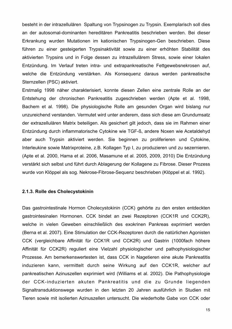
besteht in der intrazellulären Spaltung von Trypsinogen zu Trypsin. Exemplarisch soll dies
an der autosomal-dominanten hereditären Pankreatitis beschrieben werden. Bei dieser
Erkrankung wurden Mutationen im kationischen Trypsinogen-Gen beschrieben. Diese
führen zu einer gesteigerten Trypsinaktivität sowie zu einer erhöhten Stabilität des
aktivierten Trypsins und in Folge dessen zu intrazellulärem Stress, sowie einer lokalen
Entzündung. Im Verlauf treten intra- und extrapankreatische Fettgewebsnekrosen auf,
welche die Entzündung verstärken. Als Konsequenz daraus werden pankreatische
Sternzellen (PSC) aktiviert.
Erstmalig 1998 näher charakterisiert, konnte diesen Zellen eine zentrale Rolle an der
Entstehung der chronischen Pankreatitis zugeschrieben werden (Apte et al. 1998,
Bachem et al. 1998). Die physiologische Rolle am gesunden Organ wird bislang nur
unzureichend verstanden. Vermutet wird unter anderem, dass sich diese am Grundumsatz
der extrazellulären Matrix beteiligen. Als gesichert gilt jedoch, dass sie im Rahmen einer
Entzündung durch inflammatorische Cytokine wie TGF-ß, andere Noxen wie Acetaldehyd
aber auch Trypsin aktiviert werden. Sie beginnen zu proliferieren und Cytokine,
Interleukine sowie Matrixproteine, z.B. Kollagen Typ I, zu produzieren und zu sezernieren.
(Apte et al. 2000, Hama et al. 2006, Masamune et al. 2005, 2009, 2010) Die Entzündung
verstärkt sich selbst und führt durch Ablagerung der Kollagene zu Fibrose. Dieser Prozess
wurde von Klöppel als sog. Nekrose-Fibrose-Sequenz beschrieben (Klöppel et al. 1992).
2.1.3. Rolle des Cholecystokinin
Das gastrointestinale Hormon Cholecystokinin (CCK) gehörte zu den ersten entdeckten
gastrointesinalen Hormonen. CCK bindet an zwei Rezeptoren (CCK1R und CCK2R),
welche in vielen Geweben einschließlich des exokrinen Pankreas exprimiert werden
(Berna et al. 2007). Eine Stimulation der CCK-Rezeptoren durch die natürlichen Agonisten
CCK (vergleichbare Affinität für CCK1R und CCK2R) und Gastrin (1000fach höhere
Affinität für CCK2R) reguliert eine Vielzahl physiologischer und pathophysiologischer
Prozesse. Am bemerkenswertesten ist, dass CCK in Nagetieren eine akute Pankreatitis
induzieren kann, vermittelt durch seine Wirkung auf den CCK1R, welcher auf
pankreatischen Azinuszellen exprimiert wird (Williams et al. 2002). Die Pathophysiologie
der CCK-induzierten akuten Pankreat i t is und die zu Grunde l iegenden
Signaltransduktionswege wurden in den letzten 20 Jahren ausführlich in Studien mit
Tieren sowie mit isolierten Azinuszellen untersucht. Die wiederholte Gabe von CCK oder
15

dem Agonisten Cerulein wurde und wird in vielen Studien benutzt, um verschiedene
Aspekte der chronischen Pankreatitis zu induzieren, einschließlich der Aktivierung von
PSC und der Fibrogenese. Auch in diesen Studien wurde angenommen, dass die
Entstehung der Fibrose ein Produkt der wiederholten Organinflammation darstellt,
verursacht durch die Induktion multipler akuter Pankreatitiden durch CCK (sog.
Sentinelpankreatitiden). Darüber hinaus konnte in mehreren Studien gezeigt werden, dass
im Serum von Patienten welche an einer chronischen Pankreatitis litten verglichen mit
gesunden Kontrollen, erhöhte CCK-Plasmaspiegel zu messen waren (Gomez et al. 1991,
Schafmayer et al. 1985, Slaff et al. 1984). Trotz dieser experimentellen und klinischen
Ergebnisse, die auf eine Rolle von CCK in der chronischen Pankreatitis hinwiesen, hatte
bislang keine Studie untersucht, ob CCK oder eines seiner Agonisten einen direkten Effekt
auf pankreatische Sternzellen haben könnte. Selbst die Expression von CCK-Rezeptoren
auf PSC wurde bislang noch nicht untersucht.
2.1.4. Ziel der Arbeit
Um also eine mögliche direkte Rolle von CCK und CCK-Rezeptoren in der
Pathophysiologie der chronischen Pankreatitis zu untersuchen, wurden sowohl die
Expression von CCK1R und CCK2R untersucht, als auch die Effekte der Stimulation
dieser Rezeptoren auf die Kollagenexpression, die Proliferation und auf profibrogene
Signaltransduktionswege.
2.2. Zusammenfassung der Versuche und Ergebnisse
2.2.1. Expression von CCK1- und CCK2- Rezeptoren auf Sternzellen
Zunächst wurden pankreatische Sternzellen als Primärkultur aus Rattenpankreas
gewonnen. Hierzu wurde nach duktaler Injektion einer Enzymlösung und anschließendem
Verdau eine Gradientenzentrifugation durchgeführt. Die daraus gewonnenen Zellen
wurden in Kultur genommen.
Der Nachweis der CCK1- und CCK2- Rezeptoren erfolgte mittels RT-PCR (Abb.1A), durch
Immunofluoreszenz (Abb.1B) in kryokonservierten Pankreasschnitten sowie durch
immunzytologische Färbungen an kultivierten Sternzellen (nicht gezeigt). Durch einen für
Sternzellen typischen Oberflächenmarker (alpha-SMA) konnte die Spezifität der Färbung
gezeigt werden.
16

Thymidine Uptake—Rat PSC were isolated and 50,000 cells/well were seeded on 12-well plates. When reaching 60% con-fluence, cellswere serumstarved and then stimulated for 24 h inserum-free medium containing 2 !Ci/ml of [3H]thymidine(PerkinElmer Life Sciences) with no additions, 2 ng/ml of TGF,1 nM CCK, and 1 !M gastrin. After 24 h cells were harvested(Brandel Harvester, Unterfohring, Germany) and [3H]thymi-dine incorporation was measured (Hidex Plate Chameleon,Straubenhardt, Germany).
MTT Assay—Rat PSC were iso-lated and cultured. Cells weretrypsinized and 100,000 cells/wellwere seeded in 6-well plates. Whenreaching subconfluence, cells wereserum-starved and stimulated withno additions, 2 ng/ml of TGF, 1 nMCCK, or 1 !M gastrin for 24 h. Stim-ulating factors were replaced after12 h. Thereafter, 100!g/ml ofMTT(Sigma) were added and cells wereincubated for 2 h. Supernatantswere removed and 250 !l/well ofDMSO (Sigma)was added. Reactionproducts were transferred tomicro-titer plates and read in a standardmicroplate reader at 570 nm.Statistical Analysis—Data are
presented as mean ! S.E. and wereanalyzed using the Student’s t testfor unpaired data using the Prismsoftware (GraphPad). p values "0.05were considered significant.
RESULTS
Rat PSC Express CCK1 and CCK2Receptors—The presence of mRNAfor CCK1 and CCK2 receptors wasassessed by RT-PCR in PSC cul-tured for 7 days. According to themanufacturer of the primers used,specific bands of 118 (CCK1R) and93 bp (CCK2R) were expected. Asshown in Fig. 1A, expression ofCCK1R mRNA was found in PSC(lane 1), but absent in the genomicDNA (lane 2) and negative controlusing water (lane 3). Similarly,CCK2R mRNA was found in PSC(lane 4), whereas the correspondingcontrols using genomic DNA (lane5) or water (lane 6) did not show acorresponding band. We concludethat mRNA for both CCK1R andCCK2R is expressed in rat PSC.
Furthermore, the presence ofboth receptors was confirmed byimmunohistology (Fig. 1B) and im-munocytochemistry (Fig. 1C).
"-SMA as a tissue-specific marker was used to confirm local-ization on PSC. Snap frozen rat brain sections served as positivecontrol for the antibodies.CCK Causes Collagen Production in PSC—Upon stimulation
by growth factors and severalGprotein-coupled receptors, PSChave been reported to produce extracellular matrix proteinsincluding type I collagen, leading to pancreatic fibrosis, a con-stant feature of chronic pancreatitis. To date TGF-# has beenthemost potent fibrogenic stimulus described in PSC.To assess
FIGURE 1. CCK2 and CCK1 receptors are expressed on rat PSC. A, rat PSC were cultured for 7 days, RNA wasisolated, transcribed, and samples submitted to RT-PCR. Negative controls (genomic DNA and water) wereincluded. Expected product sizes are 93 (CCK2R) and 118 bp (CCK1R). A representative experiment of a total of3 independent experiments is shown. B, rat pancreatic tissue was snap frozen, fixed in acetone, and 5-!msections were immunostained. Shown is a merged image of the CCK1R/CCK2R-stained tissue (red), counter-stained with "-SMA (green). Magnification, #200. C, rat PSC were cultured, trypsinized, and plated on glasschamber slides. Cells were acetone fixed and immunostained for CCK1R/CCK2R and "-SMA. Magnification,#200.
CCK1R and CCK2R in Cells and Collagen Production
DECEMBER 10, 2010 • VOLUME 285 • NUMBER 50 JOURNAL OF BIOLOGICAL CHEMISTRY 38907
at ZENTRUM FUER M
OLEKULARE, on Decem
ber 25, 2010www.jbc.org
Downloaded from
Abb.1: Nachweis von CCK1- und CCK2- Rezeptoren mittels RT-PCR und Immunhistologie.
Es konnte so erstmals gezeigt werden, dass CCK-Rezeptoren in Pankreassternzellen
exprimiert werden.
2.2.2. Proliferation und Kollagenproduktion in pankreatischen Sternzellen nach
CCK-Stimulation
Im Anschluss daran wurde untersucht, ob die Stimulation der Zellen mit CCK funktionelle
Auswirkungen hat. Insbesondere im Hinblick auf die Funktion der Sternzellen im
erkrankten Organ, wurden die Kollagenproduktion, die Proliferation sowie die Aktivierung
profibrogener Signaltransduktionswege untersucht. Als positive Kontrolle diente TGF-ß,
welches bislang als potentester Stimulus der Kollagensynthese in PSC galt (Kordes et al.
2005).
Als Nachweis der Kollagenproduktion wurden verschiedene Methoden angewandt. Zum
einen auf Proteinebene mittels Western Blot und indirektem ELISA. Zum anderen mittles
immunzytologischer Färbungen nach Stimulation, um durch eine Kolokalisation der
17

Kollagensynthese mit alphaSMA den Nachweis zu erbringen, dass die Synthese in
Sternzellen stattfand. Wie in Abb. 2 gezeigt, stimulierten sowohl CCK als auch Gastrin
signifikant die Kollagensynthese. So konnte gezeigt werden, dass die exprimierten
Rezeptoren funktionell aktiv sind, und nach Aktivierung die Pankreasfibrose fördern
können.
whether CCK or gastrin can stimulate collagen type I produc-tion in PSC (Fig. 2) and to compare its action to TGF-! weperformed Western blot analysis and indirect ELISA uponstimulation with CCK and gastrin (Western blot, ELISA) andTGF-! (ELISA). After 24 h of stimulation, CCK (1 nM) caused a136.3% increase (p ! 0.05) in procollagen type I production.Similarly, gastrin (1 "M) caused a 136% (p ! 0.05) increase.TGF-! caused an increase in collagen production of 149%. Fur-thermore, to demonstrate that collagen type I production wasspecifically induced in PSC, we performed immunocytochem-istry for collagen type I and #-SMA (Fig. 2, right panel). Theseresults demonstrate that CCK and gastrin induced collagenproduction by rat PSC, and that collagen synthesis was compa-rable, and not statistically significantly different from TGF-!-stimulated collagen production.CCK and Gastrin Inhibit Proliferation in PSC—Some pro-
fibrogenic factors like TGF-! are also known to inhibit prolif-eration of PSC. To assess if CCK and gastrin have similar effectsin inhibiting proliferation we performed thymidine uptake andMTT assays. After 24 h of stimulation TGF-! inhibited prolif-eration by 15.91%, CCK by 18.1%, and gastrin by 14.45% overcontrols (Fig. 3A). These results demonstrate that CCK andgastrin have similar effects to TGF-! in inhibiting proliferationof PSC. To elucidate if apoptosis or cell cycle arrest were
involved in inhibition of prolifera-tion upon stimulation, we per-formed caspase-3 assays, as well asp21 and p27 analysis. The missingcleavage of caspase-3 and theup-regulation of p21 and p27 sug-gest that G1 cell cycle arrest, notapoptosis, is the likely mechanisminvolved. To address the questionwhether the up-regulation of p21and p27 was due to an increase intranscriptional level or phosphory-lation, we performed qPCR andWestern blot analysis. As shown inFig. 3C, p21 up-regulation is in partdue to an increase in the transcrip-tional level and in part due to phos-phorylation. Unlike p21, p27 ismostly regulated by phosphoryla-tion (Fig. 3D).Effect of CCK andGastrin on Cen-
tral Pro-fibrogenic Signal Transduc-tion Pathways in Rat PSC—CCKhas been reported to activate multi-ple signal transduction pathwaysincluding Akt, ERK, and Src in ratpancreatic acini 1 (7, 24). These cen-tral signal transduction pathwaysmediate activation of PSC and pan-creatic fibrogenesis (5, 16, 25, 26).We analyzedwhether CCK can acti-vate Akt, ERK, and Src signal trans-duction pathways in rat PSC. Subse-quently, we characterized the time
course of this effect using a CCK concentration that causedmaximal Akt activation (1 nM). CCKs effect on Akt was maxi-mal at 2.5 min (3.8 " 1.6-fold increase), significant at 5 min(2.3 " 1.2-fold increase) with values returning to baseline after10min (Fig. 4A). CCK also (1.7" 0.8-fold) increased ERKphos-phorylation after 2.5 min, with significant stimulation (1.8 "0.06-fold, p # 0.0018) after 5 min, stimulation slightly abovebaseline (1.4 " 0.8-fold) after 10 min, and values comparablewith baseline thereafter (Fig. 4A). Moreover, CCK caused sig-nificant stimulation (1.4 " 0.3-fold, p ! 0.05) of Src familykinases after 2.5 min with maximal stimulation (1.7 " 0.3-fold,p# 0.014) at 5min and stimulation slightly above baseline at 10and 30 min (Fig. 4A). These data demonstrate that 1 nM CCKcaused significant stimulation of the fibrogenic signal transduc-tion pathways of Akt, ERK, and Src in rat PSC. Similar to CCK,gastrin activated Akt, ERK, and Src family kinases in rat PSC(Fig. 4A). Gastrin (1 "M) causedmaximal Akt stimulation at 2.5min, significant stimulation at 5 and 10min with values return-ing to baseline after 30 min (Fig. 4A). Similarly, gastrin (1 "M)causedmaximal ERK phosphorylation after 2.5minwith valuesreturning to baseline after 3 min (Fig. 4A). Gastrin had a morecomplex effect on activation of Src family kinases with moder-ate stimulation after 2.5 and 5min, no significant stimulation at10 min, and strong stimulation at 30 min (Fig. 4A). These data
FIGURE 2. Effect of CCK and gastrin on collagen type I synthesis in rat PSC. A, rat PSC were cultured,serum-starved, and stimulated without additions, or with 1 nM CCK or 1 "M gastrin for 24 h (stimulants werereplaced every 12 h) and lysed. Lysates were analyzed by Western blot for procollagen type I. Western blotswere scanned and densitometry was performed. The values represent the mean " S.E. of 6 experiments andare expressed as percent of the pre-treatment values. (*, p ! 0.05 versus control). B, rat PSC were cultured,serum-starved, and stimulated without additions, or with 2 ng/ml of TGF, 1 nM CCK, or 1 "M gastrin for 24 h(stimulants were replaced every 12 h). Indirect ELISA was performed using cell culture supernatants. The valuesrepresent the mean " S.E. of 5 experiments and are expressed as percent of the pre-treatment value. (*, p !0.05 versus control). C, rat PSC were cultured, trypsinized, and plated on glass chamber slides. Serum-starvedcells were stimulated without additions, or with 1 nM CCK or 1 "M gastrin (stimulants were replaced every 12 h)for 24 h. Then cells were acetone fixed and immunostained for collagen type I and #-SMA. Micrographs weretaken by using same exposure time and magnification ($200).
CCK1R and CCK2R in Cells and Collagen Production
38908 JOURNAL OF BIOLOGICAL CHEMISTRY VOLUME 285 • NUMBER 50 • DECEMBER 10, 2010
at ZENTRUM FUER M
OLEKULARE, on Decem
ber 25, 2010www.jbc.org
Downloaded from
Abb. 2: Messung der Kollagensynthese in pankreatischen Sternzellen nach CCK-, Gastrin und TGF-ß -Stimulation im Western Blot (A), ELISA (B) und mittels Immunzytochemie (C)
Von einigen Stimuli wie TGF-ß ist bekannt, dass sie neben ihren profibrogenen Wirkungen
das Wachstum inhibieren (Kordes et al. 2005). Um dies für CCK und Gastrin zu
untersuchen, wurden Thymidin- und MTT-Assays durchgeführt. Nach 24-stündiger
Stimulation konnte in beiden Verfahren gezeigt werden, dass sowohl CCK als auch
Gastrin das Wachstum von PSC inhibiert, vergleichbar der Wirkung von TGF-ß (Abb.3).
18
demonstrate that, similar to CCK,gastrin stimulated Akt, ERK, andSrc in rat PSC.CCK and Gastrin Modulate Akt
Activation in a Dose-dependentManner in Rat PSC—We haverecently shown that CCK1R in-duced a biphasic modulation of Aktactivity in rat pancreatic acinidepending on the CCK concentra-tion (18). Furthermore, recent pub-lications have shown an importantrole of Akt in PSC activation (6, 19).Phosphorylation of Akt at serine473 has been shown to closely cor-relate with Akt activity in numerousstudies (20, 21). Therefore, weexamined the effect of differentdoses of CCK on Akt Ser473 phos-phorylation in rat PSC (Fig. 4B).Doses as low as 1 pM CCK caused asignificant increase in Akt Ser473phosphorylation. This response wasmaximal at 1 nM (2.36 ! 0.45-foldincrease) and then decreased to bejust slightly above control at 1 !M.These results suggest that CCK hasa complex effect on Akt activationin rat PSC: low doses of CCK inducesignificant Akt activation, whereashigher doses of CCK partiallyreverse this activation. To assess theeffect of CCK2R on Akt activationin rat PSC, we studied the effect ofthe CCK2R-preferring agonist gas-trin on Akt serine 473 phosphoryla-tion (Fig. 4B). Gastrin (0.1 nM)caused a significant (p " 0.01)increase in Akt Ser473 phosphoryla-tion and this effect wasmaximal at 1!M (2.25 ! 0.40-fold). These datasuggest that CCK2R stimulated Aktactivation in rat PSC.CCKs Action on Collagen Produc-
tion, Akt Activation Was Mediatedby Both Receptors but Primarily bythe CCK2R in Rat PSC—AlthoughCCK1R binds CCK with high affin-ity (Kd in the nanomolar range) andgastrin with low affinity (Kd in themicromolar range), the CCK2R hadalmost equal affinity for gastrin andCCK (for review, see Ref. 22). Todetermine whether CCK and gas-trin effects are mediated by CCK1Ror CCK2R, we used L364 andLY288513 as specific CCK1R andCCK2R inhibitors, respectively (for
FIGURE 3. Effect of CCK, Gastrin and TGFbeta1 on proliferation of PSC. A, thymidine uptake: 50,000 cells/wellwere seeded, stimulated for 24 h, and [3H]thymidine incorporation measured. The values represent the mean ! S.E.of 5 experiments and are expressed as percentage of the control. *, p " 0.01 versus control. MTT assay, rat PSC wereisolated and cultured for 7 days. Then 100,000 cells/well were seeded, stimulated, and the MTT assay performed. Thevalues represent the mean ! S.E. of 5 experiments and are expressed as percentage of the control. *, p " 0.01 versuscontrol. B, rat PSC were cultured, serum starved, and stimulated for 24 h with no additions, ow with 2 ng/ml of TGF,1 nM CCK, 1 !M gastrin, 100 !M cisplatin, and then lysed. Lysates were analyzed by Western blot (WB) for p21, p27,and caspase 3. "-Actin served as loading control. Shown is a representative experiment. C, rat PSC were cultured,serum starved, and stimulated for 24 h with no additions, or with 2 ng/ml of TGF, 1 nM CCK, and 1 !M gastrin. Cellswere lysed/harvested and immunoprecipitations (IP) were performed using anti-p21 Ser146. Western blots werethen analyzed for anti-p21 Ser146. RNA from harvested cells was isolated, reverse transcribed, and real time PCR forp21 mRNA was performed. The values indicated represent the mean !S.E. of 4 experiments and are expressed aspercentage of the control. (*, p"0.05 versus control). D, rat PSC were cultured, serum starved, and stimulated for 24 hwith no additionsm or with 2 ng/ml of TGF, 1 nM CCK, and 1 !M gastrin. Cells were lysed/harvested and Western blotswere performed using anti-p27 Ser10. "-Actin served as loading control. RNA from harvested cells was isolated,reverse transcribed, and real time PCR for p27 mRNA was performed. The values indicated represent the mean!S.E.of 4 experiments and are expressed as percentage of the control.
CCK1R and CCK2R in Cells and Collagen Production
DECEMBER 10, 2010 • VOLUME 285 • NUMBER 50 JOURNAL OF BIOLOGICAL CHEMISTRY 38909
at ZENTRUM FUER M
OLEKULARE, on Decem
ber 25, 2010www.jbc.org
Downloaded from
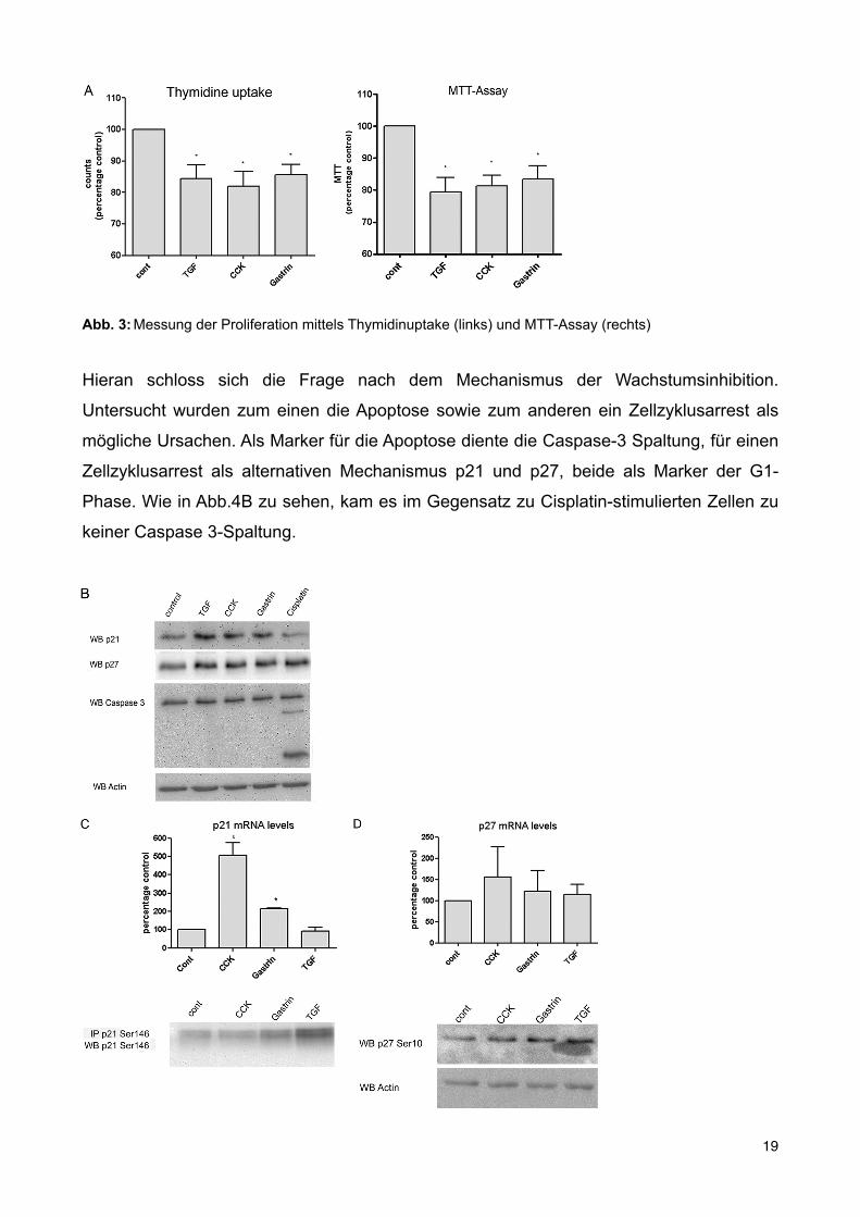
Abb. 3: Messung der Proliferation mittels Thymidinuptake (links) und MTT-Assay (rechts)
Hieran schloss sich die Frage nach dem Mechanismus der Wachstumsinhibition.
Untersucht wurden zum einen die Apoptose sowie zum anderen ein Zellzyklusarrest als
mögliche Ursachen. Als Marker für die Apoptose diente die Caspase-3 Spaltung, für einen
Zellzyklusarrest als alternativen Mechanismus p21 und p27, beide als Marker der G1-
Phase. Wie in Abb.4B zu sehen, kam es im Gegensatz zu Cisplatin-stimulierten Zellen zu
keiner Caspase 3-Spaltung.
demonstrate that, similar to CCK,gastrin stimulated Akt, ERK, andSrc in rat PSC.CCK and Gastrin Modulate Akt
Activation in a Dose-dependentManner in Rat PSC—We haverecently shown that CCK1R in-duced a biphasic modulation of Aktactivity in rat pancreatic acinidepending on the CCK concentra-tion (18). Furthermore, recent pub-lications have shown an importantrole of Akt in PSC activation (6, 19).Phosphorylation of Akt at serine473 has been shown to closely cor-relate with Akt activity in numerousstudies (20, 21). Therefore, weexamined the effect of differentdoses of CCK on Akt Ser473 phos-phorylation in rat PSC (Fig. 4B).Doses as low as 1 pM CCK caused asignificant increase in Akt Ser473phosphorylation. This response wasmaximal at 1 nM (2.36 ! 0.45-foldincrease) and then decreased to bejust slightly above control at 1 !M.These results suggest that CCK hasa complex effect on Akt activationin rat PSC: low doses of CCK inducesignificant Akt activation, whereashigher doses of CCK partiallyreverse this activation. To assess theeffect of CCK2R on Akt activationin rat PSC, we studied the effect ofthe CCK2R-preferring agonist gas-trin on Akt serine 473 phosphoryla-tion (Fig. 4B). Gastrin (0.1 nM)caused a significant (p " 0.01)increase in Akt Ser473 phosphoryla-tion and this effect wasmaximal at 1!M (2.25 ! 0.40-fold). These datasuggest that CCK2R stimulated Aktactivation in rat PSC.CCKs Action on Collagen Produc-
tion, Akt Activation Was Mediatedby Both Receptors but Primarily bythe CCK2R in Rat PSC—AlthoughCCK1R binds CCK with high affin-ity (Kd in the nanomolar range) andgastrin with low affinity (Kd in themicromolar range), the CCK2R hadalmost equal affinity for gastrin andCCK (for review, see Ref. 22). Todetermine whether CCK and gas-trin effects are mediated by CCK1Ror CCK2R, we used L364 andLY288513 as specific CCK1R andCCK2R inhibitors, respectively (for
FIGURE 3. Effect of CCK, Gastrin and TGFbeta1 on proliferation of PSC. A, thymidine uptake: 50,000 cells/wellwere seeded, stimulated for 24 h, and [3H]thymidine incorporation measured. The values represent the mean ! S.E.of 5 experiments and are expressed as percentage of the control. *, p " 0.01 versus control. MTT assay, rat PSC wereisolated and cultured for 7 days. Then 100,000 cells/well were seeded, stimulated, and the MTT assay performed. Thevalues represent the mean ! S.E. of 5 experiments and are expressed as percentage of the control. *, p " 0.01 versuscontrol. B, rat PSC were cultured, serum starved, and stimulated for 24 h with no additions, ow with 2 ng/ml of TGF,1 nM CCK, 1 !M gastrin, 100 !M cisplatin, and then lysed. Lysates were analyzed by Western blot (WB) for p21, p27,and caspase 3. "-Actin served as loading control. Shown is a representative experiment. C, rat PSC were cultured,serum starved, and stimulated for 24 h with no additions, or with 2 ng/ml of TGF, 1 nM CCK, and 1 !M gastrin. Cellswere lysed/harvested and immunoprecipitations (IP) were performed using anti-p21 Ser146. Western blots werethen analyzed for anti-p21 Ser146. RNA from harvested cells was isolated, reverse transcribed, and real time PCR forp21 mRNA was performed. The values indicated represent the mean !S.E. of 4 experiments and are expressed aspercentage of the control. (*, p"0.05 versus control). D, rat PSC were cultured, serum starved, and stimulated for 24 hwith no additionsm or with 2 ng/ml of TGF, 1 nM CCK, and 1 !M gastrin. Cells were lysed/harvested and Western blotswere performed using anti-p27 Ser10. "-Actin served as loading control. RNA from harvested cells was isolated,reverse transcribed, and real time PCR for p27 mRNA was performed. The values indicated represent the mean!S.E.of 4 experiments and are expressed as percentage of the control.
CCK1R and CCK2R in Cells and Collagen Production
DECEMBER 10, 2010 • VOLUME 285 • NUMBER 50 JOURNAL OF BIOLOGICAL CHEMISTRY 38909
at ZENTRUM FUER M
OLEKULARE, on Decem
ber 25, 2010www.jbc.org
Downloaded from
19

Abb.4: Messung der Apoptose mittels Caspase-3 (oben) und G1-Zellzyklusarrest mittels p21 und p27 realtimePCR (Mitte) und Western Blot (unten)
Stattdessen konnte mittels Western Blot und realtimePCR gezeigt werden, dass p21 und
p27 hochreguliert wurden, so dass ein G1-Arrest als Ursache für die Wachstumsinhibition
in Frage kam. Wie in Abb. 4C zu sehen, erfolgte die Regulation von p21 dabei sowohl auf
Transkriptionsebene als auch durch Phosphorylierung, wohingegen p27 hauptsächlich
durch Phosphorylierung reguliert wurde (Abde et al. 2008, Vervoorts et al. 2008).
2.2.3. Profibrogene Signaltransduktionswege nach CCK-Stimulation
Aus Untersuchungen an Azinuszel len ist bekannt, dass CCK vielfält ige
Signaltransduktionswege aktivieren kann, einschließlich Akt, ERK und Src (Masamune et
al. 2010, Pace et al. 2006). In anderen Studien zur Fibrogenese des Pankreas wurden
eben diese Signalwege untersucht und ihr profibrogener Einfluss beschrieben. Daher
wurden die Wirkungen CCKs und Gastrins auf diese Signalwege untersucht. Es zeigte
sich, dass sowohl CCK als auch Gastrin Akt, ERK und Src durch Phosphorylierung
aktivieren können (nicht gezeigt).
Die Aktivierung von ERK durch CCK und Gastrin führte zur Aktivierung des
Transkriptionsfaktors Elk-1. Dies war insofern von Interesse, da Elk-1 die Transkription von
c-fos vermittelt, einem Mechanismus, der in der Aktivierung von pankreatischen
Sternzellen durch Alkohol und Acetaldehyd involviert ist (McCarroll at al. 2003). Die ERK-
Abhängigkeit der Elk-1-Aktivierung wurde mittels spezifischer ERK-Inhibitoren untersucht
(nicht gezeigt).
2.2.4. Einfluss von Akt, ERK und Src auf Kollagensynthese und Proliferation
Wie oben beschrieben konnte zum einen gezeigt werden, dass nach CCK- und Gastrin-
Stimulation in Sternzellen die Kollagenproduktion erhöht und die Proliferation gehemmt,
sowie zum anderen die profibrogenen Signalwege PI3K/Akt, ERK und Src aktiviert
wurden. In einem nächsten Schritt wurde deshalb untersucht welche Signalwege direkt an
den beschriebenen Effekten - Kollagenproduktion und Wachstumshemmung - beteiligt
waren. Wie aus Abb.5 zu entnehmen, kam hierbei insbesondere der Src-Kinase eine
Schlüsselrolle zu. Nach Inhibition mit dem spezifischen Inhibitor PP2 konnten sowohl ein
20
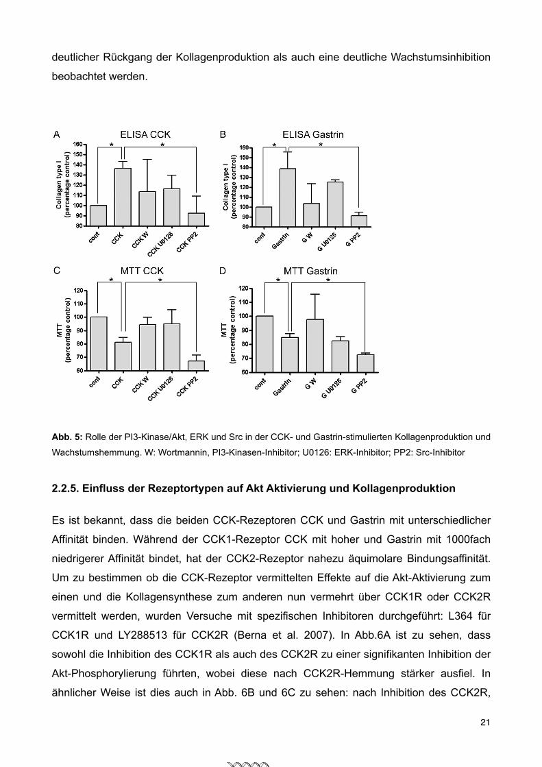
deutlicher Rückgang der Kollagenproduktion als auch eine deutliche Wachstumsinhibition
beobachtet werden.
Our data demonstrate that both CCK and gastrin cause acti-vation of the MEK-ERK pathway in rat PSC, leading to activa-tion of the transcription factor Elk-1. This is compatible withstudies showing activation of ERK by CCK in rat pancreaticacini (10) and with studies showing activation of ERK by CCKand gastrin in AR42J pancreas cancer cells (42). Our findingthat gastrin and CCK induce significant ERK activation is par-ticularly relevant because PSC activation and proliferationcaused by PDGF, one of the strongest known activators of PSC,ismediated by ERK (16, 43).We have shown that bothCCK andgastrin cause reproducible activation of Src family kinases (Fig.4A). This finding is compatible with studies showing activationof Src kinases in rat pancreatic acini by CCK (mediated byCCK1R)(24) and with studies showing activation of Src kinasesby gastrin in AR42J and Panc-1 pancreas cancer cells (44–46)aswell as in othermultiple cells. Activation of Src family kinasesby CCK and gastrin in rat PSC is interesting because Src familykinases activate the JAK2-STAT pathway after PDGF stimula-tion in PSC and thereby could be important regulators of PSCproliferation (26). Furthermore, we were able to show (Fig. 8)
that inhibition of Src reduces colla-gen synthesis and proliferation ina statistically significant manner,pointing to the Src pathway as a piv-otal pathway in CCK- and gastrin-stimulated collagen production andinhibition of proliferation.
Chronic pancreatitis is a com-plex disease and our knowledge ofthe exact pathophysiologic mecha-nisms are still incomplete. Recentstudies suggest that the develop-ment of chronic pancreatitis re-quires a first episode of pancreatitis(sentinel acute pancreatitis event),which, by the release of differentcytokines, triggers activation ofimmune cells and stellate cells,leading to chronic inflammation,fibrosis, and destruction of normalorgan architecture, resulting in lossof organ function (47, 48). In thisprocess, activation of stellate cells iscrucial, because these cells havebeen shown to be responsible forthe development of pancreaticfibrosis, which is a constant featureof chronic pancreatitis. In rodentanimal models, CCK is routinelyused in combination with otheragents to induce chronic pancreati-tis (23, 49–51). These studies havesupposed that the role of CCK inthese models was to trigger the sen-tinel pancreatitis event by interact-ing with CCK receptors on acinarcells and inducing necrosis of acinarcells (49). Our finding that CCK can
directly activate stellate cells and induce collagen productionsuggests that the role of CCK is more complex: 1) by its actionon acinar cells, it could contribute to the induction of the sen-tinel pancreatitis event; and 2) by its action on stellate cells, itcould serve as an important regulator of pancreatic fibrosis. Inconclusion, we report for the first time that rat PSC expressCCK1 and CCK2 receptors, that the natural agonists CCK andgastrin induce activation of pro-fibrotic signaling pathwaysPI3K/Akt, MEK/ERK, and Src, induce activation of the tran-scription factor Elk-1, and most notably significantly increasesynthesis of type I collagen and inhibit proliferation.
REFERENCES1. Saluja, A. K., Saluja, M., Printz, H., Zavertnik, A., Sengupta, A., and Steer,
M. L. (1989) Proc. Natl. Acad. Sci. U.S.A. 86, 8968–89712. Apte, M. V., and Wilson, J. S. (2004) Dig. Dis. 22, 273–2793. Apte, M., McCarroll, J., Pirola, R., and Wilson, J. (2007) Novartis Found.
Symp. 285, 200–2114. Apte, M. V., Phillips, P. A., Fahmy, R. G., Darby, S. J., Rodgers, S. C.,
McCaughan, G. W., Korsten, M. A., Pirola, R. C., Naidoo, D., andWilson,J. S. (2000) Gastroenterology 118, 780–794
FIGURE 8. Pathways involved in CCK and gastrin-stimulated collagen production and inhibition of pro-liferation. A, rat PSC were cultured, serum starved, and stimulated without additions, or preincubated withwortmannin (30 min, 10 !mol), PP2 (10!mol 60 min), U0126 (20!mol 60), and stimulated with 1 nM CCK for 24 h(stimulants were replaced every 12 h). Indirect ELISA was performed using cell culture supernatants. The valuesrepresent the mean ! S.E. of 4 experiments and are expressed as percent of the pre-treatment value. B, rat PSCwere cultured, serum starved, and stimulated without additions, or preincubated with wortmannin (30 min, 10!mol), PP2 (10 !mol 60 min), U0126 (20 !mol 60), and stimulated with 1 !M gastrin for 24 h (stimulants werereplaced every 12 h). Indirect ELISA was performed using cell culture supernatants. The values represent themean ! S.E. of 4 experiments and are expressed as percent of the pre-treatment value. C, MTT assay: rat PSCwere isolated and cultured for 7 days. Then 100,000 cells/well were seeded, stimulated with 1 nM CCK, and theMTT assay was performed. The values represent the mean ! S.E. of 5 experiments and are expressed aspercentage of the control. *, p " 0.01 versus control. D, MTT assay: rat PSC were isolated and cultured for 7 days.Then 100,000 cells/well were seeded, stimulated with 1 !M gastrin, and MTT assay was performed. The valuesrepresent the mean ! S.E. of 5 experiments and are expressed as percentage of the control. *, p " 0.01 versuscontrol. GW, gastrin # wortmannin.
CCK1R and CCK2R in Cells and Collagen Production
DECEMBER 10, 2010 • VOLUME 285 • NUMBER 50 JOURNAL OF BIOLOGICAL CHEMISTRY 38913
at ZENTRUM FUER M
OLEKULARE, on Decem
ber 25, 2010www.jbc.org
Downloaded from
Abb. 5: Rolle der PI3-Kinase/Akt, ERK und Src in der CCK- und Gastrin-stimulierten Kollagenproduktion und
Wachstumshemmung. W: Wortmannin, PI3-Kinasen-Inhibitor; U0126: ERK-Inhibitor; PP2: Src-Inhibitor
2.2.5. Einfluss der Rezeptortypen auf Akt Aktivierung und Kollagenproduktion
Es ist bekannt, dass die beiden CCK-Rezeptoren CCK und Gastrin mit unterschiedlicher
Affinität binden. Während der CCK1-Rezeptor CCK mit hoher und Gastrin mit 1000fach
niedrigerer Affinität bindet, hat der CCK2-Rezeptor nahezu äquimolare Bindungsaffinität.
Um zu bestimmen ob die CCK-Rezeptor vermittelten Effekte auf die Akt-Aktivierung zum
einen und die Kollagensynthese zum anderen nun vermehrt über CCK1R oder CCK2R
vermittelt werden, wurden Versuche mit spezifischen Inhibitoren durchgeführt: L364 für
CCK1R und LY288513 für CCK2R (Berna et al. 2007). In Abb.6A ist zu sehen, dass
sowohl die Inhibition des CCK1R als auch des CCK2R zu einer signifikanten Inhibition der
Akt-Phosphorylierung führten, wobei diese nach CCK2R-Hemmung stärker ausfiel. In
ähnlicher Weise ist dies auch in Abb. 6B und 6C zu sehen: nach Inhibition des CCK2R,
21

konnte eine deutlichere Hemmung der Induktion der Kollagenproduktion bzw. Aufhebung
der Wachstumsinhibition beobachtet werden, verglichen zur Inhibition des CCK1R.
Hieraus ließ sich schließen, dass die beschriebenen Effekte sowohl durch CCK1R als
auch durch CCK2R vermittelt werden, vornehmlich aber durch CCK2-Rezeptoren.
Furthermore, we were able to show that CCK and gastrin stim-ulate different signaling pathways. Thereforewe nextwanted toelucidate which pathways are involved in CCK- and gastrin-stimulated collagen production and inhibition of proliferation.As shown in Fig. 8, A (CCK) and B (gastrin), inhibition of Srcwith the specific inhibitor PP2 leads to a statistically significantdecrease in collagen synthesis in PSC, whereas inhibition ofMEK and PI3K shows only a trend to decrease collagen produc-tion without reaching statistical significance. Fig. 8, C (CCK)
and D (gastrin), shows that thesame intracellular mechanisms areinvolved in inhibiting proliferation.
DISCUSSION
Our results clearly demonstratethat both CCK1 and CCK2 recep-tors are expressed in rat PSC. First,using the RT-PCR technique, wefound that mRNA for both CCK1Rand CCK2R is present in rat PSC.For both PCR, the specificity of theprimer-target interaction was con-firmed by the presence of a specificband in agarose-gel electrophoresis(Fig. 1). Furthermore, an interactionwith possible contaminations ofgenomic DNA can be ruled out bythe absence of a specific band incontrols using genomic DNA (Fig.1A). Furthermore, we were able toshow the presence of both receptorsusing immunohistochemistry andimmunocytology, also showing co-localization with !-SMA (Fig. 1, Band C). Most importantly, the datashow that both receptors areinvolved in crucial cellular func-tions such as collagen synthesis andproliferation. Our data demonstratefor the first time that CCK and gas-trin cause a significant increase incollagen I synthesis (Fig. 2). Synthe-sis of type I collagen contributes toorgan fibrosis in the liver and pan-creas. Therefore, our finding thatCCK receptors have a direct effecton PSC by increasing collagen syn-thesis is of particular interest,because it demonstrates for the firsttime that CCK receptors can inducepancreatic fibrosis. CCK serum lev-els are often elevated in patientswith chronic pancreatitis. A directfibrogenic effect of CCK could be animportant mediator of pancreaticfibrosis, suggesting that CCK recep-tors could be an interesting thera-peutic target in patients with
chronic pancreatitis. To date, the cytokine TGF-" had been themost potent stimulator of collagen synthesis in PSC (35). Asshown in Fig. 2, the effect of CCK and gastrin on collagen syn-thesis was comparable with TGF-"-stimulated collagen pro-duction supporting their potential role in development of pan-creatic fibrosis. Although expression of other matrix proteins,such as fibronectin, TIMP-1, and MMP has been described inPSC (36), CCK and gastrin were unable to stimulate the pro-duction of fibronectin and TIMP1.
FIGURE 5. Effect of CCK1R and CCK2R inhibitor on Akt activation, collagen synthesis, and prolifera-tion mediated by CCK or gastrin in rat PSC. A, rat PSC were cultured for 7 days, serum starved, andpreincubated without additions, 1 #M CCK1R inhibitor L364 for 2.5 min, 10 nM CCK2R inhibitor LY288513,or a combination of both for 5 min. Cells were stimulated without additions (control), 1 nM CCK for 5 minor 1 #M gastrin for 5 min and then lysed. Upper panel, lysates were analyzed by Western blot for AktSer(P)473. Blots were stripped and analyzed for actin. Shown is a representative experiment of a total of 6experiments. Lower panel, Western blots (WB) were scanned and densitometry was performed. The valuesrepresent the mean ! S.E. of 6 experiments and are expressed as fold of the maximal stimulation achievedwith CCK or gastrin alone (*, p " 0.05). B, ELISA: rat PSC were cultured for 7 days, serum starved, andpreincubated with 1 #M L364 for 2.5 min, or with 10 nM CCK2R LY288513 for 5 min, or with the combina-tion. Cells were stimulated without additions, or with 1 nM CCK or 1 #M gasrin for 24 h. Stimulants andinhibitors were replaced after 12 h. Indirect ELISA using cell culture supernatants was performed. MTTassay: rat PSC were cultured for 7 days, serum starved, and preincubated with 1 #M L364 for 2.5 min, with10 nM CCK2R LY288513 for 5 min, or with the combination. Cells were stimulated without additions, orwith 1 nM CCK or 1 #M gasrin for 24 h. Stimulants and inhibitors were replaced after 12 h. MTT assays wereperformed. The values represent the mean ! S.E. of 3 experiments and are expressed as percentage of thecontrol. *, p " 0.05 versus control.
CCK1R and CCK2R in Cells and Collagen Production
DECEMBER 10, 2010 • VOLUME 285 • NUMBER 50 JOURNAL OF BIOLOGICAL CHEMISTRY 38911
at ZENTRUM FUER M
OLEKULARE, on Decem
ber 25, 2010www.jbc.org
Downloaded from
Abb. 6: Rolle der CCK1- und CCK2- Rezeptoren in der Akt-Aktivierung (A), sowie der Kollagenproduktion und Proliferation (B ELISA und MTT)
2.2.6. Zusammenfassung
Die chronische Pankreatitis ist eine komplexe Erkrankung und das Wissen über die
genaue Pathophysiologie ist nach wie vor unvollständig. Aktuelle Studien beschreiben die
Entwicklung hin zu einer chronischen Pankreatitis so, dass zuerst eine akute Pankreatitis
erforderlich ist, als sogenanntes „Sentinel acute pancreatitis event“, welches durch die
Ausschüttung verschiedener Cytokine die Aktivierung von Immunzellen und Sternzellen
22
triggert, um so der chronischen Entzündung Vorschub zu leisten. Dies führt zu Fibrose, zu
Zerstörung der normalen Pankreasarchitektur und zum Verlust der normalen
Organfunktion. Innerhalb dieses Prozesses spielt die Aktivierung pankreatischer
Sternzellen eine grundlegende Rolle, da sich herausgestellt, dass diese Zellen
verantwortlich für die Entstehung der Pankreasfibrose sind, dem typischem Merkmal der
chronischen Pankreatitis.
In Nagetiermodellen wird CCK routinemäßig allein oder in Kombination mit anderen
Stoffen eingesetzt, um eine chronische Pankreatitis zu induzieren. Diese Studien nahmen
an, dass die Rolle CCKs darin bestand durch Interaktion mit CCK-Rezeptoren auf
Azinuszellen die Sentinelpankreatitis auszulösen und in Folge dessen Nekrose zu
induzieren.
Die Ergebnisse konnten zeigen, dass Cholecystokinin Sternzellen direkt aktivieren und die
Kollagensynthese induzieren kann. Dies lässt eine komplexere Rolle von CCK in der
Pathogenese der chronischen Pankreatitis vermuten. CCK könnte zum einen durch seine
Wirkung auf Azinuszellen zu der Entstehung der Sentinelpankreatitis beitragen und zum
anderen durch seine Effekte auf Sternzellen als ein wichtiger Regulator in der Entstehung
der Fibrose fungieren.
Zusammenfassend konnte zum ersten Mal gezeigt werden, dass pankreatische
Sternzellen CCK1- und CCK2- Rezeptoren exprimieren und, dass die natürlich
vorkommenden Agonisten CCK und Gastrin die Aktivierung der profibrogenen Signalwege
PI3K/Akt, MEK/ERK/Elk-1 und Src induzieren. Am wichtigsten jedoch, dass die Stimulation
dieser Rezeptoren die Kollagensynthese signifikant erhöht sowie das Wachstum
signifikant inhibiert.
2.2.7. Literaturverzeichnis
Abde M, Abukhdeir B, Ho P (2008) Expert Rev. Mol. Med. 10, e19
Apte MV, Haber PS, Applegate TL, Norton ID, McCaughan GW, Korsten MA, Pirola RC, Wilson JS (1998) Periacinar stellate shaped cells in rat pancreas – Identification, isolation and culture. Gut 43:128–133.
Apte MV, Phillips PA, Fahmy RG, Darby SJ, Rodgers SC, McCaughan GW, Korsten MA, Pirola RC, Naidoo D, Wilson JS (2000) Gastroenterology 118, 780 –794
Bachem MG, Schneider E, Gross H, Weidenbach H, Schmid RM, Menke A, Siech M, Beger H, Grunert A, Adler G (1998) Identification, culture, and characterization of pancreatic stellate cells in rats and humans. Gastroenterology 115:421–432.
23

Berna MJ, Jensen RT (2007) Curr. Top. Med. Chem. 7, 1211–1231
Gomez Cerezo J, Codoceo R, Fernandez Calle P, Molina F, Tenias M, Vazquez JJ (1991) Digestion 48, 134 –140
Greten H, Rinninger F, Greten T (2010) Innere Medizin 13. Aufl. Thieme, Stuttgart
Hama K, Ohnishi H, Aoki H, Kita H, Yamamoto H, Osawa H, Sato K, Tamada K, Mashima H, Yasuda H, Sugano K (2006) Biochem. Biophys. Res. Commun. 340, 742–750
Klöppel G, Maillet B (1992) Virchows Archiv A Pathol Anat 420:1-4
Kordes C, Brookmann S, Häussinger D, Klonowski-Stumpe H (2005) Pancreas 31, 156 – 167
Masamune A, Satoh M, Kikuta K, Suzuki N, Shimosegawa T (2005) World J Gastroenterol. 11, 6144 – 6151
Masamune A, Kikuta K, Watanabe T, Satoh K, Hirota M, Hamada S, Shimosegawa T (2009) Gut 58, 550 –559
Masamune A, Satoh A, Watanabe T, Kikuta K, Satoh K, Suzuki N, Satoh K, Shimosegawa T (2010) Dig. Dis. Sci. 55, 204 –211
McCarroll JA, Phillips PA, Park S, Doherty E, Pirola RC, Wilson JS, Apte MV (2003) Pancreas 27, 150 –160
Pace A, Tapia JA, Garcia-Marin LJ, Jensen RT (2006) Biochim. Biophys. Acta 1763, 356 – 365
Riede U, Schaefer H, Werner M (2004) Allg. und spezielle Pathologie 5. Aufl. Thieme, Stuttgart
Riemann J, Fischbach W, Galle P, Mössner J (2008) Gastroenterologie 1. Aufl. Bd.2 Thieme, Stuttgart
Saluja AK, Saluja M, Printz H, Zavertnik A, Sengupta A, Steer ML (1989) Proc. Natl. Acad. Sci. U.S.A. 86, 8968 – 8971
Schafmayer A, Becker HD, Werner M, Fölsch UR, Creutzfeldt W (1985) Digestion 32, 136 – 139
Slaff J, Jacobson D, Tillman CR, Curington C, Toskes P (1984) Gastroenterology 87, 44 – 52
Williams JA, Sans MD, Tashiro M, Schaefer C, Bragado MJ, Dabrowski A (2002) Pharmacol. Toxicol. 91, 297–303
Vervoorts J, Lüscher B (2008) Cell Mol. Life Sci. 65, 3255–3264
24

3. Danksagung
Zunächst möchte ich mich bei meinem Doktorvater Herrn PD Dr. Michael Bläker
bedanken, für die Überlassung des Themas und die freundliche Aufnahme in die
Arbeitsgruppe.
Besonderes bedanken möchte ich bei Herrn PD Dr. Andrea Pace für die exzellente
Betreuung. Die Zusammenarbeit mit ihm hat sehr viel Spass gemacht und war
außerordentlich lehrreich.
Mein Besonderer Dank gilt Herrn Friso Nast für das Einarbeiten sowie die tolle Zeit beim
Kratzen. Weiter Frau Martina Fahl für Ihre technische Unterstützung und professionelle
Kritik, sowie Frau Dr. Dorothee Schwinge, Frau Dr. Antonella Carambia, Herrn Dr. Tassilo
Volz und Herrn Dr. Marc Lütgehetmann, die jederzeit ein offenes Ohr und gute Ratschläge
für mich hatten. Darüberhinaus möchte ich mich bei allen anderen aus den Labors der
I. Medizinischen Klinik bedanken für ihre Unterstützung und die schöne Zeit.
Zu guter Letzt möchte ich mich noch ganz besonders bei Herrn Prof. Dr. Stephen
Schröder bedanken. Seine Unterstützung und Ratschläge während des gesamten
Studiums haben all dies erst möglich gemacht. Dafür vielen Dank.
Meinen Eltern soll diese Arbeit gewidmet sein, für Ihre fortwährende Unterstützung und als
Dankeschön für dieses wunderbare Studium.
25

4. Lebenslauf
Persönliche Angaben
Geburtsdatum 13.04.1984
Geburtsort Reutlingen
Schulbildung
2003 Abitur Friedrich-List-Gymnasium Reutlingen
Zivildienst
August 2003 – Juni 2004 Universitätsklinik für Radioonkologie, Tübingen
Studium
August 2006 Erster Abschnitt der ärztlichen Prüfung, Semmelweis
Universität Budapest
Juni 2011 Zweiter Abschnitt der ärztlichen Prüfung, Universität
Hamburg
Veröffentlichungen
Hennigs JK*, Seiz O*, Spiro J, Berna MJ, Baumann HJ, Klose H, Pace A. Molecular basis
of P2-receptor-mediated calcium signaling in activated pancreatic stellate cells. Pancreas.
2011 Jul;40(5):740-6.
Berna MJ*, Seiz O*, Nast JF, Benten D, Blaeker M, Koch J, Lohse AW, Pace A. CCK1 and
CCK2 receptors are expressed on pancreatic stellate cells and induce collagen production.
J Biol Chem. 2010 Dec 10;285(50):38905-14. Epub 2010 Sep 14.
26

5. Eidesstattliche Versicherung
Ich versichere ausdrücklich, dass ich die Arbeit selbständig und ohne fremde Hilfe
verfasst, andere als die von mir angegebenen Quellen und Hilfsmittel nicht benutzt und die
aus den benutzten Werken wörtlich oder inhaltlich entnommenen Stellen einzeln nach
Ausgabe (Auflage und Jahr des Erscheinens), Band und Seite des benutzten Werkes
kenntlich gemacht habe.
Ferner versichere ich, dass ich die Dissertation bisher nicht einem Fachvertreter an einer
anderen Hochschule zur Überprüfung vorgelegt oder mich anderweitig um Zulassung zur
Promotion beworben habe.
Unterschrift: ......................................................................
27