cavity Microscopic s._Dev...mainly mucus – secreting glands providing the moisture and ......
21
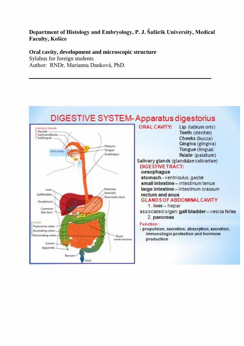
Department of Histology and Embryology, P. J. Šafárik University, Medical Faculty, Košice Oral cavity, development and microscopic structure Sylabus for foreign students Author: RNDr. Marianna Danková, PhD. _____________________________________
Transcript of cavity Microscopic s._Dev...mainly mucus – secreting glands providing the moisture and ......
Department of Histology and Embryology, P. J. Šafárik University, Medical
Faculty, Košice
Oral cavity, development and microscopic structure
Sylabus for foreign students
Author: RNDr. Marianna Danková, PhD.
_____________________________________
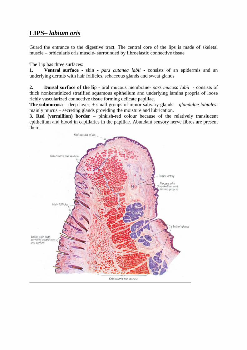
LIPS– labium oris
Guard the entrance to the digestive tract. The central core of the lips is made of skeletal
muscle – orbicularis oris muscle- surrounded by fibroelastic connective tissue
The Lip has three surfaces:
1. Ventral surface - skin - pars cutanea labii - consists of an epidermis and an
underlying dermis with hair follicles, sebaceous glands and sweat glands
2. Dorsal surface of the lip - oral mucous membrane- pars mucosa labii - consists of
thick nonkeratinized stratified squamous epithelium and underlying lamina propria of loose
richly vascularized connective tissue forming delicate papillae.
The submucosa – deep layer, + small groups of minor salivary glands – glandulae labiales-
mainly mucus – secreting glands providing the moisture and lubrication.
3. Red (vermillion) border – pinkish-red colour because of the relatively translucent
epithelium and blood in capillaries in the papillae. Abundant sensory nerve fibres are present
there.
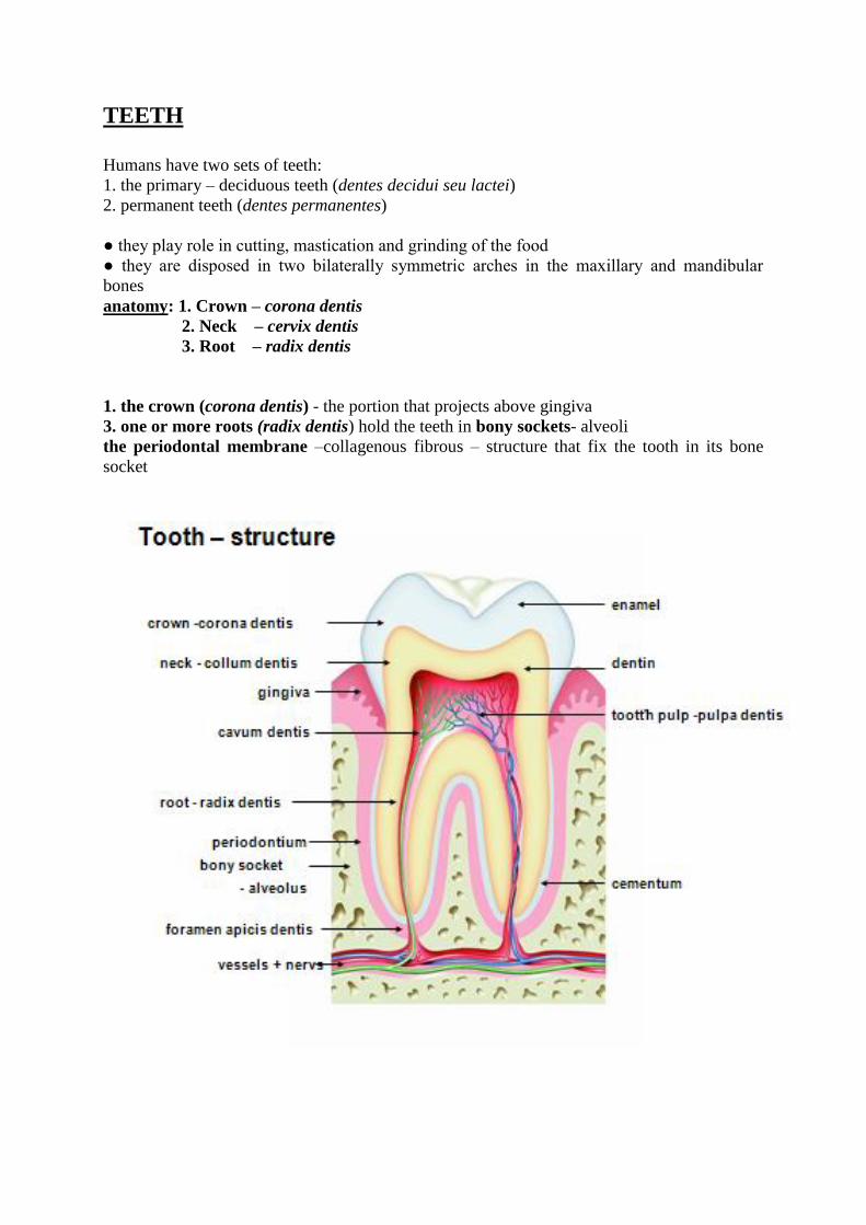
TEETH
Humans have two sets of teeth:
1. the primary – deciduous teeth (dentes decidui seu lactei)
2. permanent teeth (dentes permanentes)
● they play role in cutting, mastication and grinding of the food
● they are disposed in two bilaterally symmetric arches in the maxillary and mandibular
bones
anatomy: 1. Crown – corona dentis
2. Neck – cervix dentis
3. Root – radix dentis
1. the crown (corona dentis) - the portion that projects above gingiva
3. one or more roots (radix dentis) hold the teeth in bony sockets- alveoli
the periodontal membrane –collagenous fibrous – structure that fix the tooth in its bone
socket
Histologycal composition of the tooth –
I. Hard tissues: enamel (enamelum), dentine (dentinum), cement (cementum)
II. Soft tissue: tooth (dental) pulp (pulpa dentis)
III. Supporting tissues: periodontium (periodontium), gingiva (gingiva), alveolar bone
Enamel – enamelum
● covers the crown
● the hardest component of human body, consists of
→ 95% calcium salts – mainly hydroxyapatite
→ organc matrix – made of special glycoproteins : amelogenins and enamelins
● enamel consists of elongated columns of hydroxyapatite crystals enamel rods (prisms) that
are bound together by mineralized interrod enamel
● the prisms extend through entire thickness of enamel layer
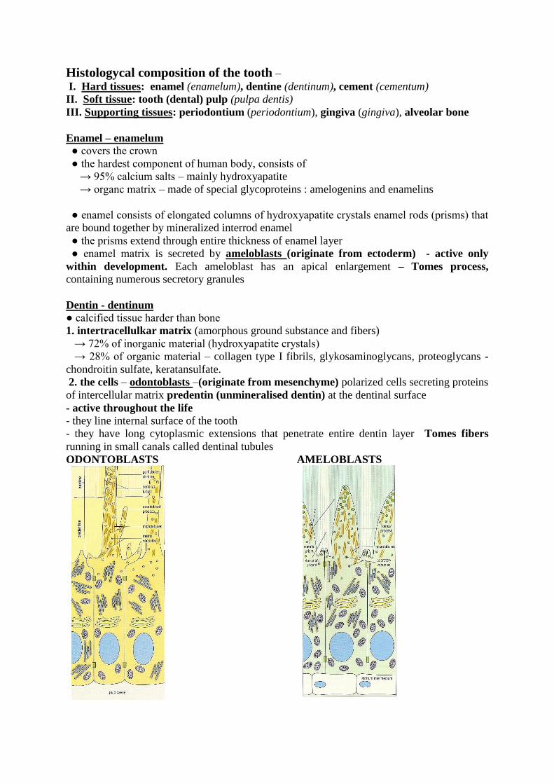
● enamel matrix is secreted by ameloblasts (originate from ectoderm) - active only
within development. Each ameloblast has an apical enlargement – Tomes process,
containing numerous secretory granules
Dentin - dentinum
● calcified tissue harder than bone
1. intertracellulkar matrix (amorphous ground substance and fibers)
→ 72% of inorganic material (hydroxyapatite crystals)
→ 28% of organic material – collagen type I fibrils, glykosaminoglycans, proteoglycans -
chondroitin sulfate, keratansulfate.
2. the cells – odontoblasts –(originate from mesenchyme) polarized cells secreting proteins
of intercellular matrix predentin (unmineralised dentin) at the dentinal surface
- active throughout the life - they line internal surface of the tooth
- they have long cytoplasmic extensions that penetrate entire dentin layer Tomes fibers
running in small canals called dentinal tubules
ODONTOBLASTS AMELOBLASTS
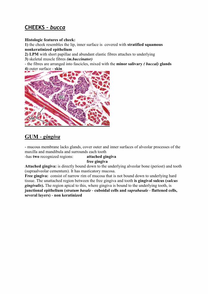
CHEEKS – bucca Histologic features of cheek:
1) the cheek resembles the lip, inner surface is covered with stratified squamous
nonkeratinized epithelium
2) LPM with short papillae and abundant elastic fibres attaches to underlying
3) skeletal muscle fibres (m.buccinator)
- the fibres are arranged into fascicles, mixed with the minor salivary ( buccal) glands
4) outer surface - skin
GUM - gingiva
- mucous membrane lacks glands, cover outer and inner surfaces of alveolar processes of the
maxilla and mandibula and surrounds each tooth
-has two recognized regions: attached gingiva
free gingiva
Attached gingiva: is directly bound down to the underlying alveolar bone (periost) and tooth
(supraalveolar cementum). It has masticatory mucosa.
Free gingiva: consist of narrow rim of mucosa that is not bound down to underlying hard
tissue. The unattached region between the free gingiva and tooth is gingival sulcus (sulcus
gingivalis). The region apical to this, where gingiva is bound to the underlying tooth, is
junctional epithelium (stratum basale - cuboidal cells and suprabasale - flattened cells,
several layers) - non keratinized
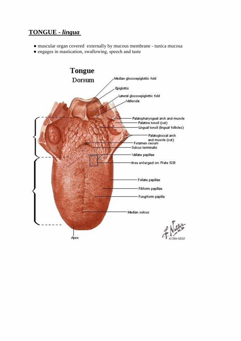
TONGUE - lingua
● muscular organ covered externally by mucous membrane - tunica mucosa
● engages in mastication, swallowing, speech and taste
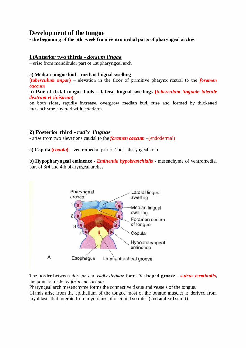
Development of the tongue
- the beginning of the 5th week from ventromedial parts of pharyngeal arches
1)Anterior two thirds - dorsum lingae – arise from mandibular part of 1st pharyngeal arch
a) Median tongue bud – median lingual swelling
(tuberculum impar) – elevation in the floor of primitive pharynx rostral to the foramen
caecum
b) Pair of distal tongue buds – lateral lingual swellings (tuberculum linguale laterale
dextrum et sinistrum)
on both sides, rapidly increase, overgrow median bud, fuse and formed by thickened
mesenchyme covered with ectoderm.
2) Posterior third - radix linguae - arise from two elevations caudal to the foramen caecum –(endodermal)
a) Copula (copula) – ventromedial part of 2nd pharyngeal arch
b) Hypopharyngeal eminence - Eminentia hypobranchialis - mesenchyme of ventromedial
part of 3rd and 4th pharyngeal arches
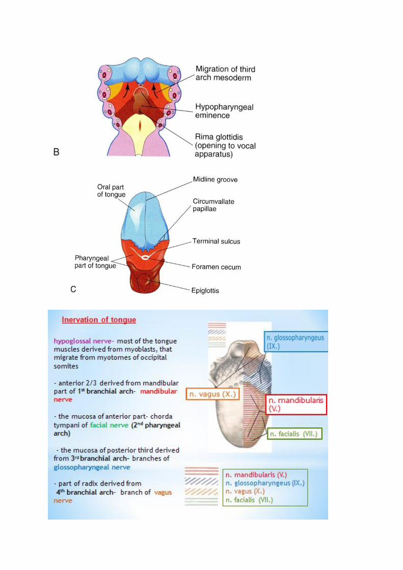
The border between dorsum and radix linguae forms V shaped groove - sulcus terminalis,
the point is made by foramen caecum.
Pharyngeal arch mesenchyme forms the connective tissue and vessels of the tongue.
Glands arise from the epithelium of the tongue most of the tongue muscles is derived from
myoblasts that migrate from myotomes of occipital somites (2nd and 3rd somit)
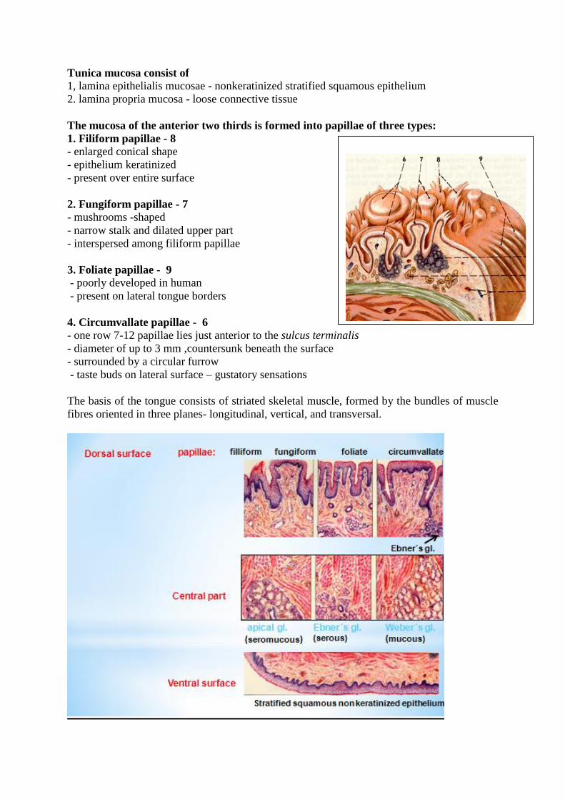
Tunica mucosa consist of 1, lamina epithelialis mucosae - nonkeratinized stratified squamous epithelium
2. lamina propria mucosa - loose connective tissue
The mucosa of the anterior two thirds is formed into papillae of three types:
1. Filiform papillae - 8
- enlarged conical shape
- epithelium keratinized
- present over entire surface
2. Fungiform papillae - 7
- mushrooms -shaped
- narrow stalk and dilated upper part
- interspersed among filiform papillae
3. Foliate papillae - 9
- poorly developed in human
- present on lateral tongue borders
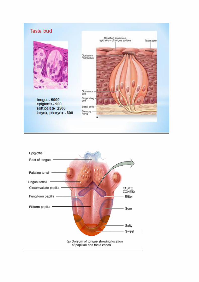
4. Circumvallate papillae - 6
- one row 7-12 papillae lies just anterior to the sulcus terminalis
- diameter of up to 3 mm ,countersunk beneath the surface
- surrounded by a circular furrow
- taste buds on lateral surface – gustatory sensations
The basis of the tongue consists of striated skeletal muscle, formed by the bundles of muscle
fibres oriented in three planes- longitudinal, vertical, and transversal.
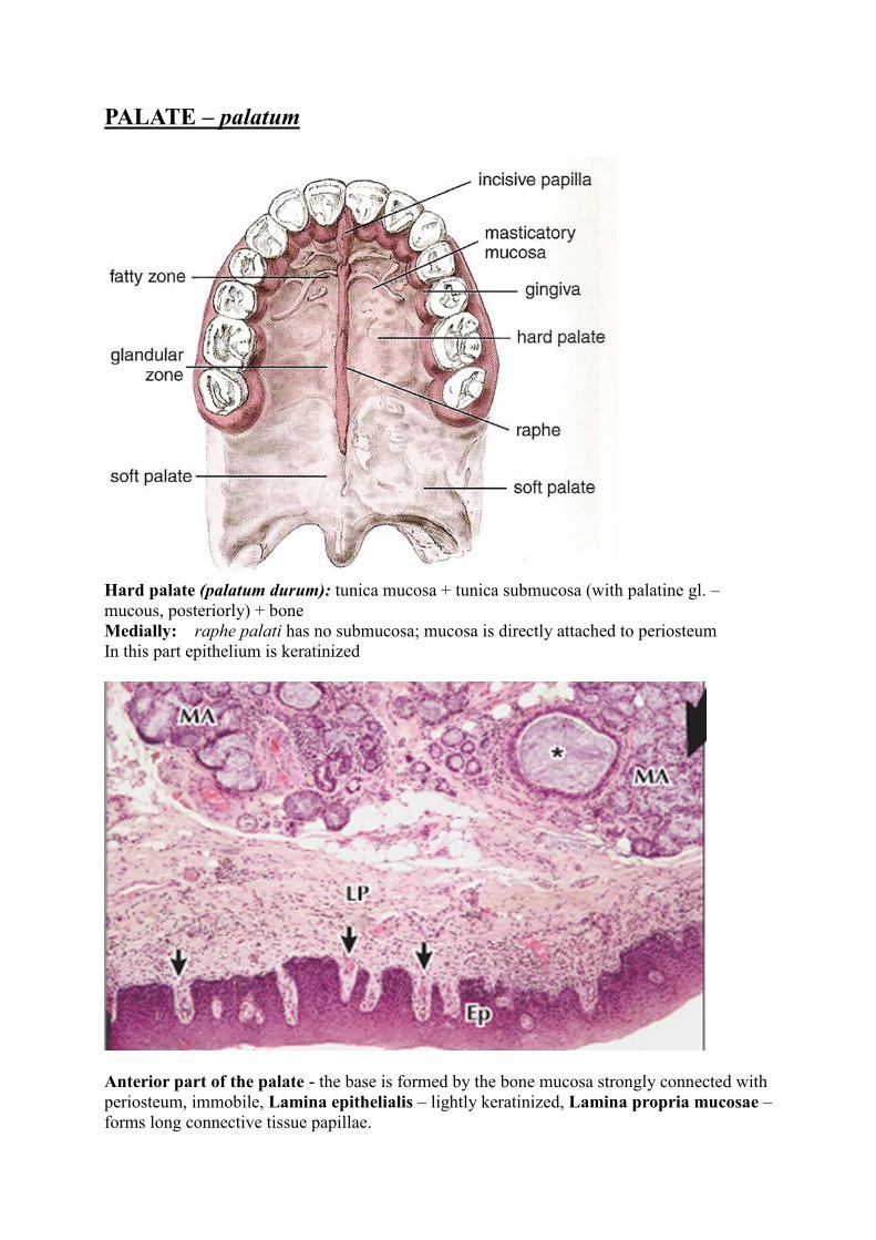
PALATE – palatum
Hard palate (palatum durum): tunica mucosa + tunica submucosa (with palatine gl. –
mucous, posteriorly) + bone
Medially: raphe palati has no submucosa; mucosa is directly attached to periosteum
In this part epithelium is keratinized
Anterior part of the palate - the base is formed by the bone mucosa strongly connected with
periosteum, immobile, Lamina epithelialis – lightly keratinized, Lamina propria mucosae –
forms long connective tissue papillae.
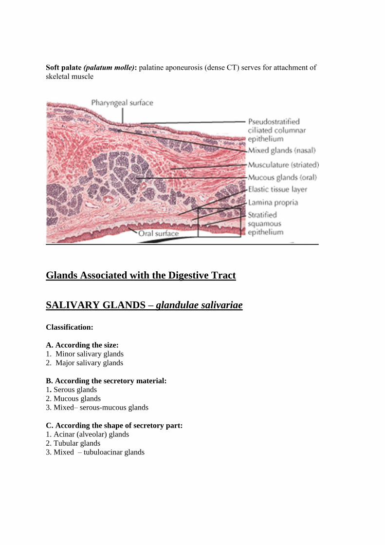
Soft palate (palatum molle): palatine aponeurosis (dense CT) serves for attachment of
skeletal muscle
Glands Associated with the Digestive Tract
SALIVARY GLANDS – glandulae salivariae
Classification:
A. According the size:
1. Minor salivary glands
2. Major salivary glands
B. According the secretory material:
1. Serous glands
2. Mucous glands
3. Mixed– serous-mucous glands
C. According the shape of secretory part:
1. Acinar (alveolar) glands
2. Tubular glands
3. Mixed – tubuloacinar glands
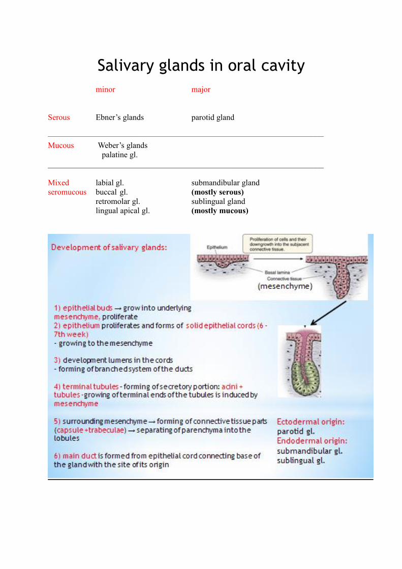
Salivary glands in oral cavity
minor major
Serous Ebner’s glands parotid gland
––––––––––––––––––––––––––––––––––––––––––––––––––––––––––––––––––––
Mucous Weber’s glands
palatine gl.
____________________________________________________________________
Mixed labial gl. submandibular gland
seromucous buccal gl. (mostly serous)
retromolar gl. sublingual gland
lingual apical gl. (mostly mucous)
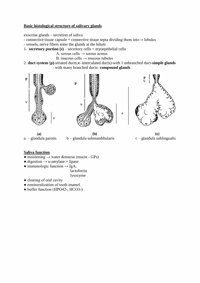
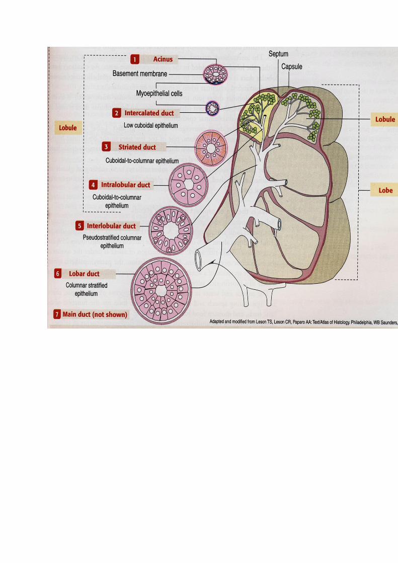
Basic histological structure of salivary glands
exocrine glands – secretion of saliva
- connective tissue capsule + connective tissue septa dividing them into→ lobules
- vessels, nerve fibers enter the glands at the hilum
1. secretory portion (s) – secretory cells + myoepithelial cells
A. serous cells → serous acinus
B. mucous cells → mucous tubules
2. duct system (p)-striated ducts,v–intercalated ducts)-with 1 unbranched duct-simple glands
- with many branched ducts- compound glands
a – glandula parotis b – glandula submandibularis c – glandula sublingualis
Saliva function
● moistening → water &mucus (mucin - GPs)
● digestion → α-amylase + lipase
● immunologic function → IgA,
lactoferrin
lysozyme
● clearing of oral cavity
● remineralization of tooth enamel
● buffer function (HPO42-, HCO3-)

Serous cells (A)
Mucous cells (B)
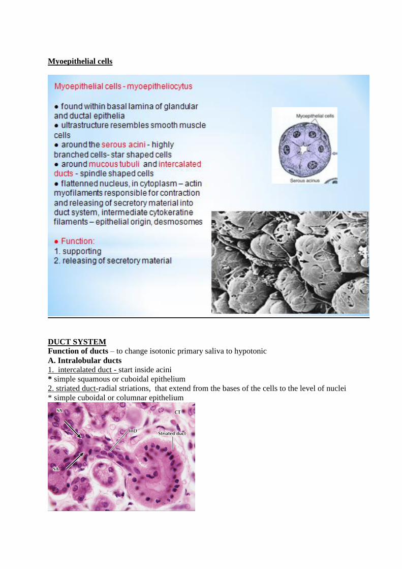
Myoepithelial cells
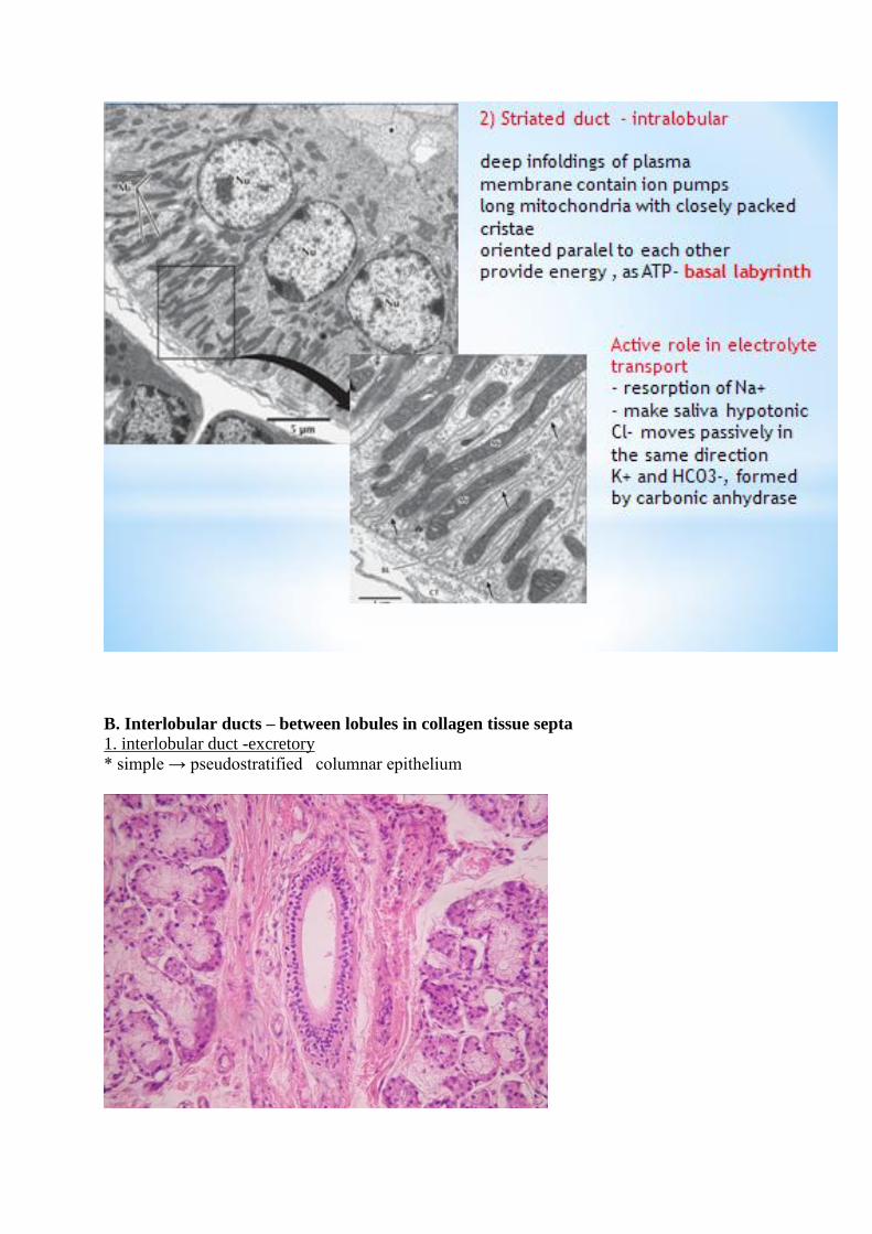
DUCT SYSTEM
Function of ducts – to change isotonic primary saliva to hypotonic
A. Intralobular ducts
1. intercalated duct - start inside acini
* simple squamous or cuboidal epithelium
2. striated duct-radial striations, that extend from the bases of the cells to the level of nuclei
* simple cuboidal or columnar epithelium
B. Interlobular ducts – between lobules in collagen tissue septa
1. interlobular duct -excretory
* simple → pseudostratified columnar epithelium
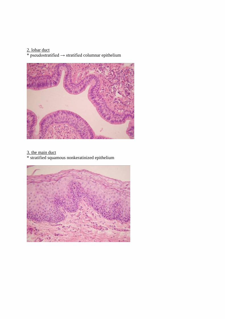
2. lobar duct
* pseudostratified → stratified columnar epithelium
3. the main duct
* stratified squamous nonkeratinized epithelium
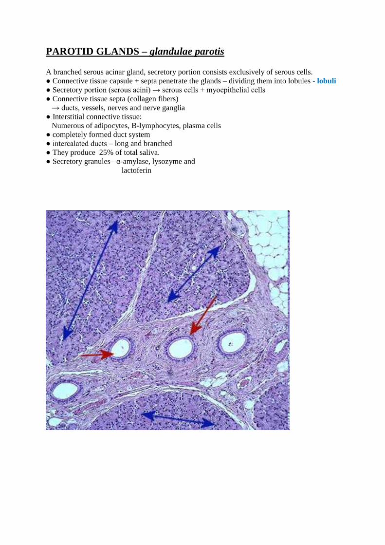
PAROTID GLANDS – glandulae parotis A branched serous acinar gland, secretory portion consists exclusively of serous cells.
● Connective tissue capsule + septa penetrate the glands – dividing them into lobules - lobuli
● Secretory portion (serous acini) → serous cells + myoepithelial cells
● Connective tissue septa (collagen fibers)
→ ducts, vessels, nerves and nerve ganglia
● Interstitial connective tissue:
Numerous of adipocytes, B-lymphocytes, plasma cells
● completely formed duct system
● intercalated ducts – long and branched
● They produce 25% of total saliva.
● Secretory granules– α-amylase, lysozyme and
lactoferin
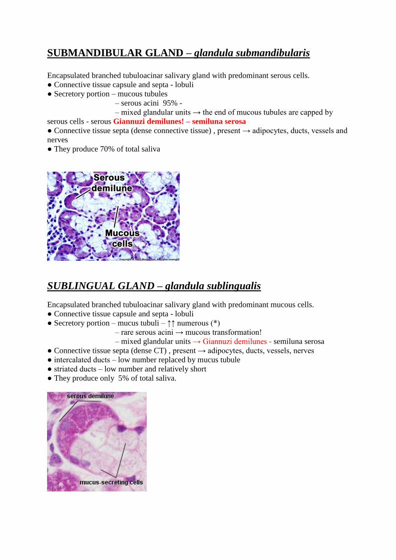
SUBMANDIBULAR GLAND – glandula submandibularis
Encapsulated branched tubuloacinar salivary gland with predominant serous cells.
● Connective tissue capsule and septa - lobuli
● Secretory portion – mucous tubules
– serous acini 95% -
– mixed glandular units → the end of mucous tubules are capped by
serous cells - serous Giannuzi demilunes! – semiluna serosa
● Connective tissue septa (dense connective tissue) , present → adipocytes, ducts, vessels and
nerves
● They produce 70% of total saliva
SUBLINGUAL GLAND – glandula sublingualis
Encapsulated branched tubuloacinar salivary gland with predominant mucous cells.
● Connective tissue capsule and septa - lobuli
● Secretory portion – mucus tubuli – ↑↑ numerous (*)
– rare serous acini → mucous transformation!
– mixed glandular units → Giannuzi demilunes - semiluna serosa
● Connective tissue septa (dense CT) , present → adipocytes, ducts, vessels, nerves
● intercalated ducts – low number replaced by mucus tubule
● striated ducts – low number and relatively short
● They produce only 5% of total saliva.