Role of proteolysis in physiological and pathological processes

1
CAVEOLIN-1: pathological and physiological implications.
Vanesa Pereira-Prado1, Gabriel Tapia2, Ronell Bologna-Molina3.
DOI: 10.22592/o2017n30a10
ABSTRACT
Caveolae are plasma membrane invaginations formed by proteins called caveolins. Of
the three caveolin isoforms, the most studied one through years has been caveolin-1
(cav-1), which has an important role in cell signaling, and its gene, CAV-1, is part of the
family of tumor suppressor genes. As its role depends on the context, the participation
and function of cav-1 in tumors is complex and remains controversial. Cav-1 interacts
with a series of receptors and molecules that regulate the initial steps of cellular
transformation to malignity. It also participates in the cell cycle, angiogenesis,
extracellular matrix remodeling, cell proliferation, among other processes. The study of
this molecule is important due to its function as a biomarker associated to the
diagnosis, prognosis and therapeutic target in pathological processes.
Keywords: caveolae; tumor suppression, cell signaling, MMPs.
1 Assistant of Molecular Pathology in Stomatology, MSc Candidate in Dental Sciences, School
of Dentistry, Universidad de la República, Uruguay ORCID: 0000-0001-7747-6718
2 Assist. Professor, Histology Department, School of Dentistry, Universidad de la República,
Uruguay. ORCID: 0000-0003-4563-9142
3 Professor of Molecular Pathology in Stomatology, School of Dentistry, Universidad de la
República, Uruguay. ORCID: 0000-0001-9755-4779
Received on: 14 Dec 2016 - Accepted on: 20 Sep 2017

2
INTRODUCTION
Caveolin-1 (Cav-1) is a plasma membrane protein that has a fundamental role in the
formation of caveolae and acts as a connective protein that organizes the
macromolecular complexes on the cell surface which participate in intracellular
signaling. Cav-1 interacts with various signaling proteins and growth factors,
intervening in various physiological and pathological processes. The aim of this review
is to collect information in relation to the expression of this protein in different biological
processes.
METHODOLOGY USED FOR DATA COLLECTION
A computer search of the literature, in Spanish and English, was conducted in Pubmed,
SciELO, Science Direct and Timbó databases for the 2005-2016 period, using
keywords such as: caveolin review, caveolin-1, cell cycle modulation, angiogenesis,
matrix remodeling. Fifty-five full-text papers available related to medicine were selected
to guide this literature review.
TUMOR SUPPRESSION: What is it?
Tumor suppressor genes are the ones that protect the cell from abandoning its normal
function and taking the carcinogenesis pathways. After their mutation, which causes
function loss or reduction, cells can progress to a malignant tumor in combination with
other genetic and metabolic changes. Tumor suppressor genes, like oncogenes, have
diverse functions in the regulation of cell growth, cell differentiation and programmed
cell death (apoptosis)(1). Both the activation of oncogenes and the inactivation of tumor
suppressor genes are critical steps in tumor initiation and progression. Over time, the
damage accumulated in several genes is responsible for changes, characteristics of
carcinomatous cells such as the increase in their ability to proliferate, to invade other
tissues and to generate metastasis(1-2). Tumor suppressor proteins encoded by these

3
genes can have a repressive effect on the regulation of cell proliferation or survival of
tumor cells(3).
CELL SIGNALLING: What is it?
Cell signaling molecules are part of a complex cellular communication system that
governs basic cellular activities and the coordination of their actions. The ability to
perceive and respond correctly to the immediate microenvironment is the basis for
development, tissue repair, immunity and homeostasis. Errors in processing
information cause different pathologies, including tumors. If we understand cell
signaling, different diseases can be addressed therapeutically more efficiently.
CAVEOLAE AND CAVEOLINS: What are they?
Caveolae are 50-100-nm invaginations located in the plasma membrane, enriched in
cholesterol and sphingolipids. Each caveola is formed by about 100-200 integral
membrane proteins called caveolins(4).
The three caveolin isoforms(1-3) are encoded by different genes, but their structures are
very similar. Their structure consists of N- and C- terminal cytosolic domains, an
oligomerization domain, including a Caveolin scaffolding domain, responsible for
dimerization and interaction with signaling partners (see figure 1)(4). Cav-1 presents two
isoforms: “alpha” with 178 24 k Da amino acids and “beta” with 148 21 k Da amino
acids(5). Cav-1 and Cav-2 are co-expressed in adipocytes, fibroblasts and endothelial
cells, while Caveolin 3 appears mainly in striated muscle tissues(6). Their activity is
highly dependent on the cellular context in which they are found, them being positive
and negative regulators of cell signaling both inside and outside the caveola. This set
of proteins and lipids accumulated in micro membrane domains can communicate a
rapid, specific and amplified signal cascade, cell proliferation, apoptosis and
migration(7).
4
Of these three caveolin proteins, the most studied one through years has been cav-1,
which has an important role in cell signaling, and its gene, CAV-1, is part of the family
of tumor suppressor genes. Due to its versatile functions, this protein is being
researched as a therapeutic and diagnostic target to understand cancer.
CAVEOLIN 1
The discovery of cav-1 as the first molecular biomarker of caveolae has allowed
biochemical, biological and genetic advances with results that have significantly
contributed to understanding these structures.
At the cellular level, various cellular components containing cav-1 have been
determined(7), among which we include: cytoplasmic membrane, Golgi apparatus,
mitochondria(8), endoplasmic reticulum, membrane vesicles(9) and nucleus(10).
Fig. 1. Caveolae structure: when cav is inserted in the lipid layers of the cell membrane,
invaginations are formed, leading to the three-dimensional structure that makes up the
caveolae.

5
Table I. Intracellular location of cav-1 related to its function
Cellular localization Normal function
Cytoplasmic membrane Signal transducer
Golgi apparatus Lipid transporter
Mitochondria Lipid transporter and metabolic regulator
Endoplasmic reticulum Lipid transporter
Plasma membrane of vesicles Endocytosis, exocytosis, transcytosis
Nucleus Tumor suppressor, genetic regulator
A cav-1 deficiency at the mitochondrial level triggers a series of disorders including lipid
dystrophy, cancer, diabetes, muscular dystrophy, heart disease and pulmonary
fibrosis(7). Some studies suggest that the role of cav-1 as a cholesterol transporter and
homeostasis regulator would affect mitochondrial function. In the absence of cav-1,
cholesterol would accumulate in the mitochondria, causing dysfunction by reducing the
membrane flow, which would decrease the efficiency of energy production by the
respiratory chain and increase the production of reactive oxygen species (ROS). These
events involve whole cell proliferation and induce apoptosis with a limited amount of
available glucose(11).
The activity of a large amount of signaling proteins can be altered by changing their
expression at the cell surface level, and this expression can be regulated by cav-1 at
the membrane level through endocytosis and exocytosis. In turn, cav-1-dependent
endocytosis regulates cell attachment by internalizing integrins and components of tight

6
junctions (occludens) and adhesion. Additionally, the mediator function of transcytosis
allows for the transport of albumin-conjugated nutrients, fatty acids and hormones
through the endothelium(7). At the nuclear level, cav-1 acts as a genetic regulator and
tumor suppressor. The CAV-1 gene that encodes cav-1 protein is a negative regulator
and tumor suppressor gene of the Ras-p42/44 MPak cascade(12). This protein is
responsible for integrin binding with tyrosine kinase prior to the start of the Ras-MAPK
pathway to promote cell cycle progression(13).
Therefore, if cav-1 does not bind the integrin with the tyrosine kinase, cell signaling is
affected, altering the cellular processes. The persistent suppression of cav-1 promotes
the emergence of pathogenic phenotypes, and several studies associate it with
advanced stages of breast cancer and prostate cancer metastasis, and a poor
prognosis(13).
CAVEOLIN-1: Does it intervene in the regulation of the cell cycle?
Cav-1 expression stops the cell cycle in G0/G1 phase. Decreased regulation of cav-1
induces cells to withdraw from G0/G1/S phase, reduces cell proliferation and lowers
the rate of DNA replication. This occurs through the p53/p21 dependent pathway. The
p53 tumor suppressor protein is an intracellular regulator involved in the modulation of
cell cycle progression; its activity is increased when cav-1 is overexpressed(14).
Cav-1 has been shown to suppress signaling in many cell types, determining that its
overexpression interferes with the vascular endothelial growth factor (VEGF) signal and
with cell proliferation in the case of endothelial cells(15).
Cav-1 negatively controls the progression of the cell cycle, leading to more advanced
cancer phases. It interacts with a number of receptor molecules that do not function
specifically in the caveolae but that initially regulate the steps of cell transformation(16).

7
Cyclin D1 expression is regulated by cav-1 expression and the aberrant activation of
the transducer and activator signals of STAT3 transcription-3. Cyclin D1 belongs to a
family of proteins that promote the progression of cancer cells through the G1-S
phases of the cell cycle. In turn, STAT3 may regulate cav-1 expression and function,
both being a reciprocal network that regulates metastasis(17).
CAVEOLIN-1: How does it participate in angiogenesis?
Angiogenesis is the process in which new vessels are formed by proliferation of
vascular endothelial cells. Cav-1 is considered a negative regulator of VEGF-R (VEGF
receptor), causing a decrease in angiogenesis. Cav-1, in endothelial cells, then
regulates angiogenesis, vascular permeability and vascular remodeling(4). VEGF
regulates angiogenesis by the interaction of cell signaling pathways, and, in cancer, it
is secreted by tumor cells and activates endothelial cells in a paracrine manner to
initiate angiogenesis processes.
Additionally, cav-1 also plays a role as a positive regulator of angiogenesis through the
PI3K/Akt pathway(18).
CAVEOLIN-1 AND ITS CONNECTION WITH ONCOGENESIS
Cav-1 is believed to have antiapoptotic and tumor-promoting characteristics, to
stimulate metastasis and to have a prognostic value of patient survival and tumor
recurrence. Conversely, other authors suggest that cav-1 can function as suppressor
and pre-apoptotic protein, so its loss promotes tumor properties by changing the
phenotype(4). Several authors state that depending on the state of the tumor, cav-1 acts
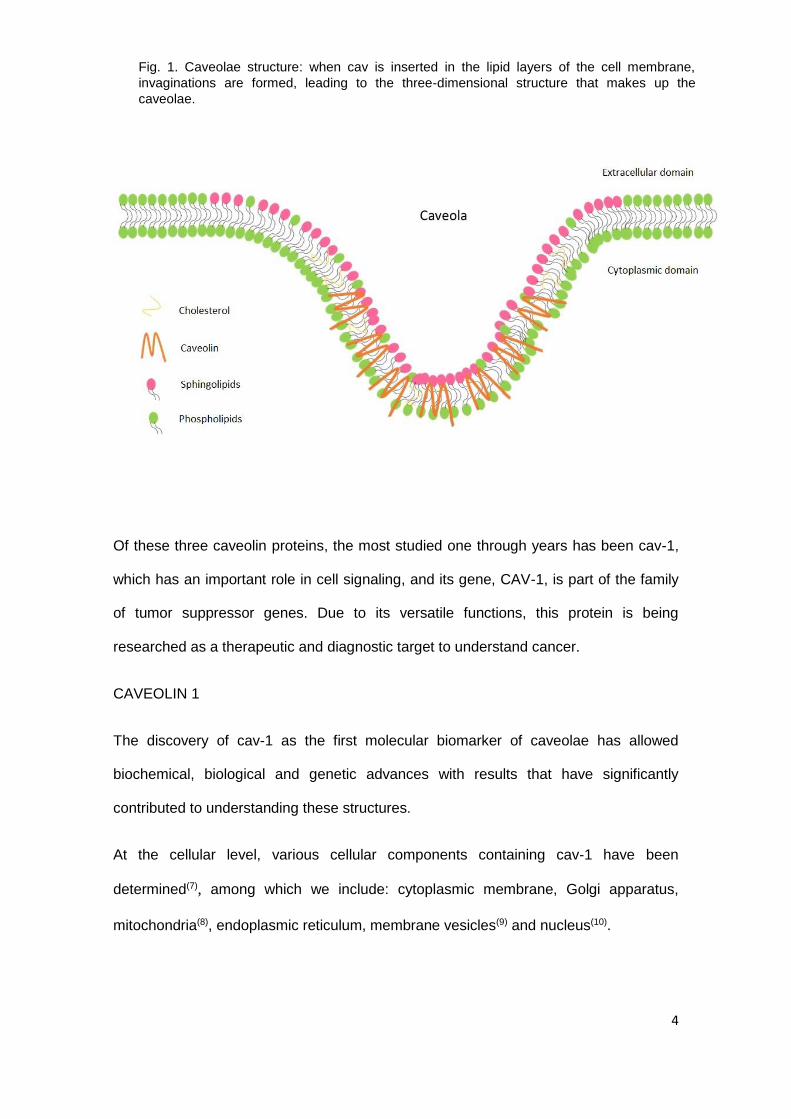
as a suppressor or an oncogene. Cav-1 is variably expressed in malignant and benign
neoplasms (Fig. 2). In early stages of tumor progression, cav-1 restricts its growth,
while in advanced stages it promotes it(19).
8
Cav-1 intervenes in one of the mechanisms involved in the oncogenic transformation of
fibroblasts. It participates in tumorigenesis through functions such as lipid transport,
membrane traffic, gene regulation and signal transduction(20). The existence of a
mutagenic domain of cav-1 can be determined, where the primary tumor formation is
due to a loss of cellular function because of cav-1 mutation, favoring the increase in the
regulation of the pathways involved in cell migration, invasion and metastasis(21).
Fig. 2. Differential immunoexpression of caveolin-1 in ameloblastoma
tumor cells. The arrows mark the immunopositivity of the tumor cells in
brown color. 40x increase.

9
CAVEOLIN-1: How is it related to matrix metalloproteinases?
The location of certain matrix metalloproteinases (MMPs) at the level of endothelial
caveolae suggests that cav-1 plays a role in their regulation. Several studies have
found that MMP-1 expression decreases after a cav-1 overexpression and vice
versa(22). Because both molecules participate in fibrosis, tumor progression and
metastasis, there have been recent publications suggesting the connection between
them(23). MMP-3, MMP-9 and MMP-14 have been found at the level of the caveolae,
suggesting this interaction. One type of MMP called “Membrane type 1”, located at the
caveolae level is negatively regulated when cav-1 causes internalization from the cell
surface(23). Physiologically, cav-1 controls cell motility by interfering with the
cytoskeleton and by modulating the interaction with the extracellular matrix, therefore, it
is involved in its remodeling by promoting its interaction with MMPs(22).
CAVEOLIN-1 and its relationship with other proteins
Cav-1 can affect cell adhesion when modulating E-cadherin, which is the major
component of intercellular junctions, maintaining a cell-cell contact and reducing the
possibility of metastasis(24). The loss of E-cadherin is a prerequisite for cell migration
and the development of an invasive and metastatic carcinogenic phenotype. Somehow,
cav-1 overexpression has been associated with abnormal expression of at least one of
the forms of E-cadherin and multiple intercellular receptors(24).
Previous studies have linked cav-1 with the epidermal growth factor (EGF) and its
EGFR receptor. Although cav-1 seems to reduce the signal transduction, its
phosphorylated form has been associated with cell migration, inducing EGF and
programmed cell death(16).
CONCLUSIONS

10
Cav-1 participates in several normal biological processes, such as angiogenesis, and
its alteration is reflected in various diseases, such as oncogenesis and tumor
progression. The study of this molecule is important given its usefulness as a
biomarker associated with diagnosis, prognosis and possible therapeutic target in
pathological processes. This forces us to continue developing research on this protein.
ACNOWLEDGEMENTS
We would like to thank Dr. Mariana Villaroel Dorrego, School of Dentistry, Universidad
Central de Venezuela, and the MSc students of the same university, Mayerlimg
Delgado and Rossana Gomez, for their collaboration in the review.
REFERENCES
1. Sherr, C.J. Principles of tumor suppression. Cell 2004; 116 (2): 235-46.
2. Yoshida, B.A.; Sokoloff, M.M.; Welch, D.R. & Rinker-Schaeffer, C.W.
Metastasis-suppressor genes: a review and perspective on an emerging field. J.
Natl. Cancer Inst. 2000; 92 (21): 1717-30.
3. Cooper, G.M. The Cell: A Molecular Approach. Tumor Suppressor Genes. 2nd
edition. Sunderland (MA): Sinauer Associates 2000.
4. Hehlgans, S. & Cordes, N. Caveolin-1: an essential modulator of cancer cell
radio and Chemoresistance. Am. J. Cancer. Res., 1 (4): 521-530, 2011.
5. Radu, V. Structure of caveolae. Biochym. et Biophy 2005; 1746, 334–348.
6. Boscher. C. & Nabi. I.R. Caveolin-1: role in cell signaling. Adv Exp Med
Biol.2012; 729: 29-50.
7. Fridolfsson, H.N.; Roth, D.M.; Insel, P.A. & Patel, H.H. Regulation of
intracellular signaling and function by caveolina. FASEB J. 2014; 28 (9): 3823-
3831.

11
8. Fridolfsson, H.N.; Kawaraguchi, Y.; Ali, S.S.; Panneerselvam, M.; Niesman,
I.R.; Finley, J.C.; et al. Mitochondria-localized caveolin in adaptation to cellular
stress and injury. FASEB J. 2012; 26 (11): 4637-49.
9. Tagawa, A.; Mezzacasa, A.; Hayer, A.; Longatti, A.; Pelkmans, L. & Helenius,
A.J. Assembly and trafficking of caveolar domains in the cell: caveolae as
stable, cargo-triggered, vesicular transporters. Cell Biol 2005; 29; 170 (5): 769-
79.
10. Jeong, K.; Kwon, H.; Lee, J.; Jang, D.; Hwang, E.M.; Park, J.Y. & Pak, Y. Rab6-
mediated retrograde transport regulates inner nuclear membrane targeting of
caveolin-2 in response to insulin. Traffic. 2012; 13 (9): 1218-33.
11. Bosch, M.; Marí, M.; Herms, A.; Fernández, A.; Fajardo, A.; Kassan, A, et al.
Caveolin-1 deficiency causes cholesterol-dependent mitochondrial dysfunction
and apoptotic susceptibility. Curr. Biol.2011; 26; 21 (8):681-6.
12. Razani, B.; Woodman, S.E. & Lisanti, M.P. Caveolae: from cell biology to
animal physiology. Pharmacol. Rev 2002; 54: 431-67.
13. Lamaze, C. & Torrino, S. Caveolae and cancer: A new mechanical perspective.
Biomed. J 2015; 38: 367-7.
14. Galbiati F.; Volonte D.; Liu J.; Capozza F.; Frank P.; Zhu L,; Pestell R.
& Michael P. Lisanti.Caveolin-1 Expression Negatively Regulates Cell Cycle
Progression by Inducing G0/G1 Arrest via a p53/p21WAF1/Cip1-dependent
Mechanism. Biol. Cell.2001; 12 (8): 2229-2244.
15. Fang, K.; Fu, W.; Beardsley, A.R.; Sun, X.; Lisanti, M.P. & Liu, J.
Overexpression of Caveolin-1 Inhibits Endothelial Cell Proliferation by Arresting
the Cell Cycle at G0/G1 Phase. Cell Cycle2007; 6 (2): 199-204.
16. Goetz, J.G.; Lajoie, P. & Wiseman, S.M. Caveolin-1 in tumor progression: the
good, the bad and the ugly. Cancer Metastasis Rev.2008; 27: 715-35.

12
17. Pancotti, F.; Roncuzzi, L. & Maggiolini, M. Caveolin-1 silencing arrests the
proliferation of metastatic lung cancer cells through the inhibition of STAT3
signaling. Cell Signal. 2012; 24: 1390-7.
18. Bai, J.; Zhao, Y.; Dou, C. & Zhang, Z. Expression and role of Caveolin-1 in
the angiogenesis of cerebral arteriovenous malformation. Zhonghua Yi Xue Za
Zhi.2014; 94 (43): 3425-8.
19. Lacroix-Triki, M,; Geyer, F.C. & Reis-Filho, J.S. Caveolin-1 P132L mutation in
human cancers: 1 CAV eat to be voiced. J Mol Diagn.2010; 12: 562-5.
20. Shan-Wei, W.; Kan-Lun, Xuc.; Shu-Qin, Ruana.; Li-Li, Zhaoa & Li-Rong, Chenc.
Overexpression of Caveolin-1 in Cancer-Associated Fibroblasts Predicts Good
Outcome in Breast Cancer. Breast Care 2012; 7: 477-483.
21. Bonuccelli, G.; Casimiro, M.C. & Sotgia, F. Caveolin-1 (P132L), a common
breast cancer mutation, confers mammary cell invasiveness and defines a
novel stem cell/metastasis-associated gene signature. Am J Pathol.2009; 174:
1650-62.
22. Haines, P.; Samuel, G.H.; Cohen, H.; Trojanowska, M. & Bujor, A.M. Caveolin-
1 is a negative regulator of MMP-1 gene expression in human dermal
fibroblasts via inhibition of Erk1/2/Ets1 signaling pathway. J. Dermatol.
Sci.2011; 64 (3): 210-6.
23. Kim, H.N. & Chung, H.S. Caveolin-1 inhibits membrane-type 1 matrix
metalloproteinase activity. BMB Rep. 2008; 31; 41 (12): 858-62.
24. Masuelli, L.; Budillon, A. & Mazocchella, L. Caveolin-1 overexpression is
associated with simultaneous abnormal expression of the E-cadherin catenins
complex and multiple ErbB receptors and with lymph nodes metástasis in head
and neck squamous cell carcinomas. J. Cell. Physiol. 2011; 227: 3344-53.
Gabriel Tapia: [email protected]