CaVenT - bmjopen.bmj.com · Protocol Catheter-directed Venous Thrombolysis in Acute Iliofemoral...
42
Protocol Catheter-directed Venous Thrombolysis in Acute Iliofemoral Vein Thrombosis - an open Randomized, Controlled, Clinical Trial The CaVenT Study Group Working Protocol - Amendment 04 – August 2007 CaVenT Catheter-directed Venous Thrombolysis
Transcript of CaVenT - bmjopen.bmj.com · Protocol Catheter-directed Venous Thrombolysis in Acute Iliofemoral...
Protocol
Catheter-directed Venous Thrombolysis in Acute Iliofemoral
Vein Thrombosis - an open Randomized, Controlled, Clinical
Trial
The CaVenT Study Group
Working Protocol - Amendment 04 – August 2007
CaVenT Catheter-directed
Venous Thrombolysis

2
3
CONTENTS
Investigators 5
CaVenT study group 7
1 Synopsis 9
2 Background 11
3 Objectives 17
4 Hypothesis 17
5 Patient population 18
6 Methods 19
7 Definitions 24
8 Statistics 27
9 Ethical considerations 28
10 Milestones 28
11 Trial organization 29
12 Publication 30
References 31
Appendix 1 (Patient Information/Approval formular) 35
Appendix 2 (VEINES-QoL and EQ-D5 formulars) 39

4

5
Investigators: Principal Investigator: Professor Per Morten Sandset, MD, PhD
Department of Hematology, Ullevål University Hospital (UUS)
NO-0407 Oslo, Norway
Tel.: +47 22119247 Fax.: +47 22119040
E-mail: [email protected]
Co-investigator: Professor Nils-Einar Kløw, MD, PhD,
Department of Cardiovascular Radiology, UUS
Tel.: +47 22119415 Fax.: +47 22119415
E-mail: [email protected]
Research Fellow: Tone Enden, MD
Departments of Hematology and Radiology, UUS
Tel.: +47 23016097 Mobile: 91716584 Fax.: +47 22119040
E-mail: [email protected]
Statistician: Professor Leiv Sandvik, PhD, Statistician
Research Forum, UUS
Tel.: +47 23015057 Fax.: +47 22118479
E-mail: [email protected]
Angiologist: Consultant Carl-Erik Slagsvold, MD, PhD,
Department of Vascular Diagnosis and Research, Oslo Vascular Center
Aker University Hospital (AUS)
Tel.: +47 22894000
E-mail: [email protected]
Hematologists: Consultant Pål Andre Holme, MD, PhD
Department of Hematology, Rikshospitalet-Radiumhospitalet (RR) HF
Tel.: +47 23070000
E-mail: [email protected]
Consultant Waleed Ghanima, MD
Department of Internal Medicine, Central Hospital in Østfold
Tel.: +47 69860000
E-mail: [email protected]
Consultant Anne Mette Njaastad, MD
Department of Hematology, AUS
Tel.: +47 22894865
E-mail: [email protected]
Radiologists: Consultant Geir Hafsahl, MD
The Interventional Centre, RR
Tel.: +47 23070000
E-mail: [email protected]
Consultant Lars Olaf Holmen, MD
Department of Radiology, Østfold Hospital Trust Fredrikstad
Tel.: +47 69860000
E-mail: [email protected]

6
Consultant Gunnar Sandbæk, MD, PhD
Department of Radiology, Aker University Hospital
Tel.: +47 22 89 40 00
E-mail: [email protected]
Research Nurse: Torill Moan
Department of Hematology, UUS
Tel.: +47 22119242
E-mail: [email protected]
Technician: Marie-Christine Mowinckel
Department of Hematology/ Hematological Res Lab, UUS
Tel.: +47 23015783
E-mail: [email protected]
7
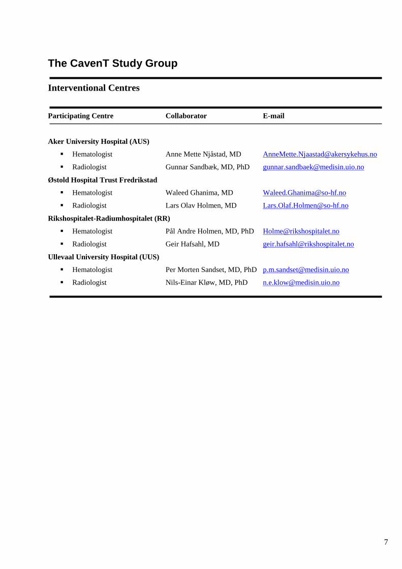
The CavenT Study Group
Interventional Centres
Participating Centre Collaborator E-mail
Aker University Hospital (AUS)
Hematologist Anne Mette Njåstad, MD [email protected]
Radiologist Gunnar Sandbæk, MD, PhD [email protected]
Østold Hospital Trust Fredrikstad
Hematologist Waleed Ghanima, MD [email protected]
Radiologist Lars Olav Holmen, MD [email protected]
Rikshospitalet-Radiumhospitalet (RR)
Hematologist Pål Andre Holmen, MD, PhD [email protected]
Radiologist Geir Hafsahl, MD [email protected]
Ullevaal University Hospital (UUS)
Hematologist Per Morten Sandset, MD, PhD [email protected]
Radiologist Nils-Einar Kløw, MD, PhD [email protected]
8
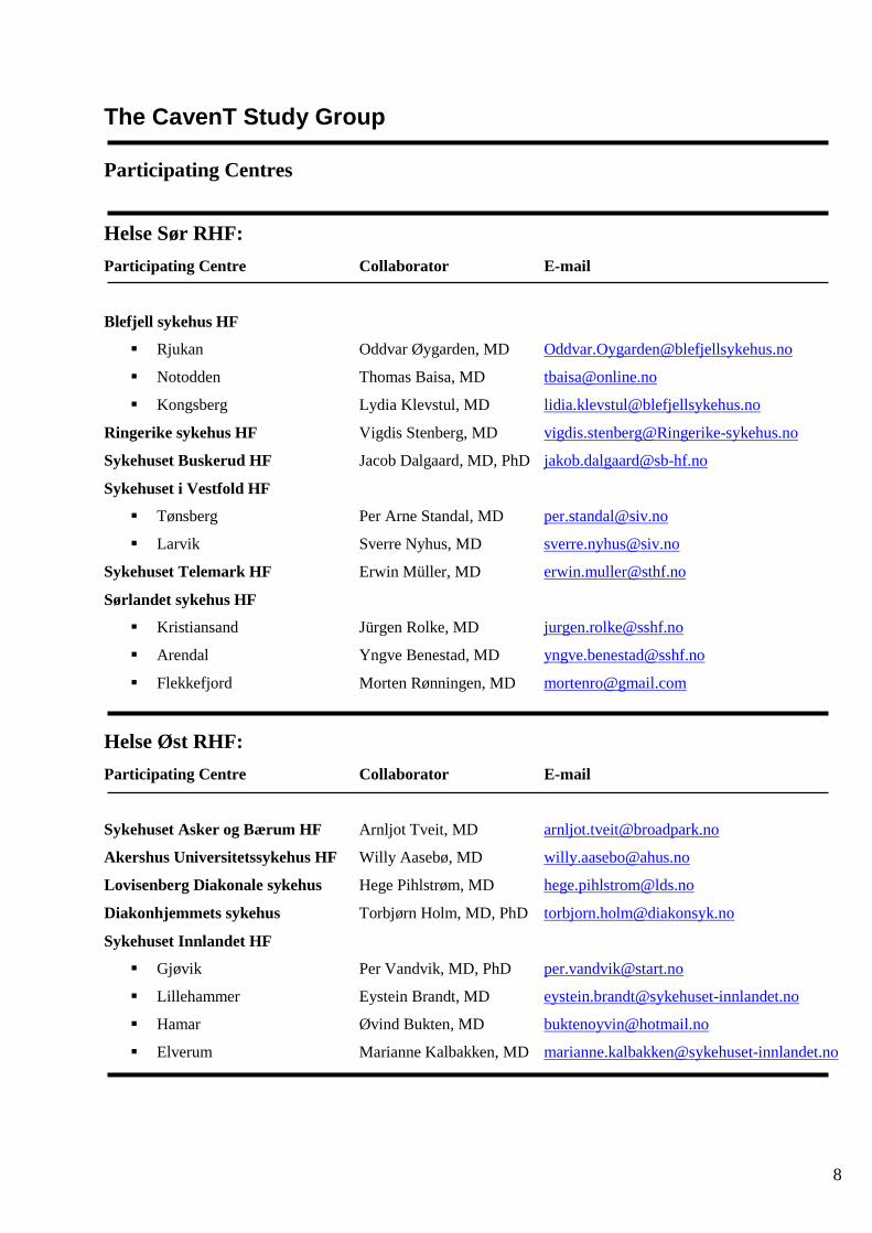
The CavenT Study Group
Participating Centres
Helse Sør RHF:
Participating Centre Collaborator E-mail
Blefjell sykehus HF
Rjukan Oddvar Øygarden, MD [email protected]
Notodden Thomas Baisa, MD [email protected]
Kongsberg Lydia Klevstul, MD [email protected]
Ringerike sykehus HF Vigdis Stenberg, MD [email protected]
Sykehuset Buskerud HF Jacob Dalgaard, MD, PhD [email protected]
Sykehuset i Vestfold HF
Tønsberg Per Arne Standal, MD [email protected]
Larvik Sverre Nyhus, MD [email protected]
Sykehuset Telemark HF Erwin Müller, MD [email protected]
Sørlandet sykehus HF
Kristiansand Jürgen Rolke, MD [email protected]
Arendal Yngve Benestad, MD [email protected]
Flekkefjord Morten Rønningen, MD [email protected]
Helse Øst RHF:
Participating Centre Collaborator E-mail
Sykehuset Asker og Bærum HF Arnljot Tveit, MD [email protected]
Akershus Universitetssykehus HF Willy Aasebø, MD [email protected]
Lovisenberg Diakonale sykehus Hege Pihlstrøm, MD [email protected]
Diakonhjemmets sykehus Torbjørn Holm, MD, PhD [email protected]
Sykehuset Innlandet HF
Gjøvik Per Vandvik, MD, PhD [email protected]
Lillehammer Eystein Brandt, MD [email protected]
Hamar Øvind Bukten, MD [email protected]
Elverum Marianne Kalbakken, MD [email protected]
9
1 SYNOPSIS
Deep vein thrombosis (DVT) is a severe disease which may cause severe disability and which is
sometimes fatal. Conventional treatment with low molecular weight heparin (LMWH) and oral
antiocoagulants is associated with some degree of long-term sequalae, i.e., post-thrombotic syndrome
(PTS), in more than 60-80% of the patients. Systemic thrombolytic therapy reduces the risk of PTS, but
is associated with an unacceptably high risk of bleeding complications, many being disabling or fatal.
Catheter-directed thrombolytic (CDT) therapy is a novel treatment modality which has been introduced
in many hospitals worldwide. Low dose fibrinolytic agents are delivered continuously and directly into
the thrombus through a catheter until thrombus has dissolved. Although many, mostly small series, have
suggested a beneficial effect of this costly treatment in terms of increased patency of the veins and
improved short term functional outcome, there are no randomized clinical trials documenting its short
and long-term efficacy and safety.
The present study is a randomized, open-label, multi-center clinical trial among hospitals in the
Eastern and Southern Norway Health Authorities (Helse Øst and Sør). Patients with acute iliofemoral
vein thrombosis will be randomized to either conventional treatment or CDT in addition to conventional
treatment. Main outcome parameters are patency rates at 6 months and prevalence of PTS at 24 months.
A number of secondary outcomes include bleeding complications, recurrent thrombosis, quality of life
(QoL), markers of importance for successful lysis and recurrent thrombosis, and whether PTS is related
to patency at the end of treatment.
Our main short-term hypothesis is that CDT of first-time acute DVT will increase patency of the
affected iliofemoral vein segments after 6 months from <50% on conventional therapy to >80% after
CDT. Our main long-term hypothesis is that CDT will improve long-term functional outcome, i.e., risk
of PTS, assessed after 2 years, from >25% on conventional treatment to <10% after CDT. The estimated
sample size is at least 100 evaluable patients in each group using a statistical significance (α) = 5% and
a statistical power (1-) = 80%.

10
11
2 BACKGROUND
Deep vein thrombosis (DVT) of the lower extremities is a common disease, which is associated with
significant morbidity. The incidence of DVT is estimated as 1 event per 1,000 per year, which ranks it
as one of the more common cardiovascular disorders 1. Furthermore, DVT is associated with several
important short- and long-term outcomes 2. Short-term there are symptoms of pain and swelling due to
inflammation and obstruction. In a small minority of cases, the condition leads to phlegmasia cerulea
dolens in which extensive venous obstruction leads to ischemia or infarction of the extremity. Lastly,
DVT can also lead to pulmonary embolism (PE), which can be fatal. Long-term sequelae of DVT
include recurrent venous thromboembolism (VTE), post-thrombotic syndrome (PTS), and chronic
thromboembolic pulmonary hypertension.
Anticoagulation therapy is the basic treatment of DVT3, which purpose is to inhibit the
thrombotic process and the inflammatory response so that the thrombus can be cleared by endogenous
fibrinolysis. Anticoagulation therapy thereby alleviates acute symptoms, prevents PE, and recurrent
events. In most cases, anticoagulation is achieved acutely with unfractionated heparin (UFH) or low
molecular weight heparin (LMWH) therapy, followed by long term anticoagulation with oral vitamin K
antagonists (eg warfarin).
Anticoagulation therapy is highly efficacious for the prevention of recurrent VTE, PE, and
death3;4
, but the ability to prevent PTS as an outcome is less clear5. PTS is thought to be a result of
residual venous stenosis and damage to the venous valves which together cause venous hypertension.
Venous hypertension leads to chronic edema and fibrin deposition in the interstitial tissues, which in
turn bring about poor oxygen exchange. Insufficient oxygenation induces skin changes, pain and, in
severe cases, chronic ulceration.
Several studies have addressed the epidemiology of PTS5;6
, i.e., the incidence of PTS over time,
its risk factors, the relationship between vein patency and development of PTS, and the usefulness of
compression stockings to prevent PTS following a first episode of acute DVT treated with
anticoagulation alone5;7-10
. The incidence of moderate or severe PTS varied across these studies, but in
general increased over time. Moderate to severe PTS developed in 2% to 11% of patients with DVT
provided that compression stockings were worn at some early point after the acute DVT. Elastic
compression stockings may reduce the risk of PTS by approximately 50%11;12
. Risk factors for severe
PTS identified by some, but not all of these studies, were recurrent ipsilateral DVT, extent of initial
thrombus, and obesity. Although the role of return of vein patency has not been established, it may still
be an appropriate surrogate for long-term outcomes.
12
Thrombolytic agents, such as streptokinase (SK), urokinase (UK), and recombinant tissue
plasminogen activator (rt-PA) are, theoretically, ideal adjuvants to standard anticoagulation therapy
because they potentially dissolve thrombi, promote early vein recanalization, and thereby, minimize
vein stenosis and valve dysfunction13;14
. Therefore, treatment strategies incorporating these agents with
anticoagulation may be more effective than those using anticoagulation alone for the prevention of PTS.
In addition, in the minority of cases with phlegmasia cerulea dolens, thrombolytic therapies may prove
limb saving. However, despite the theoretical advantages and a history of more than 30 years of use,
thrombolytic therapy has not been widely embraced for DVT treatment due to poor
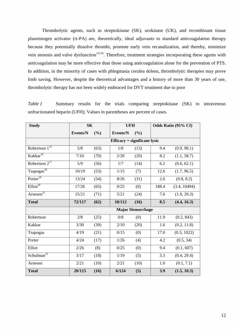
Table 1 Summary results for the trials comparing streptokinase (SK) to intravenous
unfractionated heparin (UFH); Values in parentheses are percent of cases.
Study SK
Events/N (%)
UFH
Events/N (%)
Odds Ratio (95% CI)
Efficacy = significant lysis
Robertson 115
5/8 (63) 1/8 (13) 9.4 (0.9, 98.1)
Kakkar16
7/10 (70) 2/20 (20) 8.2 (1.1, 58.7)
Robertson 217
5/9 (56) 1/7 (14) 6.2 (0.6, 62.1)
Tsapogas18
10/19 (53) 1/15 (7) 12.6 (1.7, 96.5)
Porter19
13/24 (54) 8/26 (31) 2.6 (0.8, 8.2)
Elliot20
17/26 (65) 0/25 (0) 188.4 (3.4, 10494)
Arnesen21
15/21 (71) 5/21 (24) 7.6 (1.9, 29.3)
Total 72/117 (62) 18/112 (16) 8.5 (4.4, 16.3)
Major Hemorrhage
Robertson 2/8 (25) 0/8 (0) 11.9 (0.2, 843)
Kakkar 3/30 (39) 2/10 (20) 1.6 (0.2, 11.8)
Tsapogas 4/19 (21) 0/15 (0) 17.0 (0.3, 1022)
Porter 4/24 (17) 1/26 (4) 4.2 (0.5, 34)
Elliot 2/26 (8) 0/25 (0) 9.4 (0.1, 607)
Schulman22
3/17 (18) 1/19 (5) 3.3 (0.4, 29.4)
Arnesen 2/21 (10) 2/21 (10) 1.0 (0.1, 7.1)
Total 20/115 (16) 6/124 (5) 3.9 (1.5, 10.3)
13
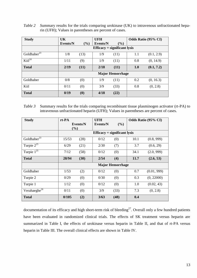
Table 2 Summary results for the trials comparing urokinase (UK) to intravenous unfractionated hepa-
rin (UFH); Values in parentheses are percent of cases.
Study UK
Events/N (%)
UFH
Events/N (%)
Odds Ratio (95% CI)
Efficacy = significant lysis
Goldhaber23
1/8 (13) 1/9 (11) 1.1 (0.1, 2.9)
Kiil24
1/11 (9) 1/9 (11) 0.8 (0, 14.9)
Total 2/19 (11) 2/18 (11) 1.0 (0.1, 7.2)
Major Hemorrhage
Goldhaber 0/8 (0) 1/9 (11) 0.2 (0, 16.3)
Kiil 0/11 (0) 3/9 (33) 0.8 (0, 2.8)
Total 0/19 (0) 4/18 (22)
Table 3 Summary results for the trials comparing recombinant tissue plasminogen activator (rt-PA) to
intravenous unfractionated heparin (UFH); Values in parentheses are percent of cases.
Study rt-PA
Events/N
(%)
UFH
Events/N (%)
Odds Ratio (95% CI)
Efficacy = significant lysis
Goldhaber23
15/53 (28) 0/12 (0) 10.1 (0.8, 999)
Turpie 225
6/29 (21) 2/30 (7) 3.7 (0.6, 29)
Turpie 125
7/12 (58) 0/12 (0) 34.1 (2.0, 999)
Total 28/94 (30) 2/54 (4) 11.7 (2.6, 53)
Major Hemorrhage
Goldhaber 1/53 (2) 0/12 (0) 0.7 (0.01, 999)
Turpie 2 0/29 (0) 0/30 (0) 0.3 (0, 22000)
Turpie 1 1/12 (0) 0/12 (0) 1.0 (0.02, 43)
Verahaeghe26
0/11 (0) 3/9 (33) 7.3 (0, 2.8)
Total 0/105 (2) 3/63 (48) 0.4
documentation of its efficacy and high short-term risk of bleeding27
. Overall only a few hundred patients
have been evaluated in randomized clinical trials. The effects of SK treatment versus heparin are
summarized in Table I, the effects of urokinase versus heparin in Table II, and that of rt-PA versus
heparin in Table III. The overall clinical effects are shown in Table IV.
14
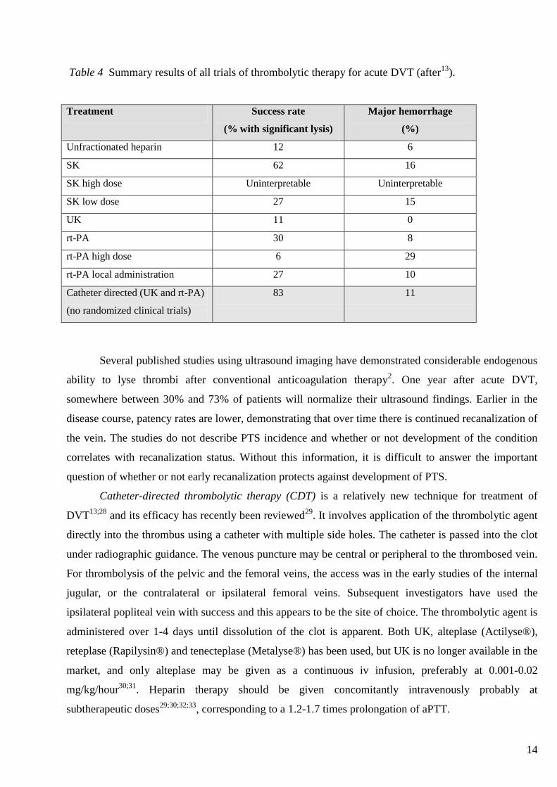
Table 4 Summary results of all trials of thrombolytic therapy for acute DVT (after13
).
Treatment Success rate
(% with significant lysis)
Major hemorrhage
(%)
Unfractionated heparin 12 6
SK 62 16
SK high dose Uninterpretable Uninterpretable
SK low dose 27 15
UK 11 0
rt-PA 30 8
rt-PA high dose 6 29
rt-PA local administration 27 10
Catheter directed (UK and rt-PA)
(no randomized clinical trials)
83 11
Several published studies using ultrasound imaging have demonstrated considerable endogenous
ability to lyse thrombi after conventional anticoagulation therapy2. One year after acute DVT,
somewhere between 30% and 73% of patients will normalize their ultrasound findings. Earlier in the
disease course, patency rates are lower, demonstrating that over time there is continued recanalization of
the vein. The studies do not describe PTS incidence and whether or not development of the condition
correlates with recanalization status. Without this information, it is difficult to answer the important
question of whether or not early recanalization protects against development of PTS.
Catheter-directed thrombolytic therapy (CDT) is a relatively new technique for treatment of
DVT13;28
and its efficacy has recently been reviewed29
. It involves application of the thrombolytic agent
directly into the thrombus using a catheter with multiple side holes. The catheter is passed into the clot
under radiographic guidance. The venous puncture may be central or peripheral to the thrombosed vein.
For thrombolysis of the pelvic and the femoral veins, the access was in the early studies of the internal
jugular, or the contralateral or ipsilateral femoral veins. Subsequent investigators have used the
ipsilateral popliteal vein with success and this appears to be the site of choice. The thrombolytic agent is
administered over 1-4 days until dissolution of the clot is apparent. Both UK, alteplase (Actilyse®),
reteplase (Rapilysin®) and tenecteplase (Metalyse®) has been used, but UK is no longer available in the
market, and only alteplase may be given as a continuous iv infusion, preferably at 0.001-0.02
mg/kg/hour30;31
. Heparin therapy should be given concomitantly intravenously probably at
subtherapeutic doses29;30;32;33
, corresponding to a 1.2-1.7 times prolongation of aPTT.

15
The decision to discontinue the drug is based on daily venographic examinations through the
indwelling catheter. Depending on the findings the catheter may be pulled out, the infusion continued, or
the catheter repositioned. To obtain flow in the veins balloon inflation may be performed at the follow-
up. Thrombolytic agents are given until there is no more evidence of thrombosis or until there is little
improvement in venographic appearance. After 72-96 hours thrombolysis is discontinued. Adjuvant
therapies include angioplasty, angioplasty with stents, thrombectomy, and surgically created arterio-
venous fistulas.
So far, there are no randomized clinical trials with long-term follow-up on the efficacy of CDT
therapy, but at least 15 case series have been reported29;34-37
. Combining the studies, 263 patients
received this type of therapy for thrombosis of the iliofemoral veins or inferior vena cava. 221 (84%)
patients were considered to have successful short-term outcomes based on venographic appearance and
13 (4.9%) patients had bleeding severe enough to warrant transfusion. Long term outcomes were not
reported, and the authors did not describe the proportion of patients requiring adjuvant therapy.
A National DVT Registry was established in North-America to analyze results in a large number
of patients treated with CDT38
. This registry included 473 patients with documented lower extremity
DVT treated with CDT, but follow-up data included only 287 patients who received 312 treatments.
Thrombi subjected to lysis included either ilio-femoral vein thrombosis in 71% of cases and femoro-
popliteal vein thrombosis in 25% of cases. The mean age of patients was 47.5 years and the mean
duration of infusion was 53 h. All patients had six months of therapy with oral anticoagulants following
CDT and many had heparin as well. Complete lysis was obtained in 31% of patients, 50-99% lysis in
52% and <50% lysis in 17%. Successful lysis was not related to location of the thrombus. The overall
primary patency rate was 80% at 12 months, with better patency for ilio-femoral segments than the
femoro-popliteal segments. Major bleeding complications occurred in 11% of patients; 39% of these at
the venous insertion site, 13% were retroperitoneal hematoma. Minor bleeding events occurred in 16%
of patients, again most often at the venous entry site. There was one fatal intracranial hemorrhage, one
subdural hematoma, and 6 pulmonary emboli of which one was fatal. Thus, the overall mortality rate
from lysis was 0.4%. There was no data on PTS.
If the PTS differs between standard therapy and thrombolytic therapy then the quality of life may
differ between patients also. Comerota assessed health-related quality of life in patients after CDT
therapy compared to a group of patients treated with standard anticoagulation therapy39
. The delayed
functional outcome and wellbeing scores were significantly better in the thrombolytic therapy group.
Although this study had some methodological shortcomings13
, the findings are still suggestive that
thrombolytic therapy may offer improved quality of life in patients who achieve successful
thrombolysis.
16
Compared to historical data of anticoagulation and intravenous thrombolysis, CDT probably has
higher recanalization rates. The studies so far, indcluding one RCT with 6 months follow-up and 35
patients40
, have been promising, but unfortunately no high-quality randomized studies with long-term
follow-up have been performed. Experimental data indicate that valves of the femoral veins may be
preserved41;42
. It is therefore possible that PTS may be reduced. However, long term studies have not
been performed. In the absence of well-designed randomized clinical studies both for early findings, the
implications of early patency for long-term clinical results, the complications, and the costs related to
treatment, CDT therapy for DVT should at present be considered experimental treatment. Still, some
Norwegian hospitals including Aker and Ullevål University Hospitals, Rikshospitalet, and the Østfold
Hospital Trust Fredrikstad, do provide this high-intensive treatment to selected patients. A case-series
with careful follow-up at Aker University Hospital has recently been published31
.
In the present study, we aim to investigate the role of CDT therapy for treatment of acute DVT
as compared with established treatment with low molecular weight heparin. The study will be an open-
label, randomized study of patients with first-time acute DVT of the affected limb, and our major
outcome parameter will be the frequency of PTS as related to early venographic patency. The results of
this study have the potential to properly define the role of this costly treatment in the future.
17
3 OBJECTIVES
3.1 PRIMARY OBJECTIVES
To investigate whether catheter-directed thrombolytic therapy for first-time acute DVT of the
iliofemoral veins may:
3.1.1 increase patency rate at 6 months.
3.1.2 reduce the risk of PTS at 2 years.
3.2 SECONDARY OBJECTIVES
3.2.1 To investigate frequency of clinically relevant bleeding related to the procedure.
3.2.2 To investigate effects on quality of life (QoL).
3.2.3 To investigate cost-effectiveness of treatment.
3.2.4 To investigate the procedural success of CDT.
3.2.5 To identify markers of importance for successful thrombolysis.
3.2.6 To investigate patency at 2 years.
3.2.7 To investigate PTS at 6 and 60 months.
3.2.8 To investigate whether presence or absence of PTS at any time point is related to patency at end
of treatment.
3.2.9 To investigate prevalence of vein anomalies (and need for angioplasty or stents).
3.2.10 To investigate prevalence of underlying thrombophilia.
3.2.11 To investigate frequency of recurrent VTE during follow-up.
3.2.12 To identify markers of importance for recurrent thrombosis.
4 HYPOTHESES
Our main short-term hypothesis is that CDT of first-time acute DVT will increase patency of the
affected iliofemoral vein segments after 6 months from <50% on conventional therapy to >80% after
CDT. Our main long-term hypothesis is that CDT will improve long-term functional outcome, i.e., risk
of PTS, assessed after 2 years, from >25% on conventional treatment to <10% after CDT.
18
5 PATIENT POPULATION
5.1 INCLUSION CRITERIA
5.1.1 Age 18-75 years.
5.1.2 Onset of symptoms <21 days.
5.1.3 Objectively verified DVT (ultrasonography, venography, computed tomography, or magnetic
resonance imaging) localized in the upper half of the thigh, the common iliac vein or the
combined iliofemoral segment.
5.1.4 Informed consent (Appendix 1).
5.2 EXCLUSION CRITERIA
5.2.1 Anticoagulant therapy prior to trial entry for >7 days.
5.2.2 Contraindications to thrombolytic therapy, including bleeding diathesis.
5.2.3 Indications for thrombolytic therapy, e.g., phlegmacia coerolia dolens or isolated vena cava
thrombosis.
5.2.4 Severe anemia (hemoglobin <8 g/dL).
5.2.5 Thrombocytopenia (platelets <80·109/L).
5.2.6 Severe renal failure – creatinine clearance <30 ml/min. Creatinine clearance will be calculated
according to the following formula:
Creatinine clearance (ml/min) = b x (140 – age (yrs)) x body weight (kg)
serum creatinine (µmol/L
b=1.23 (females); 1.04 (males)
5.2.7 Severe hypertension, i.e. persistent systolic blood pressure >160 mm Hg or diastolic blood
pressure >100 mm Hg.
5.2.8 Pregnancy and thrombosis ≤7 days post-partum (may be included after 7 days post-partum).
5.2.9 Less than 14 days post-surgery or post-trauma (may be included after 14 days).
5.2.10 History of subarachnoidal or intracerebral bleeding.
5.2.11 Disease with life expectancy <24 months.
5.2.12 Drug abuse or mental disease that may interfere with treatment and follow-up.
5.2.13 Former ipsilateral proximal DVT.
5.2.14 Malignant disease requiring chemotherapy.
5.2.15 Any thrombolytic therapy within 7 days prior to trial inclusion.
19
6 METHODS
6.1 DESIGN
Multi-center, open-label, randomized clinical study on the effect and safety of CDT therapy as
compared with conventional therapy for the treatment of acute, first-time ilio-femoral DVT. The study
will be a collaborative study of hospitals belonging to the Eastern and Southern Norway Health
Authorities (Helse Øst and Sør).
6.2 PATIENT RECRUITMENT
Eligible patients (section 5) will be invited to participate in the study. Informed consent (Appendix 1) in
accordance with the revised Helsinki Declaration must be obtained from the patient before
randomization.
6.3 RANDOMIZATION
Patients will be randomized by sealed numbered envelopes using block randomization. Each envelope
will contain information on treatment allocation. A new patient will be allocated the lowest numbered
envelope. Treatment will be open-label, but stratified for extension of DVT, i.e., only femoral or
iliofemoral DVT.
6.4 TREATMENT
6.4.1 Acute treatment
Patients will be randomized to one of the following treatment groups:
Group I Catheter-directed thrombolytic therapy with rt-PA in addition to conventional
treatment with low molecular weight heparin (for details – see 6.4.2)
Group II Conventional treatment with low molecular weight heparin (see 6.4.3)
Drugs will be ordered from the hospital’s pharmacy according to local routines.
- Group I will be given rt-PA (Actilyse®) combined with unfractionated heparin and followed by low
molecular weight heparin (LMWH) and warfarin.
- Group II, the conventional treatment arm, will be given LMWH, either sc dalteparin (Fragmin®), 200
IU/kg od, or enoxaparin (Klexane®), 1.5 mg/kg od, according to local routines, and warfarin.
20
6.4.2 Group I - Catheter-Directed Thrombolytic (CDT) therapy – procedures
Anticoagulant and fibrinolytic therapy
- Discontinue oral anticoagulants - INR should be <1.5 before the procedure.
- In case of prior sc LMWH therapy treatment should be discontinued at least 8 h before the
procedure, and in case of prior UFH treatment APTT (Cephotest®) should be adjusted to 40-60 sec
during the procedure (see below).
- An iv bolus dose of UFH, 5000 U, should be given followed by continuous iv UFH1 infusion at 15
U/kg/h. Adjust dose to keep APTT (Cephotest®) at 40-60 sec, first adjustment 6-12 h after start of
treatment.
- During the thrombolytic treatment keep APTT (Cephotest®) at 40-60 sec.
- At the completion of thrombolytic treatment:
discontinue UFH
give sc LMWH after 1 h, (either dalteparin, Fragmin®, 200 U/kg bid, or enoxaparin,
Klexane®, 1,5 mg/kg bid).
Oral warfarin (Marevan®) will be initiated according to local routines.
LMWH will be discontinued when INR has been in therapeutic range (2.0-3.0) for at least 24
hours, but should not be given for less than total 4-5 days.
Interventional procedures. In an interventional radiology unit, an introducer will be inserted into an
appropriate vein, preferentially the popliteal vein, guided by ultrasound to prevent puncture of the
artery or laceration of the vein wall and to secure only a single puncture. If possible, the wire and
catheter should be introduced above the proximal part of the thrombus (use fitting-sized perfusion
catheters, e.g., 10, 20, 30, or 50 cm). A venography should then be performed to disclose the
topography of the thrombus. CDT may be discontinued if introduction of the catheter through the
occluded segment is not successful. Catheters should be properly fixed to the skin.
The perfusion catheter (and the perfusion wire) should cover the central to peripheral part of the
thrombus. Rt-PA (Actilyse®), 20 mg diluted in 500 ml 0.9% NaCl, will be infused at 0.01 mg/kg/h.
Maximal dose infused will be 20 mg/24 h. The rt-PA dosage may be split into two catheters using
lower consentration, keeping flow the same.
1 A suitable working solution should be made to contain UFH 40 U/ml in 0.9% NaCl, e.g., mix 20000 U of UFH in 500 ml
0.9% NaCl or 40000 U in 1000 ml 0.9% NaCl. The infusion rate (ml/h) then reflects total units of UFH per 24 hrs in
thousands, e.g., 25 ml/h corresponds to 25000 U/24 h, 30 ml/h 30000 U/24 h, and so on.
21
After insertion of catheter, venography, and start of iv UFH and iv rt-PA infusion, treatment will
continue in medical wards. Blood pressure and pulse and the puncture site are assessed 4 times a
day. Hemostasis is also monitored by daily analysis of hemoglobin, fibrinogen, D-dimer, INR, and
platelet counts. APTT is monitored twice daily for adjustment of heparin dose. The patient will be
encouraged to use the muscle pump of the leg while in bed. No food and drink restrictions.
Effect of treatment will be assessed by venography at least every 24 hrs, and catheters
repositioned accordingly. Treatment should normally not continue for >96 h. At the end of
treatment, the catheters will be removed immediately and hemostasis obtained by manual
compression of the puncture site. Pressure will be continued for 2 hrs with a roll while the patient is
immobilized.
Stents. Balloon dilatation and placement of venous stents will be performed at the discretion of the
operator to establish flow and to obtain <50% residual stenosis.
Concomitant medication during procedure. During the interventional procedure concomitant use
of other antithrombotic agents should be avoided because of increased risk of bleeding. This
includes antiplatelet agents (e.g., acetylsalicylic acid, thienopyridines, GPIIb/IIIa inhibitors, non
steroidal anti-inflammatory agents, or other) or anticoagulants (e.g., low molecular weight heparin,
pentasaccharide, warfarin, or other). Concomitant use of ACE-inhibitors appears to increase the risk
of anafylactoid reactions.
6.4.3 Group II – conventional treatment with LMWH
Patients allocated the conventional treatment arm will be given sc LMWH, either dalteparin
(Fragmin®), 200 U/kg od, or enoxaparin (Klexane®), 1.5 mg/kg od, according to local hospital
routines, and simultaneous warfarin (Marevan®) according to local routines. LMWH will be
discontinued when INR has been in therapeutic range (2.0-3.0) for at least 24 hours, but should not be
given for less than total 4-5 days.
6.4.4 Subacute and chronic phase after DVT
Patients will be treated with warfarin for at least 6 months with target INR 2.0-3.0. All patients will be
adviced to use knee-high compression stockings, grade II, for 6 months.
22
6.5 VISITS AND PROCEDURES DURING FOLLOW-UP
End-point assessment will be performed by a vascular surgeon with no previous contact or knowledge
of patients’ medical history or treatment allocation. At each visit the patients will explicitly be told not
to reveal treatment allocation.
6.5.1 Visit 1 (trial entry – at hospital admission/)
6.5.1.1 Case history and general clinical examination.
6.5.1.2 Compression ultrasonography or venography, alternatively CT or MRI angiography diagnosing
acute iliofemoral DVT.
6.5.1.3 Laboratory screening (hemoglobin, platelets, leukocytes, creatinine, ASAT, ALAT, GT,
bilirubin, INR, APTT, D-Dimer, cholesterol, and CRP).
6.5.1.4 Thrombophilia screening (collection of blood samples).
6.5.1.5 Assessment of baseline QoL before treatment using VEINES-QoL and EQ-D5 (Appendix 2).
6.5.1.6 Assessment of baseline clinical score using Villalta5;43
score and the C classification of CEAP,
see Definitions.
6.5.2 Visit 2 (hospital stay)
6.5.2.1 Daily assessment of hemoglobin, platelets, fibrinogen, APTT, INR, and D-Dimer, and bilateral
leg circumference.
6.5.2.2 Daily venography will be performed in patients allocated CDT.
6.5.2.4 Bleeding complications.
6.5.3 Visit 3 – 6 m ± 2 weeks
6.5.3.1 Clinical history – recurrent thrombosis – malignancy.
6.5.3.2 Clinical PTS scores according to Villalta and CEAP. Bilateral leg circumference.
6.5.3.3 Assessment of functional venous obstruction by air-plethysmography.
6.5.3.4 Ultrasonographic assessment of postthrombotic changes, patency, and reflux 44-47
.
6.5.3.5 Quality of Life (QoL) assessment (Appendix 2).
6.5.3.6 D-dimer testing, INR, thrombophilia screening (if previously inconclusive).
6.5.4 VISIT 4 – 12 m ± 4 weeks
Telephone interview – recurrent thrombosis – malignancy.
23
6.5.5 VISIT 5 – 24 m ± 4 weeks
6.5.5.1 Clinical history – recurrent thrombosis – malignancy.
6.5.5.2 Clinical PTS scores according to Villalta and CEAP. Bilateral leg circumference..
6.5.5.3 Assessment of functional venous obstruction by air-plethysmography.
6.5.5.4 Ultrasonographic assessment of postthrombotic changes, patency, and reflux
6.5.5.5 Quality of Life (QoL) assessment (Appendix 2).
6.5.5.6 D-dimer, INR, thrombophilia screening (if previously inconclusive).
6.5.6 VISIT 6 – 36 m ± 4 weeks
Telephone interview – recurrent thrombosis – malignancy.
6.5.7 VISIT 7 – 48 m ± 4 weeks
Telephone interview – PTS screening – recurrent thrombosis – malignancy.
6.5.8 VISIT 8 – 60 m ± 8 weeks
6.5.8.1 Clinical history – recurrent thrombosis – malignancy.
6.5.8.2 Clinical PTS scores according to Villalta and CEAP. Bilateral leg circumference.
6.5.8.3 Ultrasonographic assessment of postthrombotic changes, patency, and reflux.
6.5.8.4 Assessment of functional venous obstruction by air-plethysmography.
6.5.8.5 Quality of Life (QoL) assessment (Appendix 2).
24
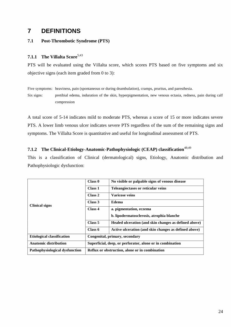
7 DEFINITIONS
7.1 Post-Thrombotic Syndrome (PTS)
7.1.1 The Villalta Score5;43
PTS will be evaluated using the Villalta score, which scores PTS based on five symptoms and six
objective signs (each item graded from 0 to 3):
Five symptoms: heaviness, pain (spontaneous or during deambulation), cramps, pruritus, and paresthesia.
Six signs: pretibial edema, induration of the skin, hyperpigmentation, new venous ectasia, redness, pain during calf
compression
A total score of 5-14 indicates mild to moderate PTS, whereas a score of 15 or more indicates severe
PTS. A lower limb venous ulcer indicates severe PTS regardless of the sum of the remaining signs and
symptoms. The Villalta Score is quantitative and useful for longitudinal assessment of PTS.
7.1.2 The Clinical-Etiology-Anatomic-Pathophysiologic (CEAP) classification48;49
This is a classification of Clinical (dermatological) signs, Etiology, Anatomic distribution and
Pathophysiologic dysfunction:
Clinical signs
Class 0 No visible or palpable signs of venous disease
Class 1 Teleangiectases or reticular veins
Class 2 Varicose veins
Class 3 Edema
Class 4 a. pigmentation, eczema
b. lipodermatosclerosis, atrophia blanche
Class 5 Healed ulceration (and skin changes as defined above)
Class 6 Active ulceration (and skin changes as defined above)
Etiological classification Congenital, primary, secondary
Anatomic distribution Superficial, deep, or perforator, alone or in combination
Pathophysiological dysfunction Reflux or obstruction, alone or in combination
25
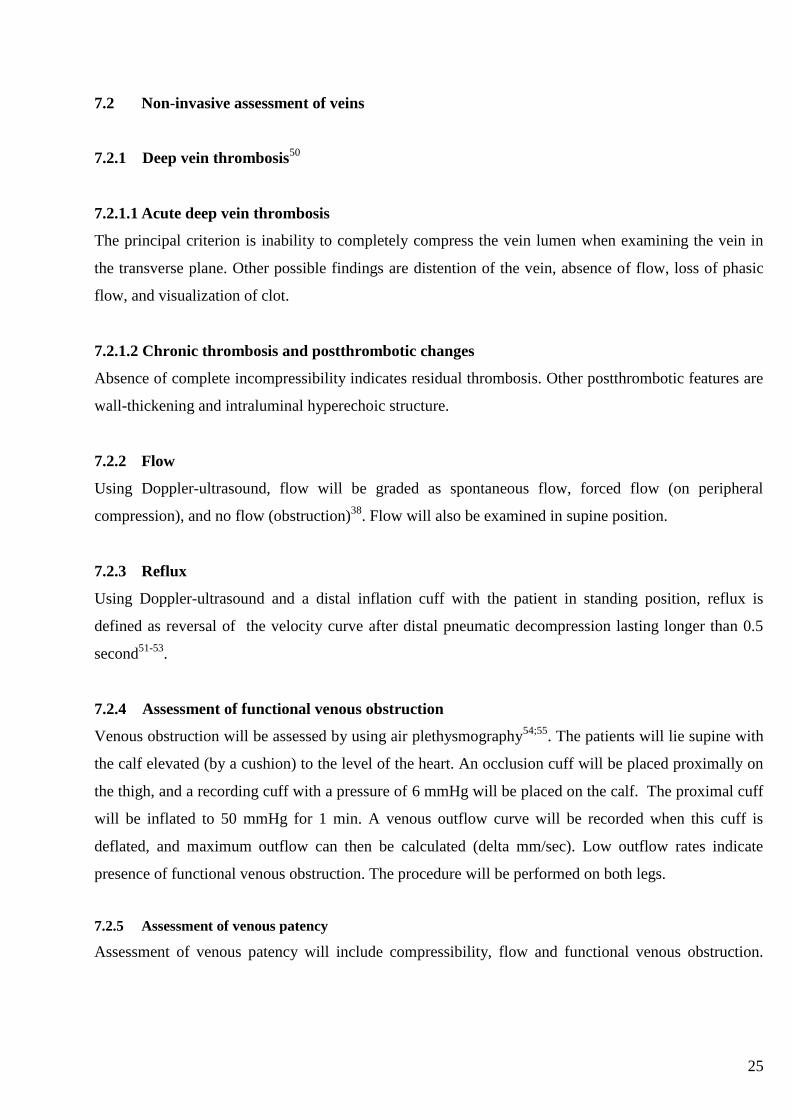
7.2 Non-invasive assessment of veins
7.2.1 Deep vein thrombosis50
7.2.1.1 Acute deep vein thrombosis
The principal criterion is inability to completely compress the vein lumen when examining the vein in
the transverse plane. Other possible findings are distention of the vein, absence of flow, loss of phasic
flow, and visualization of clot.
7.2.1.2 Chronic thrombosis and postthrombotic changes
Absence of complete incompressibility indicates residual thrombosis. Other postthrombotic features are
wall-thickening and intraluminal hyperechoic structure.
7.2.2 Flow
Using Doppler-ultrasound, flow will be graded as spontaneous flow, forced flow (on peripheral
compression), and no flow (obstruction)38
. Flow will also be examined in supine position.
7.2.3 Reflux
Using Doppler-ultrasound and a distal inflation cuff with the patient in standing position, reflux is
defined as reversal of the velocity curve after distal pneumatic decompression lasting longer than 0.5
second51-53
.
7.2.4 Assessment of functional venous obstruction
Venous obstruction will be assessed by using air plethysmography54;55
. The patients will lie supine with
the calf elevated (by a cushion) to the level of the heart. An occlusion cuff will be placed proximally on
the thigh, and a recording cuff with a pressure of 6 mmHg will be placed on the calf. The proximal cuff
will be inflated to 50 mmHg for 1 min. A venous outflow curve will be recorded when this cuff is
deflated, and maximum outflow can then be calculated (delta mm/sec). Low outflow rates indicate
presence of functional venous obstruction. The procedure will be performed on both legs.
7.2.5 Assessment of venous patency
Assessment of venous patency will include compressibility, flow and functional venous obstruction.

26
7.3 Evaluation of thrombolysis
Based on venography before and after CDT, thrombolysis will be graded by a scoring system38
. Score=0
indicates an open vein, score=1 a partly occluded vein, and score=2 a completely occluded vein.
Each of the following 7 venous segments will be given a grade (0-2): IVC, the common iliac
vein, the external iliac vein, the common femoral vein, the proximal and distal superficial femoral veins,
and the popliteal vein. A total thrombus score before and after lysis will be calculated by adding the 7
scores. The difference between the pre- and postlysis thrombus scores divided by the prelysis score
gives the grade of thrombolysis. Grade I=<50%; grade II=50-90%, and grade III=complete thrombolysis
7.4 Bleeding Complications
7.4.1 Major bleeding – any bleeding associated with a reduction in hemoglobin by ≥2 g/100 mL or
bleeding requiring transfusion of ≥2 U pack red blood cells or whole blood or bleeding in a
critical organ, intracranial, retroperitoneal or pericardial or bleeding contributing to death.
7.4.2 Clinically relevant non-major bleeding – overt bleeding not meeting criteria for major bleeding
but satisfying a priori criteria defined by the safety monitoring committee including for example
skin hematomas >100 cm2, epistaxis lasting >5 min, being repetitive (≥2/24 h) or requiring
intervention (packing, electrocoagulation), macroscopic hematuria – either spontaneous or
lasting >24 h after instrumentation (catheter or surgery) of the urogenital tract, or any other
bleeding type that is considered to have clinical consequences for the patient.
7.4.3 Trivial bleeding - all other overt bleeding episodes not meeting the criteria for clinically
relevant bleeding.
7.5 Thrombophilia screening
Includes screening for antithrombin, protein C- and protein S deficiencies, factor V Leiden
mutation, the prothrombin gene 20210GA allele variation and the methylene tetrahydrofolate
reductase (MTHFR) mutation, homocystein, lupus anticoagulants and anticardiolipin antibodies.
27
8 STATISTICS
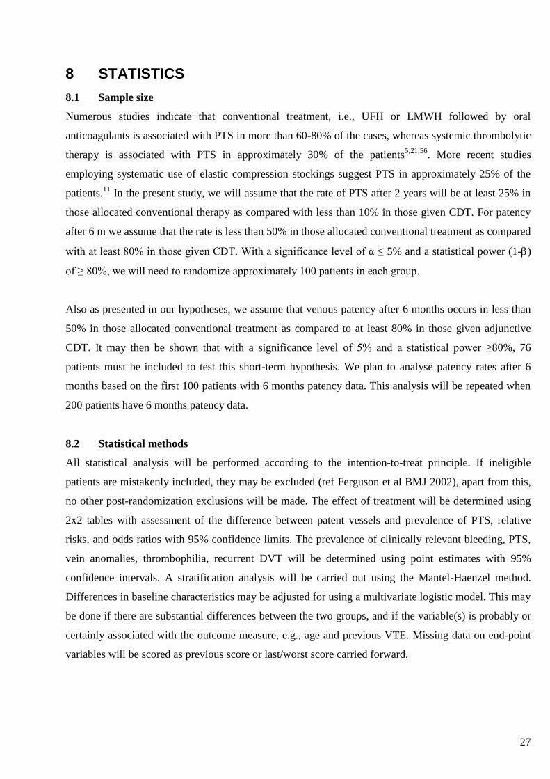
8.1 Sample size
Numerous studies indicate that conventional treatment, i.e., UFH or LMWH followed by oral
anticoagulants is associated with PTS in more than 60-80% of the cases, whereas systemic thrombolytic
therapy is associated with PTS in approximately 30% of the patients5;21;56
. More recent studies
employing systematic use of elastic compression stockings suggest PTS in approximately 25% of the
patients.11
In the present study, we will assume that the rate of PTS after 2 years will be at least 25% in
those allocated conventional therapy as compared with less than 10% in those given CDT. For patency
after 6 m we assume that the rate is less than 50% in those allocated conventional treatment as compared
with at least 80% in those given CDT. With a significance level of α ≤ 5% and a statistical power (1-)
of ≥ 80%, we will need to randomize approximately 100 patients in each group.
Also as presented in our hypotheses, we assume that venous patency after 6 months occurs in less than
50% in those allocated conventional treatment as compared to at least 80% in those given adjunctive
CDT. It may then be shown that with a significance level of 5% and a statistical power ≥80%, 76
patients must be included to test this short-term hypothesis. We plan to analyse patency rates after 6
months based on the first 100 patients with 6 months patency data. This analysis will be repeated when
200 patients have 6 months patency data.
8.2 Statistical methods
All statistical analysis will be performed according to the intention-to-treat principle. If ineligible
patients are mistakenly included, they may be excluded (ref Ferguson et al BMJ 2002), apart from this,
no other post-randomization exclusions will be made. The effect of treatment will be determined using
2x2 tables with assessment of the difference between patent vessels and prevalence of PTS, relative
risks, and odds ratios with 95% confidence limits. The prevalence of clinically relevant bleeding, PTS,
vein anomalies, thrombophilia, recurrent DVT will be determined using point estimates with 95%
confidence intervals. A stratification analysis will be carried out using the Mantel-Haenzel method.
Differences in baseline characteristics may be adjusted for using a multivariate logistic model. This may
be done if there are substantial differences between the two groups, and if the variable(s) is probably or
certainly associated with the outcome measure, e.g., age and previous VTE. Missing data on end-point
variables will be scored as previous score or last/worst score carried forward.
28
9 ETHICAL CONSIDERATIONS
This study will recruit patients with proximal DVT. Even though the efficacy and safety of CDT for the
treatment of acute proximal DVT remains to be established, some hospitals in many countries now offer
CDT to selected patients with severe DVT, especially when the DVT extends into the caval vein. In the
present study, non-trial CDT to selected patients with severe DVT will be left to the discretion of the
responsible physician.
The study will be performed in accordance with the revised Helsinki Declaration and Good
Clinical Practice (GCP). The study will only start after approval with the Regional Ethical Committee
and the Norwegian Medical Agency. All patients will be given study specific identification codes and all
data will be stored in a secured database on a secured server for research at the Ullevål University
Hospital. This server as well as data management will be controlled by the Patient Protection Ombud at
the Ullevål University Hospital. A non-linked database will provide information on the patients’ contact
information to allow follow-up. A biobank will be established at Ullevål University Hospital after
approval.
10 MILESTONES
Q1-2006 First patient randomized
Q4-2007 Last patient randomized
Q2-2008 Six months follow-up of all patients for primary efficacy parameter patency
Q2-3-2008 Reporting of study design and primary efficacy parameter patency
Q4-2009 Two-years follow-up of all patients for primary efficacy parameter PTS
Q4-Q1-09-10 Reporting of primary efficacy parameter PTS
Q4-2012 Five years follow-up of last patient for patency and PTS.
29
11 TRIAL ORGANIZATION
11.1 GENERAL ORGANIZATION
The study is an investigator initiated study which will be run independently of the pharmaceutical
industry. The study is financially supported by a grant from Eastern Norway Health Authority (doctoral
fellow; Helse Øst grant no 2005-090).
The study will be a major collaborative effort among hospitals of the Eastern and Southern
Norway Health Authorities (Helse Øst and Sør). All hospitals will be invited to participate in the study.
Patients allocated to conventional treatment will be treated at the local hospital, whereas patients
allocated CDT will be treated at Ullevål and Aker University Hospitals, the National Hospital and the
Central Hospital in Østfold.
11.2 COMMITTES
11.2.1 Executive committee
Per Morten Sandset (chair) – UUS – Hematologist
Nils-Einar Kløw – UUS – Radiologist
Leiv Sandvik – UUS – Statistician
Tone Enden – UUS – Research fellow – Resident in Radiology
Carl-Erik Slagsvold – AUS – Angiologist
Anne Mette Njåstad – AUS – Hematologist
Gunnar Sandbaek – AUS – Radiologist
Pål Andre Holme – RR – Hematologist
Geir Hafsahl – RR – Radiologist
Waleed Ghanima – Østfold Hospital Trust Fredrikstad – Hematologist
Lars Olav Holmen – Østfold Hospital Trust Fredrikstad – Radiologist
11.2.2 Steering committee
Executive committee (chair Per Morten Sandset)
One member from each collaborating hospital
11.2.3 Safety and monitoring committee
Professor emeritus Ulrich Abildgaard
Professor Frank Brosstad, Rikshospitalet-Radiumhospitalet, Oslo
30
12 PUBLICATION
Results of this study will be published in international medical journals, but will also be communicated
to the general population whenever appropriate. The results may potentially have great interest for the
scientific community, for health-providers in decision making, and for the general population.
Publication will follow the Vancouver convention. Tone Enden will be the first author of these
publications.
31
REFERENCES
1. White RH. The epidemiology of venous thromboembolism. Circulation 2003;107:I4-I8.
2. Kearon C. Natural history of venous thromboembolism. Circulation 2003;107:I22-I30.
3. Kearon C. Initial treatment of venous thromboembolism. Thromb.Haemost. 1999;82:887-891.
4. Kearon C. Duration of anticoagulation for venous thromboembolism. J.Thromb.Thrombolysis. 2001;12:59-65.
5. Kahn SR, Ginsberg JS. Relationship between deep venous thrombosis and the postthrombotic syndrome.
Arch.Intern.Med. 2004;164:17-26.
6. Prandoni P, Lensing AW, Cogo A et al. The long-term clinical course of acute deep venous thrombosis.
Ann.Intern.Med. 1996;125:1-7.
7. Lindner DJ, Edwards JM, Phinney ES, Taylor LM, Jr., Porter JM. Long-term hemodynamic and clinical sequelae of
lower extremity deep vein thrombosis. J.Vasc.Surg. 1986;4:436-442.
8. Brandjes DP, Buller HR, Heijboer H et al. Randomised trial of effect of compression stockings in patients with
symptomatic proximal-vein thrombosis. Lancet 1997;349:759-762.
9. Franzeck UK, Schalch I, Bollinger A. On the relationship between changes in the deep veins evaluated by duplex
sonography and the postthrombotic syndrome 12 years after deep vein thrombosis. Thromb.Haemost. 1997;77:1109-
1112.
10. Biguzzi E, Mozzi E, Alatri A et al. The post-thrombotic syndrome in young women: retrospective evaluation of
prognostic factors. Thromb.Haemost. 1998;80:575-577.
11. Prandoni P, Lensing AWA, Prins MH et al. Below-Knee Elastic Compression Stockings To Prevent the Post-
Thrombotic Syndrome: A Randomized, Controlled Trial. Ann Intern Med 2004;141:249-256.
12. Ginsberg JS. Routine Stocking Therapy after Deep Venous Thrombosis: A Clinical Dilemma. Ann Intern Med
2004;141:314-315.
13. Wells PS, Forster AJ. Thrombolysis in deep vein thrombosis: is there still an indication? Thromb.Haemost.
2001;86:499-508.
14. Marder VJ, Stewart D. Towards safer thrombolytic therapy. Semin.Hematol. 2002;39:206-216.
15. Robertson BR, Nilsson IM, Nylander G. Value of streptokinase and heparin in treatment of acute deep venous
thrombosis. A coded investigation. Acta Chir Scand. 1968;134:203-208.
16. Kakkar VV, Flanc C, Howe CT, O'Shea M, Flute PT. Treatment of deep vein thrombosis. A trial of heparin,
streptokinase, and arvin. Br.Med.J. 1969;1:806-810.
17. Robertson BR, Nilsson IM, Nylander G. Thrombolytic effect of streptokinase as evaluated by phlebography of deep
venous thrombi of the leg. Acta Chir Scand. 1970;136:173-180.
18. Tsapogas MJ, Peabody RA, Wu KT et al. Controlled study of thrombolytic therapy in deep vein thrombosis. Surgery
1973;74:973-984.
19. Porter JM, Seaman AJ, Common HH et al. Comparison of heparin and streptokinase in the treatment of venous
thrombosis. Am.Surg. 1975;41:511-519.
20. Elliot MS, Immelman EJ, Jeffery P et al. A comparative randomized trial of heparin versus streptokinase in the
treatment of acute proximal venous thrombosis: an interim report of a prospective trial. Br.J.Surg. 1979;66:838-843.
32
21. Arnesen H, Hoiseth A, Ly B. Streptokinase of heparin in the treatment of deep vein thrombosis. Follow-up results of a
prospective study. Acta Med.Scand. 1982;211:65-68.
22. Schulman S, Granqvist S, Juhlin-Dannfelt A, Lockner D. Long-term sequelae of calf vein thrombosis treated with
heparin or low-dose streptokinase. Acta Med.Scand. 1986;219:349-357.
23. Goldhaber SZ, Hirsch DR, MacDougall RC, Polak JF, Creager MA. Bolus recombinant urokinase versus heparin in
deep venous thrombosis: a randomized controlled trial. Am.Heart J. 1996;132:314-318.
24. Kiil J, Carvalho A, Sakso P, Nielsen HO. Urokinase or heparin in the management of patients with deep vein
thrombosis? Acta Chir Scand. 1981;147:529-532.
25. Turpie AG, Levine MN, Hirsh J et al. Tissue plasminogen activator (rt-PA) vs heparin in deep vein thrombosis.
Results of a randomized trial. Chest 1990;97:172S-175S.
26. Verhaeghe R, Besse P, Bounameaux H, Marbet GA. Multicenter pilot study of the efficacy and safety of systemic rt-
PA administration in the treatment of deep vein thrombosis of the lower extremities and/or pelvis. Thromb.Res.
1989;55:5-11.
27. O'Meara JJ, III, McNutt RA, Evans AT, Moore SW, Downs SM. A decision analysis of streptokinase plus heparin as
compared with heparin alone for deep-vein thrombosis. N.Engl.J.Med. 1994;330:1864-1869.
28. Semba CP, Dake MD. Iliofemoral deep venous thrombosis: aggressive therapy with catheter-directed thrombolysis.
Radiology 1994;191:487-494.
29. Sharafuddin MJ, Sun S, Hoballah JJ et al. Endovascular management of venous thrombotic and occlusive diseases of
the lower extremities. J.Vasc.Interv.Radiol. 2003;14:405-423.
30. Semba CP, Bakal CW, Calis KA et al. Alteplase as an Alternative to Urokinase. J Vasc Interv Radiol 2000;11:279-
287.
31. Ly B, Njaastad AM, Sandbaek G et al. [Catheter-directed thrombolysis of iliofemoral venous thrombosis].
Tidsskr.Nor Laegeforen. 2004;124:478-480.
32. Semba CP, Sugimoto K, Razavi MK. Alteplase and tenecteplase: applications in the peripheral circulation. Tech.Vasc
Interv Radiol 2001;4:99-106.
33. Benenati J, Shlansky-Goldberg R, Meglin A, Seidl E. Thrombolytic and Antiplatelet Therapy in Peripheral Vascular
Disease with Use of Reteplase and/or Abciximab: The SCVIR Consultants' Conference; May 22, 2000; Orlando, FL. J
Vasc Interv Radiol 2001;12:795-805.
34. Bjarnason H, Kruse JR, Asinger DA et al. Iliofemoral deep venous thrombosis: safety and efficacy outcome during 5
years of catheter-directed thrombolytic therapy. J Vasc Interv Radiol 1997;8:405-418.
35. Grossman C, McPherson S. Safety and efficacy of catheter-directed thrombolysis for iliofemoral venous thrombosis.
AJR Am.J.Roentgenol. 1999;172:667-672.
36. Patel NH, Stookey KR, Ketcham DB, Cragg AH. Endovascular Management of Acute Extensive Iliofemoral Deep
Venous Thrombosis Caused by May-Thurner Syndrome. J Vasc Interv Radiol 2000;11:1297-1302.
37. Grunwald MR, Hofmann LV. Comparison of Urokinase, Alteplase, and Reteplase for Catheter-directed Thrombolysis
of Deep Venous Thrombosis. J Vasc Interv Radiol 2004;15:347-352.
38. Mewissen MW, Seabrook GR, Meissner MH et al. Catheter-directed thrombolysis for lower extremity deep venous
thrombosis: report of a national multicenter registry. Radiology 1999;211:39-49.
39. Comerota AJ, Throm RC, Mathias SD, Haughton S, Mewissen M. Catheter-directed thrombolysis for iliofemoral deep
venous thrombosis improves health-related quality of life. J.Vasc.Surg. 2000;32:130-137.
40. Elsharawy M, Elzayat E. Early results of thrombolysis vs anticoagulation in iliofemoral venous thrombosis. A
33
randomised clinical trial. Eur.J.Vasc.Endovasc.Surg. 2002;24:209-214.
41. Cho JS, Martelli E, Mozes G, Miller V, Gloviczki P. Effects of thrombolysis and venous thrombectomy on valvular
competence, thrombogenicity, venous wall morphology, and function. J Vasc Surg 1998;28:787-799.
42. Rhodes(a) J, Cho JS, Gloviczki P et al. Thrombolysis for experimental deep venous thrombosis maintains valvular
competence and vasoreactivity. J Vasc Surg 2000;31:1193-1205.
43. Villalta S, Prandoni P, Cogo A et al. The utility of non-invasive tests for detection of previous proximal-vein
thrombosis. Thromb.Haemost. 1995;73:592-596.
44. Baker SR, Burnand KG, Sommerville KM et al. Comparison of venous reflux assessed by duplex scanning and
descending phlebography in chronic venous disease. The Lancet 1993;341:400-403.
45. Gaitini D, Torem S, Pery M, Kaftori JK. Image-directed Doppler ultrasound in the diagnosis of lower-limb venous
insufficiency. J Clin.Ultrasound 1994;22:291-297.
46. Magnusson M, Kalebo P, Lukes P, Sivertsson R, Risberg B. Colour Doppler ultrasound in diagnosing venous
insufficiency. A comparison to descending phlebography. Eur.J Vasc Endovasc.Surg 1995;9:437-443.
47. Mantoni M, Larsen L, Lund JO et al. Evaluation of chronic venous disease in the lower limbs: comparison of five
diagnostic methods. Br J Radiol 2002;75:578-583.
48. Rutherford R, Padberg F, Comerota A et al. Venous severity scoring: An adjunct to venous outcome assessment. J
Vasc Surg 2000;31:1307-1312.
49. Eklof B, Rutherford RB, Bergan JJ et al. Revision of the CEAP classification for chronic venous disorders: Consensus
statement. Journal of Vascular Surgery 2004;40:1248-1252.
50. Fraser JD, Anderson DR. Deep Venous Thrombosis: Recent Advances and Optimal Investigation with US. Radiology
1999;211:9-24.
51. Sarin S, Sommerville K, Farrah J, Scurr JH, Coleridge Smith PD. Duplex ultrasonography for assessment of venous
valvular function of the lower limb. Br J Surg. 1994;81:1591-1595.
52. Labropoulos N, Tiongson J, Pryor L et al. Definition of venous reflux in lower-extremity veins. Journal of Vascular
Surgery 2003;38:793-798.
53. Coleridge-Smith P, Labropoulos N, Partsch H et al. Duplex Ultrasound Investigation of the Veins in Chronic Venous
Disease of the Lower Limbs--UIP Consensus Document. Part I. Basic Principles. European Journal of Vascular and
Endovascular Surgery;In Press, Corrected Proof:
54. Stranden E, Laerum F. [Plethysmographic diagnosis of deep vein thrombosis]. Tidsskr.Nor Laegeforen.
1982;%20;102:321-323.
55. Nicolaides AN. Investigation of Chronic Venous Insufficiency : A Consensus Statement. Circulation 2000;102:126e-
163.
56. Heldal M, Seem E, Sandset PM, Abildgaard U. Deep vein thrombosis: a 7-year follow-up study. J Intern.Med.
1993;234:71-75.

34
35
Appendix 1
FORESPØRSEL OM Å DELTA I EN FORSKNINGSSTUDIE:
CaVenT-studien – kateterbasert trombolyse ved akutt dyp venetrombose
Denne forespørselen om å delta i forskningsprosjektet ”CaVenT” går til pasienter som legges inn med
akutt blodpropp i lår- og bekkenvener ved sykehus i Helseregion Sør og Øst.
Du bestemmer selv
Det er frivillig å delta i studien. Dersom du velger å ikke delta, trenger du ikke oppgi noen grunn for
dette. Dersom du ikke ønsker å delta i studien, vil behandlingen din være den vanlige behandlingen som
pasienter med din sykdom mottar. Du kan når som helst trekke deg underveis uten begrunnelse.
Bakgrunn
Undersøkelsene viser at du har fått en blodpropp i en samleblodåre (vene) i låret og/eller i bekkenet.
Tilstanden kalles dyp venetrombose. Standardbehandlingen ved akutt dyp venetrombose er
blodfortynnende medisin, først sprøyter med lavmolekylært heparin (inneholder legemidlene Fragmin
eller Klexane) i 4-8 dager og deretter tabletter (legemidlet Marevan) i minst 3-6 måneder. Målet med
behandlingen er å stoppe utviklingen av blodproppen, forhindre at blodproppen løsner og går til lungene
og å redusere plagsomme senfølger i form av smerter, hevelse og hudforandringer. Slike senfølger kalles
posttrombotisk syndrom. Om lag en fjerdedel av pasientene utvikler posttrombotisk syndrom i løpet av
de første 2 årene etter standardbehandling for blodropp.
De siste årene er det utviklet en ny behandling for å løse opp blodpropp som kalles kateterbasert
trombolyse. Behandlingen er beskrevet i detalj under. Foreløpige resultater tyder på at denne
behandlingen kan løse opp blodproppen raskere og forebygge senplagene, men så langt har det ikke vært
gjennomført studier som kan gi gode svar på dette.
Prosjektets formål
Hensikten med dette forskningsprosjektet er å avklare om tilleggsbehandling med kateterbasert
trombolyse gir bedre resultat i akutt fase og færre plager på lang sikt uten økt risiko for bivirkninger
sammenliknet med standard blodfortynnende medisin alene.
Om kateterbasert trombolyse/blodproppløsende behandling
Behandlingen gjennomføres i samarbeid mellom hematologisk/indremedisinsk avdeling og
røntgenavdelingen. Selve prosedyren blir utført ved røntgenavdelingen. Du får først lokalbedøvelse.
Deretter fører vi inn et 2 mm tykt plastrør i venen (blodåren) i knehasen og inn i selve blodproppen. Så
gir vi kontinuerlig en lav dose av et blodproppløsende medikament (legemidlet Actilyse) gjennom
plastrøret i inntil 3-4 dager. Samtidig gir vi også en lav dose blodfortynnende medisin (legemidlet
heparin) som drypp intravenøst. Blodproppen løser seg langsomt opp, og tidspunktet for å avslutte
behandlingen blir bestemt ut fra daglige kontroller med røntgen kontrastundersøkelse. Mens
behandlingen pågår må man holde sengen.
Dersom det i forløpet av behandlingen påvises en unormal blodåre (vene), oftest en medfødt
innsnevring, som kan forklare hvorfor blodpropp oppsto, vil vi vurdere å gi tilleggsbehandling ved å
36
utvide blodåren ved hjelp av et ballongkateter, eventuelt legge inn en stent (forsterkning). Dette vil sikre
normal blodstrøm etter behandlingen.
Behandling med blodpropp-oppløsning utføres ved flere av de store sykehusene i regionen, og dersom
ditt sykehus ikke kan utføre behandlingen, vil du bli overført til et av disse.
Etter avsluttet kateterbasert behandling vil du få vanlig behandling med lavmolekylært heparin og
Marevan og bli fulgt opp etter gjeldende retningslinjer ved ditt lokalsykehus.
Gjennomføring
For å kunne gjøre en vitenskapelig sammenlikning av resultatene, vil det bli foretatt en trekning slik at
halvparten av pasientene vil få standard behandling, mens den andre halvparten vil få kateterbasert
trombolyse i tillegg. Du gis skriftlig og muntlig informasjon om forskningsprosjektet når du legges inn.
Deltagelse i studien medfører i tillegg til vanlig behandling og oppfølging, ekstra samtaler med lege
(noen som telefonkonsultasjon) og enkelte undersøkelser (ultralyd, blodprøver) ved ulike tidspunkt i de
påfølgende 2 år. Uansett behandling vil vi kontakte deg regelmessig, enten per telefon (etter 12, 36 og
48 måneder) eller ved kontrollundersøkelse (etter 6, 24 og 60 måneder). Undersøkelsene omfatter
ultralydundersøkelse og blodprøver.
Risiko ved behandlingen
Kateterbasert trombolyse medfører en litt økt risiko for blødning sammenliknet med den vanlige
behandlingen. Det vanligste er mindre blødning ved innstikksstedet der plastrøret er lagt inn. Hos noen
få pasienter har det vært rapportert blødninger andre steder, mest alvorlig er blødninger i tarm og hode.
Dersom slik blødning oppstår, vil vi stoppe den trombolytiske behandlingen og sette i gang tiltak for å
behandle blødningen etter gjeldende rutiner ved sykehusene.
Blodprøver og biobank
Blodprøvene som blir tatt og informasjonen utledet av dette materialet vil bli lagret i en såkalt
”forskningsbiobank” ved Ullevål universitetssykehus HF. Hvis du sier ja til å delta i studien, gir du også
samtykke til at det biologiske materialet og analyseresultater inngår i biobanken. Blodprøvene vil bli
lagret i fryseboks ved hematologisk forskningslaboratorium i tråd med interne retningslinjer.
Viseadministrerende direktør ved sykehuset er ansvarlig for biobanken. Biobanken planlegges å vare til
2027. Etter dette vil materiale og opplysninger bli destruert/slettet etter interne retningslinjer.
Slik ivaretas dine prøver og personopplysninger
Personvernet ivaretas i samsvar med betingelser gitt i konsesjon fra Datatilsynet/melding til sykehusets
personvernombud. Forskningsdata, inklusive opplysninger utledet av det biologiske materialet, lagres på
eget, sikret datasystem ved sykehuset. Alle opplysningene vil bli behandlet konfidensielt. I prosjektet
har du et prosjektnummer som knytter deg som person til prosjektet gjennom en adresseliste. Kun
prosjektansvarlig har adgang til adresselisten.
37
Hvem som har vurdert prosjektet
Regional komité for medisinsk forskningsetikk, Øst-Norge, har vurdert prosjektet, og har ingen
innvendinger mot at det gjennomføres. Forskningsbiobanken er meldt til Sosial- og helsedirektoratet,
som ikke har innsigelser til opprettelse av biobanken.
Økonomi
Forskningsprosjektet er et samarbeid mellom sykehusavdelinger i Helse Sør og Øst. Prosjektet er delvis
finansiert gjennom forskningsmidler fra Helse Øst. Det er ikke aktuelt å samarbeide med industri, og det
er heller ikke aktuelt med kommersialisering av produkter. Prosjektansvarlig og andre som arbeider med
prosjektet har ingen form for økonomisk vinning knyttet til prosjektet.
Dine rettigheter
Hvis du sier ja til å delta i studien, har du rett til å få innsyn i hvilke opplysninger som er registrert om
deg. Du har videre rett til å få korrigert evt. feil i de opplysningene vi har registrert. Hvis du senere
trekker deg fra studien, kan du kreve at materialet destrueres. Du kan også kreve å få slettet
opplysninger vi har registrert. Ved henvendelse til prosjektansvarlig kan du få nærmere opplysninger
om dette. Du kan ikke få slettet opplysninger eller destruert materiale dersom de er anonymisert, er
viderebehandlet og inngår i et annet biologisk produkt eller dersom opplysningene allerede har inngått i
et vitenskapelig arbeid. Adgangen til destruksjon gjelder heller ikke dersom det ved lov er fastsatt at
materialet eller opplysningene skal oppbevares.
Prosjektansvarlig – mer informasjon
Dersom du har flere spørsmål om studien eller biobanken kan du kontakte en av de prosjektansvarlige
legene (se under) eller legen som er ansvarlig for oppfølging ved ditt sykehus (se under).
---------------------------------------- --------------------------------------------
Per Morten Sandset Nils Einar Kløw
Avd. overlege, professor, dr. med Seksjonsoverlege, professor, dr. med
Prosjektansvarlig
Hematologisk avdeling, UUS Hjerte- og karradiologisk avdeling, UUS
----------------------------------------
Tone Enden
Lege, stipendiat
Prosjektleder, UUS
Tlf UUS 22 11 80 80, calling nr. 581 78389
e-mail: [email protected]
Prosjektansvarlig lege ved ditt sykehus er:
Navn:
Tittel:
Adresse:
Telefon:
38
CaVenT-studien
Samtykke – prosjektdeltaker
Deltakelse i studien er basert på ditt frivillige, informerte samtykke. Dersom du ønsker informasjon
utover det som framkommer i dette informasjonsskrivet og den muntlige informasjonen du har
mottatt/vil få, har du full anledning til å be om dette.
Dersom du etter å ha fått den informasjon du synes er nødvendig, sier ja til å delta i studien, må du
signere samtykkeerklæringen.
Jeg, (navn med blokkbokstaver), bekrefter at jeg har
mottatt skriftlig informasjon om studien, har fått anledning til å innhente den informasjon jeg har hatt
behov for, og er villig til å delta i prosjektet.
Signatur Dato .
(sign. prosjektdeltaker) (datert av prosjektdeltaker)
Informasjon om studien er gitt av:
Lege,______________________________________(navn med blokkbokstaver)
Signatur Dato .
(sign. lege)
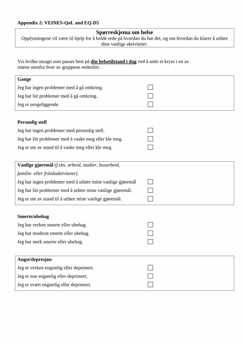
Appendix 2: VEINES-QoL and EQ-D5
Spørreskjema om helse Opplysningene vil være til hjelp for å holde rede på hvordan du har det, og om hvordan du klarer å utføre
dine vanlige aktiviteter.
Vis hvilke utsagn som passer best på din helsetilstand i dag ved å sette et kryss i en av
rutene utenfor hver av gruppene nedenfor.
Gange
Jeg har ingen problemer med å gå omkring.
Jeg har litt problemer med å gå omkring.
Jeg er sengeliggende.
Personlig stell
Jeg har ingen problemer med personlig stell.
Jeg har litt problemer med å vaske meg eller kle meg.
Jeg er ute av stand til å vaske meg eller kle meg.
Vanlige gjøremål (f.eks. arbeid, studier, husarbeid,
familie- eller fritidsaktiviteter).
Jeg har ingen problemer med å utføre mine vanlige gjøremål
Jeg har litt problemer med å utføre mine vanlige gjøremål.
Jeg er ute av stand til å utføre mine vanlige gjøremål.
Smerte/ubehag
Jeg har verken smerte eller ubehag.
Jeg har moderat smerte eller ubehag.
Jeg har sterk smerte eller ubehag.
Angst/depresjon
Jeg er verken engstelig eller deprimert.
Jeg er noe engstelig eller deprimert.
Jeg er svært engstelig eller deprimert.

40
Besvar hvert spørsmål nedenfor ved å krysse av svaret som angitt. Hvis du er usikker på hva du skal svare, vennligst
svar etter beste evne.
Disse spørsmålene er om din oppfatning av beina dine.
1.
I løpet av de 4 siste ukene, hvor ofte har du hatt noen av disse plagene i beina?
(Sett ett kryss på hver linje)
Daglig
Flere
ganger i
uka
Omtrent én
gang i uka
Sjeldnere
enn én gang
i uka
Aldri
1. Tunge bein 1 2 3 4 5
2. Vondt i beina 1 2 3 4 5
3. Hevelse 1 2 3 4 5
4. Kramper om natta 1 2 3 4 5
5. Varme eller brennende følelse 1 2 3 4 5
6. Urolige bein 1 2 3 4 5
7. Banking 1 2 3 4 5
8. Kløe 1 2 3 4 5
9. Prikking 1 2 3 4 5
2.
Når på dagen er plagene i beina mest uttalte? (Sett ett kryss)
1 Når jeg våkner 4 Om natta
2
2
Midt på dagen 5 Når som helst i løpet av dagen
3 På slutten av dagen 6 Aldri
3.
Sammenlignet med for ett år siden, hvordan vil du vurdere dine plager i beina nå? (Sett ett kryss)
1 Mye bedre nå enn for ett år siden
4 Noe verre nå enn for ett år siden
2 Noe bedre nå enn for ett år siden 5 Mye verre nå enn for ett år siden
3 Omtrent det samme nå som for ett år siden 6 Jeg hadde ingen plager i beina i fjor
4.
Følgende spørsmål gjelder daglige aktiviteter. Setter plagene i beina begrensninger for dine daglige
aktiviteter? Hvis « ja », i hvilken grad?

41
(Sett ett kryss på hver linje)
Jeg jobber ikke
JA, begrenser meg mye
JA, begrenser meg litt
NEI, begrenser meg ikke
a. Daglige aktiviteter på jobb. 0 1 2 3
b. Daglige aktiviteter hjemme (husarbeid, småjobber,
hagearbeid, o.l.)
1 2 3
c. Fritidsaktiviteter hvor du må stå lenge (selskap, ta buss, handle
o.l.)
1 2 3
d. Fritidsaktiviteter hvor du må sitte lenge (kino, teater, på reise o.l.)
1 2 3
5.
3. I løpet av de 4 siste ukene, har du hatt noen av disse problemene i jobb eller i daglige aktiviteter på
grunn av plagene i beina?
(Sett ett kryss på hver linje)
JA NEI
a. Redusert arbeidstid eller tid til andre aktiviteter 1 2
b. Gjennomført mindre enn du skulle ønsket 1 2
c. Blitt begrenset i type jobb eller aktiviteter 1 2
d. Hatt vanskeligheter med å utføre jobben eller andre aktiviteter (f eks det krevde større anstrengelse)
1 2
6.
I løpet av de 4 siste ukene, i hvilken grad har plagene i beina kommet i veien for samvær med familie, venner, naboer eller grupper? (Sett ett kryss)
1 Ikke i det hele tatt 4 Ganske stor
2 Lett 5 Svær
3 Moderat
7.
Hvor mye smerter har du hatt i beina i løpet av de 4 siste ukene? (sett ett kryss)
1 Ingen 4 Moderat
2 Svært lite 5 Mye
3 Lite 6 Svært mye
8.
Disse spørsmålene er om hvordan du føler deg, og om hvordan du har hatt det de siste 4 ukene som følge av plagene i beina. For hvert spørsmål, kryss av for det svaret som passer best med hvordan du har følt deg. Hvor mye i løpet av de 4 siste ukene-
(Sett ett kryss på hver linje)
Hele tiden
Det meste
av tiden
Ganske ofte
Av og til
Sjelde
n
Aldri

42
a. har du vært bekymret for hvordan beina
dine ser ut?
1 2 3 4 5 6
b. har du følt deg irritabel 1 2 3 4 5 6
c. har du følt at du har vært til byrde for familie eller venner?
1 2 3 4 5 6
d. har du vært bekymret for å skumpe borti ting?
1 2 3 4 5 6
e. har dine beins utseende påvirket ditt klesvalg ?
1 2 3 4 5 6
Vennligst oppgi dato for utfyllingen: _____/_____/_______ (dag/måned/år)