CA_TG Sequence at 5 End of Oligo(A)-tract.pdf - Prof. Manju Bansal
10
THE JOURNAL OF BIO~ICAL CHEMISTRY 0 1994 by The American Society for Biochemistry and Molecular Biology, Inc Vol. 269, No. 10, Issue of March 11, pp. 7824-7833, 1994 Printed in USA. CAITG Sequence at the 5' End of Oligo(A)-tracts Strongly Modulates DNA Curvature* (Received for publication, June 23, 1993, and in revised form, November 12, 1993) Akhilesh K. NagaichSO, Dhanaqjay BhattacharyyaS, Samir K. BrahmachariSO, and Manju Bansdm From the Wolecular BioDhvsics Unit and the #Centre for Genetic Engineering, Indian Institute of Science, Bangalom-560 012, Indid An analysis of the base pair doublet geometries in available crystal structures indicates that theoften re- ported intrinsic curvature of DNA containing oligo- (dfA)-d(T)) tracts may also depend on the nature of the flanking sequences. The presence of CAPTG doublet in particular at the 5' end of these tracts is expected to enhance their intrinsic bending property. To test this proposition, three oligonucleotides, d(GAAAAAC- CCCCC), d(CCCCCCAAAAAG), d(GAAAAATTMTC), and their complementary sequences were synthesized to study the effect of various flanking sequences, at the 5' and 3' ends of the A-tracts, on the curvatureof DNA in solution. An analysis of the polyacrylamide gel electro- phoretic mobilities of these sequences under different conditions of salts and temperatures (below their melt- ing points) clearly showed that the oligomer with CMG sequence in the center was always more retarded than the oligomer with AClGT sequence, as well as the oligo- mer with AT/AT sequence. Hydroxylradical probing of the sequences with AC/GT and CAPTG doublet junctions gives a similar cutting pattern in the A-tracts, which is quite different from that in the C-tracts, indicating that the oligo(A)-tracts have similar structures in the two oligomers. KMn04 probing shows that the oligomer with a CAPTG doublet junction forms a kink that is respon- sible for its inherent curvature and unusual electropho- retic mobility. UV melting shows a reduced thermal sta- bility of the duplex with CAPTG doublet junction, and circular dichroism (CD) studies indicate that a premelt- ing transition occurs in the oligomer with CA/TG dou- blet step before global melting but not in the oligomer with AC/GT doublet step, which may correspond to ther- mally induced unbending of the oligomer. These obser- vations indicate that the CAPTG doublet junction at the 5' end of the oligo(A)-tract has a crucial role in modulat- ing the overall curvature in DNA. The anomalous electrophoreticmobility ofA-tract-containing DNA has become closely identified with DNA curvature (1-6). There are several reports of DNA curvature in a wide variety of biological systems; in many cases, the loci of curvature are found in regions of ~ n c t i o n a l importance, i.e. origin of replica- tion, promoters, etc., suggesting the functional importance of curvature per se (7-8). The effect of oligo(A)-tracts has been * This work was supported by grants from the Council for Scientific and Industrial Research, India and the Department of Biotechnology, India. The costs of publication of this article were defrayed in part by the payment of page charges. This article must therefore be hereby marked "adoertisement" in accordance with 18 U.S.C. Section 1734 solely to indicate this fact. ll To whomcorrespondenceshould be addressed.Tel.: 80-3344411 (Ext. 2534); Fax: 91-80-3341683; Telex: 0845-9349 IISc IN; E-mail: [email protected]. attributed to the intrinsic features of the AA doublet sequence, namely the "wedge model" (91, as well as to discontinuities at the junction between random sequence DNA and oligo(A)- tracts, the so-called "junction model" (10-12). Theoretical Monte Carlo simulations of oligo(A)-tract-containing polymers also indicate that these tractsshow features that are in agree- ment with both these models (13, 14). Thus, while it is now generally accepted that the presence of A-tracts, repeating in a phasedmanner, coinciding with the helical repeatin DNA causes the helix axis to curve, the actual physical nature of the bending locus is not yet clearly elucidated.Recently it has been suggested that non-(A).)-tract containing sequences can also have slight curvature (15,16), and presence of CAEG dinucleo- tide steps has been shown to be important in DNA curvature (17). Since the oligonucl~tide crystal structures give the most accurate details of DNA structure, we have analyzed the local geometries of the various base pairdoublets from all available crystal structures and used this data to predict the curvature of some well characterized DNA sequences (18, 19). Our analysis indicated that the sequences flanking the A-tract are also quite crucial in determining the overall curvature of DNA. In par- ticular the presence of a C m G doublet at the 5' end can considerably enhance the intrinsic bending traits of a n oligo(A1- tract. It is interesting to note that the CAEG step is present in both the naturally occurring bent DNA from kinetoplasts and the synthetic sequences studied by Crothers and co-workers that are characterized as being intrinsically curved (10). In addition, the syntheticpromoter sequences that have been re- ported to have a bending locus and in turn a strong influence on transcription activation are also preceded by a C residue at the 5' end of A-tracts (20). A detailed sequence analysis of the natural DNA indicates that oligo(A)-tracts are preferentially flanked by the complementary base T, but these sequences have not generally been characterized as curved (21). Most of the gel mobility studies have been carried out using polymers with a variety of sequences in the non-A-tracts and by their very nature show the cumulative effects of oligo(A)-tracts, as well as the junctions at both 5' and 3' ends. An investigation of multimers of d(Cz&CZ)n, d(C2&G2),, and d(Gz&G& shows a decreasing order of anomaly in gel mobility, indicating the im- portance of the sequences flanking the A-tract in inducing the overall curvature (22). However, in this case also it is diEcult to differentiate the effects due to various junctions. An oligo- meric sequence with an oligo(A)-tract, juxtaposed on one side only with other sequences, can help in understanding the role of doublet sequences at the 5' and 3' ends. An earlier study (23) hasalready shown that even small oligomers such as the decamers d(G&T4C) and d(GT4A&) have different electropho- retic mobilities, a feature attributed to the differences in cur- vature between the two oligomers, arising due to AT and TA sequences at the center. We therefore decided to investigate the effect of C m G doublet sequence on the mobility of short oli- 7824 by on January 21, 2009 www.jbc.org Downloaded from
Transcript of CA_TG Sequence at 5 End of Oligo(A)-tract.pdf - Prof. Manju Bansal

THE JOURNAL OF B I O ~ I C A L CHEMISTRY 0 1994 by The American Society for Biochemistry and Molecular Biology, Inc
Vol. 269, No. 10, Issue of March 11, pp. 7824-7833, 1994 Printed in U S A .
CAITG Sequence at the 5' End of Oligo(A)-tracts Strongly Modulates DNA Curvature*
(Received for publication, June 23, 1993, and in revised form, November 12, 1993)
Akhilesh K. NagaichSO, Dhanaqjay BhattacharyyaS, Samir K. BrahmachariSO, and Manju Bansdm From the Wolecular BioDhvsics Unit and the #Centre for Genetic Engineering, Indian Institute of Science, Bangalom-560 012, Indid
An analysis of the base pair doublet geometries in available crystal structures indicates that the often re- ported intrinsic curvature of DNA containing oligo- (dfA)-d(T)) tracts may also depend on the nature of the flanking sequences. The presence of CAPTG doublet in particular at the 5' end of these tracts is expected to enhance their intrinsic bending property. To test this proposition, three oligonucleotides, d(GAAAAAC- CCCCC), d(CCCCCCAAAAAG), d(GAAAAATTMTC), and their complementary sequences were synthesized to study the effect of various flanking sequences, at the 5' and 3' ends of the A-tracts, on the curvature of DNA in solution. An analysis of the polyacrylamide gel electro- phoretic mobilities of these sequences under different conditions of salts and temperatures (below their melt- ing point s ) clearly showed that the oligomer with C M G sequence in the center was always more retarded than the oligomer with AClGT sequence, as well as the oligo- mer with AT/AT sequence. Hydroxyl radical probing of the sequences with AC/GT and CAPTG doublet junctions gives a similar cutting pattern in the A-tracts, which is quite different from that in the C-tracts, indicating that the oligo(A)-tracts have similar structures in the two oligomers. KMn04 probing shows that the oligomer with a CAPTG doublet junction forms a kink that is respon- sible for its inherent curvature and unusual electropho- retic mobility. UV melting shows a reduced thermal sta- bility of the duplex with CAPTG doublet junction, and circular dichroism (CD) studies indicate that a premelt- ing transition occurs in the oligomer with CA/TG dou- blet step before global melting but not in the oligomer with AC/GT doublet step, which may correspond to ther- mally induced unbending of the oligomer. These obser- vations indicate that the CAPTG doublet junction at the 5' end of the oligo(A)-tract has a crucial role in modulat- ing the overall curvature in DNA.
The anomalous electrophoretic mobility ofA-tract-containing DNA has become closely identified with DNA curvature (1-6). There are several reports of DNA curvature in a wide variety of biological systems; in many cases, the loci of curvature are found in regions of ~ n c t i o n a l importance, i.e. origin of replica- tion, promoters, etc., suggesting the functional importance of curvature per se (7-8). The effect of oligo(A)-tracts has been
* This work was supported by grants from the Council for Scientific and Industrial Research, India and the Department of Biotechnology, India. The costs of publication of this article were defrayed in part by the payment of page charges. This article must therefore be hereby marked "adoertisement" in accordance with 18 U.S.C. Section 1734 solely to indicate this fact.
ll To whom correspondence should be addressed. Tel.: 80-3344411 (Ext. 2534); Fax: 91-80-3341683; Telex: 0845-9349 IISc IN; E-mail: [email protected].
attributed to the intrinsic features of the AA doublet sequence, namely the "wedge model" (91, as well as to discontinuities at the junction between random sequence DNA and oligo(A)- tracts, the so-called "junction model" (10-12). Theoretical Monte Carlo simulations of oligo(A)-tract-containing polymers also indicate that these tracts show features that are in agree- ment with both these models (13, 14). Thus, while it is now generally accepted that the presence of A-tracts, repeating in a phased manner, coinciding with the helical repeat in DNA causes the helix axis to curve, the actual physical nature of the bending locus is not yet clearly elucidated. Recently it has been suggested that non-(A).)-tract containing sequences can also have slight curvature (15,16), and presence of CAEG dinucleo- tide steps has been shown to be important in DNA curvature (17). Since the oligonucl~tide crystal structures give the most accurate details of DNA structure, we have analyzed the local geometries of the various base pair doublets from all available crystal structures and used this data to predict the curvature of some well characterized DNA sequences (18, 19). Our analysis indicated that the sequences flanking the A-tract are also quite crucial in determining the overall curvature of DNA. In par- ticular the presence of a C m G doublet at the 5' end can considerably enhance the intrinsic bending traits of an oligo(A1- tract. It is interesting to note that the CAEG step is present in both the naturally occurring bent DNA from kinetoplasts and the synthetic sequences studied by Crothers and co-workers that are characterized as being intrinsically curved (10). In addition, the synthetic promoter sequences that have been re- ported to have a bending locus and in turn a strong influence on transcription activation are also preceded by a C residue at the 5' end of A-tracts (20). A detailed sequence analysis of the natural DNA indicates that oligo(A)-tracts are preferentially flanked by the complementary base T, but these sequences have not generally been characterized as curved (21). Most of the gel mobility studies have been carried out using polymers with a variety of sequences in the non-A-tracts and by their very nature show the cumulative effects of oligo(A)-tracts, as well as the junctions at both 5' and 3' ends. An investigation of multimers of d(Cz&CZ)n, d(C2&G2),, and d(Gz&G& shows a decreasing order of anomaly in gel mobility, indicating the im- portance of the sequences flanking the A-tract in inducing the overall curvature (22). However, in this case also it is diEcult to differentiate the effects due to various junctions. An oligo- meric sequence with an oligo(A)-tract, juxtaposed on one side only with other sequences, can help in understanding the role of doublet sequences at the 5' and 3' ends. An earlier study (23) has already shown that even small oligomers such as the decamers d(G&T4C) and d(GT4A&) have different electropho- retic mobilities, a feature attributed to the differences in cur- vature between the two oligomers, arising due to AT and TA sequences at the center. We therefore decided to investigate the effect of C m G doublet sequence on the mobility of short oli-
7824
by on January 21, 2009 w
ww
.jbc.orgD
ownloaded from

CAITG Sequence Modulates DNA Curvature 7825
gonucleotides containing oligo(A)-tracts and to compare it with the related ACfGT and the earlier characterized ATfAT se- quences. We chose dodecamer fragments to increase the length to diameter ratio for the molecules and, hence, their anisotropic shape. We find that oligomers d(GAscs) and d(C&sG), which have identical base composition and A-tract length, show dif- ferent el~trophoretic mobilities, as well as differences in the geometry at their central junction doublet as indicated by hy- droxyl radical and KMn04 probing. This sequence-dependent structural distortion in oligomer d(C&,G) is also reflected in its thermodynamic behavior as revealed by W melting and CD analysis. These observations confirm that the origin of greater bending in the oligomer d(C&&) as compared to the oligomer d(G&cs) lies in the unique geometry of its central c m junction doublet.
MA'IERIALS AND METHODS Model Building of Sequence-dependent Polymeric DNA Mole-
cules-In an earlier analysis, we have shown that the mean values of local doublet parameters (24), obtained by considering the B-DNA crys- tal structures data, available in the January 1991 release of the Brookhaven ptotein Data Bank (25), can provide a stxuctural basis for the relative electrophoretic migration of some well characterized genomic sequences (19). Similar analysis has now been carried out for synthetic polynucleotide sequences, using the data from the June 1993 update of the Nucleic Acid Data Base (26), which includes crystal struc- ture data for several new oligonucleotides. The mean values of the local doublet parameters for the 10 unique doublet sequences (Considering parameters for CC, TT, TG, TC, GT, and CT doublets as equivalent to their complementary base sequences, i.e. GG, AA, CA, GA, AC, and AG respectively) are given in Table I. It is found that inclusion of data from protein-DNA complexes does not lead to any significant differences in the mean parameter values, indicating that the local parameters are quite well defined unless the DNA undergoes a large protein-induced distortion. Hence the protein-DNA complex data have been included to obtain the parameters for the sequences AC and AG, for which only a few data points are available even in the current oligonucl~tide data base. Model structures of several polynucleotide sequences have been generated following the procedure described earlier (27).
The generated structures have been characterized by two different parameters. (i) The ratio of the end-to-end distanced to the actual path length l,, was calculated; (ii) the three principle moments of inertia were calculated, and the ratio of the largest to the smallest was taken as an indicator of the length to diameter of the inertia ellipsoid fitted to the model structure (14, 28).
Preparation and Purification of Oligonucleotides-Tko pairs of complementary oligonucleotides d(GA&J-d(G6T5C) and d(C&G). d(CT5G6), as well as the self-complementmy sequence d(G&T5C), were synthesized on a Pharmaeia automated DNA synthesizer Gene Assem- bler Plus, employing P-cyanoethyl phosphoramidite chemistry. Oligo- nucleotides were cleaved from the support and deprotected using 25% aqueous ammonia at 55 "C for 16 h. The deprotected oligonucieo~des were purified on 20% PAGE' containing 8 M urea using TriSmOratd EDTA, pH 8.0, as the running buffer. The gel was W-shadowed, and the desired bands were eluted in sterile water. The oligonucleotides were desalted by passing twice through Pharmacia NAP columns and C-18 Sep-Pak cartridges.
Polyacrylamide Gel Electrophoresis-Equimolar amounts of comple- mentary oligonucleotides were dissolved in 2 m~ sodium cacodylate, 0.1 nm EDTA, and 50 m~ NaCl or 12 m~ MgCl,, denatured at 90 "C for 3 min, and slowly cooled down to room temperature to achieve efficient annealing. Polyacrylamide slab gels (25%, w/v) with acrylamide to bis- acrylamide ratio of 19:l were made and run in 1 x TBE buffer contain- ing 50 m~ NaCl or 12 f l l ~ MgCIZ. We have found that 25% acrylamide concentration in the gel gives the best resolution of dodecamers. The gels were prerun for a fairly long time until the current became con- stant. Samples were loaded on to the gel preequilib~ted at indicated temperatures for 1 h, and electrophoresis was performed at 10 Vkm till the bromphenol blue in the control lane moved up to 22 cm. The gels were stained in ethidium bromide solution in water (1 pgfml) for 10 min and photographed.
d(C&G), and d(G&T,C) were radiolabeled using [y-32P]dATP and Hydroxyl Radical Probingal-purified oligonucleotides d(GA5C6),
The abbreviation used is: PAGE, polyacrylamide gel electrophoresis.
polynucleotide kinase. The phosphorylated oligomers were run on a 20% polyacrylamide gel containing 8 M urea, eluted in water, and de- salted as described previously. Labeled oligonucleotides of appropriate radioactivity were mixed with their complementary oligonucleotides, phosphorylated with cold dATP and dissolved in 2 m sodium cacody- late, 0.1 m~ EDTA, and 50 m NaCl, pH 7.2 135 pl), heated at 90 "C for 3 min, and slowly cooled to room temperature to achieve efficient an- nealing. The oligomers were preincubated at 4 and 20 "C for 15 min before reaction. The hydroxyi radical cleavage reactions were i ~ t ~ a t e d by adding 15 p1 of a mixture containing 0.2 m FeS04(NH4)zS04-6Hz0 and 0.4 m~ EDTA (5 pl), 0.6% hydrogen peroxide (5 $1. and 20 m~ sodium salt of L-ascorbic acid (5 pl). The reactions were carried out a t appropriate temperatures for 5 min and terminated by adding 0.1 M thiourea (20 pl). The DNA was precipitated using 3 M sodium acetate (25 pl), ethanol (750 pl), and tRNA (10 pgf at -70 "C overnight. The pre- cipitated DNA was pelleted and washed three times with 70% ethanol, dried, and suspended in DNA sequencing gel loading buffer containing (80% formamide in 1 x TBE, bromphenol blue, and xylene cyano1 dyes). A part of the reaction mixture was loaded on a 25% denaturing poiy- acrylamide gel containing 8 M urea. The electrophoresis was performed at 40 Vkm, and the gels were autoradiographed. Each hydroxyl radical probing experiment was repeated three times to ensure reproducibility of the results. ~ n 0 ~ Pr~&gng-Oligonu~eotides d(G6T,C1, d(CT&& and
d(G&T,C) were radiolabeled with [y-32P]dATP and gel-purified as de- scribed previously. Equimolar amounts of their complementary se- quences were mixed and dissolved in 2 m sodium cacodylate, 0.1 nm EDTA, and 50 m~ NaCl, pH 7.2 (19 pl), heated at 90 "C for 3 min, and annealed at room temperature. Samples were incubated at 4 and 20 "C for 15 min and reacted with 1 pl of KMnO, (100 pmoVml) for exactly 5 min. Reactions were stopped by adding allyl alcohol (1 IJX and oligo- mers were ethanol-precipitated along with 10 pg of Escherichia coli tRNA. The oligomers were pelleted, washed with 70% ethanol, and dried. The samples were resuspended in 100 pl of 1 M piperidine and heated to 90 "C for 30 min. Piperidine was removed by repeated drying with water. The DNA cleavage products obtained were resuspended in sequencing gel loading buffer of composition discussed previously, heat- denatured at 90 "C for 3 min, and loaded on a 25% denaturing PAGE containing 8 M urea. The gels were run at 40 Vfcm in 1 x TEE buffer and auto~diographed. KMnO, probing reactions were carried out three times far each oligonucleotide, and the results were found to be highly reproducible.
W Melting Analysis-The temperature-dependent absorption spec- tra were measured at 260 nm using a Beckman DUdB spectrophotom- eter equipped with a Peltier temperature-controlled and programmed cell holder that accommodates five cuvettes. Melting profiles of oligo- mers were obtained at a strand concentration of 8 x M by increasing temperature at the rate of 0.5 "Chin with the absorbance and the temperature being recorded every 30 s. All the samples were prepared at the same buffer concentration used to measure CD spectra for better comparison. We recorded the melting profiles of the oligomers in 2 m sodium cacodylate, 0.1 nm EDTA, and 50 m NaC1. The thermodynamic parameters were calculated based on temperature-dependent W ab- sorption data assuming a two-state model of helix to coil transition considering a molec~arity of 2 and the T, values were deduced from daldl"" uersus temperature plots.
Circular Dichroism-CD spectra were recorded in the spectral range of 220 to 320 nm on a JASCO J-5OOA automatic spectropolarimeter equipped with a DP 501 data processor and thermostatically controlled cuvette holder. The samples were prepared by heating at 95 "C for 3 min in 5 x M strand concentration followed by gradual cooling to room temperature. Prior to scan, samples were diluted to give a strand con- centration of 8 x loA M and equilibrated in a CD cell holder for 1 h at room temperature to obtain equilibrium spectra. The samples were then cooled to 5 "C, and CD spectra was recorded after equilibrating the samples for 20 min at each temperature. All the CD spectra were re- corded in a cuvette of path length 2 mm. In order to increase signal to noise ratio, four scans were accumulated and the average spectrum was recorded after subtracting the background scan of buf€er.
~ ~ i ~ o ~ t ~ ~ c Scanning-Scanning laser de~i tometry of autoradi~ graphs was done using LKB-2202 Ultroscan densitometer using an aperture width of 0.05 cm.
RESULTS AND DISCUSSION Theoretical Analysis of DNA Bending-Model structures
were generated (using the set of base pair step parameters listed in Table I) for polynucleotide sequences, with decamer
by on January 21, 2009 w
ww
.jbc.orgD
ownloaded from

TABLE I
of all available crystal structures of B-form DNA for the 10 unique The mean local doublet parameters (18, 24) obtained from analysis
dinucleotide sequences
correspond to steps with the BiI c o ~ o ~ a ~ o n (18). The oligonucleotide The CA doublet parameters listed here and used in the generation
data was taken from the June 1993 release of Nucleic Acid Data Base (26), and the number of data points available for each sequence is given in parentheses. The average value for each parameter is given in the last line and is close to the value for the fiber model (18).
Tilt (7) Roll ( p ) Twist fo,) Shift (0,) Slide (By) Rise (DZ)
CG (72) 0.1 3.2 35.1 0.1 0.5 3.53 GC (39) -0.2 -6.5 38.6 -0.2 0.5 3.34 AA(49) -1.7 2.6 35.6 -0.1 -0.2 3.23
AT (30) 0.2 -0.3 32.5 GA(35) -0.9 0.3 38.5 -0.0 -0.1 3.10
GG (20) -1.8 5.8 32.4 -0.1 0.1 -0.4 3.29
CA (10) 0.6 -7.5 47.9 0.1 2.9 3.40 AC (15) -0.2 -0.7 33.3 -0.2 -0.1 3.35 AG (13) -0.5 7.1 29.5 -0.1 0.4 3.48 TA(O7) 0.0 0.5 39.0 -0.2 0.2 3.22
0.7 3.47
Average -0.5 0.5 36.1 -0.1 0.5 3.35
repeats containing oligo(A)-tracts of varying length. The values obtained for the ratios of end-to-end distance to the actual path length (d/lmw) traced by the model structures generally show an approximately inverse correlation with their experimentally observed relative mobility (RL) in polyacrylamide gel (Table 11). The calculated values of the ratios of the largest to smallest principal moments (ZmJZmin) also show a similar relation, Le., both dllmax and ImJZmin values are relatively large for those sequences that are straight (with RL values close to 1.0) and are smaller for the curved fragments.
Model structures were also generated for the restriction frag- ments of kinetoplast DNA from Crithidia fusciculatu (I, 11, and 111) and Leishmania tarentolue (N) and are shown in Fig. 1, while the bending parameters are included in Table 11. It is clearly seen that fragments I1 and N, which show abnormally retarded mobility, are predicted to be curved, while fragments I and I11 are straight as expected from their RL values (29,301. A few models of random nucleotide sequences with different percentages of AT content have also been built for comparison and the relevant parameters for a few representative sequences are given in Table 11. It is clearly seen that random sequence polynucleotide models have large values for d/Zma, (i.e. in the range of 0.93 to 1.0) as well for I m ~ ~ I ~ i n (2301, indicating that these model structures are nearly straight but can occasionally be slightly bent. This is in agreement with the deduction from their gel migration data (31) that the random sequence struc- tures can also show a slight amount of curvature.
Thus, the mean local doublet geometries, as obtained from all the available B-DNA crystal structures, can explain the anomalous gel migration data of p o l ~ u c l ~ t i d e s of repetitive sequences, as well as that of the well characterized genomic sequences. In view of the static model considered by us, the correlation between the theoretical models and the gel retar- dation data is quite good, indicating that the average molecular shape is correctly predicted. The extensive Monte Carlo simu- lation studies also show only a qualitative agreement with the experimental data, with the theoretical models for the few se- quences studied, showing a much smaller variation than ex- pected from the gel mobility data (13). The values of local pa- rameters reported by other workers were deduced from theoretical consideration or by empirical fitting of cyclization and gel mobility data. It is interesting to note that the roll and twist values for dinucleotide steps other than AA, CA, and GG are found to be very similar in crystal structures and from the gel data (11, 12). However, there is a basic assumption that the
TABLE I1 The structural parameters describing the bending of the predicted structural models of 150-base pair fragments for a few synthetic
sequences with decamer repeats and some genomic DNA sequences are listed, along with the experimentally observed relative mobility
iRJ of the fragments in ~ ~ y ~ r y l a m ~ d e gel A few computer-generated random sequences of 150 base pairs in
length with varying AT content are also included for comparison.
Sequence
Synthetic polymers
Percentage AT content R~
GGCCAAACCG 30 1.23 GGCCAAAACG 40 1.60 GGCAAAAACG 50 2.00 GGCARAARAC 60 2.31 GC- 80 2.21 C A A ? A U A M 90 1.73 CAAAATTTTG 80 2.23 CTTTTAAAAG 80 1.04
I 45 1.09 I1 63 3.18 I11 52 1.00 I V 57 3.78
Computer-generated 150-mer sequences CCCCCAAAAA 50 Random 1 20 Random 2 50 Random 3 80
Genomic sequences"
d I L a x -~
0.48 0.52 0.42 0.38 0.35 0.57 0.50 0.78
0.92 0.89 0.97 0.77
0.32 0.93 0.97 0.99
I",dmin
3.9 4.4 3.6 3.2 3.1 4.8 2.4 9.8
55.8 16.3 90.7 8.0
2.5 32.7 59.7
139.5
plast DNAof C. fasciculata (I, 11, and 111) (29) and L. tarentolae (IV) (30) a The genomic sequences are the restriction fragments from kinetc-
of length 274, 211, 150, and 414 base pairs, respectively.
1 I 1 I11 IV
listed in Table If, generated using mean local doublet param- FIG. 1. Models for the four genomic sequences (Z, ZZ, ZZZ and rV)
eters obtained from B-DNA crystal structures (as given in Table I). The C1' atoms as representatives of each nucleotide have been joined to simulate the base-paired double helix. The differences between the
fragments I and III are clearly seen. curvature of fragments N and N as compared to the relatively straight
AA step has a negative roll angle (10-13) and the CAA'G step has a roll angle opposite to that of AAlTT step, so that a CA step occurring out of phase with the center of the oligo(A)-tract implicitly enhances the curvature, although it was not explic- itly mentioned by the authors of these studies. On the contrary, the mean roll angle for the 49 A" steps observed in the oligonucleotide crystal structures, solved using high resolution data, has a small positive mean value (+2.63" with a standard deviation of 3.7") and hence a better agreement with the gel data is obtained if the accompanying CA steps are taken to have the negative roll, high twist combination (18, 32) that is
by on January 21, 2009 w
ww
.jbc.orgD
ownloaded from
CA i TG Sequence M ~ u ~ a t e s DNA Curvature 7827
3'
5 3, (a) (b)
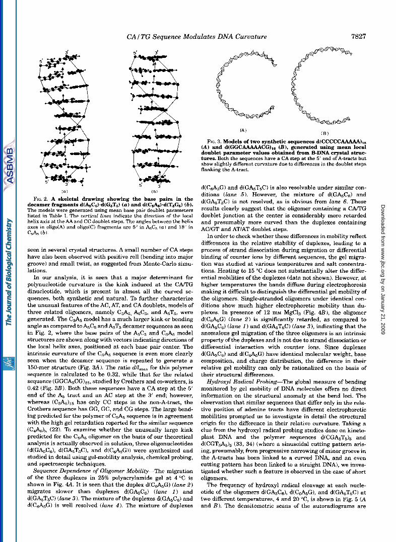
decamer hragments d(&C5)-d(G5T5) (a) and dfC&f-d(T6G6) (b). FIG. 2. A skeletal drawing showing the base pairs in the
The models were generated using mean base pair doublet parameters
helix axis at the AA and CC doublet steps. The angles between the helix listed in Table I. The uerticut lines indicate the direction of the local
axes in oligo(A) and o1igoiC) fragments are 5" in A& { a ) and 18" in C A , (b) .
seen in several crystal structures. A small number of CA steps have also been observed with positive roll (bending into major groove) and small twist, as suggested from Monte-Carlo simu- lations.
In our analysis, it is seen that a major determinant for polynucleotide curvature is the kink induced at the CA/TG dinucleotide, which is present in almost all the curved se- quences, both synthetic and natural. To further characterize the unusual features of the AC, AT, and CA doublets, models of three related oligomers, namely C5&, A5C5, and A5T5, were generated. The C d 5 model has a much larger kink or bending angle as compared toA5C5 and A5T5 decamer sequences as seen in Fig. 2, where the base pairs of the A5C5 and C5A5 model structures are shown along with vectors indicating directions of the local helix axes, positioned at each base pair center. The intrinsic curvature of the C5A5 sequence is even more clearly seen when the decamer sequence is repeated to generate a 150-mer structure (Fig. 3A). The ratio d/lmax for this polymer sequence is calculated to be 0.32, while that for the related sequence (GGCA5CG)15, studied by Crothers and co-workers, is 0.42 (Fig. 3B). Both these sequences have a CA step at the 5' end of the A5 tract and an AC step at the 3' end however, whereas fC5A5)15 has only CC steps in the non-A-tract, the Crothers sequence has GG, GC, and CG steps. The large bend- ing predicted for the polymer of C5A5 sequence is in agreement with the high gel retardation reported for the similar sequence (CdA& (22). To examine whether the unusually large kink predicted for the C d 5 oligomer on the basis of our theoretical analysis is actually observed in solution, three oligonucleotides (d(GA&& d(GA5T5C), and d(Cd5G)) were synthesized and studied in detail using gel-mobility analysis, chemical probing, and spectroscopic techniques.
Sequence Dependence of Oligomer Mobility-The migration of the three duplexes in 25% polyacrylamide gel at 4 "C is shown in Fig. 4A. It is seen that the duplex d(C&G) (lane 2) migrates slower than duplexes d(GA5C6) (lane 1 ) and d(GAsT5C) (lane 3) . The mixture of the duplexes d(GA5C6) and d(C&G) is well resolved (lane 4 ) . The mixture of duplexes
(A ) f B )
(A) and d(GGCAAAAACGfl6 (B) , generated using mean local FIG. 3. Models of two synthetic sequences d(CCCCCAAAAA),,
doublet parameter values obtained fiwm B-DNA crystal struc- tures. Both the sequences have a CA step at the 5' end of A-tracts but show slightly different curvature due to differences in the doublet steps flanking the A-tract.
d(Cd5G) and d(GA5T5C) is also resolvable under similar con- ditions (Zane 5 ) . However, the mixture of d(GA5Cs) and d(GA5T5C) is not resolved, as is obvious from lane 6. These results clearly suggest that the oligomer containing a CAlTG doublet junction at the center is considerably more retarded and presumably more curved than the duplexes containing AC/GT and AT/AT doublet steps.
In order to check whether these differences in mobility reflect differences in the relative stability of duplexes, leading to a process of strand dissociation during migration or differential binding of counter ions by different sequences, the gel migra- tion was studied at various temperatures and salt concentra- tions. Heating to 15 "C does not substantially alter the differ- ential mobilities of the duplexes (data not shown). However, at higher temperatures the bands diffuse during electrophoresis making it difficult to distinguish the differential gel mobility of the oligomers. Single-stranded oligomers under identical con- ditions show much higher electrophoretic mobility than du- plexes. In presence of 12 mM MgClz (Fig. 4 2 3 1 , the oligomer d(C&G) (lane 2) is significantly retarded, as compared to d(GA5Cs) (lane 1 ) and d(GA5T5C) (lane 3 ), indicating that the anomalous gel migration of the three oligomers is an intrinsic property of the duplexes and is not due to strand dissociation or differential interaction with counter ions. Since duplexes d(GA5Ce) and d(Cd5G) have identical molecular weight, base composition, and charge distribution, the difference in their relative gel mobility can only be rationalized on the basis of their structural differences.
Hydroxyl Radical Probing-The global measure of bending monitored by gel mobility of DNA molecules offers no direct i n f o ~ a t i o n on the structural anomaly at the bend loci. The observation that similar sequences that differ only in the rela- tive position of adenine tracts have different electrophoretic mobilities prompted us to investigate in detail the structural origin for the difference in their relative curvature. Taking a clue from the hydroxyl radical probing studies done on kineto- plast DNA and the polymer sequences d(CGA5T5I5 and d(CGT&)5 (33, 34) (where a sinusoidal cutting pattern aris- ing, presumably, from progressive narrowing of minor groove in the A-tracts has been linked to a curved DNA, and an even cutting pattern has been linked to a straight DNA), we inves- tigated whether such a feature is observed in the case of short oligomers.
The frequency of hydroxyl radical cleavage at each nucle- otide of the oligomers d(GA5C& d(C&G), and d(GA5T5C) at two different temperatures, 4 and 20 "C, is shown in Fig. 5 ( A and B). The densitometric scans of the autoradiograms are
by on January 21, 2009 w
ww
.jbc.orgD
ownloaded from

7828 CAI TG Sequence Modulates DNA Curvature
4 5 6
B 1 2 3
FIG. 4. A, electrophoretic mobility of three dodecamers in 25% PAGE at 4 "C in 50 mM NaCI. Lanes 1, 2, and 3 contain duplexes d(GASC6), d(C&\,G), and d(GA,T,C), respectively. Lanes 4 , 5, and 6 contain the mixtures of duplexes d(GASC6) + d(C&,G), d(C&,G) + d(GAST,C), and d(GA,T,C) + d(GA,C,), respectively. B, electrophoretic mobility of do- decamer duplexes d(GA, c,) (lane l ), d(C&,G) (lane 2) , and d(GA,T,C) (lane 3 ) in the presence of 12 mu MgCI,.
shown in Fig. 5 (C-E). The A-tracts in both the oligomers d(C&,G) and d(GAsC6) show reduced cleavage at 4 "C (lanes 1 and 2, respectively, in panel A), as compared to the C-tracts, indicating the unique nature of the oligo(A)-tracts in both the sequences, particularly a t low temperature. The central CA step in the oligomer d(C&,G) shows a remarkably high sus- ceptibility to cleavage a t 4 "C as compared to any other doublet step, which may indicate its unusual structure (Fig. 5C). At 20 "C the A-tracts and the C-tracts of both the oligomers show nearly uniform hydroxyl radical cleavage (Fig. 5A, lanes 3 and 4 ), indicating the presence of sequence-independent structures.
The oligomer d(GAsTsC) shows a marginally reduced cutting around the central region a t 4 "C (lane 1 ) and a fairly even cutting frequency a t 20 "C (Fig. 5B, lane 2 ). It is interesting to note that this temperature-induced transition in the A-tracts of all the oligomers is observed well below their respective T, values under identical NaCl concentration. A premelting tran- sition in A-tracts has been reported earlier by chemical probing studies in the case of longer synthetic sequences indicating that oligo(A)-tracts are capable of forming more than one structure before global melting (35, 36). However, we do not observe a progressive decrease in the intensity of bands in the A-tracts, similar to that reported previously (33, 34) for polymeric se- quences and attributed to the narrowing of minor groove in the region. In a separate hydroxyl radical probing study of 30-mers containing three oligo(A)-tracts of varying lengths, phased with the helical repeat, we have found a progressive decrease in band intensity in the A-tracts.2 Since the oligomers reported here are smaller and oligo(A)-tracts are flanked only on one side, the groove width in their A-tracts may not be severely constrained.
KMn04 Probing-KMn04 has been used as a probe to study the conformational flexibility of short adenine tracts present in DNA (35, 36). An in vivo probing of B-Z junction sequences having a T residue at the junction has also been reported using KMn04 as a probe (37). Recently, oligonucleotides d(G4T,G4) (n = 2 4 ) , adopting a hairpin G-quartet structure by dimerization of hairpin loops, have also been probed in our laboratory using KMn04 (38). Since KMn04 reacts with the DNA primarily via oxidation of the C5-C6 double bond of pyrimidines (T>>C), stacking of the bases causes protection from this reaction (39- 40). When the base stacking is disrupted, the susceptibility of thymines, to react with KMn04 increases significantly (41,42). We investigated the geometry of the dinucleotide junction steps AC/GT and C m G present in oligomers d(GAsCd and d(CdSG) using KMn04 as a probe. The oligomer d(GAsCd does not react with KMn04 at 4 "C, while in the oligomer d(C&G), the T a t the CA/TG junction shows a remarkable reactivity a t this temperature (Fig. 6 A , lanes 1 and 2 ). At 20 "C the T-tracts in both the oligomers show reactivity with KMn04 (Fig. 6 A , lanes 3 and 4) . Similarly, in the duplex d(GAsT&), all the T residues remain totally unreactive a t 4 "C, but at 12 "C some reactivity is observed, which is considerably enhanced a t 20 "C (Fig. 6B, lunes 1, 2, and 3 ) . All the T residues in the single stranded oligonucleotides show very high reactivity under simi- lar conditions. This clearly indicates that, a t low temperatures, the sequence d(C,&G) has a conformation having a more pro- nounced kink a t CARG junction compared to that of the AC/GT junction in the oligomer d(GASCs) or the AT/AT junction in oligomer d(GAsT5C). In order to see whether the sequence- dependent structural distortion of the oligomers leading to dif- ferent electrophoretic mobilities, as well as the difference in the geometry of central junction doublet, as revealed by KMn04 and hydroxyl radical probing, is reflected in their global con- formation and thermodynamic properties, we analyzed their melting behavior, as well as CD spectra.
U V Melting Analysis-The UV melting profiles of the three dodecamer sequences d(GA5C6), d(C&G) and d(GASTsC) at 260 nm, in 50 mM NaCl are shown in Fig. 7A. This absorbance versus temperature data was converted into a fractional helic- ity (a) versus T melting curve (Fig. 7B) and the transition midpoint (T,) of each oligomer was determined by d d d T " versus temperature ("C) plot (Fig. 7C). The van't Hoff transi- tion enthalpy was calculated from the general form of the van't Hoff equation,
2 A . K. Nagaich, S. K. Brahmachari, and M. Bansal, unpublished data.
by on January 21, 2009 w
ww
.jbc.orgD
ownloaded from

FIG. 5. A, autoradiograph of the gel comparing the hydroxyl radical cleavage pattern of the oligomers d(C,&G) (lane 1 ) and d(G&CR) (lane 2 ) at 4 "C and d(C&G) (lane 3) and d(G&C,) (lane 4 ) at 20 OC. The arrow indicates the enhanced cleavage at the CA/"G junction of the duplex d(C,+ISG) a t 4 "C. The absolute intensities of the bands in different lanes cannot be compared, as they result from two separate reactions. The relative intensities of the bands within the lane can be compared, since they result from a single reaction (33.34). B , autoradiograph of the gel comparing the hydroxyl radical cleavage pattern of the duplex d(GA,T,C) a t 4 "C (lane 1 ) and a t 20 "C (lane 2). respectively. C , densito- metric scans of the hydroxyl radical cleav- age pattern of the duplex d(C,+ISG) a t 4 and 20 "C, i.e. lanes I and lane 3 of panel A. D, densitometric scans of the hydroxyl radical cleavage pattern of the duplex d(GASC6) at 4 and 20 "C, i.e. lanes 2 and 4 of panel A. The A-tract shows much re- duced cleavage as compared to C-tract a t 4 "C. E , densitometric scans of the hy- droxyl radical cleavage pattern of the du- plex d(GA,T,C) a t 4 and 20 "C, i.e. lanes 1 and 2 of panel B.
CAI TG Sequence Modulates DNA Curvature 7829
A
*
C
1 2 3 4 1 2
B
D
cc c c c
20°( C
by on January 21, 2009 w
ww
.jbc.orgD
ownloaded from

7830 CAITG Sequence Modulates DNA Curvature
A I 2 3 4
B I z 3 "-
( lam I ) , d(C&G) at 4 "C (lane 2). d(G&CG) at 20 "C (lane 3), and FIG. 6. A, KMnO, cleavage pattern of the duplexes d(G&Cs) at 4 "C
d(C&G) at 20 "C (lane 4) , respectively. B , KMn0, cleavage pattern of
(Eq. 1)
where n is the molecularity and T,,, is the transition midpoint (43). The values of AG were calculated by determining Keq at T,, extrapolating it to 25 "C and substituting this value in the standard equation AG= -RT In KZgR. Assuming AC, = 0, these values were substituted in the equation AG= AH - TAS to calculate entropy and the values obtained are listed in Table 111. I t is clear from Fig. 7A that helix to coil transition in the oligomer d(Cd5G) is broader and less cooperative than that in oligomers d(GA5Cs) and d(GA5T5C). The curved oligomer d(Cd5G) also displays thermal activity in the premelting do- main, as is obvious in Fig. 7C. The duplex d(Cd5G) has lower T, (40.3 "C) as compared to duplex d(GA5Cs) (44.8 "C), while the duplex d(GA5T5C) has a much lower T,,, value (34 "C), as expected from its higher AT content. The oligomer d(GA5Cs) also shows large changes in enthalpy ( A H = -55.7 kcaVmol), as well as free energy (AG = -11.7 kcaVmo1) as compared to the oligomer d(Cd5G) ( A H = -35.8 kcaVmol and AG = -10 kcaV mol), indicating its higher thermal stability. The duplex d(GA5T5C) also shows a large change in enthalpy ( A H = -56.9 kcaVmol), as well as free energy (AG = -9.5 kcaVmo1) due to the highly cooperative helix to coil transition observed for this oligomer (Fig. 7A). Since the oligomers d(GA5C6) and d(Cd5G) differ only in the sequence a t the central junction step, a dif- ference of about 20 kcaVmol in AH can only be correlated to the loss of stacking energy at the C m G step in the oligomer d(C45G).
Circular Dichroism Studies-We have employed circular di- chroism measurements to study the equilibrium melting and temperature-dependent conformational change in all three do- decamers. The CD spectra of the three duplexes have been recorded in 50 mM NaCl in cacodylate buffer at different tem- peratures. The spectra at a few selected temperatures have been plotted in Fig. 8 ( A X ) . At low temperature (5 "C), the curved duplex d(C&G), with a C m G doublet at the junction, displays a spectrum characteristic of B-DNA, with a positive peak a t 265 nm and a negative peak a t 242 nm (Fig. 8A). Increasing the temperature up to 35 "C causes an increase in positive ellipticity accompanied by a small decrease in the negative ellipticity. Further increase in temperature up to 75 "C causes a decrease in positive, as well as negative, ellip- ticity, which indicates melting of the duplex structure. The spectrum of the relatively straight oligomer d(GAsCs), with AC/TG junction, has a broad positive peak with maxima a t 260 nm and a shoulder at 280 nm and a negative peak at 245 nm at 5 "C (Fig. 8B). Increasing temperature up to 35 "C does not cause any substantial change in the positive ellipticity, and further increase in the temperature causes global melting of the duplex. Considering the transition midpoint of both these oligomers under identical salt concentration, it can be postu- lated that the duplex d(Cd5G) undergoes a premelting tran- sition before global melting. The self complementary duplex d(GA5T5C) shows some of the features of poly(d(A)).poly(d(T)) and exhibits a positive peak a t 282 nm, a shoulder at 262 nm, and a negative peak a t 247 nm at 5 "C (Fig. 8C). The shoulder at 262 nm was also observed earlier for the polymer d(G&T,C), and has been attributed to the base pairs a t the ATIAT junction having large propeller twist (44). The disap- pearance of this shoulder in the duplex d(GA5T5C) as the tem- perature increases indicates a thermally induced rearrange- ment in the oligomer before global melting. It is interesting to note that the oligomer d(Cd5G), which is kinked, displays
by on January 21, 2009 w
ww
.jbc.orgD
ownloaded from

CA/ TG Sequence ~ ~ d u l a t e s DNA Curvature 783 1
A 1.10
1.05
1 .bo ,-. E 5: 0.95 N CI
0.90 t 0 n $ 0.85 2
0.80
0.75
0.70 0
/ /
/ ." ".-
6 1 . 2 0
1.00 - 1
0.20 1 -0.00
~
6oool 5000
4000
3000 v
>: 5 2000 D
0
? 000
0
-looc:ma..,~~,, . I . , I , . , , , , , I , , , I , , , ,.,, ,,,, 0 10 2b i o i o do sb 7b do 9b
Ternperatcrr ( " C )
FIG. 7. A, absorbance versus temperature ( " 0 profiles for d(GA5C6) ("-), d(C&Gt (...), and d(G&T5C) (- - -) at 260 nm annealed in 2 m~ sodium cacodylate, 0.1 m~ EDTA, and 50 m~ NaCI. The strand concen- tration for all the oligomers is 8 x lo4 M. E , van't Hoff plot of fractional helicity (a) versus temperature ("C) for d(G&C6) (-), dCC&G) (...I,
TABLE I11 Spectroscopically derived thermodynamic parameters for the melting
of oligomers d(GA,Cd, d(C&G), and d(GA,T&)
Duplex T, AH TLLS AG
"C k ~ l i ~ dtG&Cd 44.8 0.2 -55.7 -44.0 -11.7
40.3 * 0.3 -35.8 -25.8 -10.0 d(GAET6C) 34.0 f 0.5 -56.9 -47.4 -9.5 d(C&G)
spectral features of normal B-DNA, as revealed by CD spectra, suggesting that there are only marginal differences in the con- f o ~ a t i o n of olig~A)-tracts and C-tracts as compared to ran- dom sequence DNA and that the bending in the oligomer arises mainly due to the characteristic geometry at the central junc- tion step, which is not necessarily reflected in the CD spectra. The oligomers d(C,&,G) and d(GA5C6) also do not undergo a B + A transition on lowering the water activity up to 60% etha- nol, despite the presence of oligo C-tracts (data not shown). I t has been proposed earlier that ~mperature-de~ndent change in negative ellipticity in the DNA usually monitors duplex to single-strand transition, whereas change in the positive ellip- ticity with temperature gives an indication about the intramo- lecular helix to helix transition (45). In order to facilitate a comparison in the premelting behavior of all the three oligo- mers, the change in positive ellipticity a t [elmw was carefully measured. Results of four such measurements along with their mean deviations are given in Table IV. The increase in positive ellipticity from 8.1 x lo3 degrees . cm2 dmol" to 9.7 x lo3 degrees * cm2 dmol" in the duplex d(C,&G) at 265 nm, with increase in the temperature from 5 "C to 35 "C, clearly shows a premelting transition before global melting. Our results are in partial agreement with the earlier spectroscopic and calorimet- ric studies done on poly(d(A)).poly(d(T)), kinetoplast DNA, and bent decamer d(G&T,C), which show a premelting behavior in the oligo A-tracts prior to the global melting (45, 46). A com- parison in the melting behavior of sequences d(G&T,C) and d(GT&C) had shown that the former had greater thermal stability than the latter, a feature that was attributed to the spine of hydration associated with the A-tract in this sequence. However, in our study the bent oligomer d(C,+45G) shows a premelting behavior and lower thermal stability as compared to the straight oligomer d(GA5C6). The trends seen in the tem- perature-dependent gel mobility and the change in the elliptic- ity of the positive peak in the spectra of all the three dodecam- ers suggest that the structural features responsible for the unusual electrophoretic migration of duplexes d(C&G) and d(GA5T&) also account for their premelting behavior prior to global melting and strand dissociation. This lends further sup- port to our gel mobility data that the oligomers during the electrophoretic migration are not the kinetically trapped spe- cies but thermodynamically stable structures.
Role of CAiTG Sequence in DNA Bending-The predictions of relative bending of DNA oligomers on the basis of observed local geometries of various doublet sequences in crystal struc- tures are corroborated by our studies on the dodecamer se- quences. In particular, they highlight the important role of the CATG sequence at the 5' end of an oligo(A)-tract. This doublet seems to have structural features that are different from the other two p ~ m i d i n e . p u ~ n e doublets TA and CG. It has been suggested from empirical data analysis (12) that both C m G and AC/GT doublets play a passive role by allowing large tran-
ture-dependent W absorption data at 260 nm shown in panel A . C, and d(GAST6C) (- - -1. These points were evaluated from the tempera-
differential melting curves for the dodeeamers d(GA,C,) (-), d(CAG1 (.-,),,and d(G&T,C) (- - -1. The presence of a broad shoulder in the case of olxgomer d(Cd6G) indicates a premelting before denatur- ation.
by on January 21, 2009 w
ww
.jbc.orgD
ownloaded from

7832 CA f TG Sequence Modulates DNA Curvature
A
10
7 5 ; N
E O
'0
; Q
m
x -5 m c_
u
-1 0
240 260 320 Wavelength inrn)
24 0 280 320 Wavelength (nrn)
240 Wavelength (nm)
FIG. 8. A, CD spectra of duplex d(C&G) at 5 "c (-), 25 "c f- - -), 35 "c (-.-.-), 55 "C (...), and 75 "C (-) in 2 m~ sodium cacodylate, 0.1 mM EDTA, and 50 mhp NaCl. €2, CD spectra of the duplex d(G&Cd at
"C degcm2 ,dmol" 5 9.9 2 0.1
15 10.2 t 0.05 25 10.2 f 0.05 35 10.0 0.05 45 8.3 f 0.2 55 6.8 f 0.2
degcm2 .dmol" deg.cm2 .dmol" 8.1 f 0.1 8.2 t 0.1 9.2 f 0.05 8.9 f 0.05 9.0 2 0.05 8.2 f 0.05 9.7 0.05 7.5 f 0.1 8.8 * 0.1 5.4 f 0.1 7.2 2 0.1 3.8 f 0.1
sient kinks. While this may be true in general, our results indicate that in fact these two sequences exhibit completely different properties when they flank an o1igofA)-tract, with the CAplyf sequence giving rise to a relatively static kink.
A gel retardation study on the polymer of 5'd(GGG- CAAAAAC13' base-paired with its complementary sequence 5'd(GlTMYGCCC)3' also shows that base stacking is the dominating force in the formation and stabilization of curved DNA (47). The authors of the study have studied the effect of various modifications in A-tracts, like methylation of the complementary pyrimidine residues and substitution of the adenosine residues with inosine, etc., on the formation of al- tered B' structure, which is believed to cause curvature. How- ever, we observe that in this study the A-tracts are also flanked on both sides by C residues and, in fact, all the sequences show s i~ i f icant curvature, as expected from our studies. It is likely that the polypurine tracts have essentially similar features in these curved oligonucleotides, with only minor differences aris- ing due to variations in propeller twist and pyrimidine methy- lation etc. This gives added support to our conclusion that the kink induced at CA/TG step at the 5' end of the oligo(A)-tracts is a major determinant of the overall curvature in DNA. Of course any intrinsic curvature of oligo(A)-tracts, as well as the dynamic flexibility of various doublet steps (including CA) and hence the polynucleotide as a whole, is also relevant to the mobility of DNA through the gels.
While the observation that isolated CA steps in random se- quence DNA modulate DNA bending has been made by Bolshoy et al. (121, they in fact suggested that it is a locus for a flexible hinge and should reduce bending. A recent gel circularization study by Harrington et al. (48) on point mutations and single base mismatches in the OR3 site of A phage and its complex with the Cro protein also indicates that oligonucleotides with CA, CAC, and CACA sequence elements are anisotropically flexible and facilitate protein-induced DNA bending. Circular dichroism studies carried out by these authors show that the middle of the OR3 operator region, which has a CAA sequence element, is sensitive to overwinding upon Cro binding. NMR studies carried out by Pate1 et al. (49) suggest an unusual structure for CAC/GTG triplet, possibly involving a partial un- stacking in one strand. Our KMn04 probing results indicate large destacking at CAlTG doublet step in the oligomer d(C$I,G), making the T residue at the junction accessible to the probe. The destacking seems to be enhanced by the pres- ence of a neighboring A-tract, but it remains to be seen to what extent it is affected by the length of the adjoining A-tract. We believe that CAPI'G step in the sequence CAA can readily adopt the dinucleotide step geometry involving negative roll, large positive slide, and very large twist. This conformation with a
5 o c (-), 35 oc (- - -), 45 "C (--.-), 55 "C (-..), and 75 "C (-1 in 2 sodium cacodylate, 0.1 m~ EDTA and 50 m~ NaCl. C, CD spectra of the duplex d(G&T,C) at 5 "C +", 15 "C (- - -), 35 "C (----), 45 "C f...), and 75 "C (-) in 2 rn sodium cacodylate, 0.1 m~ EDTA, and 50 mhf NaCl.
by on January 21, 2009 w
ww
.jbc.orgD
ownloaded from

CAITG Sequence Modulates DNA Curvature 7833
large twist can explain Harrington's observation that the CAA sequence in the middle of the OR3 sequence facilitates over- winding of the DNA upon binding with Cro protein, as well as our observation that a CA sequence at the 5'end of A-tracts causes a static kink with base destacking.
Acknowledgments-We thank the oligonucleotide synthesis facility at the Indian Institute of Science. We thank Dr. M. S. Shaila and P. Balagurumoorthy for careful editing of the manuscript.
REFERENCES
1. Marini, J. C., Levene, S . D., Crothers, D. M., and Englund, P. T. (1982) Proc.
2. Wu, H. M., and Crothers, D. M. (1984) Nature 308, 509-513 3. Hageman, P. J. (1984) Proc. Natl. Acad. Sci. U. S. A. 81, 46324636 4. Hagerman, P. J. (1986) Nature 321, 449-450 5. Koo, H. S . , Wu, H. M., and Crothers, D. M. (1986) Nature 320, 501-606 6. Jernigan, R. L., Sarai, A,, Shapiro, B., Nussinov, R. (1987) J. Biomol. Struct.
7. Zahn, K., and Blattner, F. R. (1987) Science 236, 416422 8. Bossi, L., and Smith, D. M., (1984) Cell 39, 643452 9. Ulanovsky, L. E., and Trifonov, E. N. (1987) Nature 326, 72C-722
Natl. Acad. Sci. U. S. A. 79, 7664-7668
Dyn. 4,561-567
10. Koo, H. S . , and Crothers, D. M. (1988) Proc. Natl. Acad. Sci. U. S. A. 86,
11. De Santis, P., Palleschi, A,, Savino, M., and Scipioni, A. (1990) Biochemistry 29
12. Bolshoy, A,, McNamara, P., Harrington, R. E., and Trifonov, E. N. (1991) Proc.
13. Zhurkin, V. B., Ulyanov, N. B., Gorin, A. A,, and Jernigan, R. L.(1991) Proc.
14. Olson, W. K., Marky, N. L., Jernigan, R. L., and Zhurkin, V. B. (1993) J. Mol.
1763-1767
,9269-9273
Natl. Acad. Sci. U. S. A. 88, 23122316
Natl. Acad. Sci. U. S. A. 8 8 , 704G7050
Biol. 232, 53C-554 15. Milton, D. L., Casper, M. L., Wills, N. M., and Gesteland, R. F. (1990) Nucleic
16. Goodsell, D. S . , Kopka, M. L., Cascio, D., and Dickerson, R. E. (1993) Proc.
17. Beutel, B. A,, and Gold, L.(1992) J. Mol. Biol. 228, 803412 18. Bhattacharyya, D., and Bansal, M. (1990) J. Biomol. Struct. Dyn. 8, 539-572 19. Bansal, M., Bhattacharyya, D., and Mohanty, D. (1991) in Molecular Confor-
mation and Biological Interactions (Balaram, P., and Ramaseshan, S . , eds) pp. 347-362, Indian Academy of Sciences, Bangalore, India
20. Bracco, L., Kotlarz, D., Kolb, A,, Diekmann, S . , and Buc, H. (1989) EMBO J. 8, 13,4289-4296
21. Nussinov, R., Sarai, A,, Smythers, G. W., and Jernigan, R. L. (1988) J. Biomol. Struct. Dyn. 6, 3, 543-562
Acids Res. 18, 4, 817-820
Natl. Acad. Sci. U. S. A. SO, 293CL2934
22. Abagyan, R. A,, Mironov, V. N., Chernov, B. K. , Chuprina, V. P., and Ulyanov, A. V (1989) Nucleic Acids Res. 16, 4, 989-992
23. Chen, J . H., Seeman, N. C., andKallenbach, N. R. (1988) NucleicAcidsRes. 16, 14,68034812
24. Cambridge Convention for Definition and Nomenclature of Nucleic Acid Struc- ture Parameters (1989) J. Mol. Biol. 205, 787-791
25. Bernstein, F. C., Koetzle, T. F., Williams, G. J. B., Meyer, E. F., Brice, M. D., Rcdgers, J. R., Kennard, O., Shimanouchi, T., and Tasumi, M. (1977) J. Mol.
26. Berman, H. M., Olson, W. K., Beveridge, D. L., Westbrook, J., Gelbin, A,, Biol. 112, 535-542
Demeny, T., Hsieh, S.-H., Srinivasan, A. R., and Schneider, B. (1992) Bio- phys. J . 63,751-759
27. Bhattacharyya, D., and Bansal, M. (1988) J. Biomol. Struct. Dyn. 6,93-104 28. Srinivasan, A. R., Torres, R., Clark, W., and Olson, W. K. (1987) J. Biomol.
29. Kitchin, P. A., Klein, V. A., Ryan, K. A,, Gann, K. L., Rauch, C. A,, Kang, D. S. ,
30. Diekmann, S . , and Wang, J. C. (1985) J. Mol. Bid . 186, 1-11 31. Stellwagen, N. C. (1983) Biochemistry 22,61864X93 32. Yanagi, K., Prive, G. G., and Dickerson, R. E. (1991)J. Mol. Biol. 217,201-214 33. Burkhoff, A. M., and Tullius, T. D. (1987) Cell 48,935-943 34. Burkhoff, A. M., and Tullius, T. D. (1988) Nature 331,455-457 35. McCarthy, J. G., Williams, L. D., and Rich, A. (1990) Biochemistry 29, 6071-
36. McCarthy, J. G., and Rich, A. (1991) Nucleic Acids Res. 19, 12,3421-3429 37. Jiang, H., Zacharias, W., and Amirhaeri, S . (1991) Nucleic Acids Res. 19, 24,
69434948 38. Balagurumoorthy, P., Brahmachari, S . K., Mohanty, D., Bansal, M., and Sa-
39. Howgate, p., Jones, A. S. , and Tittensor, J. J. (1968) J. chem. Soc. (C), 275279 sisekharan, V. (1992) Nucleic Acids Res. 20, 15, 40614067
40. Iida, S., and Hayatsu, H. (1971) Biochim. Biophys. Acta 240,370-375 41. Hayatsu, H., and Ukita, T. (1967) Biochem. Biophys. Res. Commun. 29,556-
42. Rubin, C. M., and Sehmid, C. W. (1980) Nucleic Acids Res. 8, 46134619 43. Marky, L. A., and Breslauer, K. J. (1987) Biopolymers 26, 1601-1620 44. Brahms, S . , and Brahms, J. G. (1990) Nucleic Acids Res. 18,6, 1559-1564 45. Chan, S . S. , Breslauer, K. J., Hogan, M. E., Kessler, D. J., Austin, R. H.,
Ojemann, J., Passner, J. M., and Wiles, N. C. (1990) Biochemistry 29, 61614171
46. Park, Y. W., and Breslauer, K. J. (1991) Proc. Natl. Acad. Sci. U, S. A. 8 8 ,
47. Diekmann, S. , Mazzarelli, J. M., McLaughlin, L. W., Von Kitzing, E., and 1551-1555
48. Lyubchenko, Y. L., Shlyakhtenko, L. S. , Appella, E., and Hamngton, R. E. Travers, A. A. (1992) J. Mol. Biol. 226, 729-738
49. Patel, D. J., Shapim, L., and Hare, D. (1987) in Unusual DNA Structures (1993) Biochemistry 32,41214127
(Wells, R. D., and Harvey, S . C., eds) pp. 115-161, Springer-Verlag, New York
Struct. Dyn. 6, 459496
Wells, R. D., and Englund, P. T. (1986) J. Biol. Chem. 261, 11302-11309
6081
561
by on January 21, 2009 w
ww
.jbc.orgD
ownloaded from


![Manju Final[1]](https://static.fdocuments.net/doc/165x107/53fd15a5dab5ca4c2c8b460c/manju-final1.jpg)