Catecholamines as outcome markers in isolated traumatic ... · of circulating catecholamines...
10
Catecholamines as outcome markers in isolated traumatic brain injury: the COMA-TBI study Sandro B. Rizoli 1,3 , Blessing N. R. Jaja 2,8 , Alex P. Di Battista 3 , Shawn G. Rhind 4 , Antonio Capone Neto 5 , Leodante da Costa 6 , Kenji Inaba 7 , Luis Teodoro da Luz 6 , Bartolomeu Nascimento 6 , Adic Perez 1,6 , Andrew J. Baker 1,2,3,8 and Airton Leonardo de Oliveira Manoel 1,2,8* Abstract Background: Elevated catecholamine levels might be associated with unfavorable outcome after traumatic brain injury (TBI). We investigated the association between catecholamine levels in the first 24 h post-trauma and functional outcome in patients with isolated moderate-to-severe TBI. Methods: A cohort of 174 patients who sustained isolated blunt TBI was prospectively enrolled from three Level-1 Trauma Centers. Epinephrine (Epi) and norepinephrine (NE) concentrations were measured at admission (baseline), 6, 12 and 24 h post-injury. Outcome was assessed at 6 months by the extended Glasgow Outcome Scale (GOSE) score. Fractional polynomial plots and logistic regression models (fixed and random effects) were used to study the association between catecholamine levels and outcome. Effect size was reported as the odds ratio (OR) associated with one logarithmic change in catecholamine level. Results: At 6 months, 109 patients (62.6%) had an unfavorable outcome (GOSE 5–8 vs. 1–4), including 51 deaths (29.3%). Higher admission levels of Epi were associated with a higher risk of unfavorable outcome (OR, 2.04, 95% CI: 1.31–3.18, p = 0.002) and mortality (OR, 2.86, 95% CI: 1.62–5.01, p = 0.001). Higher admission levels of NE were associated with higher risk of unfavorable outcome (OR, 1.59, 95% CI: 1.07–2.35, p = 0.022) but not mortality (OR, 1. 45, 95% CI: 0.98–2.17, p = 0.07). There was no relationship between the changes in Epi levels over time and mortality or unfavorable outcome. Changes in NE levels with time were statistically associated with a higher risk of mortality, but the changes had no relation to unfavorable outcome. Conclusions: Elevated circulating catecholamines, especially Epi levels on hospital admission, are independently associated with functional outcome and mortality after isolated moderate-to-severe TBI. Keywords: Traumatic brain injury, Catecholamines, Epinephrine, Norepinephrine, Functional outcome Background Traumatic brain injury (TBI) is the leading cause of dis- ability and mortality among young adults worldwide, with a major socio-economic impact and costs of more than US$60 billion per year in the USA alone [1–3]. Trauma elicits a complex systemic response, character- ized by profound alterations in neuroendocrine and immune function geared toward restoring homeostasis [4]. Activation of the hypothalamic-pituitary-adrenal axis and the sympathetic nervous system (SNS) leads to the secretion of glucocorticoids and catecholamines respect- ively, along with complex neuroimmune interactions [4]. These changes are recognized as central pathways in the pathogenesis of post-traumatic complications [4, 5]. Traumatic brain injury, in particular, leads to immediate and profound SNS activation with massive release of cat- echolamines [epinephrine (Epi), norepinephrine (NE)] [6]. While the adrenergic response is essential for sur- vival – hypotension doubles mortality of patients with * Correspondence: [email protected] 1 St. Michael’s Hospital, 30 Bond Street, Toronto, ON M5B 1W8, Canada 2 Keenan Research Centre for Biomedical Science of St. Michael’s Hospital, 30 Bond Street, Toronto, ON M5B 1W8, Canada Full list of author information is available at the end of the article Rizoli et al. Critical Care (2017) 21:37 DOI 10.1186/s13054-017-1620-6 DRDC-RDDC-2017-P017 DRDC Date of Publication: April 2017 © Her Majesty the Queen in Right of Canada, as represented by the Minister of National Defence, 2017 © Sa Majesté la Reine (en droit du Canada), telle que représentée par le ministre de la Défense nationale, 2017
Transcript of Catecholamines as outcome markers in isolated traumatic ... · of circulating catecholamines...

Catecholamines as outcome markersin isolated traumatic brain injury:the COMA-TBI studySandro B. Rizoli1,3, Blessing N. R. Jaja2,8, Alex P. Di Battista3, Shawn G. Rhind4, Antonio Capone Neto5,Leodante da Costa6, Kenji Inaba7, Luis Teodoro da Luz6, Bartolomeu Nascimento6, Adic Perez1,6,Andrew J. Baker1,2,3,8 and Airton Leonardo de Oliveira Manoel1,2,8*
Abstract
Background: Elevated catecholamine levels might be associated with unfavorable outcome after traumatic braininjury (TBI). We investigated the association between catecholamine levels in the first 24 h post-trauma andfunctional outcome in patients with isolated moderate-to-severe TBI.
Methods: A cohort of 174 patients who sustained isolated blunt TBI was prospectively enrolled from three Level-1Trauma Centers. Epinephrine (Epi) and norepinephrine (NE) concentrations were measured at admission (baseline),6, 12 and 24 h post-injury. Outcome was assessed at 6 months by the extended Glasgow Outcome Scale (GOSE)score. Fractional polynomial plots and logistic regression models (fixed and random effects) were used to study theassociation between catecholamine levels and outcome. Effect size was reported as the odds ratio (OR) associatedwith one logarithmic change in catecholamine level.
Results: At 6 months, 109 patients (62.6%) had an unfavorable outcome (GOSE 5–8 vs. 1–4), including 51 deaths(29.3%). Higher admission levels of Epi were associated with a higher risk of unfavorable outcome (OR, 2.04, 95% CI:1.31–3.18, p = 0.002) and mortality (OR, 2.86, 95% CI: 1.62–5.01, p = 0.001). Higher admission levels of NE wereassociated with higher risk of unfavorable outcome (OR, 1.59, 95% CI: 1.07–2.35, p = 0.022) but not mortality (OR, 1.45, 95% CI: 0.98–2.17, p = 0.07). There was no relationship between the changes in Epi levels over time andmortality or unfavorable outcome. Changes in NE levels with time were statistically associated with a higher risk ofmortality, but the changes had no relation to unfavorable outcome.
Conclusions: Elevated circulating catecholamines, especially Epi levels on hospital admission, are independentlyassociated with functional outcome and mortality after isolated moderate-to-severe TBI.
Keywords: Traumatic brain injury, Catecholamines, Epinephrine, Norepinephrine, Functional outcome
BackgroundTraumatic brain injury (TBI) is the leading cause of dis-ability and mortality among young adults worldwide,with a major socio-economic impact and costs of morethan US$60 billion per year in the USA alone [1–3].Trauma elicits a complex systemic response, character-ized by profound alterations in neuroendocrine and
immune function geared toward restoring homeostasis[4]. Activation of the hypothalamic-pituitary-adrenal axisand the sympathetic nervous system (SNS) leads to thesecretion of glucocorticoids and catecholamines respect-ively, along with complex neuroimmune interactions [4].These changes are recognized as central pathways in thepathogenesis of post-traumatic complications [4, 5].Traumatic brain injury, in particular, leads to immediateand profound SNS activation with massive release of cat-echolamines [epinephrine (Epi), norepinephrine (NE)][6]. While the adrenergic response is essential for sur-vival – hypotension doubles mortality of patients with
* Correspondence: [email protected]. Michael’s Hospital, 30 Bond Street, Toronto, ON M5B 1W8, Canada2Keenan Research Centre for Biomedical Science of St. Michael’s Hospital, 30Bond Street, Toronto, ON M5B 1W8, CanadaFull list of author information is available at the end of the article
Rizoli et al. Critical Care (2017) 21:37 DOI 10.1186/s13054-017-1620-6
DRDC-RDDC-2017-P017DRDC Date of Publication: April 2017
© Her Majesty the Queen in Right of Canada, as represented by the Minister of National Defence, 2017 © Sa Majesté la Reine (en droit du Canada), telle que représentée par le ministre de la Défense nationale, 2017

severe TBI [7] – it also increases oxygen demand by theheart and brain causing cardiovascular dysfunction andmay lead to further brain damage. We hypothesized thatin patients with moderate-to-severe TBI, elevated levelsof circulating catecholamines measured on admissionare associated with unfavorable 6-month functional out-come. Therefore, we conducted a prospective, observa-tional cohort study to evaluate the association betweencirculating catecholamine levels and functional outcomeafter isolated blunt moderate-to-severe TBI.
MethodsSelection of participantsA prospective, observational cohort study was conductedin three Level-1 Trauma Centers, two centers in Canadaand one center in USA, from November 2011 to Sep-tember 2013. Inclusion criteria: (a) adult patients (age ≥16 years); (b) isolated blunt moderate-to-severe TBI,
defined by a Glasgow Coma Scale [8] (GCS) score <13;and (c) non-head Abbreviated Injury Scores (AIS) ≤ 2.Exclusion criteria included: (a) elapsed time between in-jury and admission to the Emergency Department (ED)exceeding 3 hours; (b) age <16 years; (c) pregnancy; (d)absence of vital signs prior to ED admission; and (e)penetrating head injury.Figure 1 illustrates the study enrollment process
and follow-up according to the STROBE statement:guidelines for reporting observational studies [9].From September 2011 to June 2013, 3264 patientswere screened in two trauma centers in Toronto[2216 at Sunnybrook Health Sciences Centre (SHSC),1048 at St. Michael’s Hospital (SMH)] and from Janu-ary 2013 to March 2013, 1750 patients were screenedat the LA County General Hospital and the Universityof Southern California Medical Center (LA County).Two thousand and ninety-five patients did not meet
Fig. 1 Flow diagram of the screening process. LA Los Angeles County General Hospital and the University of Southern California Medical Center,SHSC Sunnybrook Health Sciences Centre, SMH St. Michael’s Hospital, WC withdrawal of consent
Rizoli et al. Critical Care (2017) 21:37 Page 2 of 10

the inclusion criteria at SHSC, 978 at SMH and 1737at LA County. The final cohort was of 189 patientswith isolated moderate-to-severe TBI enrolled in thestudy (121 patients from SHSC, 55 patients fromSMH and 13 patients from LA County). In total 15patients were excluded from the final analysis: onepatient was excluded later after enrollment, because itwas noticed that the age was less than 16 years old;three patients at SHSC and four patients at LACounty were removed from the cohort due to with-drawal of consent post enrollment, by the patient’spower of attorney. Additionally, five patients withpenetrating TBI, and four outliers with discrepantlyhigh catecholamine levels were also excluded. Theoutliers had levels that were above the upper limits ofdetection of the assay. Two patients were not locatedusing previous contact information and within theprovincial registries, and we could not assess theirlong-term outcome.Control group: following informed consent, peripheral
venous blood samples were also collected once from 50healthy volunteers [age 30.3 ± 7.7 years (mean ± SD)] usinga 21-gauge needle following a resting period of 30 minutes.Their catecholamine levels were used as the control/base-line catecholamine levels. Control participants were re-cruited locally by advertisement and excluded if they hadany previous history of TBI or co-morbidities.
Procedures and data collectionClinical, laboratory and imaging data were collectedupon hospital arrival and throughout the hospitalstay. It included baseline demographics, trauma infor-mation [i.e., mechanism of injury, elapsed time fromthe injury to hospital, Injury Severity Score (ISS), Ab-breviated Injury Scores (AIS), computed tomography(CT) Marshall Classification [10] (Additional file 1:Table S1), laboratory values, neurological and clinicalstatus, and past medical history]. A complete list ofprocedures and data collection can be found in theAdditional file 1.
Sample collection and preservationVenous blood samples for plasma catecholamine ana-lyses were drawn into 10-mL K2EDTA vacutainers(Vacutainer, Becton Dickinson, Rutherford, NJ, USA)as soon as possible after admission to the traumaroom (baseline) and again at 6, 12 and 24 h post ad-mission. Specimens were immediately centrifuged at1600 × g for 15 minutes (4 °C), the plasma separatedinto aliquots and frozen at −70 °C until analyses. Theteams caring for the patients were blinded to the re-sults of all research assays and consequently the re-sults were not available for treatment decisions.
Determination of plasma catecholamines concentrationsPlasma Epi and NE concentrations (pmol/L) were deter-mined from duplicate samples using a direct competitiveenzyme immunoassay method according to the manu-facturer’s instructions (Bi-CAT EIA, Alpco Diagnostics,Salem, NH, USA). Briefly, plasma Epi and NE wereextracted by using a cis-diol-specific affinity gel,acylated and then derivatized enzymatically into N-acylmetanephrine and N-acylnormetanephrine, respect-ively. Antibody bound to the solid-phase catecholamineswas detected by an anti-rabbit IgG-peroxidase conjugateusing tetramethylbenzidine as a substrate. This coloro-metric reaction was terminated by the addition of 0.25 MH2SO4 and the absorbance measured at 450 nanometers(nm) and 630 nm using a multi-detection microplatereader (VICTOR 3, PerkinElmer, Waltham, MA, USA).Quantification of unknown samples was achieved by com-paring their absorbance with a reference curve preparedwith known standard concentrations included in the kit.Detected antibody was inversely proportional to catechol-amine concentrations of the sample.
First 24-hour eventsAll significant clinical/surgical events during the first24 hours were recorded, including any treatment withvasoactive drugs, neurosurgical procedures, hypotensionand intracranial hypertension episodes, respiratory fail-ure, changes in chest radiography, electrocardiogramand head CT.
Outcome assessmentThe primary outcome was the association between cir-culating catecholamine levels measured on hospital ad-mission with mortality and functional outcome assessedby the extended Glasgow Outcome Scale (GOSE) at6 months. The outcome assessment was performed bystructured telephone interviews [11, 12] with the patientor his/her caregiver. The interviewers were blinded tothe patients’ catecholamine levels. For the analysis,GOSE outcome was dichotomized into favorable (GOSE5–8) and unfavorable (GOSE 1–4) outcome (Additionalfile 1: Table S2).
Statistical analysesDemographic and clinical characteristics were summa-rized to compare patients who experienced unfavor-able outcome with those who had favorable outcomeusing mean ± standard deviation (SD) for continuousvariables, or frequency distributions and percentagesfor categorical variables. Statistical differences wereassessed with one-way ANOVA, Mann–Whitney U orX2 tests as applicable. Bar charts were plotted toexamine changes in catecholamine levels over the first24 hours post-injury. Fractional polynomial plots were
Rizoli et al. Critical Care (2017) 21:37 Page 3 of 10
obtained to investigate the relation of catecholaminelevels to severity of brain injury on CT scans basedon the Marshall scoring system. The association be-tween baseline catecholamine levels and outcome at6-month follow-up was investigated by fitting fixed-effects logistic regression models. An adjusted analysisaccounted for the core prognostic factors of TBIincluding age, GCS score, pupillary reactivity andMarshal CT score of brain injury [10]. Logarithmictransformation was performed to correct for the ex-treme skewness in the distribution of catecholaminevalues prior to inclusion in the regression models.Furthermore, we investigated the effect of temporalchanges in catecholamine levels on outcome using arandom-effects logistic regression model to disaggre-gate the within-subject effect of catecholamine, whichreflects the effect of the temporal changes in cat-echolamine levels, from the between-subject effect ofcatecholamine. Effect size was reported as the odds
ratio (OR) associated with one logarithmic change incatecholamine level. Statistical significance was set ata p value <0.05. All data were analyzed using GraphPadPrism Version 6.0d (GraphPad Inc., San Diego, CA, USA)and Stata version 13.1 (StataCorp, College Station,TX, USA).
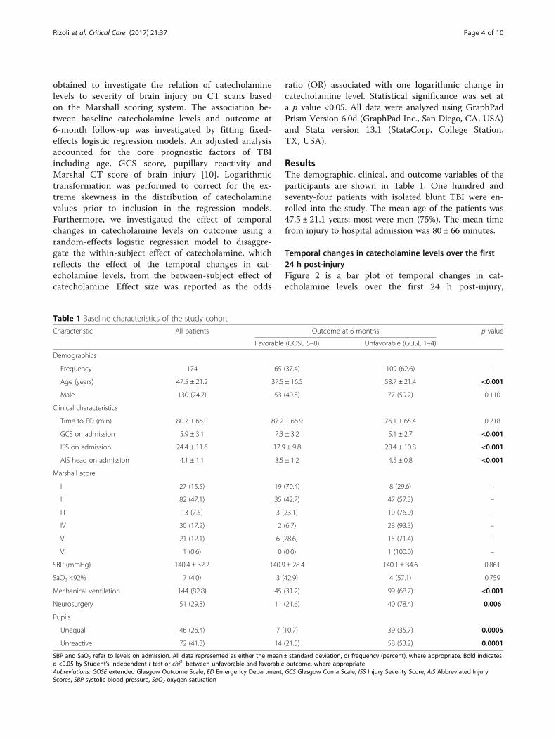
ResultsThe demographic, clinical, and outcome variables of theparticipants are shown in Table 1. One hundred andseventy-four patients with isolated blunt TBI were en-rolled into the study. The mean age of the patients was47.5 ± 21.1 years; most were men (75%). The mean timefrom injury to hospital admission was 80 ± 66 minutes.
Temporal changes in catecholamine levels over the first24 h post-injuryFigure 2 is a bar plot of temporal changes in cat-echolamine levels over the first 24 h post-injury,
Table 1 Baseline characteristics of the study cohort
Characteristic All patients Outcome at 6 months p value
Favorable (GOSE 5–8) Unfavorable (GOSE 1–4)
Demographics
Frequency 174 65 (37.4) 109 (62.6) –
Age (years) 47.5 ± 21.2 37.5 ± 16.5 53.7 ± 21.4 <0.001
Male 130 (74.7) 53 (40.8) 77 (59.2) 0.110
Clinical characteristics
Time to ED (min) 80.2 ± 66.0 87.2 ± 66.9 76.1 ± 65.4 0.218
GCS on admission 5.9 ± 3.1 7.3 ± 3.2 5.1 ± 2.7 <0.001
ISS on admission 24.4 ± 11.6 17.9 ± 9.8 28.4 ± 10.8 <0.001
AIS head on admission 4.1 ± 1.1 3.5 ± 1.2 4.5 ± 0.8 <0.001
Marshall score
I 27 (15.5) 19 (70.4) 8 (29.6) –
II 82 (47.1) 35 (42.7) 47 (57.3) –
III 13 (7.5) 3 (23.1) 10 (76.9) –
IV 30 (17.2) 2 (6.7) 28 (93.3) –
V 21 (12.1) 6 (28.6) 15 (71.4) –
VI 1 (0.6) 0 (0.0) 1 (100.0) –
SBP (mmHg) 140.4 ± 32.2 140.9 ± 28.4 140.1 ± 34.6 0.861
SaO2 <92% 7 (4.0) 3 (42.9) 4 (57.1) 0.759
Mechanical ventilation 144 (82.8) 45 (31.2) 99 (68.7) <0.001
Neurosurgery 51 (29.3) 11 (21.6) 40 (78.4) 0.006
Pupils
Unequal 46 (26.4) 7 (10.7) 39 (35.7) 0.0005
Unreactive 72 (41.3) 14 (21.5) 58 (53.2) 0.0001
SBP and SaO2 refer to levels on admission. All data represented as either the mean ± standard deviation, or frequency (percent), where appropriate. Bold indicatesp <0.05 by Student’s independent t test or chi2, between unfavorable and favorable outcome, where appropriateAbbreviations: GOSE extended Glasgow Outcome Scale, ED Emergency Department, GCS Glasgow Coma Scale, ISS Injury Severity Score, AIS Abbreviated InjuryScores, SBP systolic blood pressure, SaO2 oxygen saturation
Rizoli et al. Critical Care (2017) 21:37 Page 4 of 10

including the levels measured in 50 healthy volun-teers. The levels of Epi and NE were highest at base-line measurement immediately following admission. Themean levels dropped at a rate of approximately 50% at re-peat measurements.
Association of catecholamine levels and severity of thebrain injuryFigure 3a and b shows the relation of baseline catechol-amine levels and the Marshall CT score of brain injuryseverity [10]. Epi levels rose sharply with increasing
Fig. 2 Bar plot showing temporal changes in mean catecholamine levels. a Epinephrine levels. b Norepinephrine levels
Fig. 3 a and b Fractional polynomial plots of the relation of hospital-admission catecholamine levels to Marshall CT score of brain injury. c and dFractional polynomial plots of the relation of catecholamine levels to outcomes at the different time points. Epi epinephrine, NE norepinephrine
Rizoli et al. Critical Care (2017) 21:37 Page 5 of 10

Marshall score up to a score of 3, and then slowly plat-eaued beyond a Marshall score of 3 (Fig. 3a). We noteda similar rise in NE levels with higher Marshall scores.However, NE levels dropped sharply beyond a Marshallscore of 4 (Fig. 3b).
Effect of baseline cathecolamine levels on outcomeAt 6 months, 109 patients (62.6%) had an unfavorableoutcome, including 51 deaths (29.3%). Sixty-five patients(37.4%) had a favorable outcome and two patients (1.1%)were lost in the follow-up. Compared with patients whoexperienced favorable outcomes, those with unfavorableoutcomes had significantly higher median baseline levelsof both Epi (1216 vs. 4280 pmol/L, p <0.0001) and NE(5298 vs. 28,492 pmol/L, p <0.0001) at admission. Theresults of a fixed-effect logistic regression analysis areshown in Table 2. In the unadjusted analysis, higherbaseline levels of Epi and NE were strongly associatedwith a higher risk of mortality. In the adjusted analysis,the effect of Epi remained statistically significant [OR,2.86, 95% confidence interval (CI): 1.62–5.01, p = 0.001],but the effect of NE was not statistically significant(1.45, 95% CI: 0.98–2.17, p = 0.07). Higher Epi and NElevels were statistically associated with higher risk of un-favorable outcome in the unadjusted analyses (Table 2),and after adjustment for age, GCS, and pupillary reactiv-ity (Epi: OR, 2.04, 95% CI: 1.31–3.18, p = 0.002; NE: 1.59,95% CI: 1.07–2.35, p = 0.022).
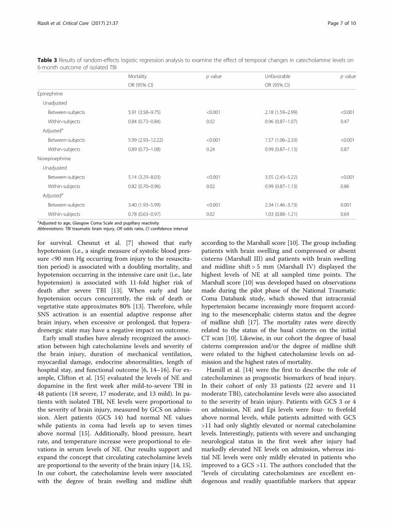
Effect of temporal changes in catecholamine levels onoutcomeFigure 3c and d shows fractional polynomial plots of therelationship between catecholamine levels and outcomesat the different time points. The graph demonstrates alinear relationship between catecholamine levels andoutcome, and a complex interaction with time. Admis-sion Epi and NE levels had the strongest prognostic ef-fect. The effects of Epi and NE on outcome weakenedwith time. Likelihood ratio tests of one-way interactionwith time was significant only for mortality outcome(mortality: Epi, p = 0.004; NE, p <0.001; unfavorable out-come: Epi, p = 0.73; NE, p = 0.50). Table 3 shows results
from the random-effects logistic regression analysis toexamine the effect on outcome of the changes with timein catecholamine levels. The estimated conditionalbetween-subject effects of Epi on mortality and unfavor-able outcomes were statistically significant in the un-adjusted and adjusted analysis (p <0.001). The within-subject effects, which reflects the effects of changes inEpi level with time, was statistically significant for mor-tality but not for unfavorable outcome in the unadjustedanalysis (p = 0.02). In the adjusted analysis, the within-subject effects were not significant for mortality (p =0.24) or unfavorable outcome (p = 0.87). The estimatedconditional between-subject effects of NE on mortalityas well as unfavorable outcome were significant in un-adjusted and adjusted analysis (p <0.001). The within-subject effect of NE was significant for mortality in bothunadjusted and adjusted analysis, indicating a drop inNE levels with time was associated with a lower risk ofmortality. The within-subject effect was not significantfor unfavorable outcome in the unadjusted and adjustedanalysis (p = 0.86 and 0.69 respectively).
DiscussionIn this study, we investigated the timeline of catechol-amine release during the initial 24 h post-injury in alarge prospective cohort of isolated moderate-to-severeTBI patients and its association with mortality and long-term functional outcome. In total, 174 patients with iso-lated TBI were enrolled (79% severe and 21% moderate),with an excellent long-term follow-up (99%) at 6 months.Our main findings were: (1) TBI patients displayed apattern of peripheral catecholamine release over the first24 h of injury characterized by a massive release into theperipheral circulation early in the course of disease,followed by a gradual decline over time. (2) Both Epiand NE levels on admission demonstrated an independ-ent association with functional outcome, measured byGOSE at 6 months post-injury in a dose–responsefashion.Traumatic brain injury leads to an immediate and pro-
found SNS activation with massive release of both cen-tral and peripheral catecholamines [6], which is essential
Table 2 Results of fixed-effect logistic regression analysis for the effect of hospital-admission catecholamine levels on 6-month out-come of isolated TBI
Catecholamine Mortality outcome pvalue
Unfavorable outcome pvalueOR (95% CI)
Epi unadjusted 2.90 (1.93–4.36) <0.001 1.93 (1.41–2.64) <0.001
Epi adjusteda 2.86 (1.62–5.01) <0.001 1.59 (1.07–2.35) 0.022
NE unadjusted 2.15 (1.55–2.98) <0.001 2.66 (1.79–3.94) <0.001
NE adjusteda 1.5 (0.98–2.17) 0.070 2.04 (1.31–3.18) 0.002aAdjusted to age, Glasgow Coma Scale and pupillary reactivityAbbreviations: OR odds ratio, CI confidence interval, Epi epinephrine, NE norepinephrine
Rizoli et al. Critical Care (2017) 21:37 Page 6 of 10
for survival. Chesnut et al. [7] showed that earlyhypotension (i.e., a single measure of systolic blood pres-sure <90 mm Hg occurring from injury to the resuscita-tion period) is associated with a doubling mortality, andhypotension occurring in the intensive care unit (i.e., latehypotension) is associated with 11-fold higher risk ofdeath after severe TBI [13]. When early and latehypotension occurs concurrently, the risk of death orvegetative state approximates 80% [13]. Therefore, whileSNS activation is an essential adaptive response afterbrain injury, when excessive or prolonged, that hypera-drenergic state may have a negative impact on outcome.Early small studies have already recognized the associ-
ation between high catecholamine levels and severity ofthe brain injury, duration of mechanical ventilation,myocardial damage, endocrine abnormalities, length ofhospital stay, and functional outcome [6, 14–16]. For ex-ample, Clifton et al. [15] evaluated the levels of NE anddopamine in the first week after mild-to-severe TBI in48 patients (18 severe, 17 moderate, and 13 mild). In pa-tients with isolated TBI, NE levels were proportional tothe severity of brain injury, measured by GCS on admis-sion. Alert patients (GCS 14) had normal NE valueswhile patients in coma had levels up to seven timesabove normal [15]. Additionally, blood pressure, heartrate, and temperature increase were proportional to ele-vations in serum levels of NE. Our results support andexpand the concept that circulating catecholamine levelsare proportional to the severity of the brain injury [14, 15].In our cohort, the catecholamine levels were associatedwith the degree of brain swelling and midline shift
according to the Marshall score [10]. The group includingpatients with brain swelling and compressed or absentcisterns (Marshall III) and patients with brain swellingand midline shift > 5 mm (Marshall IV) displayed thehighest levels of NE at all sampled time points. TheMarshall score [10] was developed based on observationsmade during the pilot phase of the National TraumaticComa Databank study, which showed that intracranialhypertension became increasingly more frequent accord-ing to the mesencephalic cisterns status and the degreeof midline shift [17]. The mortality rates were directlyrelated to the status of the basal cisterns on the initialCT scan [10]. Likewise, in our cohort the degree of basalcisterns compression and/or the degree of midline shiftwere related to the highest catecholamine levels on ad-mission and the highest rates of mortality.Hamill et al. [14] were the first to describe the role of
catecholamines as prognostic biomarkers of head injury.In their cohort of only 33 patients (22 severe and 11moderate TBI), catecholamine levels were also associatedto the severity of brain injury. Patients with GCS 3 or 4on admission, NE and Epi levels were four- to fivefoldabove normal levels, while patients admitted with GCS>11 had only slightly elevated or normal catecholaminelevels. Interestingly, patients with severe and unchangingneurological status in the first week after injury hadmarkedly elevated NE levels on admission, whereas ini-tial NE levels were only mildly elevated in patients whoimproved to a GCS >11. The authors concluded that the“levels of circulating catecholamines are excellent en-dogenous and readily quantifiable markers that appear
Table 3 Results of random-effects logistic regression analysis to examine the effect of temporal changes in catecholamine levels on6-month outcome of isolated TBI
Mortality p value Unfavorable p value
OR (95% CI) OR (95% CI)
Epinephrine
Unadjusted
Between-subjects 5.91 (3.58–9.75) <0.001 2.18 (1.59–2.99) <0.001
Within-subjects 0.84 (0.73–0.84) 0.02 0.96 (0.87–1.07) 0.47
Adjusteda
Between-subjects 5.99 (2.93–12.22) <0.001 1.57 (1.06–2.33) <0.001
Within-subjects 0.89 (0.73–1.08) 0.24 0.99 (0.87–1.13) 0.87
Norepinephrine
Unadjusted
Between-subjects 5.14 (3.29–8.03) <0.001 3.55 (2.43–5.22) <0.001
Within-subjects 0.82 (0.70–0.96) 0.02 0.99 (0.87–1.13) 0.86
Adjusteda
Between-subjects 3.40 (1.93–5.99) <0.001 2.34 (1.46–3.73) 0.001
Within-subjects 0.78 (0.63–0.97) 0.02 1.03 (0.88–1.21) 0.69aAdjusted to age, Glasgow Coma Scale and pupillary reactivityAbbreviations: TBI traumatic brain injury, OR odds ratio, CI confidence interval
Rizoli et al. Critical Care (2017) 21:37 Page 7 of 10

to reflect the extent of brain injury and that may predictthe likelihood of recovery.” In another study, Woolfet al. [16] analyzed the catecholamine response to multi-system trauma. They found NE levels were significantlycorrelated with severity of injury only if the injury in-cluded the brain. The same group had described that pa-tients with high levels of NE (>900 pg/mL) remained inpoor clinical status (with a low GCS) or died, while pa-tients with NE levels <900 pg/mL improved to a GCS of11 within 1 week [6].Regarding the pattern of catecholamine release into
the peripheral circulation, contrary to what Hamill et al.[14] described in their cohort “that catecholamine levelsremained relatively stable within the first 48 hours of in-jury”, our measurements demonstrate that circulatingcatecholamines display a characteristic pattern of releaseover the first 24 h of injury. This pattern is characterizedby a massive release of NE and Epi into the peripheralcirculation, with peak levels detected on admission,followed by gradual decline over the subsequent 24 h.However, these levels remained significantly higher bythe end of the 24 h period, when compared to measure-ments done in healthy volunteers. In addition, when TBIpatients were divided into favorable and unfavorableoutcome groups, both groups displayed a similar patternover the first 24 h, and both Epi and NE levels remainedsignificantly higher in the unfavorable outcome group(Fig. 3a and b).Our present study further demonstrates that admis-
sion Epi and NE levels are independently associated with6-month outcome measured by GOSE. Our analysisseems to suggest that both Epi and NE levels are associ-ated with unfavorable outcome, while the levels of Epiare only related to mortality outcome. A dose–responserelationship was seen, with higher catecholamine levelson admission being related to outcome. Though the cat-echolamine levels decreased with time, we found thistemporal change had no relation to outcome; rather itwas the absolute level of catecholamines at baseline (ad-mission) that was associated with outcome.Several possible pathophysiological mechanisms may
explain the relationship between high catecholaminelevels and worse outcomes after TBI:
1. Animal models of cardiac arrest have demonstratedthat Epi injection during cardiopulmonaryresuscitation (CPR) has detrimental effects throughits alpha-1-adrenergic receptor actions on cerebralmicrovascular blood flow [18, 19]. It reduces corticalmicrocirculatory blood flow, which increases the se-verity of cerebral ischemia during CPR [19], andafter restoration of spontaneous circulation [18].
2. The systemic inflammatory response is mediated bythe increased catecholamine levels [20]. Additionally,
the sympathoadrenal activation drives coagulopathyand endotheliopathy [21], through endothelial damage/dysfunction, mostly glycocalyx disruption. This complexinteraction between the SNS, endotheliopathy,inflammation and coagulation remains to be completelyunderstood in the acute phase of TBI [20, 21].
3. Increased cardiac and cerebral oxygen demands [22–25].4. Hypermetabolism, protein catabolism and muscle
wasting [26–29]. Hypermetabolism is a commonmetabolic response of trauma, and follows inconcert the increased sympathetic system activity. Itis associated with altered lipid and proteinmetabolism, leading to loss of lean body mass [30].
5. Increased intracapillary hydrostatic pressure leadingto vasogenic cerebral edema [31, 32].
Our findings underscore the need for further studies todetermine whether a causal relationship exists betweencatecholamine levels and clinical outcome after TBI, assuch a relationship may represent an opportunity for tar-geted pharmacological therapy against secondary injuryafter TBI. Presently, no specific pharmacological treat-ment exists that effectively prevents or limits the progres-sion of secondary brain injury [33]. Given the presentfindings, adrenergic blockade, may therefore, be a poten-tial therapeutic intervention worthy of further exploration.A recent meta-analysis has demonstrated that exposure tobeta-blockers after TBI was associated with a profound re-duction of in-hospital mortality by 65% (pooled adjustedodds ratio 0.35; 95% CI 0.27–0.45) [34]. Despite these re-sults, the benefits of the use of beta-blockers in the acutephase of TBI remain unproven and in need of a more ro-bust evaluation in a randomized clinical trial.
ConclusionsThis multicenter, prospective, observational cohort studyhas demonstrated that circulating catecholamine levelsare markedly elevated in moderate and severe isolatedblunt TBI patients. This elevation follows a pattern,characterized by massive release into peripheral circula-tion early after injury, decreasing thereafter over the first24 hours. Peak levels of catecholamines are markers ofbrain injury severity and are independently associatedwith functional outcome measured by the 6-monthGOSE, in a dose-dependent fashion. Also, elevated Epilevels on admission were independently associated withan increased risk of death.
Additional file
Additional file 1: Additional description of Procedures, Data Collection,and Routine Clinical Hematology Analyses. Table S1 - CT Classification -Traumatic Coma Data Bank. Table S2 - Functional Outcome according tothe extended Glasgow Outcome Scale. (DOCX 18 kb)
Rizoli et al. Critical Care (2017) 21:37 Page 8 of 10
AbbreviationsAIS: Abbreviated Injury Scores; CI: Confidence interval; CPR: Cardiopulmonaryresuscitation; CT: Computed tomography; ED: Emergency Department;Epi: Epinephrine; GCS: Glasgow Coma Scale; GOSE: Extended GlasgowOutcome Scale; ISS: Injury Severity Score; NE: Norepinephrine; OR: Odds ratio;SD: Standard deviation; SNS: Sympathetic nervous system; TBI: Traumaticbrain injury
AcknowledgementsThe COMA-TBI study group would like to gratefully acknowledge the tech-nical and administrative assistance of Marlene Santos, Sandy Trpcic, JaneTopolovec-Vranic, Yangmei Li, Maria Shiu, and Shahid Hassan.
FundingThis study was funded by a Research Grant awarded to Dr. Sandro Rizoli bythe Physician’s Services Incorporation Foundation.
Availability of data and materialsThe raw data supporting our findings is available upon request.
Authors’ contributionsSBR, SGR, ACN, AJB and ALOM conceived and designed the study. AP, LCand KI carried out the data acquisition. BNRJ, APDB, LTDL, SBR and ALOManalyzed and interpreted the data. ALOM, SBR, APDB and BNRJ drafted themanuscript. SBR, BNRJ, APDB, SGR, ACN, LC, KI, BN and AJB critically revisedthe manuscript for important intellectual content. BNRJ, APDB and LTDLperformed the statistical analysis. All authors read and approved the finalmanuscript.
Competing interestsThe authors declare that they have no competing interests.
Consent for publicationNot applicable.
Ethics approval and consent to participateThe study was approved by the Research Ethics and Institutional ReviewBoards of the following institutions that enrolled patients: St. Michael’sHospital, Sunnybrook Health Sciences Centre, Los Angeles County GeneralHospital and the University of Southern California Medical Center. Informedwritten consent was obtained by patients or next-of-kin. In the impossibilityof patient’s or familial consent, consent was delayed in accordance with theTri-Council Policy Agreement for Research in Emergency Health Situation(Article 2.8), and was subsequently obtained by either the patient or next-of-kin. Written informed consent was also obtained from healthy volunteersprior to blood sampling.
Author details1St. Michael’s Hospital, 30 Bond Street, Toronto, ON M5B 1W8, Canada.2Keenan Research Centre for Biomedical Science of St. Michael’s Hospital, 30Bond Street, Toronto, ON M5B 1W8, Canada. 3Institute of Medical Science,University of Toronto, Toronto, ON, Canada. 4Defence Research andDevelopment Canada (DRDC), Toronto Research Centre, Toronto, ON,Canada. 5Hospital Israelita Albert Einstein, Avenida Albert Einstein, 627/701 -Morumbi, São Paulo, SP 05652-900, Brazil. 6Department of Surgery,Sunnybrook Health Sciences Centre, University of Toronto., 2075 BayviewAve., Toronto, ON M4N 3M5, Canada. 7University of Southern California, 2051Marengo Street, IPT, C5L100, Los Angeles, CA 90033, USA. 8NeuroscienceResearch Program, Keenan Research Centre of the Li Ka Shing KnowledgeInstitute, St. Michael’s Hospital, 30 Bond Street, Toronto, ON M5B 1W8,Canada.
Received: 9 November 2016 Accepted: 27 January 2017
References1. Boto GR. Severe head injury and the risk of early death. J Neurol Neurosurg
Psychiatry. 2006;77:1054–9.2. Shackford SR, Mackersie RC, Holbrook TL, et al. The epidemiology of
traumatic death. A population-based analysis. Arch Surg. 1993;128:571–5.
3. Langlois JA, Rutland-Brown W, Wald MM. The epidemiology and impact oftraumatic brain injury: a brief overview. J Head Trauma Rehabil. 2006;21:375–8.
4. Molina PE. Neurobiology of the stress response: contribution of thesympathetic nervous system to the neuroimmune axis in traumatic injury.Shock. 2005;24:3–10.
5. Elenkov IJ, Wilder RL, Chrousos GP, Vizi ES. The sympathetic nerve: anintegrative interface between two supersystems: the brain and the immunesystem. Pharmacol Rev. 2000;52:595–638.
6. Woolf PD, Hamill RW, Lee LA, Cox C, McDonald JV. The predictive value ofcatecholamines in assessing outcome in traumatic brain injury. J Neurosurg.1987;66:875–82.
7. Chesnut RM, Marshall LF, Klauber MR, et al. The role of secondary brain injuryin determining outcome from severe head injury. J Trauma. 1993;34:216–22.
8. Teasdale G, Jennett B. Assessment of coma and impaired consciousness. Apractical scale. Lancet. 1974;2:81–84.
9. Elm VE, Altman DG, Egger M, Pocock SJ, Gøtzsche PC, Vandenbroucke JP. TheStrengthening the Reporting of Observational Studies in Epidemiology (STROBE)statement: guidelines for reporting observational studies. Lancet. 2007;370:1453–57.
10. Marshall LF, Marshall SB, Klauber MR, Clark MB. A new classification of headinjury based on computerized tomography. J Neurosurg. 1991;1:S14–20.
11. Wilson JT, Pettigrew LE, Teasdale GM. Structured interviews for the GlasgowOutcome Scale and the extended Glasgow Outcome Scale: guidelines fortheir use. J Neurotrauma. 1998;15:573–85.
12. Wilson JTL, Edwards P, Fiddes H, Stewart E, Teasdale GM. Reliability of postalquestionnaires for the Glasgow Outcome Scale. J Neurotrauma. 2002;19:999–1005.
13. Chesnut RM, Marshall SB, Piek J, Blunt BA, Klauber MR, Marshall LF. Early andlate systemic hypotension as a frequent and fundamental source of cerebralischemia following severe brain injury in the Traumatic Coma Data Bank.Acta Neurochir Suppl (Wien). 1993;59:121–5.
14. Hamill RW, Woolf PD, McDonald JV, Lee LA, Kelly M. Catecholamines predictoutcome in traumatic brain injury. Ann Neurol. 1987;21:438–43.
15. Clifton GL, Ziegler MG, Grossman RG. Circulating catecholamines andsympathetic activity after head injury. Neurosurgery. 1981;8:10–4.
16. Woolf PD, McDonald JV, Feliciano DV, Kelly MM, Nichols D, Cox C. Thecatecholamine response to multisystem trauma. Arch Surg. 1992;127:899–903.
17. Toutant SM, Klauber MR, Marshall LF, et al. Absent or compressed basalcisterns on first CT scan: ominous predictors of outcome in severe headinjury. J Neurosurg. 1984;61:691–4.
18. Ristagno G, Sun S, Tang W, et al. Effects of epinephrine and vasopressin oncerebral microcirculatory flows during and after cardiopulmonaryresuscitation. Crit Care Med. 2007;35:2145–9.
19. Ristagno G, Tang W, Huang L, et al. Epinephrine reduces cerebral perfusionduring cardiopulmonary resuscitation. Crit Care Med. 2009;37:1408–15.
20. Di Battista AP, Rhind SG, Hutchison MG, et al. Inflammatory cytokine andchemokine profiles are associated with patient outcome and the hyperadrenergicstate following acute brain injury. J Neuroinflammation. 2016;13:1–14.
21. Di Battista AP, Rizoli SB, Lejnieks B, et al. Sympathoadrenal activation isassociated with acute traumatic coagulopathy and endotheliopathy inisolated brain injury. Shock. 2016;46:93–106.
22. Hammerle AF, Hackl JM, Brücke T, Rumpl E, Hörtnagl H. The activity of thesympathetic nervous system following severe head injury. Intensive CareMed. 1980;6(3):169–7.
23. McLeod AA, Neil-Dwyer G, Meyer CH, Richardson PL, Cruickshank J, BartlettJ. Cardiac sequelae of acute head injury. Br Heart J. 1982;47:221–6.
24. Larremore T, Markovchick V. Cardiac sequelae of acute head injury. Br HeartJ. 1983;49:101–2.
25. Dunser MW, Hasibeder WR. Sympathetic overstimulation during critical illness:adverse effects of adrenergic stress. J Intensive Care Med. 2009;24:293–316.
26. Dunser MW, Ruokonen E, Pettilä V, et al. Association of arterial bloodpressure and vasopressor load with septic shock mortality: a post hocanalysis of a multicenter trial. Crit Care. 2009;13:R181.
27. Herndon DN, Hart DW, Wolf SE, Chinkes DL, Wolfe RR. Reversal of catabolismby beta-blockade after severe burns. N Engl J Med. 2001;345:1223–9.
28. Arbabi S, Ahrns KS, Wahl WL, et al. Beta-blocker use is associated with improvedoutcomes in adult burn patients. J Trauma. 2004;56:265–9. discussion 269–71.
29. Diaz EC, Herndon DN, Porter C, Sidossis LS, Suman OE, Børsheim E. Effectsof pharmacological interventions on muscle protein synthesis andbreakdown in recovery from burns. Burns. 2015;41:649–57.
30. Monk DN, Plank LD, Franch-Arcas G, Finn PJ, Streat SJ, Hill GL. Sequentialchanges in the metabolic response in critically injured patients during thefirst 25 days after blunt trauma. Ann Surg. 1996;223:395–405.
Rizoli et al. Critical Care (2017) 21:37 Page 9 of 10
31. Asgeirsson B, Grände PO, Nordström CH. A new therapy of post-traumabrain oedema based on haemodynamic principles for brain volumeregulation. Intensive Care Med. 1994;20:260–7.
32. Naredi S, Eden E, Zäll S, Stephensen H, Rydenhag B. A standardizedneurosurgical neurointensive therapy directed toward vasogenic edemaafter severe traumatic brain injury: clinical results. Intensive Care Med. 1998;24:446–51.
33. Park E, Bell JD, Baker AJ. Traumatic brain injury: can the consequences bestopped? CMAJ. 2008;178:1163–70.
34. Alali AS, McCredie VA, Golan E, Shah PS, Nathens AB. Beta blockers for acutetraumatic brain injury: a systematic review and meta-analysis. NeurocritCare. 2014;20:514–23.
• We accept pre-submission inquiries
• Our selector tool helps you to find the most relevant journal
• We provide round the clock customer support
• Convenient online submission
• Thorough peer review
• Inclusion in PubMed and all major indexing services
• Maximum visibility for your research
Submit your manuscript atwww.biomedcentral.com/submit
Submit your next manuscript to BioMed Central and we will help you at every step:
Rizoli et al. Critical Care (2017) 21:37 Page 10 of 10