Catechol O-Methyltransferase: Glucuronidation of Inhibitors and
59
Catechol O-Methyltransferase: Glucuronidation of Inhibitors and Methylation of Substrates by Pia Lautala Pharmaceutical Chemistry Division Department of Pharmacy University of Helsinki Finland Academic Dissertation To be presented with the permission of the Faculty of Science of the University of Helsinki, for public criticism in Auditorium 1041 of Viikki Biocentre on October 28 th , 2000, at 12 o’clock noon Helsinki 2000
Transcript of Catechol O-Methyltransferase: Glucuronidation of Inhibitors and
Catechol O-Methyltransferase:Glucuronidation of Inhibitorsand Methylation of Substrates
by
Pia Lautala
Pharmaceutical Chemistry DivisionDepartment of Pharmacy
University of HelsinkiFinland
Academic DissertationTo be presented with the permission of the Faculty of Science of the
University of Helsinki, for public criticism in Auditorium 1041of Viikki Biocentre on October 28th, 2000, at 12 o’clock noon
Helsinki 2000
Supervisor:
Prof. Jyrki TaskinenDivision of Pharmaceutical ChemistryDepartment of PharmacyUniversity of HelsinkiFinland
Reviewers:
Prof. Olavi PelkonenDepartment of Pharmacology and ToxicologyFaculty of MedicineUniversity of OuluFinland
Docent Seppo AuriolaDepartment of Pharmaceutical ChemistryFaculty of PharmacyUniversity of KuopioFinland
Opponent:
Prof. Matti LangDepartment of Pharmaceutical BiochemistryFaculty of PharmacyUniversity of UppsalaSweden
ISBN 951-45-9558-0 (nid.)ISBN 952-91-2654-9 (pdf)ISBN 952-91-2655-7 (html)
ISSN 1239-9469YliopistopainoHelsinki 2000
Contents
LIST OF ORIGINAL PUBLICATIONS 1
LIST OF ABBREVIATIONS 2
ABSTRACT 3
1. INTRODUCTION 4
2. REVIEW OF THE LITERATURE 5
2.1. Role of conjugation reactions in drug and xenobiotic metabolism 5
2.2. Conjugation of catechols 7
2.2.1. Glucuronidation 9
2.2.2. Methylation 12
2.2.3. Sulphation 15
2.3. In vitro studies on conjugation reactions 15
2.3.1. UGT assays 16
2.3.2. COMT assays 16
3. AIMS OF THE STUDY 18
4. MATERIALS AND METHODS 19
4.1. Chemicals 19
4.2. Enzyme sources 19
4.3. Reaction mixtures 20
4.4. Analytical methods 20
4.4.1. Thin-layer chromatography 20
4.4.2 High-performance liquid chromatography 21
4.4.3. Method validation 22
4.5. Enzyme kinetic analysis 22
4.6. QSAR and molecular modelling 22
5. RESULTS AND DISCUSSION 23
5.1. Development and validation of enzyme assays 23
5.1.1. Thin-layer chromatographic UGT assay for the determination
of nitrocatechol glucuronidation 23
5.1.2 Radiochemical high-performance liquid chromatographic
COMT assay 24
5.2. Glucuronidation of nitrocatechols 26
5.2.1. Glucuronidation of nitrocatechols by rat liver microsomes 26
5.2.2. Glucuronidation of entacapone and tolcapone by human liver
microsomes and recombinant UGT isoforms 28
5.2.3. Species differences 30
5.3. Substrate selectivity of rat and human S-COMT 31
5.4. Methylation of structurally diverse compounds by human
S-COMT 32
5.4.1. Enzyme kinetic parameters 32
5.4.2. Effect of molecular structure on binding affinity and reactivity 36
5.4.3. Predictive models 38
6. CONCLUSIONS 39
ACKNOWLEDGEMENTS 40
REFERENCES 41
APPENDIX: ORIGINAL PUBLICATIONS I-V
1
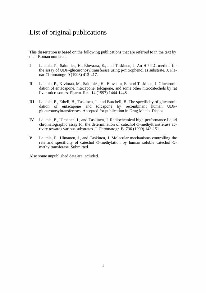
List of original publications
This dissertation is based on the following publications that are referred to in the text bytheir Roman numerals.
I Lautala, P., Salomies, H., Elovaara, E., and Taskinen, J. An HPTLC method forthe assay of UDP-glucuronosyltransferase using p-nitrophenol as substrate. J. Pla-nar Chromatogr. 9 (1996) 413-417.
II Lautala, P., Kivimaa, M., Salomies, H., Elovaara, E., and Taskinen, J. Glucuroni-dation of entacapone, nitecapone, tolcapone, and some other nitrocatechols by ratliver microsomes. Pharm. Res. 14 (1997) 1444-1448.
III Lautala, P., Ethell, B., Taskinen, J., and Burchell, B. The specificity of glucuroni-dation of entacapone and tolcapone by recombinant human UDP-glucuronosyltransferases. Accepted for publication in Drug Metab. Dispos.
IV Lautala, P., Ulmanen, I., and Taskinen, J. Radiochemical high-performance liquidchromatographic assay for the determination of catechol O-methyltransferase ac-tivity towards various substrates. J. Chromatogr. B. 736 (1999) 143-151.
V Lautala, P., Ulmanen, I., and Taskinen, J. Molecular mechanisms controlling therate and specificity of catechol O-methylation by human soluble catechol O-methyltransferase. Submitted.
Also some unpublished data are included.
2
List of abbreviations
AdoMet S-Adenosyl-L-methionine14C-AdoMet S-Adenosyl-L-[methyl-14C]methionineAdoHcy S-Adenosyl-L-homocysteinecDNA Complementary deoxyribonucleic acidCYP Cytochrome P450COMT Catechol O-methyltransferaseDHBA 3,4-Dihydroxybenzoic acidDDC Dopa decarboxylaseGST Glutathione S-transferaseHPLC High-performance liquid chromatographyHPTLC High-performance thin-layer chromatographyMB-COMT Membrane-bound catechol O-methyltransferaseMEP Molecular electrostatic potentialNAT N-acetyltransferase4NPG 4-Nitrophenyl-β-D-glucuronideNSAID Non-steroidal anti-inflammatoric drugQSAR Quantitative structure-activity relationshipsRSD Relative standard deviationS-COMT Soluble catechol O-methyltransferaseSN2 Bimolecular nucleophilic substitutionSULT SulphotransferaseTLC Thin-layer chromatographyUDPGA Uridine diphosphoglucuronic acid14C-UDPGA Uridine diphospho[U-14C]glucuronic acidUGT Uridine diphosphoglucuronosyltransferaseUV Ultraviolet

3
Abstract
Catechol structures can be found in many endogenous compounds including catechol-amines and catechol estrogens and in various drugs and drug candidates. Catecholic hy-droxyls provide reactive groups for phase II metabolic enzymes of which differentforms of UGTs, SULTs and COMTs compete for their conjugation. Little is known,however, about the factors determining the substrate acceptance of these enzymes andtheir relative contribution to the metabolism of catechols with diverse structures.
In this study, two novel analytical methods were developed: an HPTLC methodcombining radioactivity measurement and densitometry for the assay of UGT, and a ra-diochemical HPLC method for the assay of COMT. The respective methods were util-ised in studying the glucuronidation properties of a set of nitrocatechols in rat liver mi-crosomes and in determining the apparent enzyme kinetic parameters of methylation for41 structurally diverse catechols catalysed by human recombinant S-COMT. In addi-tion, the in vitro glucuronidation of the COMT inhibitors entacapone and tolcapone wascompared by determining the kinetic parameters using human liver microsomes and therelevant human recombinant UGT isoforms.
The results on the glucuronidation of nitrocatechols indicated that although they maybe excellent UGT substrates, this property is greatly affected by the nature and positionof substituents. Tolcapone was a slightly better substrate than entacapone in rat livermicrosomes, whereas entacapone showed a 14-fold Vmax/Km value in human liver micro-somes. Consequently, rat might be a poor animal model in predicting the glucuronida-tion of this type of compound in humans. The higher glucuronidation rate of entacaponecompared with tolcapone in human microsomes may explain part of its approximatelyseven times faster elimination half-life in vivo. Both compounds, especially entacapone,were excellent substrates of UGT1A9, which knowledge may be useful in evaluatingrisks for metabolic interactions.
A great variation was detected in the methylation ability of structurally diverse cate-chols. For instance, among drugs used in the treatment of Parkinson’s disease no meth-ylation of entacapone or tolcapone was observed, L-dopa and carbidopa appeared to bepoor COMT substrates, whereas benserazide exhibited a relatively high affinity and re-activity. The best endogenous substrate was 2-hydroxyestradiol. For QSAR analysis,the experimental data were combined with the calculation of substituent physico-chemical properties and modelling of the compounds to the active site of rat S-COMT.The most decisive factor increasing affinity and simultaneously decreasing reactivitywas the electron-withdrawing effect of substituents. In general, hydrophobic substitu-ents increased and hydrophilic groups reduced the affinity, but the orientation of theside chains greatly affected the extent of interactions formed with the hydrophobic sur-roundings of the binding site. Most important of the several ortho-effects discovered,that bulky ortho-substituents worsened affinity and reactivity, was demonstrated byapomorphine that was not methylated under the conditions applied. Predictive modelsfor affinity and reactivity were constructed, and they may be utilised, in conjunctionwith modelling of the active site, in assessing interactions between endogenous cate-chols and catecholic drugs and in designing catecholic drugs with controlled metabolicmethylation.
4
1. Introduction
Once an orally administered drug has entered the body, it has to be absorbed, distrib-uted, metabolised and finally excreted. All these stages can have a significant effect onthe bioavailability of the drug and must be taken into account in drug development (Linand Lu, 1997). Although drugs may be extensively excreted as such by the kidney, mostdrugs undergo some kind of metabolic transformation before excretion. Metabolic reac-tions can be divided into phase I and phase II reactions (Gibson and Skett, 1994). PhaseI reactions, such as oxidation, reduction or hydrolysis, produce functional groups thatare subsequently capable of conjugation in phase II reactions. Conjugation with en-dogenous compounds usually results in more water-soluble molecules, which facilitatesexcretion in the bile and urine. The most important enzyme family catalysing phase Ireactions is cytochrome P-450 (CYP). UDP-glucuronosyltransferases (UGTs), sulpho-transferases (SULTs), glutathione-S-transferases (GSTs) and different acetyltransferasesand methyltransferases are mainly responsible for the phase II reactions. Different en-zymes and enzyme families may contribute to the metabolism of a given drug and therate and route can greatly affect the duration of action and safety of the drug. Besidespoor absorption, inappropriate metabolism is one of the main reasons preventing theclinical use of many promising drug candidates (Prentis et al., 1988).
Due to the complexity of metabolic enzymes and the many internal and externalfactors influencing them, the rate and route of metabolism is difficult to predict (Lin andLu, 1997). Metabolic studies are traditionally initiated by measuring the eliminationhalf-life of a drug candidate in laboratory animals and identifying the metabolites fromplasma and urine samples. However, numerous examples show that remarkable differ-ences exist between species and problems in metabolism may appear only in admini-stration to humans. The increasing availability of human tissues and the advances ofgene technology in producing individual enzymes have brought in vitro methods toroutine use in drug metabolism studies. In vitro studies are suited for early assessmentof metabolism and selection of the animal model for toxicity studies and for identifica-tion of the individual enzyme forms contributing to the metabolism of a drug candidate.In vitro data may be further utilised in drug interaction studies and sometimes even inpredicting in vivo clearance.
Cloning and expression of individual human enzymes that catalyse metabolic reac-tions has enabled investigations on their structures and mechanisms underlying theircatalytical actions and substrate selectivities (Lin and Lu, 1997). At the moment, it isnot possible to predict metabolism on the basis of molecular structure, yet the ability tocontrol affinity to certain metabolic enzymes or alter the rate of metabolism by rationalstructure modification would be useful in the drug discovery and development process.In order to aspire after that goal, however, great efforts in investigating individual en-zyme forms and other factors influencing drug metabolism in vivo are required.
In this study in vitro methods have been utilised in the study of the glucuronidationof nitrocatecholic catechol O-methyltransferase (COMT) inhibitors and in the develop-ment of predictive models for methylation catalysed by human soluble COMT.
5
2. Review of the literature
2.1. Role of conjugation reactions in drug andxenobiotic metabolism
Phase II enzymes play an important role in the biotransformation of endogenous andxenobiotic compounds to more easily excretable forms as well as in the metabolic inac-tivation of pharmacologically active compounds. An especially important function is todetoxify carcinogenic compounds formed in phase I reactions. For example, carcino-genic diol epoxides, formed in phase I reactions from polycyclic aromatic hydrocar-bons, are normally conjugated with glutathione and thereby readily excreted from thebody (Guengerich, 1992). However, reduced capacity of the phase II enzymes may leadto the appearance of toxic compounds. For instance, 2-hydroxybiphenyl, an antimicro-bial agent used to protect edible crops, is metabolised to non-toxic glucuronide and sul-phate conjugates at low doses, whereas at high doses these pathways become saturatedand oxidative metabolism starts to produce toxic compounds capable of initiating blad-der cancer (Reitz et al., 1983). Although phase II reactions are basically detoxifying, theformed conjugates may also mediate adverse effects, for example acting as carriers forcarcinogenic compounds. 2-Naphthylamine, found in cigarette smoke, is N-hydroxylated to a carcinogen in the liver, but subsequently glucuronidated to inactiveN-hydroxy-N-glucuronide. The glucuronide is, however, hydrolysed in the slightlyacidic environment of the bladder and decomposed to a nitrenium ion that can bind toDNA and initiate cancer (Kadlubar et al., 1981, Miller and Miller, 1981). The site ofconjugation may have undesirable effects, for example biotransformation of a drug to amore hydrophilic conjugate in the gastrointestinal tract usually deteriorates its absorp-tion. A special case of potentially harmful phase II metabolites is 1-O-acylglucuronidesin which UDP-glucuronic acid is conjugated with a carboxyl acid group by ester link-age. These compounds are chemically labile and the glucuronic acid moiety may bedisplaced by nucleophiles. This leads to either hydrolysis of the glucuronide, in-tramolecular rearrangement by acyl migration or intermolecular transacylation. Cova-lent binding of 1-O-acylglucuronides and ester isomers to proteins by transacylation, orglycosylation, are suspected to cause cytotoxicity, carcinogenecity and allergic reactions(Fenselau, 1994).
The fact that interindividual differences in metabolic response occur is not related tophase I enzymes only, but external as well as internal factors including age, sex, dis-eases and genetics are known to influence also phase II enzymes. A well-known exam-ple of the effect of age on glucuronidation is the 'grey baby' syndrome that is caused bythe decreased excretion of chloramphenicol glucuronide in new-borns (Weiss et al.,1960). In old age the metabolic clearance sometimes declines, but this is caused morelikely by the lowered liver blood flow than by the decreased activity of metabolic en-zymes (Miners and Mackenzie, 1991). However, decreased clearance of codeine to its6-O-glucuronide has been observed in the elderly (Bochner et al., 1990). Higher capac-ity in males than in females for glucuronidation of some drugs, including paracetamol(Abernethy et al., 1982, Miners et al., 1983) diflunisal (Macdonald et al., 1990) andpropranolol (Walle et al., 1989), has been reported suggesting that the activity of someUGTs may be affected by sex hormones. Risks for adverse effects caused by polymor-phism in drug-metabolising enzymes are mainly associated with the CYP isoforms.
6
However, many phase II metabolic enzymes are genetically polymorphic as well, themost familiar being N-acetyltransferase (NAT). Polymorphism of this enzyme was firstdiscovered when isoniazid was used in the treatment of tuberculosis (Evans et al.,1960). The incidence of rapid and slow acetylators varies considerably between bothethnic groups and individuals in these groups, giving rise to different metabolic re-sponses towards drugs metabolised via NAT (Evans, 1989). Other phase II metabolicenzymes exhibiting genetically polymorphic forms include GST (Laisney et al., 1984),COMT (Weinshilboum, 1984, 1988, Boudiková et al, 1990, Grossman et al., 1992),UGT (Clarke and Burchell, 1994) SULT (Coughtrie et al., 1999) and thiopurine meth-yltransferase (Weinshilboum and Sladek, 1980). Numerous studies suggesting theircontribution to diseases and variations in drug responses have been carried out, but theclinical significance of most of the polymorphic phase II enzymes remains rather un-clear.
External factors causing interindividual variations in drug metabolism includesmoking, medication, nutrition and environmental chemicals (Pelkonen and Breimer,1994). Binding of polycyclic aromatic hydrocarbons, abundant in tobacco smoke, to thecorresponding receptor (Ah) has been found to induce not only certain CYP forms(Whitlock et al., 1996) but also some UGTs and GSTs (Bock et al., 1990). For example,heavy smoking has been reported to accelerate the glucuronidation of mexiletine andpropranolol by 30 and 55%, respectively (Grech-Bélanger et al. 1985, Walle et al.,1987). Certain drugs are able to induce or inhibit some phase II metabolic enzymesthereby increasing or decreasing the plasma clearance of other drugs metabolised viathe same enzymes. For instance, the anticonvulsant agents phenobarbitone, phenytoinand carbamazepine are known to enhance the glucuronidation of various drugs includ-ing oxazepam, paracetamol and valproic acid, (Scott et al., 1983, Miners et al., 1984,Panesar et al., 1989), while probenecid has been found to inhibit the glucuronidation ofmany drugs, especially those forming acyl glucuronides (Miners and Mackenzie, 1991).
Although metabolism normally inactivates a drug and transforms it to an easily ex-cretable form, in some cases a metabolite has been even more potent than the parentcompound. Two well-known examples of phase II metabolites exhibiting high pharma-cological activity are the N-sulphate conjugate of minoxidil (Johnson et al., 1982),which, rather than minoxidil itself, causes the relaxation of smooth muscles (Kauffmanet al., 1994), and morphine 6-O-glucuronide, which is a more potent mu-opioid receptoragonist than morphine (Pasternak et al., 1987, Paul et al., 1989, Frances et al., 1990).Entero-hepatic circulation, in which the formed glucuronide is excreted in the intestine,hydrolysed by bacterial β-glucuronidase, reabsorbed, and transported back to the liver,may markedly prolong the pharmacological action of a drug. Even though the mostcommon reason for prodrug design is to enhance the absorption of hydrophilic drugs,also glucuronide conjugates have been developed as prodrug candidates. For example, aglucuronide conjugate of p-hydroxyaniline mustard, delivered in conjunction with thehydrolytic, tumour-targeted β-glucuronidase, has been tested for its ability to kill tu-mour cells (Cheng et al., 1999).
In addition to prodrug discovery, drug designers have recently been able to take ad-vantage of metabolic reactions using another approach. In Parkinson’s disease, wherethe symptoms are caused by the lack of dopamine in the striatum, patients have gener-ally been treated with L-dopa, a dopamine precursor capable of penetrating the blood-brain barrier. To decrease the decarboxylation of L-dopa to dopamine already in the pe-riphery, it is normally administered in conjunction with a dopa decarboxylase (DDC)inhibitor (benserazide or carbidopa) (Männistö et al., 1992). However, when DDC isinhibited, the predominant metabolic pathway of L-dopa turns out to be the COMT-
7
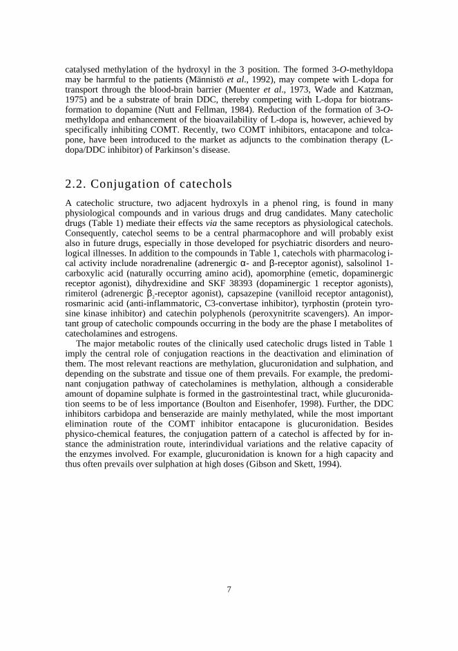
catalysed methylation of the hydroxyl in the 3 position. The formed 3-O-methyldopamay be harmful to the patients (Männistö et al., 1992), may compete with L-dopa fortransport through the blood-brain barrier (Muenter et al., 1973, Wade and Katzman,1975) and be a substrate of brain DDC, thereby competing with L-dopa for biotrans-formation to dopamine (Nutt and Fellman, 1984). Reduction of the formation of 3-O-methyldopa and enhancement of the bioavailability of L-dopa is, however, achieved byspecifically inhibiting COMT. Recently, two COMT inhibitors, entacapone and tolca-pone, have been introduced to the market as adjuncts to the combination therapy (L-dopa/DDC inhibitor) of Parkinson’s disease.
2.2. Conjugation of catechols
A catecholic structure, two adjacent hydroxyls in a phenol ring, is found in manyphysiological compounds and in various drugs and drug candidates. Many catecholicdrugs (Table 1) mediate their effects via the same receptors as physiological catechols.Consequently, catechol seems to be a central pharmacophore and will probably existalso in future drugs, especially in those developed for psychiatric disorders and neuro-logical illnesses. In addition to the compounds in Table 1, catechols with pharmacolog i-cal activity include noradrenaline (adrenergic α- and β-receptor agonist), salsolinol 1-carboxylic acid (naturally occurring amino acid), apomorphine (emetic, dopaminergicreceptor agonist), dihydrexidine and SKF 38393 (dopaminergic 1 receptor agonists),rimiterol (adrenergic β2-receptor agonist), capsazepine (vanilloid receptor antagonist),rosmarinic acid (anti-inflammatoric, C3-convertase inhibitor), tyrphostin (protein tyro-sine kinase inhibitor) and catechin polyphenols (peroxynitrite scavengers). An impor-tant group of catecholic compounds occurring in the body are the phase I metabolites ofcatecholamines and estrogens.
The major metabolic routes of the clinically used catecholic drugs listed in Table 1imply the central role of conjugation reactions in the deactivation and elimination ofthem. The most relevant reactions are methylation, glucuronidation and sulphation, anddepending on the substrate and tissue one of them prevails. For example, the predomi-nant conjugation pathway of catecholamines is methylation, although a considerableamount of dopamine sulphate is formed in the gastrointestinal tract, while glucuronida-tion seems to be of less importance (Boulton and Eisenhofer, 1998). Further, the DDCinhibitors carbidopa and benserazide are mainly methylated, while the most importantelimination route of the COMT inhibitor entacapone is glucuronidation. Besidesphysico-chemical features, the conjugation pattern of a catechol is affected by for in-stance the administration route, interindividual variations and the relative capacity ofthe enzymes involved. For example, glucuronidation is known for a high capacity andthus often prevails over sulphation at high doses (Gibson and Skett, 1994).
8
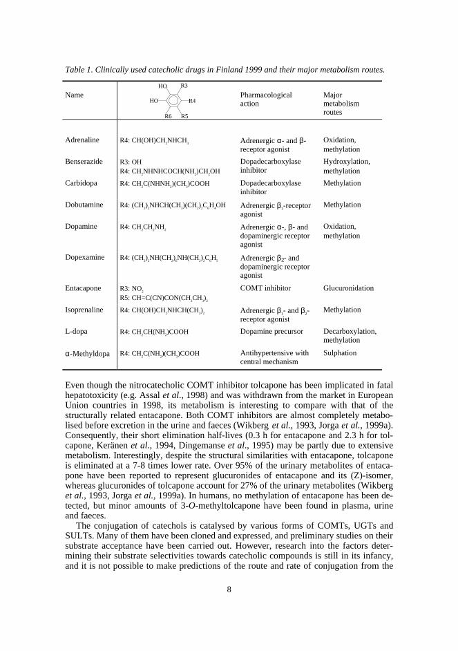
Table 1. Clinically used catecholic drugs in Finland 1999 and their major metabolism routes.
Name Pharmacologicalaction
Majormetabolismroutes
Adrenaline R4: CH(OH)CH2NHCH
3 Adrenergic α- and β-receptor agonist
Oxidation,methylation
Benserazide R3: OHR4: CH
2NHNHCOCH(NH
2)CH
2OH
Dopadecarboxylaseinhibitor
Hydroxylation,methylation
Carbidopa R4: CH2C(NHNH
2)(CH
3)COOH Dopadecarboxylase
inhibitorMethylation
Dobutamine R4: (CH2)
2NHCH(CH
3)(CH
2)
2C
6H
4OH Adrenergic β1-receptor
agonistMethylation
Dopamine R4: CH2CH
2NH
2 Adrenergic α-, β- anddopaminergic receptoragonist
Oxidation,methylation
Dopexamine R4: (CH2)
2NH(CH
2)
6NH(CH
2)
2C
6H
5 Adrenergic β2- anddopaminergic receptoragonist
Entacapone R3: NO2
R5: CH=C(CN)CON(CH2CH
3)
2
COMT inhibitor Glucuronidation
Isoprenaline R4: CH(OH)CH2NHCH(CH
3)
2 Adrenergic β1- and β2-receptor agonist
Methylation
L-dopa R4: CH2CH(NH
2)COOH Dopamine precursor Decarboxylation,
methylation
α-Methyldopa R4: CH2C(NH
2)(CH
3)COOH Antihypertensive with
central mechanismSulphation
Even though the nitrocatecholic COMT inhibitor tolcapone has been implicated in fatalhepatotoxicity (e.g. Assal et al., 1998) and was withdrawn from the market in EuropeanUnion countries in 1998, its metabolism is interesting to compare with that of thestructurally related entacapone. Both COMT inhibitors are almost completely metabo-lised before excretion in the urine and faeces (Wikberg et al., 1993, Jorga et al., 1999a).Consequently, their short elimination half-lives (0.3 h for entacapone and 2.3 h for tol-capone, Keränen et al., 1994, Dingemanse et al., 1995) may be partly due to extensivemetabolism. Interestingly, despite the structural similarities with entacapone, tolcaponeis eliminated at a 7-8 times lower rate. Over 95% of the urinary metabolites of entaca-pone have been reported to represent glucuronides of entacapone and its (Z)-isomer,whereas glucuronides of tolcapone account for 27% of the urinary metabolites (Wikberget al., 1993, Jorga et al., 1999a). In humans, no methylation of entacapone has been de-tected, but minor amounts of 3-O-methyltolcapone have been found in plasma, urineand faeces.
The conjugation of catechols is catalysed by various forms of COMTs, UGTs andSULTs. Many of them have been cloned and expressed, and preliminary studies on theirsubstrate acceptance have been carried out. However, research into the factors deter-mining their substrate selectivities towards catecholic compounds is still in its infancy,and it is not possible to make predictions of the route and rate of conjugation from the
R6 R5
R4
R3HO
HO
9
molecular structure. That would, however, be useful in assessing the capacity of cate-cholic drug candidates for interactions with physiological catechols, and other cate-cholic drugs, already at early stages of the drug development process.
2.1.1. Glucuronidation
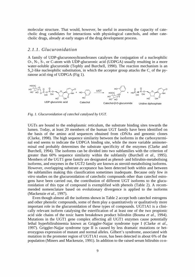
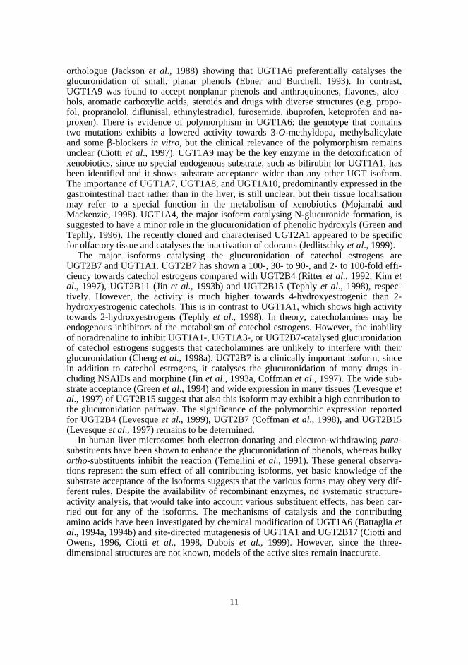
A family of UDP-glucuronosyltransferases catalyses the conjugation of a nucleophilicO-, N-, S-, or C-atom with UDP-glucuronic acid (UDPGA) usually resulting in a morewater-soluble glucuronide (Tephly and Burchell, 1990). The reaction mechanism is anSN2-like nucleophilic substitution, in which the acceptor group attacks the C1 of the py-ranose acid ring of UDPGA (Fig 1).
Fig. 1. Glucuronidation of catechol catalysed by UGT.
UGTs are bound to the endoplasmic reticulum, the substrate binding sites towards thelumen. Today, at least 20 members of the human UGT family have been identified onthe basis of the amino acid sequences obtained from cDNAs and genomic clones(Clarke, 1998). The high sequence similarity between the isoforms in the carboxytermi-nal end seems to indicate the UDPGA binding site, while the more variable aminoter-minal end probably determines the substrate specificity of the enzymes (Clarke andBurchell, 1994). The isoforms can be divided into two subfamilies with the criterion ofgreater than 60% sequence similarity within the subfamily (Burchell et al., 1995).Members of the UGT1 gene family are designated as phenol- and bilirubin-metabolisingisoforms, and enzymes in the UGT2 family are known as steroid-metabolising isoforms.However, overlapping substrate acceptance has been detected both within and betweenthe subfamilies making this classification sometimes inadequate. Because only few invitro studies on the glucuronidation of catecholic compounds other than catechol estro-gens have been carried out, the contribution of different UGT isoforms to the glucu-ronidation of this type of compound is exemplified with phenols (Table 2). A recom-mended nomenclature based on evolutionary divergence is applied to the isoforms(Mackenzie et al., 1997).
Even though almost all the isoforms shown in Table 2 accept both catechol estrogensand other phenolic compounds, some of them play a quantitatively or qualitatively moreimportant role in the glucuronidation of these types of compounds. UGT1A1 is a clini-cally relevant isoform catalysing the esterification of at least one of the two propionicacid side chains of the toxic haem breakdown product bilirubin (Bosma et al., 1994).Mutations in the UGT1 gene complex affecting all UGT1 enzymes cause potentiallylethal hyperbilirubinemia known as Griggler-Najjar syndrome type I (Clarke et al.,1997). Griggler-Najjar syndrome type II is caused by less dramatic mutations or het-erozygous expression of mutant and normal alleles. Gilbert’s syndrome, associated withmutation in the promoter region of UGT1A1 exon, has been detected in about 6% of thepopulation (Miners and Mackenzie, 1991). In addition to the raised serum bilirubin co n-
+
O
O P OOH
OP O
OH
OHOOC
OHOH OH
OH
O N
OHO
O
NH
UDP-glucuronic acidUridine diphosphate
OH
OH
OH
OOHOOC
OHOH OH
+
Catechol Catechol-β-D-glucuronide
UGTP O
OH
OP O
OH
O
OH
O N
OHO
O
NHOH
10
centrations, a decrease in the glucuronidation of for example acetaminophen has beenobserved in people suffering from this syndrome (De Morais et al., 1992). In addition tobilirubin, UGT1A1 catalyses the glucuronidation of catechol estrogens, phenols, an-thraquinones and flavones with diverse structures (Senafi et al., 1994). Interestingly,octylgallate, which has a pyrogallol structure, has appeared to be a better substrate ofUGT1A1 than bilirubin itself. The important function of UGT1A1 in eliminating biliru-bin in conjunction with the wide substrate acceptance and genetic polymorphism mayimply possibilities for interactions between compounds glucuronidated via this isoform.On the other hand, UGT1A1 is expressed at higher levels in the liver compared with theother UGT1 isoforms and possibly exhibits a high capacity (Sutherland et al., 1992).
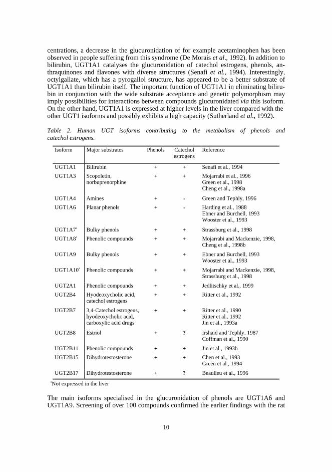
Table 2. Human UGT isoforms contributing to the metabolism of phenols andcatechol estrogens.
Isoform Major substrates Phenols Catecholestrogens
Reference
UGT1A1 Bilirubin + + Senafi et al., 1994
UGT1A3 Scopoletin,norbuprenorphine
+ + Mojarrabi et al., 1996Green et al., 1998Cheng et al., 1998a
UGT1A4 Amines + - Green and Tephly, 1996
UGT1A6 Planar phenols + - Harding et al., 1988Ebner and Burchell, 1993Wooster et al., 1993
UGT1A7a Bulky phenols + + Strassburg et al., 1998
UGT1A8a Phenolic compounds + + Mojarrabi and Mackenzie, 1998,Cheng et al., 1998b
UGT1A9 Bulky phenols + + Ebner and Burchell, 1993Wooster et al., 1993
UGT1A10a Phenolic compounds + + Mojarrabi and Mackenzie, 1998,Strassburg et al., 1998
UGT2A1 Phenolic compounds + + Jedlitschky et al., 1999
UGT2B4 Hyodeoxycholic acid,catechol estrogens
+ + Ritter et al., 1992
UGT2B7 3,4-Catechol estrogens,hyodeoxycholic acid,carboxylic acid drugs
+ + Ritter et al., 1990Ritter et al., 1992Jin et al., 1993a
UGT2B8 Estriol + ? Irshaid and Tephly, 1987Coffman et al., 1990
UGT2B11 Phenolic compounds + + Jin et al., 1993b
UGT2B15 Dihydrotestosterone + + Chen et al., 1993Green et al., 1994
UGT2B17 Dihydrotestosterone + ? Beaulieu et al., 1996
aNot expressed in the liver
The main isoforms specialised in the glucuronidation of phenols are UGT1A6 andUGT1A9. Screening of over 100 compounds confirmed the earlier findings with the rat
11
orthologue (Jackson et al., 1988) showing that UGT1A6 preferentially catalyses theglucuronidation of small, planar phenols (Ebner and Burchell, 1993). In contrast,UGT1A9 was found to accept nonplanar phenols and anthraquinones, flavones, alco-hols, aromatic carboxylic acids, steroids and drugs with diverse structures (e.g. propo-fol, propranolol, diflunisal, ethinylestradiol, furosemide, ibuprofen, ketoprofen and na-proxen). There is evidence of polymorphism in UGT1A6; the genotype that containstwo mutations exhibits a lowered activity towards 3-O-methyldopa, methylsalicylateand some β-blockers in vitro, but the clinical relevance of the polymorphism remainsunclear (Ciotti et al., 1997). UGT1A9 may be the key enzyme in the detoxification ofxenobiotics, since no special endogenous substrate, such as bilirubin for UGT1A1, hasbeen identified and it shows substrate acceptance wider than any other UGT isoform.The importance of UGT1A7, UGT1A8, and UGT1A10, predominantly expressed in thegastrointestinal tract rather than in the liver, is still unclear, but their tissue localisationmay refer to a special function in the metabolism of xenobiotics (Mojarrabi andMackenzie, 1998). UGT1A4, the major isoform catalysing N-glucuronide formation, issuggested to have a minor role in the glucuronidation of phenolic hydroxyls (Green andTephly, 1996). The recently cloned and characterised UGT2A1 appeared to be specificfor olfactory tissue and catalyses the inactivation of odorants (Jedlitschky et al., 1999).
The major isoforms catalysing the glucuronidation of catechol estrogens areUGT2B7 and UGT1A1. UGT2B7 has shown a 100-, 30- to 90-, and 2- to 100-fold effi-ciency towards catechol estrogens compared with UGT2B4 (Ritter et al., 1992, Kim etal., 1997), UGT2B11 (Jin et al., 1993b) and UGT2B15 (Tephly et al., 1998), respec-tively. However, the activity is much higher towards 4-hydroxyestrogenic than 2-hydroxyestrogenic catechols. This is in contrast to UGT1A1, which shows high activitytowards 2-hydroxyestrogens (Tephly et al., 1998). In theory, catecholamines may beendogenous inhibitors of the metabolism of catechol estrogens. However, the inabilityof noradrenaline to inhibit UGT1A1-, UGT1A3-, or UGT2B7-catalysed glucuronidationof catechol estrogens suggests that catecholamines are unlikely to interfere with theirglucuronidation (Cheng et al., 1998a). UGT2B7 is a clinically important isoform, sincein addition to catechol estrogens, it catalyses the glucuronidation of many drugs in-cluding NSAIDs and morphine (Jin et al., 1993a, Coffman et al., 1997). The wide sub-strate acceptance (Green et al., 1994) and wide expression in many tissues (Levesque etal., 1997) of UGT2B15 suggest that also this isoform may exhibit a high contribution tothe glucuronidation pathway. The significance of the polymorphic expression reportedfor UGT2B4 (Levesque et al., 1999), UGT2B7 (Coffman et al., 1998), and UGT2B15(Levesque et al., 1997) remains to be determined.
In human liver microsomes both electron-donating and electron-withdrawing para-substituents have been shown to enhance the glucuronidation of phenols, whereas bulkyortho-substituents inhibit the reaction (Temellini et al., 1991). These general observa-tions represent the sum effect of all contributing isoforms, yet basic knowledge of thesubstrate acceptance of the isoforms suggests that the various forms may obey very dif-ferent rules. Despite the availability of recombinant enzymes, no systematic structure-activity analysis, that would take into account various substituent effects, has been car-ried out for any of the isoforms. The mechanisms of catalysis and the contributingamino acids have been investigated by chemical modification of UGT1A6 (Battaglia etal., 1994a, 1994b) and site-directed mutagenesis of UGT1A1 and UGT2B17 (Ciotti andOwens, 1996, Ciotti et al., 1998, Dubois et al., 1999). However, since the three-dimensional structures are not known, models of the active sites remain inaccurate.
12
2.2.2 Methylation
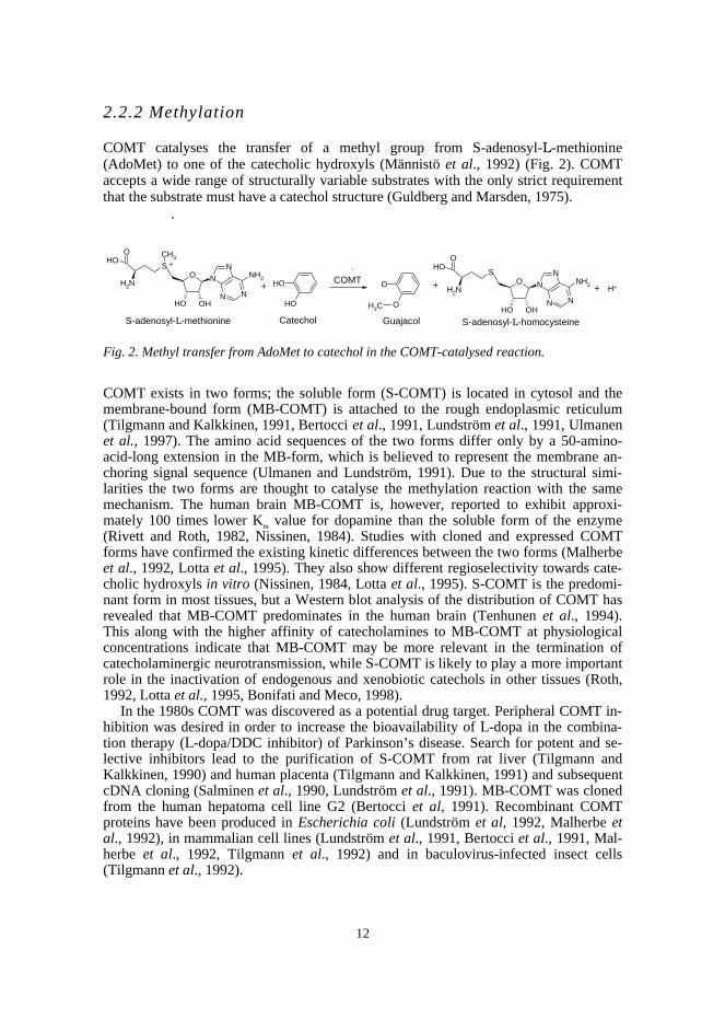
COMT catalyses the transfer of a methyl group from S-adenosyl-L-methionine(AdoMet) to one of the catecholic hydroxyls (Männistö et al., 1992) (Fig. 2). COMTaccepts a wide range of structurally variable substrates with the only strict requirementthat the substrate must have a catechol structure (Guldberg and Marsden, 1975).
Fig. 2. Methyl transfer from AdoMet to catechol in the COMT-catalysed reaction.
COMT exists in two forms; the soluble form (S-COMT) is located in cytosol and themembrane-bound form (MB-COMT) is attached to the rough endoplasmic reticulum(Tilgmann and Kalkkinen, 1991, Bertocci et al., 1991, Lundström et al., 1991, Ulmanenet al., 1997). The amino acid sequences of the two forms differ only by a 50-amino-acid-long extension in the MB-form, which is believed to represent the membrane an-choring signal sequence (Ulmanen and Lundström, 1991). Due to the structural simi-larities the two forms are thought to catalyse the methylation reaction with the samemechanism. The human brain MB-COMT is, however, reported to exhibit approxi-mately 100 times lower Km value for dopamine than the soluble form of the enzyme(Rivett and Roth, 1982, Nissinen, 1984). Studies with cloned and expressed COMTforms have confirmed the existing kinetic differences between the two forms (Malherbeet al., 1992, Lotta et al., 1995). They also show different regioselectivity towards cate-cholic hydroxyls in vitro (Nissinen, 1984, Lotta et al., 1995). S-COMT is the predomi-nant form in most tissues, but a Western blot analysis of the distribution of COMT hasrevealed that MB-COMT predominates in the human brain (Tenhunen et al., 1994).This along with the higher affinity of catecholamines to MB-COMT at physiologicalconcentrations indicate that MB-COMT may be more relevant in the termination ofcatecholaminergic neurotransmission, while S-COMT is likely to play a more importantrole in the inactivation of endogenous and xenobiotic catechols in other tissues (Roth,1992, Lotta et al., 1995, Bonifati and Meco, 1998).
In the 1980s COMT was discovered as a potential drug target. Peripheral COMT in-hibition was desired in order to increase the bioavailability of L-dopa in the combina-tion therapy (L-dopa/DDC inhibitor) of Parkinson’s disease. Search for potent and se-lective inhibitors lead to the purification of S-COMT from rat liver (Tilgmann andKalkkinen, 1990) and human placenta (Tilgmann and Kalkkinen, 1991) and subsequentcDNA cloning (Salminen et al., 1990, Lundström et al., 1991). MB-COMT was clonedfrom the human hepatoma cell line G2 (Bertocci et al, 1991). Recombinant COMTproteins have been produced in Escherichia coli (Lundström et al, 1992, Malherbe etal., 1992), in mammalian cell lines (Lundström et al., 1991, Bertocci et al., 1991, Mal-herbe et al., 1992, Tilgmann et al., 1992) and in baculovirus-infected insect cells(Tilgmann et al., 1992).
NO
N
N
OHOHN
NH2
CH3
+
S
CH3
NH2
OOH
OH
OH
O
O
+ NO
N
N
OHOHN
NH2
S
NH2
OOH
+ H+COMT
S-adenosyl-L-methionine GuajacolCatechol S-adenosyl-L-homocysteine
+
13
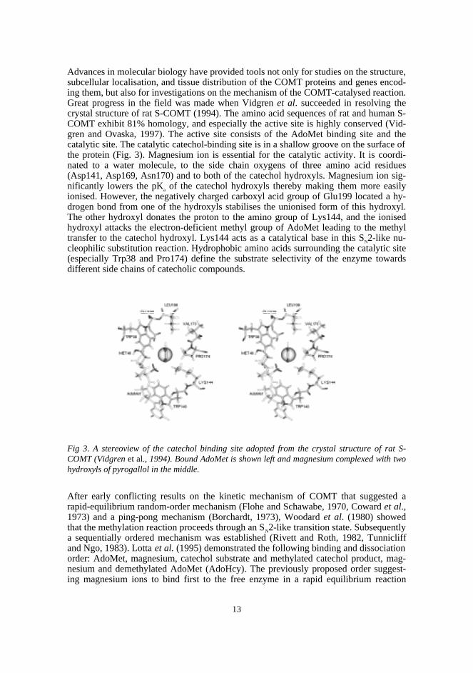
Advances in molecular biology have provided tools not only for studies on the structure,subcellular localisation, and tissue distribution of the COMT proteins and genes encod-ing them, but also for investigations on the mechanism of the COMT-catalysed reaction.Great progress in the field was made when Vidgren et al. succeeded in resolving thecrystal structure of rat S-COMT (1994). The amino acid sequences of rat and human S-COMT exhibit 81% homology, and especially the active site is highly conserved (Vid-gren and Ovaska, 1997). The active site consists of the AdoMet binding site and thecatalytic site. The catalytic catechol-binding site is in a shallow groove on the surface ofthe protein (Fig. 3). Magnesium ion is essential for the catalytic activity. It is coordi-nated to a water molecule, to the side chain oxygens of three amino acid residues(Asp141, Asp169, Asn170) and to both of the catechol hydroxyls. Magnesium ion sig-nificantly lowers the pKa of the catechol hydroxyls thereby making them more easilyionised. However, the negatively charged carboxyl acid group of Glu199 located a hy-drogen bond from one of the hydroxyls stabilises the unionised form of this hydroxyl.The other hydroxyl donates the proton to the amino group of Lys144, and the ionisedhydroxyl attacks the electron-deficient methyl group of AdoMet leading to the methyltransfer to the catechol hydroxyl. Lys144 acts as a catalytical base in this SN2-like nu-cleophilic substitution reaction. Hydrophobic amino acids surrounding the catalytic site(especially Trp38 and Pro174) define the substrate selectivity of the enzyme towardsdifferent side chains of catecholic compounds.
Fig 3. A stereoview of the catechol binding site adopted from the crystal structure of rat S-COMT (Vidgren et al., 1994). Bound AdoMet is shown left and magnesium complexed with twohydroxyls of pyrogallol in the middle.
After early conflicting results on the kinetic mechanism of COMT that suggested arapid-equilibrium random-order mechanism (Flohe and Schawabe, 1970, Coward et al.,1973) and a ping-pong mechanism (Borchardt, 1973), Woodard et al. (1980) showedthat the methylation reaction proceeds through an SN2-like transition state. Subsequentlya sequentially ordered mechanism was established (Rivett and Roth, 1982, Tunnicliffand Ngo, 1983). Lotta et al. (1995) demonstrated the following binding and dissociationorder: AdoMet, magnesium, catechol substrate and methylated catechol product, mag-nesium and demethylated AdoMet (AdoHcy). The previously proposed order suggest-ing magnesium ions to bind first to the free enzyme in a rapid equilibrium reaction
14
(Jeffery and Roth, 1987) could be argued on the basis of the crystal structure of COMT;the AdoMet binding site is located behind the magnesium binding site thereby forcingAdoMet to bind before magnesium. Catecholamines are predominantly methylated atthe meta-position, which in early studies was suggested to be due to the polar ionic sidechain favouring that binding orientation (Creveling et al., 1970, 1972). This interpreta-tion was later verified by computer-aided modelling of one low-activity conformer ofdopamine to the active site of COMT (Lotta et al., 1995). Modelling revealed that, indeed, binding in the orientation leading to para-methylation causes more unfavourableinteractions with the hydrophobic residues surrounding the active site compared withthe other binding mode. This might not, however, be the only explanation, because onlyone low-energy conformer was studied and the flexible side chain may adopt variableorientations. In addition to the hydrophobic interactions, the nature of substituents atvarious positions may affect the regioselectivity with other mechanisms as well. For e x-ample, in ring-fluorinated catecholamines variable preference to the hydroxyls has beenexplained by changes in nucleophilicity (Firnau et al., 1981, Creveling et al., 1981,Thakker et al., 1986).
COMT is usually thought to possess a very wide acceptance of structurally diversecatecholic compounds. The clinical importance of COMT inhibition has directed studieson the structure-activity relationships to the factors affecting inhibition potency ratherthan to the methylation reaction itself. Two research groups, both of which would intro-duce its own molecule to clinical use later, independently synthesised potent and selec-tive COMT inhibitors with 3-nitrocatechol as the key structure (Borgulya et al., 1989,Bäckström et al., 1989). Further studies showed that inhibition activity is enhanced byelectron-withdrawing substituents at positions 3 and 5 and that binding affinity is im-proved by a hydrophobic substituent at position 5 (Taskinen et al., 1989, Lotta et al.,1992). Compounds containing electronegative groups, such as 3,5-dinitrocatechol, de-crease the nucleophilicity of the ionised catechol hydroxyls and strongly stabilise theionised catechol-COMT complex thus making the energy barrier for the methylationhigh (Vidgren and Ovaska, 1997, Ovaska and Yliniemelä, 1998). The few studies on thesubstrate selectivity of COMT have emphasised the relevance of hydrophobicity in theincrement of affinity (Raxworthy and Gulliver, 1982, Youde et al., 1984). However,these conclusions have been derived on the basis of a small number of related com-pounds rather than a proper structure-activity analysis.
Existence of polymorphic COMT forms, a thermolabile low activity and a thermo-stabile high activity COMT, has been reported (Scanlon et al., 1979, Weinshilboum,1984, 1988, Boudiková et al, 1990, Grossman et al., 1992). The molecular basis of thepolymorphism is variation at the 108 amino acid residue (Val-108 being thermostabileand Met-108 thermolabile) (Lotta et al., 1995). The two alleles result in homozygousindividuals with high or low COMT activity and heterozygous individuals with inter-mediate activity (Bonfati and Meco, 1999). A markedly higher frequency of the highactivity allele has been reported among Chinese and Japanese people (~75%) thanamong Caucasians (50%) (Xie et al., 1997, Kunugi et al., 1997). Previous studies havealso shown that Orientals and black Americans exhibit a higher COMT activity than doCaucasians (Rivera-Calimlim and Reilly, 1984, McLeod et al., 1994). In contrast, alower COMT activity has been reported among the Saami population (Klemetsdal et al.,1994). Several studies have suggested the association of COMT polymorphism withneurological and psychiatric disorders (e.g. Kunugi et al., 1997, Strous et al., 1997a,Chen et al., 1997, Lachman et al., 1996) and with breast cancer risk (Lavigne et al.,1997, Thompson et al., 1998), but also contradicting findings have been published (e.g.Xie et al., 1997, Strous et al., 1997b, Ohara et al., 1998, Millikan et al., 1998).
15
2.2.3. Sulphation
The conjugation of 3’-phosphoadenosine 5’-phosphosulphate with an O-, N- or S- ac-ceptor group is catalysed by cytosolic sulphotransferases. The SULT enzyme familyincludes enzymes that catalyse the sulphation of phenolic xenobiotics (P-PST), cate-cholamines (M-PST), estrogens (EST), and steroids (HST) (Coughtrie, 1998). Differentisoforms exhibit overlapping substrate specificities, although many of them show pref-erence towards some type of substrates. Structure-activity relationships have not beenfully investigated for any of the isoforms. However, in human liver samples neutral sub-stituents, usually rather in the ortho- than in the para-position, have been found to fa-vour phenol sulphation (Temellini et al., 1991). SULT1A3 (M-PST), which is responsi-ble for the sulphation of catecholamines, is predominantly expressed in the small intes-tinal mucosa and may have a special role in the detoxification of dietary xenobiotics(Coughtrie, 1998). As a result of the action of SULT1A3, circulating catecholaminesappear mostly as sulphate conjugates thus facilitating their transportation to other tis-sues. At least SULT1A1 (P-PST) is genetically polymorphic, but further investigationson its significance are needed (Coughtrie et al., 1999).
2.3. In vitro studies on catechol conjugation
In vitro studies on conjugation reactions are usually performed by incubating the stud-ied compound, in the presence of a cosubstrate, with the enzyme and by measuring thereaction velocity. The most common enzyme sources from different species are tissuehomogenates, subcellular fractions and recombinant enzymes. Since following the dis-appearance of the substrate is insensitive and unspecific, reaction velocity is normallydetermined on the basis of formation of the products. In kinetic studies, the initial reac-tion velocity is determined as a function of substrate concentration at the saturating con-centration of the cosubstrate (Cornish-Bowden, 1995). Most enzymes follow Michaelis-Menten kinetics; reaction velocity increases almost linearly at low substrate concentra-tions after which it starts to reach the maximum asymptotically. The enzyme kinetic pa-rameters Vmax and Km, describing the capacity of the reaction and the affinity of the sub-strate to the enzyme, are derived by fitting the Michaelis-Menten equation (1) to theinitial velocity values obtained. At low substrate concentrations, normally occurring invivo, the reaction is best described by the ratio of Vmax and Km.
(1) V = (Vmax x [s]) / (Km + [s])V = reaction velocityVmax = maximum reaction velocity[s] = substrate concentrationKm = substrate concentration at half the maximum velocity
In addition to studies on substrate selectivity and reaction mechanisms, in vitro methodsmay be utilised in the selection of the animal model for toxicity studies and in the earlyassessment of metabolism and metabolic interactions in humans. Attempts to predict invivo clearance based on in vitro data have been made almost exclusively utilising com-pounds that are metabolised by phase I enzymes (Houston and Carlile, 1997). However,a study on tolcapone shows that reasonably accurate predictions are possible also forcompounds metabolised mainly by phase II enzymes (Lave et al., 1996).
16
2.3.1. UGT assays
The major difficulty of in vitro studies on UGT is its latency, due to its subcellular lo-calisation in the rough endoplasmic reticulum, the active site towards the lumen. Activ-ity may be enhanced by damaging the membrane by detergents or sonication, whichimproves the access of the substrates to the active site. Optimum concentrations of de-tergents (e.g. digitonin, Lubrol WX, Brij 58, CHAPS, Tergitol NP-10 and Triton X-100)have been shown to increase the UGT activity by 2- to 5-fold (Winsnes, 1969, Mag-dalou et al., 1979, Thomassin et al., 1985, Lawrence et al., 1992, Lett et al., 1992, Mar-tin and Black, 1994). However, detergents have been reported to alter not only the Vmax
but also the Km values (Thomassin et al., 1985) and excessive detergent concentrationsdeactivate the enzyme. There is no direct evidence of the contribution of Mg2+ ion to theglucuronidation reaction, although magnesium has been found to increase the glucu-ronidation rate of for example morphine (Lawrence et al., 1992). β-Glucuronidase,which is located in the lumen of endoplasmic reticulum, as are UGTs, catalyses the hy-drolysis of glucuronides, but it can be selectively inhibited by saccharolactone. In addi-tion, the pH normally used in the glucuronidation studies (6-8) does not favour the ac-tion of β-glucuronidase that exhibits a pH optimum at 4-5 (Kauffman, 1994).
Assays for UGT include spectrometric and fluorometric methods, for instance con-tinuous fluorometric monitoring of reaction products (Väisänen et al., 1983), but mostlycommon chromatographic techniques have been utilised. Most methods have been de-veloped for specific substrates. However, a very widely used method, introduced byBansal and Gessner (1980), is suited for structurally diverse compounds including sim-ple phenols and hormones. The universality of the method is based on uridine diphos-pho[U-14C]glucuronic acid (14C-UDPGA) that is conjugated with the acceptor substratesforming 14C-labelled products, which makes authentic reference standards unnecessary.In this method, the formed glucuronides are separated from unreacted UDPGA onpreparative silica gel TLC plates with a mixture of n-butanol, water, acetone, glacialacetic acid and 30% ammonia (70:60:50:18:1.5). The glucuronide spots are identifiedby autoradiography and quantitated by liquid scintillation counting after scraping fromthe plate. The method with minor modifications has been especially useful in studies onsubstrate selectivity of UGTs. The tedious and time-consuming autoradiography andscraping with subsequent liquid scintillation counting have been replaced by quantita-tion of the radioactive glucuronides directly from the TLC plates by a digital autoradio-graph or a radioanalytical imaging system (Ritter et al., 1990, Ebner and Burchell,1993). The main disadvantage of the method is its inability to separate multiple glucu-ronides originating from the same parent compound. Therefore a general HPLC method,based on 14C-UDPGA and on-line radioactivity detection (Coughtrie et al., 1986), re-cently published in an improved form (Ethell et al., 1998), may overcome the old TLCassay.
2.3.2. COMT assays
As explained previously, Mg2+ ions are essential for the COMT-catalysed methylationreaction. Some other divalent cations, such as Cd2+, Hg2+, Mn2+ and Cu2+, have beenfound to promote methylation as well (Axelrod and Tomchick, 1958, Senoh et al., 1962,Flohe 1974, Boadi et al., 1991). In contrast, Ca2+ ions seem to inhibit COMT (Weinshil-boum and Raymond, 1976). Purified human S-COMT has been shown to require cys-teine as a reducing agent to maintain its activity (Tilgmann and Kalkkinen, 1991). Otheragents capable of inhibiting the deactivation, probably caused by oxidation of the sul-
17
phydryl groups of the protein, include mercaptoethanol and dithiothreitol (Tilgmannand Ulmanen, 1996). Reducing agents in the reaction mixture may also protect the cate-cholic hydroxyls from oxidation during the reaction. AdoHcy, the demethylated endproduct of AdoMet, has been found to inhibit COMT (Coward et al., 1973), but at lowsubstrate and enzyme and saturating AdoMet concentrations its effect becomes negligi-ble.
Many early COMT assays relied on fluorometric (e.g. Axelrod and Tomchick, 1958),spectrophotometric (e.g. Coward and Wu, 1973, Borchardt, 1974) or most commonlyradiochemical methods (e.g. Jonas and Gershon, 1974, Raymond and Weinshilboum,1975, Gulliver and Tipton, 1978, Bates et al., 1979, Zürcher and Da Prada, 1982).Adding a radioactively labelled substrate or cosubstrate (S-adenosyl-L-[methyl-14C]methionine or S-adenosyl-L-[methyl-3H]methionine) to the reaction mixture resultedin radioactive end products that could be separated from the parent compounds by liq-uid-liquid extraction or thin-layer chromatography, and that could be subsequentlyquantitated in a liquid scintillation counter. Radiochemical methods are simple and sen-sitive, applicable for various catechol substrates and require no reference standards.However, impurities in radiochemicals and variable recovery in the extraction proce-dure impair their reliability. In addition, regioisomeric O-methylated metabolites, pro-duced from many compounds in vitro, cannot be quantitated separately. Development ofgas chromatographic (Creveling et al., 1972, Lin and Narasimhachari, 1974, Koh et al.,1991) and liquid chromatographic COMT assays has, however, enabled separation ofthe regioisomeric products. High-performance liquid chromatography has been coupledwith various detection devices including UV (Pennings and Van Kempen, 1979), elec-trochemical (Borchardt et al., 1978, Shoup et al., 1980, Koh et al., 1981, Nissinen andMännistö, 1984), fluorometric (Zaitsu et al., 1981, Nohta et al., 1984, Smit et al., 1990,Zürcher et al., 1996) and radiochemical detectors (Nissinen, 1985). Most of the assaysare intended for the measurement of COMT activity in different tissues including thosewith a low level of COMT expression, such as brain or erythrocytes. Especially assaysutilising electrochemical, radioactivity and fluorescence detectors are specific and sen-sitive (respective limits of detection 0.5, 0.04 pmol and 11 fmol reaction product perinjection) (Reenilä et al., 1995, Tuomainen et al., 1996, Nissinen, 1985, Zürcher et al.,1996). These methods use a specific substrate and do not allow determination of COMTactivity towards catechols with diverse structures.
18
3. Aims of the study
The primary aims of this study were 1) to evaluate the susceptibility of nitrocatecholicCOMT inhibitors to glucuronidation in vitro and 2) to characterise the structural fea-tures of catecholic compounds that determine their properties as S-COMT substrates.
The specific aims were:�to develop and validate analytical methods for the in vitro studies on the glucuronida-tion and methylation of catechols�to evaluate the glucuronidation of various nitrocatechols in rat liver microsomes�to characterise the human UGT isoforms that are mainly responsible for the glucu-ronidation of the COMT inhibitors entacapone and tolcapone�to compare the glucuronidation kinetics of entacapone and tolcapone in rat and humanliver microsomes as well as by human UGT isoforms�to compare the substrate selectivity of rat and human S-COMT�to construct predictive models for the methylation of catechols by human S-COMT
19
4. Materials and methods
The original publications contain more detailed descriptions of the materials and meth-ods utilised.
4.1. Chemicals
Most of the 57 catecholic compounds were purchased from commercial sources andwere of the highest grade available. Nitrocatechol derivatives 3-nitrocatechol, 3,5-dinitrocatechol, entacapone, entacapone(Z)-isomer, nitecapone and tolcapone werekindly supplied by Orion Pharma (Espoo, Finland). UDPGA was obtained from SigmaChemical Company (St. Louis, Missouri, USA) or Boehringer-Mannheim (Mannheim,Germany), AdoMet from Boehringer-Mannheim and 14C-UDPGA and 14C-AdoMet fromNEN Du Pont (Boston, USA). 4-Nitrophenyl-β-D-glucuronide (4NPG) was purchasedfrom Sigma Chemical Company and vanillic acid from Aldrich (Sigma-AldrichChemie, Steinheim, Germany). Reference standards of the 3-O-glucuronides of entaca-pone and tolcapone were synthesised at the Department of Pharmaceutical Chemistry,University of Helsinki, Finland (Luukkanen et al., 1999).
4.2. Enzyme sources
Rat liver microsomes were prepared from liver homogenates of male Wistar rats by dif-ferential centrifugation at the Finnish Institute of Occupational Health, Helsinki (I, II).The rats were pre-treated with creosote (200 mg in 4 ml olive oil/kg) or did not receiveany pre-treatment. Protein concentrations of the microsomal suspensions were deter-mined by the method of Lowry et al. (1951). Human liver microsomes were purchasedfrom Human Biologics Inc. (Arizona, USA) (III). Microsomes were stored at -70oC be-fore being used.
Recombinant V79 cell lines expressing human UGT isoforms were grown up andmaintained at the Department of Molecular and Cellular Pathology, Ninewells Hospitaland Medical School, Dundee, Scotland, as described previously (Ethell et al, 1998)(III). The harvested cells were stored at -70oC and, before use, disrupted by sonication.The protein concentrations were determined using bovine serum albumin as a standard(Lowry et al., 1951). Recombinant rat and human S-COMT proteins were produced inE. coli at the Department of Molecular Biology and Target Protein Research, OrionPharma, Finland, as described in detail earlier (Lundström et al., 1992) (IV, V). Theharvested cells were disrupted by sonication and kept at -70oC before being used for theenzyme assays. The total protein concentrations of the lysates were determined accord-ing to Bradford (1976).
20
4.3. Reaction mixtures
In the UGT assays the reaction mixture contained MgCl2 (5 mM), UDPGA (2-5 mM),substrate (0.010-2.5 mM), and the UGT source (microsomes or lysates from cells ex-pressing UGT isoforms) in 50-100 mM phosphate or Tris/maleate buffer (pH 7.4) (I-III). When radioactivity was utilised in the quantitation, 0.1 µCi 14C-UDPGA was addedto the reaction mixture (II, III). The samples were incubated at 37°C for 15-60 minutesbefore the reactions were terminated by adding organic solvent (methanol or acetoni-trile) or 4 M perchloric acid. The precipitated proteins were removed by centrifugation.
The COMT assays were performed in 100 mM Na2HPO4/NaH2PO4 buffer (pH 7.4)containing MgCl2 (5 mM), L-cysteine (20 mM), AdoMet (150 µM), 14C-AdoMet (0.1µCi), a catechol substrate (0.25-3000 µM) and human or rat S-COMT bacterial lysate(IV, V). In kinetic studies, the amount of enzyme was chosen separately for each sub-strate on the basis of the concentration range used in order to maintain appropriate con-ditions for Michaelis-Menten kinetics. The samples were pre-incubated at 37oC for 5min before the reactions were started by adding the catechol substrate or theAdoMet/14C-AdoMet mixture. After the 15- to 30-min-long incubation period the reac-tions were terminated by adding cold 4 M perchloric acid. The samples were centri-fuged before the HPLC analysis.
4.4. Analytical methods
4.4.1. Thin-layer chromatography (I-II)
The glucuronides of nitrocatechols were separated from the parent compounds andUDPGA on RP-18 HPTLC plates using a horizontal development mode. The eluentconsisted of 50 mM NaH2PO4 (pH 2.2) and acetonitrile (6:4 v/v). The sample applica-tion was performed utilising the Linomat IV spray-on technique (Camag, Muttenz,Switzerland). The glucuronides were quantitated with the aid of one sample in which14C-UDPGA had been added before incubation. Four different volumes of this samplewere applied to the plate in order to obtain a calibration curve. After development, theair-dried plates were scanned with a Camag TLC Scanner II, controlled by the CamagCats program version 3.17, at wavelengths specific for each nitrocatechol. Three of theradioactivity-containing glucuronide spots were scraped from the plates and, after addi-tion of liquid scintillation cocktail (Optiphase Hisafe 2, FSA Laboratory Supplies,Loughborough, UK), quantitated in a liquid scintillation counter (Wallac 1410, Turku,Finland). The amounts of the glucuronides in the standard spots, calculated from themean value derived from the radioactivity measurements, were fed into the Cats pro-gram and the glucuronides in the other samples were quantitated on the basis of the den-sitometric analysis.
21
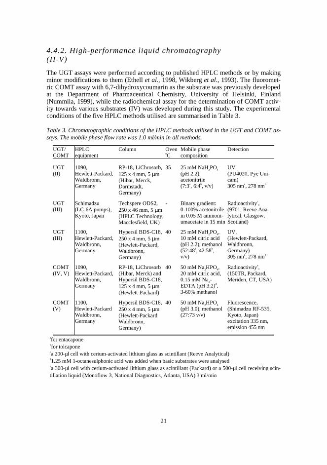
4.4.2. High-performance liquid chromatography(II-V)
The UGT assays were performed according to published HPLC methods or by makingminor modifications to them (Ethell et al., 1998, Wikberg et al., 1993). The fluoromet-ric COMT assay with 6,7-dihydroxycoumarin as the substrate was previously developedat the Department of Pharmaceutical Chemistry, University of Helsinki, Finland(Nummila, 1999), while the radiochemical assay for the determination of COMT activ-ity towards various substrates (IV) was developed during this study. The experimentalconditions of the five HPLC methods utilised are summarised in Table 3.
Table 3. Chromatographic conditions of the HPLC methods utilised in the UGT and COMT as-says. The mobile phase flow rate was 1.0 ml/min in all methods.
UGT/COMT
HPLCequipment
Column OvenoC
Mobile phasecomposition
Detection
UGT(II)
1090,Hewlett-Packard,Waldbronn,Germany
RP-18, LiChrosorb,125 x 4 mm, 5 µm(Hibar, Merck,Darmstadt,Germany)
35 25 mM NaH2PO4
(pH 2.2),acetonitrile(7:3a, 6:4b, v/v)
UV(PU4020, Pye Uni-cam)305 nma, 278 nmb
UGT(III)
Schimadzu(LC-6A pumps),Kyoto, Japan
Techspere ODS2,250 x 46 mm, 5 µm(HPLC Technology,Macclesfield, UK)
- Binary gradient:0-100% acetonitrilein 0.05 M ammoni-umacetate in 15 min
Radioactivityc,(9701, Reeve Ana-lytical, Glasgow,Scotland)
UGT(III)
1100,Hewlett-Packard,Waldbronn,Germany
Hypersil BDS-C18,250 x 4 mm, 5 µm(Hewlett-Packard,Waldbronn,Germany)
40 25 mM NaH2PO4,10 mM citric acid(pH 2.2), methanol(52:48a, 42:58b,v/v)
UV,(Hewlett-Packard,Waldbronn,Germany)305 nma, 278 nmb
COMT(IV, V)
1090,Hewlett-Packard,Waldbronn,Germany
RP-18, LiChrosorb(Hibar, Merck) andHypersil BDS-C18,125 x 4 mm, 5 µm(Hewlett-Packard)
40 50 mM Na2HPO4,20 mM citric acid,0.15 mM Na2-EDTA (pH 3.2)d,3-60% methanol
Radioactivitye,(150TR, Packard,Meriden, CT, USA)
COMT(V)
1100,Hewlett-PackardWaldbronn,Germany
Hypersil BDS-C18,250 x 4 mm, 5 µm(Hewlett-PackardWaldbronn,Germany)
40 50 mM Na2HPO4
(pH 3.0), methanol(27:73 v/v)
Fluorescence,(Shimadzu RF-535,Kyoto, Japan)excitation 335 nm,emission 455 nm
afor entacaponebfor tolcaponeca 200-µl cell with cerium-activated lithium glass as scintillant (Reeve Analytical)d1.25 mM 1-octanesulphonic acid was added when basic substrates were analysedea 300-µl cell with cerium-activated lithium glass as scintillant (Packard) or a 500-µl cell receiving scin-tillation liquid (Monoflow 3, National Diagnostics, Atlanta, USA) 3 ml/min
22
4.4.3. Method validation
The HPTLC method applied in the investigation of the glucuronidation of nitrocate-chols by rat liver microsomes was validated with 4-nitrophenol as the model substrate(I). The validation procedure comprised specificity, limit of quantitation, repeatabilityof sample application (n=7), recovery (3 concentrations, n=6), reproducibility of themethod (6 concentrations, n=6), and reproducibility of the determination of the enzymekinetic parameters (n=6). The reliability of quantitation based on the method combiningdensitometry and radioactivity measurement was tested by comparing the results ob-tained from the same samples by this method and by the densitometric method thatutilises 4NPG as the reference standard (6 concentrations).
3,4-Dihydroxybenzoic acid (DHBA) was used as the model substrate in the valida-tion of the new radiochemical HPLC method developed for the assay of COMT activitytowards various substrates (IV). In this method the methylated products were quanti-tated by comparing their peak areas in the radioactivity detector with the total area ofradioactive peaks. The accuracy of this quantitation method was investigated by ana-lysing samples containing different initial concentrations of DHBA simultaneously byradioactivity and UV detection. In the UV detection, the 3-O-methylated product ofDHBA, vanillic acid, was used as the reference standard. Recovery was determined bycomparing the sum of AdoMet, vanillic and isovanillic acid peak areas in the radioac-tivity detector with the peak area of unincubated AdoMet (n=6). The limit of quantita-tion and the limit of detection were estimated for vanillic acid by using the criteria forsignal to noise ratio of ten and three. The same samples containing six initial concentra-tions of DHBA (n=6) were used for investigating the reproducibility of the method andthe reproducibility of the determination of the enzyme kinetic parameters.
4.5. Enzyme kinetic analysis (I-V)
The apparent enzyme kinetic parameters Vmax and Km were determined by measuring theinitial reaction velocity as a function of catechol concentration at fixed cosubstrate con-centration. The Michaelis-Menten equation was fitted to the initial velocity values usingthe Leonora Steady-state Enzyme Kinetics program version 1.0 by A. Cornish-Bowden(1994). In some cases equations for substrate inhibition and competitive or mixed inhi-bition were used.
4.6. QSAR and molecular modelling (V)
The octanol-water distribution coefficients were calculated using the LOGKOW method(Meylan and Howard, 1995), and the SPSS 8.0.1 program (SPSS Inc., Chicago, IL,USA) was utilised in statistical analyses. The catechols were modelled by the Spartan5.0 program (Wavefunction Inc., Irvine, CA, USA) and optimised by using the semiem-pirical AM1 method. The molecular electrostatic potentials (MEP) were computed bysingle point ab initio calculations at the 3-21G(*) level. The Insight II program (Mo-lecular Simulations Inc., San Diego, CA, USA) was utilised in superimposing the cate-chols on 3,5-dinitrocatechol in the active site model of rat S-COMT (Brookhaven Pro-tein Data Bank entry 1VID).
23
5. Results and discussion
5.1. Development and validation of enzymeassays
5.1.1. Thin-layer chromatographic UGT assay for the determi-nation of nitrocatechol glucuronidation (I, II)
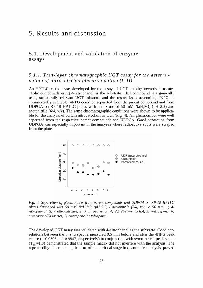
An HPTLC method was developed for the assay of UGT activity towards nitrocate-cholic compounds using 4-nitrophenol as the substrate. This compound is a generallyused, structurally relevant UGT substrate and the respective glucuronide, 4NPG, iscommercially available. 4NPG could be separated from the parent compound and fromUDPGA on RP-18 HPTLC plates with a mixture of 50 mM NaH2PO4 (pH 2.2) andacetonitrile (6/4, v/v). The same chromatographic conditions were shown to be applica-ble for the analysis of certain nitrocatechols as well (Fig. 4). All glucuronides were wellseparated from the respective parent compounds and UDPGA. Good separation fromUDPGA was especially important in the analyses where radioactive spots were scrapedfrom the plate.
Fig. 4. Separation of glucuronides from parent compounds and UDPGA on RP-18 HPTLCplates developed with 50 mM NaH2PO4 (pH 2.2) / acetonitrile (6/4, v/v) to 50 mm. 1; 4-nitrophenol, 2; 4-nitrocatechol, 3; 3-nitrocatechol, 4; 3,5-dinitrocatechol, 5; entacapone, 6;entacapone(Z)-isomer, 7; nitecapone, 8; tolcapone.
The developed UGT assay was validated with 4-nitrophenol as the substrate. Good cor-relations between the in situ spectra measured 0.5 mm before and after the 4NPG peakcentre (r=0.9805 and 0.9847, respectively) in conjunction with symmetrical peak shape(T0.05=1.0) demonstrated that the sample matrix did not interfere with the analysis. Therepeatability of sample application, often a critical stage in quantitative analysis, proved
Compound
Mig
ratio
n di
stan
ce (
mm
)
0
10
20
30
40
50
UDP-glucuronic acid GlucuronideParent compound
1 2 3 4 5 6 7 8
24
to be satisfactory (RSD=2.2%, 50 µM 4NPG, n=7). The limit of quantitation, 15 pmol4NPG on the plate, representing the lowest concentration at which the RSD of sampleapplication (n=3) did not exceed 5% (Ferenczi-Fodor et al., 1993), enabled reactionvelocity measurements at low 4NP concentrations. The recoveries of various 4NPGconcentrations (12.5, 50 and 75 µM) spiked in the sample matrix were rather consistent(95.2±3.4, 102.8±2.5 and 102.0±2.2%, respectively, n=6) and showed a mean value of100%. The relatively low RSD values (between 4.1 and 11.7%, n=6) achieved at sixinitial concentrations of 4-nitrophenol suggested that the within-laboratory reproduci-bility of the method was at a tolerable level. The good reproducibility of the determina-tion of the enzyme kinetic parameters Km and Vmax (RSD 6.1 and 6.3%, respectively,n=6) indicated that the method was suitable for enzyme kinetic studies.
Lack of reference standards of the different nitrocatechol glucuronides necessitatedthe utilisation of 14C-labelled UDPGA in quantitation. However, to decrease the costsand risks of the analysis, the radioactive cosubstrate was added to just one of the sam-ples in each kinetic series. The concentration level of the glucuronides was obtainedwith this sample and the glucuronides in the other samples were quantitated on the basisof densitometric analysis. The good correlation (r=0.9920) between the results obtainedfor the same 4-nitrophenol samples by this method and by the method utilising 4NPGdemonstrated that reliable results could be obtained in a wide range of glucuronide con-centrations. In general, the benefits of TLC include low solvent consumption, off-linecharacter and possibility to analyse several samples simultaneously. Compared with thetraditional UGT assay developed by Bansal and Gessner (1980), the new method wascost-effective and did not require time-consuming exposure to x-ray films. Although itis only applicable for compounds exhibiting a reasonably high molecular absorptivity,all the nitrocatecholic compounds could be analysed under the same conditions. Themain disadvantage of the method, that multiple glucuronides could not be separated, isshared by all TLC-based UGT assays. However, the idea of a using radioactively la-belled cosubstrate only for calibrating the product level is applicable for HPLC as well.This approach was used in a study on chrysene and benzo(a)pyrene phenols in which,after HPLC separation, quantitation was based on the linear relationship observed be-tween radioactivity, fluorescence and UV absorption (Bock et al., 1992). Improvementin sensitivity by combining UV or fluorescence detection with radioactivity measure-ment may be desired in kinetic studies where a high UDPGA concentration is required,which decreases the formation of radioactively labelled glucuronides.
5.1.2. Radiochemical high-performance liquidchromatographic COMT assay (IV, V)
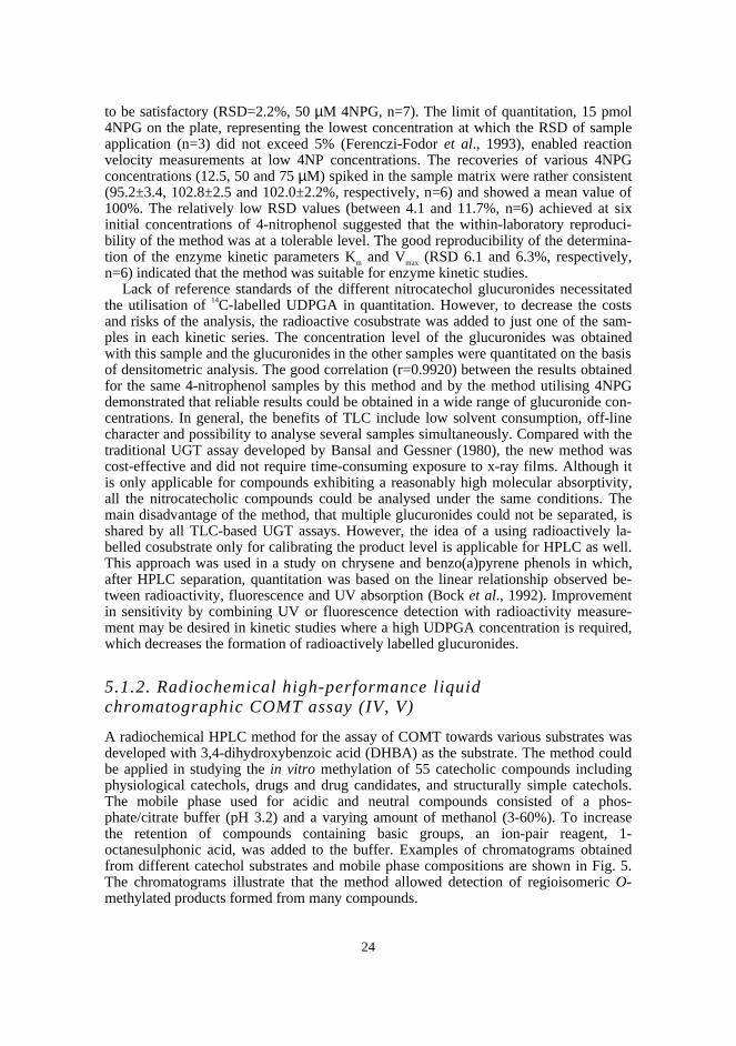
A radiochemical HPLC method for the assay of COMT towards various substrates wasdeveloped with 3,4-dihydroxybenzoic acid (DHBA) as the substrate. The method couldbe applied in studying the in vitro methylation of 55 catecholic compounds includingphysiological catechols, drugs and drug candidates, and structurally simple catechols.The mobile phase used for acidic and neutral compounds consisted of a phos-phate/citrate buffer (pH 3.2) and a varying amount of methanol (3-60%). To increasethe retention of compounds containing basic groups, an ion-pair reagent, 1-octanesulphonic acid, was added to the buffer. Examples of chromatograms obtainedfrom different catechol substrates and mobile phase compositions are shown in Fig. 5.The chromatograms illustrate that the method allowed detection of regioisomeric O-methylated products formed from many compounds.
25
mV mV mV mV
0 5 10 min 0 5 10 min 0 5 10 15 min 0 5 min
Fig. 5. Chromatograms in the radioactivity detector obtained from samples in whichAdoMet/14C-AdoMet and recombinant human S-COMT were incubated with a) 3,4-dihydroxybenzoic acid b) dopamine c) L-dopa d) 2-hydroxyestradiol. The column used was aHypersil BDS-C18, 125x4 mm, 5 µm, and the proportion of phosphate/citrate buffer (pH 3.2)and methanol was a) 90:10 b) 85 (including 1.25 mM 1-octanesulphonic acid) :15 c) 95 (in-cluding 1.25 mM 1-octanesulphonic acid) :5 d) 40:60.
The COMT assay was validated with DHBA as the substrate and recombinant human S-COMT as the enzyme source. The O-methylated metabolites were quantitated by com-paring their peak areas in the radioactivity detector with the total area of radioactivepeaks. The results of the validation experiments based on the quantitation of vanillicacid are summarised in Table 4. The quantitation based on comparison of the area ofradioactive peaks correlated well with the method relying on UV detection. The recov-ery and the repeatability of the method were good, and the good repeatability of the de-termination of the enzyme kinetic parameters showed that the method was well suitedfor its intended use. When applying the method to other compounds, however, morevariation in for example recovery and stability may be expected. Fortunately, no loss oftotal radioactivity, represented by the sum of peak areas in the radioactivity detector,could be detected among the compounds studied. Stability problems were minimised byusing fresh solutions only, adding antioxidant (L-cysteine) to the reaction mixture andalways analysing the samples immediately after preparation. Earlier published Km val-ues for DHBA, dopamine, noradrenaline and L-dopa determined by using HPLC withelectrochemical detection are well comparable with those obtained by this method(Lotta et al., 1995).Most of the compounds could be analysed with a solid scintillant in the radioactivitydetector. A solid scintillant, earlier utilised in UGT assay (Ethell et al., 1998), has someadvantages compared with on-line liquid scintillation counting. No scintillation liquidor additional pump is needed, obviating the need to optimise the scintillant/mobilephase flow rate. The use of a solid scintillant not only reduces costs but also markedlydecreases the production of waste. However, compounds exhibiting low Km values re-quired improved sensitivity. The limit of detection could be lowered from 9 to 1pmol/injection by substituting the 300-µl cerium-activated lithium glass cell by a 500-µlflow cell that received scintillation liquid 3 ml/min. With this modification the sensitiv-ity level (0.45 pmol) of an earlier COMT assay utilising S-adenosyl-L-[methyl-3H]methionine and on-line radiochemical detection (Nissinen, 1985) could be reached.Considerably lower metabolite levels, however, are detectable by electrochemical andfluorescence detectors (Reenilä et al., 1995, Tuomainen et al., 1996, Zürcher et al.,
A B C D
AdoMet
Vanillicacid Iso-
vanillicacid
AdoMet AdoMet AdoMet
M1
M2
M
M2M1

26
1996), and assays relying on them are methods of choice when COMT activity has to bemeasured in erythrocytes or other tissues with low COMT expression. Nevertheless, thenew method utilising 14C-AdoMet and on-line radiochemical detection is suitable forstudies on structure-activity relationships and mechanisms of regioselectivity in whichstructurally diverse catecholic compounds are included. The method provides also arapid screening assay for the in vitro methylation of for instance new catecholic drugcandidates.
Table 4. Validation of the radiochemical HPLC method for the assay of COMT with 3,4-dihydroxybenzoic acid as the substrate.
Validation parameter Result
Accuracy of quantitation (5-300 µM DHBA) compared to UV-detection, r2=0.9988 (n=6)
Recovery (300 µM DHBA) 97.5 ± 2.5%, (n=6)
Repeatability-at different DHBA concentrations (5-300 µM)-determination of kinetic parameters
RSD= 3.30-7.30% (6 concentrations, n=6)Km: RSD=7.94%, Vmax: RSD =3.57% (n=3)
Limit of detection-solid cell-liquid cell
9 pmol/injection at signal/noise = 31 pmol/injection at signal/noise = 3
5.2. Glucuronidation of nitrocatechols
5.2.1. Glucuronidation of nitrocatechols by rat liver micr o-somes (II)
In order to assess the properties of nitrocatechol-type compounds as UGT substrates,enzyme kinetic parameters were determined for a nitrocatechol series and the modelsubstrate 4-nitrophenol in rat liver microsomes (Table 5). Microsomes were derivedfrom male Wistar rats treated with creosote, which, similar to 3-methylcholantherene,has been found to cause a two-fold induction of 4-nitrophenol glucuronidation (Luuk-kanen et al., 1997). In this study creosote was found to increase the glucuronidation rateof entacapone and tolcapone, by approximately two-fold.
Disubstituted COMT inhibitors showed approximately one order of magnitude lowerVmax/Km values compared with 4-nitrophenol and 4-nitrocatechol. This may be due toUGT1*06, an isoform known to be induced by 3-methylcholantherene (and possibly bycreosote) and reported to exhibit a restricted specificity towards small and planar phe-nols (Jackson et al., 1988). Consequently, other isoforms are likely to contribute to theglucuronidation of the compounds with bulkier substituents. Existence of a parabolicdependence between glucuronidation rate and lipophilicity has been suggested, with anoptimum value of logP=2.25 for phenolic compounds in rats (Kim, 1991). Although theglucuronidation rates of 4-n-propylphenol and 4-tert-butylphenol (respective estimatedlogPow=3.04 and 3.42) have been reported to be higher than that of 4-nitrophenol (logP ow
27
=1.91) in rat liver microsomes (Jackson et al., 1988), in this study entacapone and tol-capone (respective logPow=2.22 and 3.13) exhibited approximately five times lower Vmax
values than 4-nitrophenol. Apparently other structural features, not identified in thisstudy, affected the glucuronidation of these COMT inhibitors. Nevertheless, the morelipophilic nature of tolcapone compared with entacapone and especially nitecapone(logPow=1.04) may explain its higher affinity towards rat UGTs. The especially high Km
value obtained for nitecapone may indicate a contribution of a distinct low-affinity iso-form to the glucuronidation of this compound.
Table 5. Apparent enzyme kinetic parameters for the glucuronidation of 4-nitrophenol and somenitrocatechols by liver microsomes from creosote-treated rats. The values represent mean ± SDof three or four independent experiments.
Compound Vmax
(nmol min-1 mg-1)Km
(mM)Vmax/Km
(ml min-1 mg-1)
4-Nitrophenol 68.8±3.8 0.11±0.02 0.63
4-Nitrocatechol 153.0±26.4 0.19±0.05 0.79
3-Nitrocatechol 127.7±15.5 0.72±0.11 0.18
3,5-Dinitrocatechol 36.2±7.7 1.04±0.27 0.035
Entacapone 13.4±2.1 0.75±0.27 0.018
Entacapone(Z)-isomer 15.5±3.4 0.40±0.11 0.039
Nitecapone 52.9±5.9 2.40±0.19 0.022
Tolcapone 11.0±1.5 0.29±0.06 0.038
4-nitrocatechol was a better substrate than 4-nitrophenol, which demonstrates that anortho-hydroxyl does not interfere with the binding to the enzyme, and that catecholsmay be excellent substrates of UGTs. Substituents in the benzene ring, however, mark-edly influence the properties catechols exhibit as substrates of UGTs. The four-fold in-crease in the Km value observed with 3-nitrocatechol compared with 4-nitrocatecholmay be due to steric hindrance. Accordingly, bulky ortho-substituents have been foundto decrease the tendency of phenol glucuronidation in rat and human liver microsomes(Boutin et al., 1985, Temellini et al., 1991). The even higher Km value of 3,5-dinitrocatechol compared with 3-nitrocatechol may be caused by the increased nucleo-philicity of the catecholic hydroxyls. Conclusions about the electronic effects of sub-stituents cannot, however, be drawn on the basis of this study in which only nitrocate-chols were included. In general, contradicting results have been published concerningthe electronic effects of substituents (Magdalou et al., 1982, Jackson et al., 1988, Te-mellini et al., 1991, Kim, 1991, Yin et al., 1994) and proper structure-activity analyseswith individual UGT isoforms are needed to explain them.
The Vmax/Km value, which describes the reaction at low substrate concentration, wasapproximately two times higher for tolcapone than the respective value for entacapone.Consequently, the longer elimination half-life of tolcapone observed in humans in vivocould not be explained by their glucuronidation kinetics in rat liver microsomes.
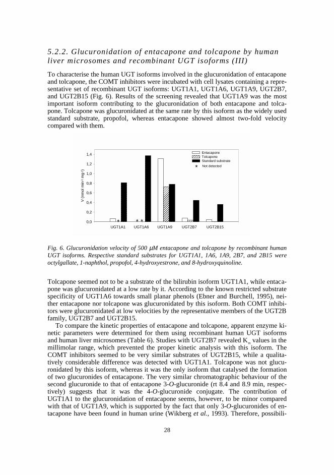
28
5.2.2. Glucuronidation of entacapone and tolcapone by humanliver microsomes and recombinant UGT isoforms (III)
To characterise the human UGT isoforms involved in the glucuronidation of entacaponeand tolcapone, the COMT inhibitors were incubated with cell lysates containing a repre-sentative set of recombinant UGT isoforms: UGT1A1, UGT1A6, UGT1A9, UGT2B7,and UGT2B15 (Fig. 6). Results of the screening revealed that UGT1A9 was the mostimportant isoform contributing to the glucuronidation of both entacapone and tolca-pone. Tolcapone was glucuronidated at the same rate by this isoform as the widely usedstandard substrate, propofol, whereas entacapone showed almost two-fold velocitycompared with them.
Fig. 6. Glucuronidation velocity of 500 µM entacapone and tolcapone by recombinant humanUGT isoforms. Respective standard substrates for UGT1A1, 1A6, 1A9, 2B7, and 2B15 wereoctylgallate, 1-naphthol, propofol, 4-hydroxyestrone, and 8-hydroxyquinoline.
Tolcapone seemed not to be a substrate of the bilirubin isoform UGT1A1, while entaca-pone was glucuronidated at a low rate by it. According to the known restricted substratespecificity of UGT1A6 towards small planar phenols (Ebner and Burchell, 1995), nei-ther entacapone nor tolcapone was glucuronidated by this isoform. Both COMT inhibi-tors were glucuronidated at low velocities by the representative members of the UGT2Bfamily, UGT2B7 and UGT2B15.
To compare the kinetic properties of entacapone and tolcapone, apparent enzyme ki-netic parameters were determined for them using recombinant human UGT isoformsand human liver microsomes (Table 6). Studies with UGT2B7 revealed Km values in themillimolar range, which prevented the proper kinetic analysis with this isoform. TheCOMT inhibitors seemed to be very similar substrates of UGT2B15, while a qualita-tively considerable difference was detected with UGT1A1. Tolcapone was not glucu-ronidated by this isoform, whereas it was the only isoform that catalysed the formationof two glucuronides of entacapone. The very similar chromatographic behaviour of thesecond glucuronide to that of entacapone 3-O-glucuronide (rt 8.4 and 8.9 min, respec-tively) suggests that it was the 4-O-glucuronide conjugate. The contribution ofUGT1A1 to the glucuronidation of entacapone seems, however, to be minor comparedwith that of UGT1A9, which is supported by the fact that only 3-O-glucuronides of en-tacapone have been found in human urine (Wikberg et al., 1993). Therefore, possibili-
V (
nmol
min
-1 m
g-1 )
0,0
0,2
0,4
0,6
0,8
1,0
1,2
1,4
����
� �������
�������
����������
������
���
EntacaponeTolcapone
��Standard substrate��
UGT1A1 UGT1A6 UGT1A9 UGT2B7 UGT2B15 *
*
* *
Not detected
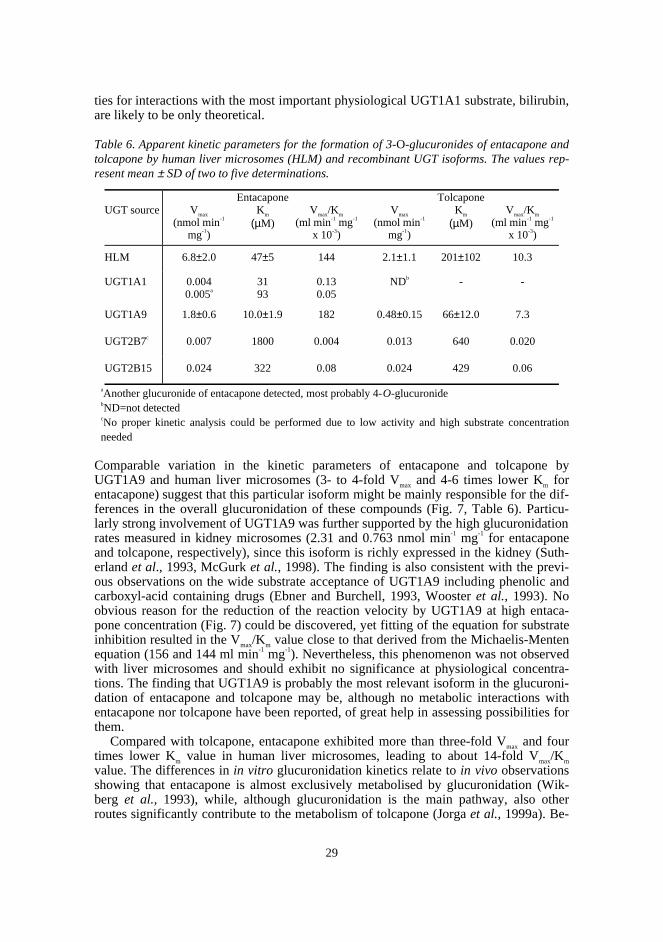
29
ties for interactions with the most important physiological UGT1A1 substrate, bilirubin,are likely to be only theoretical.
Table 6. Apparent kinetic parameters for the formation of 3-O-glucuronides of entacapone andtolcapone by human liver microsomes (HLM) and recombinant UGT isoforms. The values rep-resent mean ± SD of two to five determinations.
UGT source Vmax
(nmol min-1
mg-1)
EntacaponeKm
(µM)Vmax/Km
(ml min-1 mg-1
x 10-3)
Vmax
(nmol min-1
mg-1)
TolcaponeKm
(µM)Vmax/Km
(ml min-1 mg-1
x 10-3)
HLM 6.8±2.0 47±5 144 2.1±1.1 201±102 10.3
UGT1A1 0.0040.005a
3193
0.130.05
NDb - -
UGT1A9 1.8±0.6 10.0±1.9 182 0.48±0.15 66±12.0 7.3
UGT2B7c 0.007 1800 0.004 0.013 640 0.020
UGT2B15 0.024 322 0.08 0.024 429 0.06
aAnother glucuronide of entacapone detected, most probably 4-O-glucuronidebND=not detectedcNo proper kinetic analysis could be performed due to low activity and high substrate concentrationneeded
Comparable variation in the kinetic parameters of entacapone and tolcapone byUGT1A9 and human liver microsomes (3- to 4-fold Vmax and 4-6 times lower Km forentacapone) suggest that this particular isoform might be mainly responsible for the dif-ferences in the overall glucuronidation of these compounds (Fig. 7, Table 6). Particu-larly strong involvement of UGT1A9 was further supported by the high glucuronidationrates measured in kidney microsomes (2.31 and 0.763 nmol min-1 mg-1 for entacaponeand tolcapone, respectively), since this isoform is richly expressed in the kidney (Suth-erland et al., 1993, McGurk et al., 1998). The finding is also consistent with the previ-ous observations on the wide substrate acceptance of UGT1A9 including phenolic andcarboxyl-acid containing drugs (Ebner and Burchell, 1993, Wooster et al., 1993). Noobvious reason for the reduction of the reaction velocity by UGT1A9 at high entaca-pone concentration (Fig. 7) could be discovered, yet fitting of the equation for substrateinhibition resulted in the Vmax/Km value close to that derived from the Michaelis-Mentenequation (156 and 144 ml min-1 mg-1). Nevertheless, this phenomenon was not observedwith liver microsomes and should exhibit no significance at physiological concentra-tions. The finding that UGT1A9 is probably the most relevant isoform in the glucuroni-dation of entacapone and tolcapone may be, although no metabolic interactions withentacapone nor tolcapone have been reported, of great help in assessing possibilities forthem.
Compared with tolcapone, entacapone exhibited more than three-fold Vmax and fourtimes lower Km value in human liver microsomes, leading to about 14-fold Vmax/Km
value. The differences in in vitro glucuronidation kinetics relate to in vivo observationsshowing that entacapone is almost exclusively metabolised by glucuronidation (Wik-berg et al., 1993), while, although glucuronidation is the main pathway, also otherroutes significantly contribute to the metabolism of tolcapone (Jorga et al., 1999a). Be-
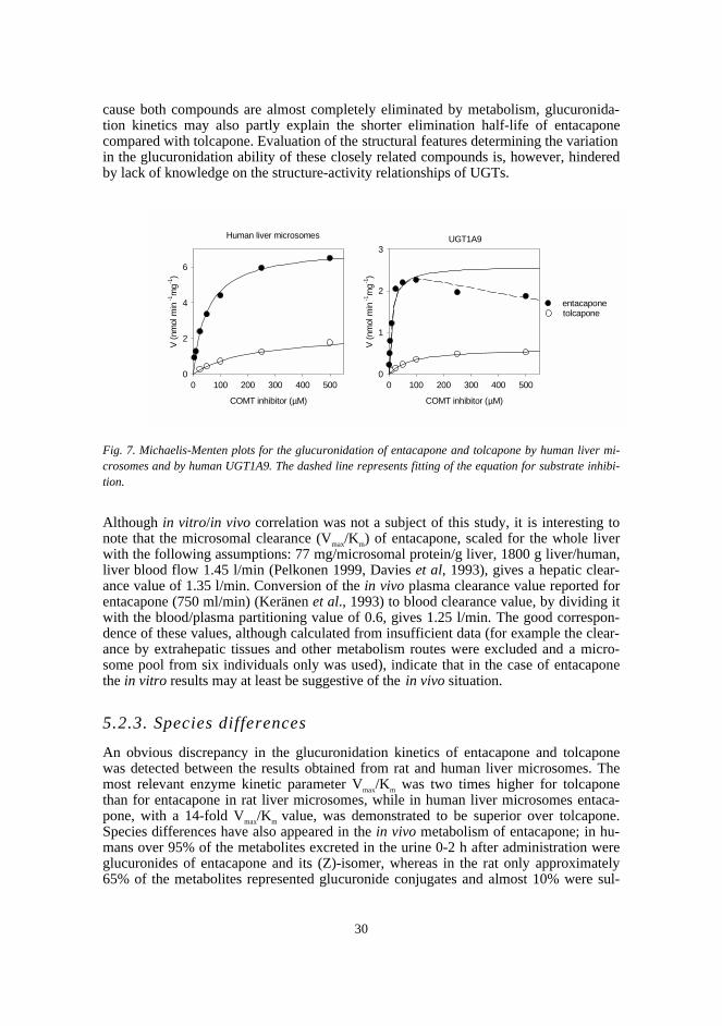
30
cause both compounds are almost completely eliminated by metabolism, glucuronida-tion kinetics may also partly explain the shorter elimination half-life of entacaponecompared with tolcapone. Evaluation of the structural features determining the variationin the glucuronidation ability of these closely related compounds is, however, hinderedby lack of knowledge on the structure-activity relationships of UGTs.
Fig. 7. Michaelis-Menten plots for the glucuronidation of entacapone and tolcapone by human liver mi-crosomes and by human UGT1A9. The dashed line represents fitting of the equation for substrate inhibi-tion.
Although in vitro/in vivo correlation was not a subject of this study, it is interesting tonote that the microsomal clearance (Vmax/Km) of entacapone, scaled for the whole liverwith the following assumptions: 77 mg/microsomal protein/g liver, 1800 g liver/human,liver blood flow 1.45 l/min (Pelkonen 1999, Davies et al, 1993), gives a hepatic clear-ance value of 1.35 l/min. Conversion of the in vivo plasma clearance value reported forentacapone (750 ml/min) (Keränen et al., 1993) to blood clearance value, by dividing itwith the blood/plasma partitioning value of 0.6, gives 1.25 l/min. The good correspon-dence of these values, although calculated from insufficient data (for example the clear-ance by extrahepatic tissues and other metabolism routes were excluded and a micro-some pool from six individuals only was used), indicate that in the case of entacaponethe in vitro results may at least be suggestive of the in vivo situation.
5.2.3. Species differences
An obvious discrepancy in the glucuronidation kinetics of entacapone and tolcaponewas detected between the results obtained from rat and human liver microsomes. Themost relevant enzyme kinetic parameter Vmax/Km was two times higher for tolcaponethan for entacapone in rat liver microsomes, while in human liver microsomes entaca-pone, with a 14-fold Vmax/Km value, was demonstrated to be superior over tolcapone.Species differences have also appeared in the in vivo metabolism of entacapone; in hu-mans over 95% of the metabolites excreted in the urine 0-2 h after administration wereglucuronides of entacapone and its (Z)-isomer, whereas in the rat only approximately65% of the metabolites represented glucuronide conjugates and almost 10% were sul-
COMT inhibitor (µM)
0 100 200 300 400 500
V (
nmol
min
-1m
g-1)
0
2
4
6
COMT inhibitor (µM)
0 100 200 300 400 500
V (
nmol
min
-1m
g-1)
0
1
2
3
Human liver microsomes UGT1A9
entacaponetolcapone
31
phates (Wikberg et al, 1993). Therefore, based on both in vivo and in vitro data, ratseems not to be a good animal model for predicting the glucuronidation of this type ofcompound in humans. This may concern all the compounds glucuronidated mainly byUGT1A9, since predominant in vivo excretion as sulphate conjugates and low glucu-ronidation activity in liver microsomes have been reported for propofol in the rat (LeGuellec et al., 1995, Simons et al., 1991), while in humans its main metabolic pathwayis glucuronidation, which is also supported by the high in vitro glucuronidation ratemeasured in liver microsomes (Simons et al., 1988, Le Guellec et al., 1995).
5.3. Substrate selectivity of rat and human S-COMT (IV,V)
The three dimensional structure of rat S-COMT has been solved by crystallography(Vidgren et al., 1994). In order to evaluate its applicability for modelling human S-COMT, enzyme kinetic parameters of 17 catecholic compounds were determined usingrecombinant rat and human S-COMT. For the first time substrate selectivity of rat andhuman S-COMT was determined under the same experimental conditions thus allowinga proper comparison. The enzymes exhibited very similar regioselectivities and both theKm and Vmax values, determined for the same catechols, were highly correlated (r2=0.988and r2=0.927, respectively, n=25, including regioisomers). This is in agreement with thehigh sequence homology (81%) of the enzymes and the almost completely conservedamino-acid residues in the active site (Vidgren and Ovaska, 1997). The absolute Vmax
values obtained using rat and human S-COMT were not comparable, since concentra-tions of active enzyme were not known, but the Km values showed 3-4 times higher fig-ures for the rat enzyme, on average. It is possible that the rat enzyme, although ex-pressing similar mechanisms for substrate selectivity, possesses a somehow looser ac-tive site structure thereby decreasing the affinity of substrate binding.
Fig. 8. Correlation of reaction velocities (nmol min-1 mg-1) between human and rat S-COMT(r2=0.858, n=30). The dashed line represents the correlation without gallic acid, pyrogallol andcatechin (r2=0.960, n=27).
Human S-COMT
0 5 10 15 20 25 30 35
Rat
S-C
OM
T
0
10
20
30
40
50
60
70
80
Pyrogallol
Gallic acid
Catechin
32
When rat and human S-COMT activity was screened at one relatively high concentra-tion (IV, 500 µM) discarding three outliers (gallic acid, pyrogallol and catechin) fromthe analysis improved correlation from r2=0.858 (n=30) to r2=0.960 (n=27) (Fig. 8). Py-rogallol derivatives are low-affinity inhibitors of COMT and at the high concentrationused they inhibited their own methylation. The higher affinity towards the human en-zyme appeared as a more pronounced inhibition of the human enzyme than of the ratenzyme, which probably explains the deviation from the correlation. Despite the kineticdifference, rat S-COMT proved to be an excellent model for the human enzyme therebyvalidating its use as a template in computer-aided molecular modelling.
5.4. Methylation of structurally diverse compounds byhuman S-COMT (V)
5.4.1. Enzyme kinetic parameters
Methylation of 46 structurally diverse catechols was investigated using recombinanthuman S-COMT to obtain data for structure-activity analysis. The enzyme kinetic pa-rameters could be determined for 41 compounds including simple substituted andphysiological catechols and catecholic drugs and drug candidates (Tables 7-8).
Under the conditions used, no methylation of 3,5-dinitrocatechol, 2,3-dihydroxybenzoic acid, tolcapone, entacapone, or apomorphine was observed. Two re-gioisomeric products could be detected from many of the compounds, yet the analyticalmethod utilising 14C-AdoMet did not allow elucidation of the metabolite structures.However, the enzyme kinetic parameters were determined separately for both isomerswhen possible and useful experimental data about regioselectivity, that supported ob-servations derived from molecular modelling, could be gathered. Pyrogallol and cate-chol showed the highest Vmax values (53.2 and 48.9 nmol min-1 mg-1), while the lowestmeasurable Vmax was 0.15 nmol min-1 mg-1 for 3-nitrocatechol. Among the series of sub-stituted catechols in Table 7, catechol exhibited the highest Km value (50 µM). Thisvalue was more than three orders of magnitude higher than the lowest value obtained(29 nM, for tetrachlorocatechol). Nevertheless, many endogenous and other catecholswith pharmacological activity exhibited several-fold higher Km values than catechol(Table 8). The Vmax/Km values were highest for tetrachlorocatechol and 2,3-dihydroxynaphthalene.Among the endogenous catechols 2- and 4-hydroxyestradiols were superior COMT sub-strates; they exhibited high Vmax values and low Km values leading to, for instance, ap-proximately 60- and 10-fold Vmax/Km values compared with dopamine, respectively.Among drugs used in the treatment of Parkinson’s disease, entacapone and tolcaponewere not methylated in vitro and L-dopa and carbidopa were poor substrates with re-spective Vmax/Km values of 0.053 and 0.024 ml min-1 mg-1, while the other DDC inhibitor,benserazide, showed Vmax and Km values equal to those of catechol. Differences foundbetween carbidopa and benserazide are in agreement with the previous in vitro resultsobtained with COMT purified from pig liver (Hagan et al., 1980, Gordonsmith et al.,1982). In vitro results also support the in vivo observations concerning the effect ofCOMT inhibition by tolcapone on the pharmacokinetics of these compounds; no inter-actions were detected with carbidopa, whereas the bioavailability of benserazide wasenhanced by tolcapone at large L-dopa/benserazide doses (Sedek et al., 1993, Jorga et
33
al., 1999b, Jorga et al., 1999c). Among the other clinically used drugs dobutamine wasa relatively good and isoprenaline a moderately good COMT substrate, whereas α-methyldopa showed the highest Km value (1.8 mM) among all 41 compounds. Thesefindings are in agreement with the knowledge on their preferential metabolism routes invivo (Table 1). Even though apomorphine has been reported to be a good substrate of ratliver COMT in vitro (Cannon et al., 1972), and its elimination half-life in rats is in-creased by tolcapone (Coudore et al., 1997), no methylation was detected under the ex-perimental conditions applied. In vitro observations may not always correspond to thein vivo situation, as exemplified by tolcapone that, despite showing no implications formethylation in our study, has been reported to undergo biotransformation to 3-O-tolcapone in vivo (e.g. Dingemanse et al., 1995). On the other hand, in a study on themetabolism of intravenously administered apomorphine in a Parkinsonian patient, nomethylated products were found in plasma or urine (van der Geest et al., 1997). In ourstudy, apomorphine was demonstrated to inhibit the methylation of DHBA (Ki=240 µMfor competitive inhibition) and thus shown to be capable of binding to the enzyme.
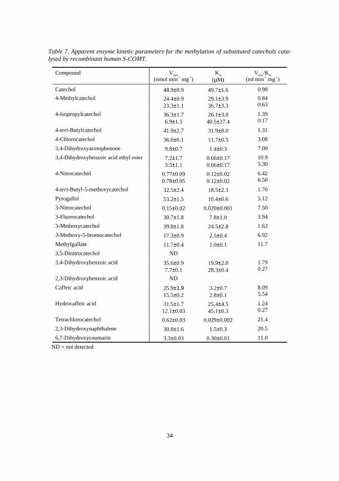
34
Table 7. Apparent enzyme kinetic parameters for the methylation of substituted catechols cata-lysed by recombinant human S-COMT.
Compound Vmax
(nmol min-1 mg-1)Km
(µM)Vmax/Km
(ml min-1 mg-1)
Catechol 48.9±0.9 49.7±1.6 0.98
4-Methylcatechol 24.4±0.923.3±1.1
29.1±3.936.7±3.3
0.840.63
4-Isopropylcatechol 36.3±1.76.9±1.5
26.1±3.040.5±27.4
1.390.17
4-tert-Butylcatechol 41.9±2.7 31.9±8.0 1.31
4-Chlorocatechol 36.0±0.1 11.7±0.5 3.08
3,4-Dihydroxyacetophenone 9.8±0.7 1.4±0.3 7.00
3,4-Dihydroxybenzoic acid ethyl ester 7.2±1.73.5±1.1
0.66±0.170.66±0.17
10.95.30
4-Nitrocatechol 0.77±0.090.78±0.05
0.12±0.020.12±0.02
6.426.50
4-tert-Butyl-5-methoxycatechol 32.5±2.4 18.5±2.3 1.76
Pyrogallol 53.2±1.5 10.4±0.6 5.12
3-Nitrocatechol 0.15±0.02 0.020±0.001 7.50
3-Fluorocatechol 30.7±1.8 7.8±1.0 3.94
3-Methoxycatechol 39.8±1.8 24.5±2.8 1.62
3-Methoxy-5-bromocatechol 17.3±0.9 2.5±0.4 6.92
Methylgallate 11.7±0.4 1.0±0.1 11.7
3,5-Dinitrocatechol ND
3,4-Dihydroxybenzoic acid 35.6±0.97.7±0.1
19.9±2.028.3±0.4
1.790.27
2,3-Dihydroxybenzoic acid ND
Caffeic acid 25.9±1.915.5±0.2
3.2±0.72.8±0.1
8.095.54
Hydrocaffeic acid 31.5±1.712.1±0.03
25.4±4.545.1±0.3
1.240.27
Tetrachlorocatechol 0.62±0.03 0.029±0.002 21.4
2,3-Dihydroxynaphthalene 30.8±1.6 1.5±0.3 20.5
6,7-Dihydroxycoumarin 3.3±0.03 0.30±0.01 11.0
ND = not detected

35
Table 8. Apparent enzyme kinetic parameters for the methylation of some endogenous cate-chols, catecholic drugs and drug candidates catalysed by recombinant human S-COMT.
Compound Vmax
(nmol min-1 mg-1)Km
(µM)Vmax/Km
(ml min-1 mg-1)
L-Noradrenaline 33.2±0.8 256±20 0.13
3,4-Dihydroxymandelic acid 43.7±2.0 132±11 0.33
3,4-Dihydroxyphenylglycol 37.2±1.4 46.4±4.7 0.80
L-Adrenaline 44.0±1.0 132±13 0.33
Dopamine 35.9±1.4 188±16 0.19
5-Hydroxydopamine 46.7±1.1 61.6±4.1 0.76
6-Hydroxydopamine 25.3±2.0 244±32 0.10
3,4-Dihydroxyphenyl acetic acid 34.0±2.24.0±0.1
68.8±10.853.7±4.0
0.490.074
2-Hydroxyestradiol 32.4±2.613.7±1.8
3.7±1.14.2±2.2
8.763.26
4-Hydroxyestradiol 31.5±2.5 12.4±2.6 2.54
α-Methyldopa 21.4±0.72.6±0.3
1789±1081170±309
0.0120.002
L-dopa methylester 44.2±0.7 51.1±2.9 0.86
L-dopa 29.8±0.9 564±28 0.053
S(-)-Carbidopa 14.3±0.8 599±63 0.024
Benserazide 41.8±3.6 50.2±7.2 0.83
Tolcapone ND
Entacapone ND
Isoprenaline 25.8±0.63.1±0.3
145±8.5170±31
0.180.018
Dobutamine 29.0±0.96.9±0.5
23.7±3.128.6±7.5
1.220.24
1-Methyl-6,7-dihydroxy-1,2,3,4-tetrahydroisoquinoline
18.3±0.910.6±0.1
29.4±3.736.2±0.2
0.620.29
SKF 38393 5.0±0.0410.5±0.5
67.2±1.273.6±8.3
0.0740.14
Dihydrexidine 10.1±2.310.5±6.2
25.0±14.553.1±50.6
0.400.20
Apomorphine ND
ND = not detected
36
5.4.2. Effect of molecular structure on binding affinity and r e-activity
Combining experimental data, molecular modelling and calculated data on substituentphysico-chemical properties, mechanisms affecting binding affinity and reactivity couldbe elucidated. The most important substituent effects identified in this study were theelectronic effect on catecholic hydroxyls, interactions with the surroundings of the cate-chol binding groove, steric hindrance, especially by ortho-substituents, and some otherortho-effects.
In this study, the most important factor affecting reactivity appeared to be the elec-tron-withdrawing effect of substituents. The electronic effect of substituents is bestdemonstrated by a set of small 4-substituted compounds that exhibit a minimum amountof direct interactions with the amino acid residues in the binding site (the first eightcompounds in Table 7). Their Vmax values were highly correlated with parameters de-scribing electronic effects of the substituents. Hammet σ - value, which quantitates theeffect of a 4-substituent on the ionisation of phenol, explained 94% of the variation inthe Vmax values, and even better correlation (r2=0.98) was achieved when the lower mo-lecular electrostatic potentials (MEP) were calculated for the monoanionic forms of thecompounds. A semiempirical study on COMT inhibition has suggested that the reasonfor lowered reactivity by electronegative substituents is stabilisation of the catecholateanion, formed via proton transfer to the amino group of Lys 144 (Ovaska andYliniemelä, 1998). The catecholate anion-enzyme complex, rather than the transitionstate complex, is stabilised leading to a high energy barrier for methylation. As expectedon the basis of QSAR studies on COMT inhibition (Taskinen et al., 1989, Lotta et al.,1992), results of this study showed that binding affinity was increased by electronega-tive substituents as well. The Hammet σ - value and MEP explained 94 and 97%, re-spectively, of the variation in log(1/Km) values among the eight 4-substituted catechols.Direct interactions of the catechols with the surroundings of the binding site were eluci-dated by superimposing the compounds on 3,5-dinitrocatechol in the crystal structure ofrat S-COMT. Favourable and unfavourable interactions with the hydrophobic aminoacid residues lining the catechol binding site could be observed. In general, hydrophobicsubstituents increased the affinity, as exemplified by the lowered Km values of 4-alkylsubstituted catechols and 2,3-dihydroxynaphathlene compared with catechol. Themost favourable interactions were formed with 2-hydroxyestradiol that has a flat fusedring structure filling optimally the binding pocket (Fig. 9). The higher Km values ob-tained for other compounds with a fused ring structure compared with hydroxyestradiolswere obviously due to the unfavourable interactions caused by the ionised amino group.In apomorphine the amino group is in such an orientation that it should not affect thebinding. However, its bulky ortho-substituent overlaps severely one of the surroundingresidues (Trp143). The methyl group of AdoMet is also overlapped, which may causethe observed loss of reactivity. Inhibition of the COMT-catalysed methylation of DHBAby apomorphine revealed that binding of apomorphine is possible, yet conformationalchanges of the enzyme are required.
The positively charged side chain of catecholamines (e.g. dopamine) has been sug-gested to interact unfavourably with the hydrophobic amino acids in the active sitethereby lowering the affinity (Lotta et al., 1995). However, the side chain is flexible andmay be in an orientation in which interactions are avoided. In this study, hydrocaffeicacid exhibited almost one order of magnitude lower Km compared with that of dopa-mine, although the flexibility of the oppositely charged side chains of these compoundsshould be similar. Fitting of 27 low energy conformers of dopamine to the active site
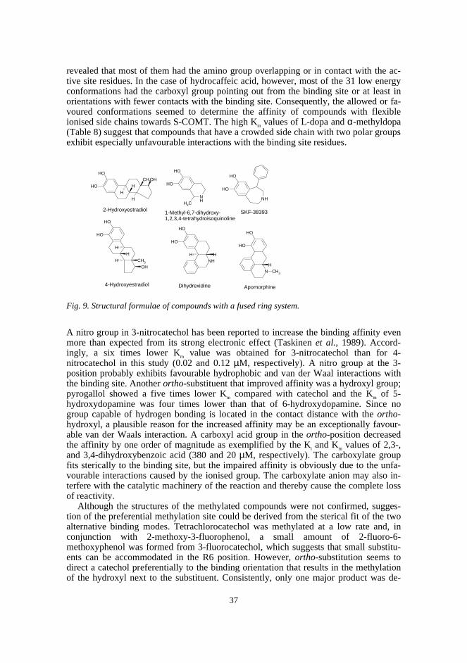
37
revealed that most of them had the amino group overlapping or in contact with the ac-tive site residues. In the case of hydrocaffeic acid, however, most of the 31 low energyconformations had the carboxyl group pointing out from the binding site or at least inorientations with fewer contacts with the binding site. Consequently, the allowed or fa-voured conformations seemed to determine the affinity of compounds with flexibleionised side chains towards S-COMT. The high Km values of L-dopa and α-methyldopa(Table 8) suggest that compounds that have a crowded side chain with two polar groupsexhibit especially unfavourable interactions with the binding site residues.
Fig. 9. Structural formulae of compounds with a fused ring system.
A nitro group in 3-nitrocatechol has been reported to increase the binding affinity evenmore than expected from its strong electronic effect (Taskinen et al., 1989). Accord-ingly, a six times lower Km value was obtained for 3-nitrocatechol than for 4-nitrocatechol in this study (0.02 and 0.12 µM, respectively). A nitro group at the 3-position probably exhibits favourable hydrophobic and van der Waal interactions withthe binding site. Another ortho-substituent that improved affinity was a hydroxyl group;pyrogallol showed a five times lower Km compared with catechol and the Km of 5-hydroxydopamine was four times lower than that of 6-hydroxydopamine. Since nogroup capable of hydrogen bonding is located in the contact distance with the ortho-hydroxyl, a plausible reason for the increased affinity may be an exceptionally favour-able van der Waals interaction. A carboxyl acid group in the ortho-position decreasedthe affinity by one order of magnitude as exemplified by the Ki and Km values of 2,3-,and 3,4-dihydroxybenzoic acid (380 and 20 µM, respectively). The carboxylate groupfits sterically to the binding site, but the impaired affinity is obviously due to the unfa-vourable interactions caused by the ionised group. The carboxylate anion may also in-terfere with the catalytic machinery of the reaction and thereby cause the complete lossof reactivity.
Although the structures of the methylated compounds were not confirmed, sugges-tion of the preferential methylation site could be derived from the sterical fit of the twoalternative binding modes. Tetrachlorocatechol was methylated at a low rate and, inconjunction with 2-methoxy-3-fluorophenol, a small amount of 2-fluoro-6-methoxyphenol was formed from 3-fluorocatechol, which suggests that small substitu-ents can be accommodated in the R6 position. However, ortho-substitution seems todirect a catechol preferentially to the binding orientation that results in the methylationof the hydroxyl next to the substituent. Consistently, only one major product was de-
OH
OH
OHCH3
H
H
H
NHCH3
OH
OH
NH
OH
OH
OH
OH
OH
CH3
H
H
H NH
OH
OH
H
N
OH
OH
HCH3
2-Hydroxyestradiol 1-Methyl-6,7-dihydroxy-1,2,3,4-tetrahydroisoquinoline
SKF-38393
4-Hydroxyestradiol Dihydrexidine Apomorphine
H
38
tected for pyrogallol derivatives and other 3-substituted catechols. One metabolite wasformed also from 4-hydroxyestradiol, while two metabolites with similar affinity weredetected for 2-hydroxyestradiol. The relatively bulky ortho-substituent in 4-hydroxyestradiol seems to direct the binding to the orientation that leads to the meth-ylation of the hydroxyl next to it. Different kinds of interactions of the side chain withthe binding site residues at different binding modes can explain the regioselectivity ofmany compounds. For example, catecholamines obviously bind predominantly in theorientation resulting in the m-methylation because of less unfavourable interactions inthis orientation, as previously suggested by Lotta et al. (1995). Compared with the stericfactors, the electronic effects of the substituents seem to have a minor contribution toregioselectivity. In studies on ring-fluorinated catecholamines, ortho-fluorine has beensuggested to facilitate methylation of the adjacent hydroxyl by increasing its ionisation(Firnau et al., 1981, Creveling et al., 1981, Thakker et al., 1986). However, it is nowknown that electronegative substituents conversely decrease reactivity, and the reasonfor the altered regioselectivity may be the steric ortho-effect discussed above.
5.4.3. Predictive models
The most decisive factor determining the reactivity of a compound was the electroniceffect of the substituents on the catechol hydroxyls. The volume of an isoenergy MEPwas used to describe the electronic effects, because this parameter can be readily calcu-lated for any substrate. In the case of neutral compounds (n=20), MEP, calculated forthe catecholate monoanion exhibiting the lower minimum, could explain over 93% ofthe variation in the Vmax values. The correlation clearly deteriorated when compoundscontaining ionisable side chains were included (r2=0.705, n=38). However, ionisablegroups are expected to affect the acidity of the catechol hydrogens to a small extentonly, and the Vmax of this kind of compound can be predicted to be near as that of unsub-stituted catechol. Deterioration of correlation between MEP and Vmax was probablycaused by the poor steric fit of some compounds to the active site. Therefore, for pre-dicting Vmax both calculation of the electronic effect and modelling of the compound tothe active site are required.
Besides the electronic effect of the substituents, hydrophobicity of a catechol ap-peared to be an important factor in predicting affinity. LogKow values were calculated bythe LOGKOW method, because the method is rapid and has been shown to correlatewell with experimental values (Meylan and Howard, 1995). In the two parameters’ re-gression model (2), the logKow values were calculated for the compounds with their sidechain in ionised form.
(2) log(1/Km) = -0.30(±0.03) MEPvol + 0.21(±0.03) logKow + 6.99(±0.23)
The correlation between the calculated and observed log(1/Km) values was r2=0.833(n=38), and the leave-five-out cross-validation gave r2=0.782. Although the model ex-cludes for example the pronounced unfavourable effects of polar groups caused bysteric crowding (e.g. α-methyldopa) and the increased affinity caused by an ortho-hydroxyl, the affinity of a catechol may be predicted with reasonable accuracy. Moreo-ver, most steric and conformational effects may be evaluated by fitting the compound tothe crystal structure of rat S-COMT.
The predictive models constructed in this study, supported by modelling of the activesite, may be utilised in evaluating the interactions between endogenous and exogenous
39
catechols (e.g. dietary compounds and drugs) and in designing new catecholic drugswith controlled metabolic methylation.
Conclusions
An HPTLC method combining densitometry and radioactivity measurement was devel-oped for the study of the in vitro glucuronidation of nitrocatechols. These types of com-pounds may be excellent substrates of rat liver UGTs, but the position and nature ofsubstituents greatly affect their glucuronidation. Nitrocatechols with large substituents,such as entacapone and tolcapone, exhibited one order of magnitude lower Vmax/Km val-ues compared with smaller ones in microsomes from creosote-treated rats. Tolcaponeexhibited a two-fold Vmax/Km value compared with entacapone. In contrast, entacaponewas shown to be a clearly better UGT substrate in human liver microsomes suggestingthat the rat is a poor model for predicting the glucuronidation of these compounds inhumans. Since both entacapone and tolcapone are rapidly glucuronidated in vivo, the14-fold Vmax/Km for entacapone in human liver microsomes may explain some of its ap-proximately seven times shorter elimination half-life. Both compounds, but particularlyentacapone, appeared to be excellent substrates of UGT1A9, knowledge which may beutilised in assessing the risks for metabolic interactions. More knowledge on the struc-ture-activity relationships of individual UGT isoforms is required for the evaluation ofthe structural features turning these COMT inhibitors to such good UGT substrates andcausing the differences between them.
A radiochemical HPLC method for the assay of COMT activity towards structurallydiverse catechols was developed. Enzyme kinetic parameters were determined for 41catechols using human S-COMT. Of all the compounds, tetrachlorocatechol and 2,3-dihydroxynaphthalene exhibited the highest Vmax/Km values, while hydroxyestrogenswere the most specific physiological substrates. Catecholic drugs showed variablemethylation ability. For instance among drugs used in the treatment of Parkinson’s dis-ease, entacapone and tolcapone were not detectably methylated, L-dopa and carbidopashowed low affinity towards COMT, while benserazide appeared to be a good substrateof COMT. Electronic factors were found to be the most decisive substituent effect de-termining the reactivity of the catecholic hydroxyls. Electron-withdrawing substituentsnot only decreased reactivity but also increased affinity. The affinity was improved byhydrophobic substituents as well, whereas hydrophilic side chains deteriorated it. In thecase of substrates with flexible side chains the favoured conformations seemed to de-termine the affinity. As demonstrated with apomorphine, bulky ortho-substituents de-creased the affinity and reactivity. In contrast, an ortho-hydroxyl enhanced the meth-ylation. An ortho-substituent generally directed the methylation to the hydroxyl next toit. Predictive models were constructed for reactivity, comprising the electronic effect oncatechol hydroxyls, and for affinity, including also a factor describing hydrophobicity.The models may be utilised, supported by modelling of the active site, in evaluating in-teractions between catecholic compounds or in designing catecholic drugs with con-trolled metabolic methylation.
40
Acknowledgements
This study was carried out mainly in the Pharmacutical Chemistry Division, Departmentof Pharmacy, University of Helsinki, during the years 1995-2000. Part of the work withhuman recombinant UGT isoforms was carried out in Ninewells Hospital and MedicalSchool, University of Dundee, Scotland.
My sincerest gratitude belongs to Professor Jyrki Taskinen, who suggested the sub-ject of this dissertation and significantly contributed to the studies with wise ideas andcomments. His long experience and wide knowledge in different branches of science aswell as never-failing enthusiasm, which was more like passion in the case of COMT,made the completion of this study possible.
I express my sincerest appreciation to Professor Olavi Pelkonen and Docent SeppoAuriola for constructive criticism of the manuscript, which lead to its improvement.
I am deeply grateful to the whole staff of the Pharmaceutical Chemistry Division forproviding a very pleasant and understanding environment in which to work. DocentHannele Salomies deserves extra thanks for being real support pillar, both in scientificand other concerns. I extend my warm thanks to Mrs Inkeri Huttunen, Helena Keski-Hynnilä (Lic. Pharm.), Leena Luukkanen (Lic. Pharm.) and Tiia Kuuranne (M. Sc.,Pharm.) for their friendship and support. I express warm appreciation to Maija Kivimaa(M. Sc., Pharm.) for excellent technical assistance during her pro gradu studies and toJukka-Pekka Salo (Lic. Pharm.) for kindly drawing Fig. 3.
I am most grateful to Professor Brian Burchell, University of Dundee, for giving methe opportunity to visit his laboratory, world-famous in UGT research. I am especiallythankful to Dr. Brian Ethell for pleasant and successful collaboration, and for takingcare of practical arrangements. I warmly thank Docent Eivor Elovaara for providingwith the rat liver microsomes and guiding me in the first steps of performing enzymeassays, as well as for her continuous encouragement throughout the work. I thank thestaff of Molecular Biology and Target Protein Research, Orion Pharma, for teaching methe basics of molecular biology during my PhD studies. Special thanks are due to Do-cent Ismo Ulmanen, former head of the group, whose great expertise and friendly andrelaxed attitude will long be remembered, and to Mrs Raija Savolainen, the superb re-search assistant, who was never too busy to lend a helping hand.
I express my gratitude to Docent Tom Wikberg for valuable advice and encourage-ment at the final stages of this work. I also thank Mr. Harri Salonen for revising the la n-guage of the manuscript.
Finally, my warmest thanks belong to friends and family members, especially Mr.Vesa-Pekka Pihlavisto, for continuous support and understanding.
This work was part of the project “ Development and prevalidation of predictivemodels for catechol drug conjugation and their evaluation for rational drug design”(BMH4-CT97-2621) supported by the Biotechnology and Biological Sciences ResearchCouncil, the Commission of the European Communities. Financial support provided bythe Council and by the Finnish Pharmaceutical Society is gratefully acknowledged.
41
References
Abernethy, D.R., Divoll, M., Greenblatt, D.J., and Ameer, B. Obesity, sex, and acetaminophen
disposition. Clin. Pharmacol. Ther. 31 (1982) 783-790.
Assal, F., Spahr, L., Hadengue, A., Rubbici-Brandt, L., and Burkhard, P.R. Tolcapone and fulminant
hepatitis, Lancet 352 (1998) 958.
Axelrod, J. and Tomchick, R. J. Enzymatic O-methylation of epinephrine and other catechols. Biol.
Chem. 233 (1958) 702-705.
Bansal, S.K. and Gessner, T. A unified method for the assay of uridine diphospho-glucuronyltransferase
activities toward various aglycones using uridine diphospho[U-14C]glucuronic acid. Anal. Biochem. 109
(1980) 321-329.
Bates, G.W., Edman, C.D., Porter, J.C., Johnston, J.M, and MacDonald, P.C. An assay for human
erythrocyte catechol-O-methyltransferase activity using a catechol estrogen as the substrate. Clin. Chim.
Acta 94 (1979) 63-71.
Battaglia, E., Pritchard, M., Ouzzine, M., Fournel-Gigleux, S., Radominska, A., Siest, G., and Magdalou,
J. Chemical modification of human UDP-glucuronosyltransferase UGT1*6 by diethyl pyrocarbonate:
possible involvement of a histidine residue in the catalytic process. Arch. Biochem. Biophys. 309 (1994a)
266-272.
Battaglia, E., Senay, C., Fournel-Gigleux, S., Herber, R., Siest, G., and Magdalou, J. The chemical
modification of human liver UDP-glucuronosyltransferase UGT1*6 reveals the involvement of a
carboxyl group in catalysis. FEBS Lett. 346 (1994b) 146-150.
Beaulieu, M., Levesque, E., Hum, D.W., and Bélanger, A. Isolation and characterization of a novel
cDNA encoding a human UDP-glucuronosyltransferase active on C19 steroids. J. Biol. Chem. 271 (1996)
22855-22862.
Bertocci, B., Miggiano, V., Da Prada, M., Dembic, Z., Lahm, H.-W., and Malherbe, P. Human catechol-
O-methyltransferase: Cloning and expression of the membrane-associated form. Proc. Natl. Acad. Sci.
USA 88 (1991) 1416-1420.
Boadi, W.Y., Urbach, J., Barnea, E.R., Brandes, J.M., and Yannai, S. In vitro effect of mercury on aryl
hydrocarbon hydroxylase, quinone reductase, catecholamine-O-methyltransferase and glucose-6-
phosphate dehydrogenase activities in term human placenta. Pharmacol. Toxicol. 68 (1991) 317-321.
Bochner, F., Chen, Z.R., Young, G., Somogyi, A., and Reynolds, G. Codeine disposition and metabolism:
comparison in young and elderly subjects. Eur. J. Pharmacol. 183 (1990) 630-631 (Abstract).
Bock, K.W., Lipp, H.-P., and Bock-Henning, B.S. Induction of drug-metabolising enzymes by
xenobiotics. Xenobiotica 20 (1990) 1101-1111.
Bock, K.W., Gschaidmeier, H., Seidel, A., Baird, S., and Burchell, B. Mono- and diglucuronide
formation from chrysene and benzo(a)pyrene phenols by 3-methylcholanthrene-inducible phenol UDP-
glucuronosyltransferase (UGT1A1). Mol. Pharmacol. 42 (1992) 613-618.
42
Bonifati, V. and Meco, G. New, selective catechol-O-methyltransferase inhibitors as therapeutic agents in
Parkinson’s disease. Pharmacol. Ther. 81 (1999) 1-36.
Borchardt, R.T. Catechol O-methyltransferase 1. Kinetics of tropolone inhibition. J. Med. Chem. 16
(1973) 377-382.
Borchardt, R.T. A rapid spectrophotometric assay for catechol-O-methyltransferase. Anal. Biochem. 58
(1974) 382-389.
Borchardt, R.T., Hegazi, M.F., and Schowen, R.L. Determination of O-methylated metabolites of
catecholamines using high-performance liquid chromatography and electrochemical detection. J.
Chromatogr. 152 (1978) 255-259.
Borgulya, J., Bruderer, H., Bernauer, K., Zürcher, G., and Da Prada, M. Catechol-O-methyltransferase-
inhibiting pyrocatechol derivatives: synthesis and structure-activity studies. Helv. Chim. Acta 72 (1989)
952-968.
Bosma, P.J., Seppen, J., Goldhoorn, B., Bakker, C., Elferink, R.P.J.O., Chowdhury, J.R., Chowdhury,
N.R., and Jansen, P.L.M. Bilirubin UDP-glucuronosyltransferase 1 is the only relevant bilirubin
glucuronidating isoform in man. J. Biol. Chem. 269 (1994) 17960-17964.
Boudiková, B., Szumlanski, C., Maidak, B., and Weinshilboum, R. Human liver catechol-O-
methyltransferase pharmacogenetics. Clin. Pharmacol. Ther. 48 (1990) 381-389.
Boulton, A.A. and Eisenhofer, G. Catecholamine metabolism, in Advances in pharmacology vol 42,
Catecholamines bridging basic science with clinical medicine, Goldstein, D.S., Eisenhofer, G., and Mc
Carty, R. (Eds.), Academic Press, San Diego, CA (1998) 273-292.
Boutin, J.A., Thomassin, J., Siest, G., and Cartier, A. Heterogeneity of hepatic microsomal UDP-
glucuronosyltransferase activities. Biochem. Pharmacol. 34 (1985) 2235-2249.
Bradford, M.M. A rapid and sensitive method for quantitation of microgram quantities of protein utilizing
the principle of protein-dye binding. Anal. Biochem. 72 (1976) 248-254.
Burchell, B., Brierley, C.H., and Rance, D. Specificity of human UDP-glucuronosyltransferases and
xenobiotic glucuronidation. Life Sci. 57 (1995) 1819-1831.
Bäckström, R., Honkanen, E., Pippuri, A., Kairisalo, P., Pystynen, J., Heinola, K., Nissinen, E., Lindén,
I.-B., Männistö, P.T., Kaakkola, S., and Pohto, P. Synthesis of some novel potent and selective catechol
O-methyltransferase inhibitors. J. Med. Chem. 32 (1989) 841-846.
Cannon, J.G., Smith, R.V., Modiri, A., Sood, S.P., Borgman, R.J., Aleem, M.A., and Long, J.P. Centrally
acting emetics. 5. Preparation and pharmacology of 10-hydroxy-11 methoxyaporphine (isoapocodeine).
In vitro enzymatic methylation of apomorphine. J. Med. Chem. 15 (1972) 273-276.
Chen, F., Ritter, J.K., Wang, M.G., McBride, O.W., Lubet, R.A., and Owens, I.S. Characterization of a
cloned human dihydrotestosterone/androstanediol UDP-glucuronosyl-transferase and its comparison to
other steroid isoforms. Biochemistry 32 (1993) 10648-10657.
Chen, C.H., Lee, Y.R., Wei, F.C., Koong, F.J., Hwu, H.G., and Hsiao, K.J. Association study of NlaIII
and MspI genetic polymorphisms of catechol-O-methyltransferase gene and susceptibility to
schizophrenia. Biol. Psych. 41 (1997) 985-987.
43
Cheng, Z.O., Rios, G.R., King, C.D., Coffman, B.L., Green, M.D., Mojarrabi, B., Mackenzie P.I., and
Tephly, T.R. Glucuronidation of catechol estrogens by expressed human UDP-glucuronosyltransferases
(UGTs) 1A1, 1A3, and 2B7. Toxicol. Sci. 45 (1998a) 52-57.
Cheng, Z.O., Radominska-Pandya, A., and Tephly, T.R. Cloning and expression of human UDP-
glucuronosyltransferase (UGT) 1A8. Arch. Biochem. Biophys. 356 (1998b) 301-305.
Cheng, T.L., Wei, S.L., Chen, B.M., Chern, J.W., Wu, M.F., Liu, P.W., and Roffler, S.R. Bystander
killing of tumour cells by antibody-targeted enzymatic activation of a glucuronide prodrug. Br. J. Cancer
79 (1999) 1378-1385.
Ciotti, M. and Owens, I.S. Evidence for overlapping active sites for 17α-ethinylestradiol and bilirubin in
the human major bilirubin UDPglucuronosyltransferase. Biochemistry 35 (1996) 10119-10124.
Ciotti, M., Marrone, A., Potter, C., and Owens, I.S. Genetic polymorphism in the human UGT1A6
(planar phenol) UDP-glucuronosyltransferase – pharmacological implications. Pharmacogenetics 7
(1997) 485-495.
Ciotti, M., Cho, J.W., George, J., and Owens, I.S. Required buried α-helical structure in the bilirubin
UDP-glucuronosyltransferase, UGT1A1, contains a nonreplaceable phenylalanine. Biochemistry 37
(1998) 11018-11025.
Clarke, D.J. and Burchell, B. The uridine diphosphate glucuronosyltransferase multigene family: function
and regulation, in Conjugation-deconjugation reactions in drug metabolism and toxicity, Kauffman, F.
(Ed.) Springer-Verlag Berlin-Heidelberg (1994) 3-43.
Clarke, D.J., Moghrabi, N., Monaghan, G., Cassidy, A., Boxer, M., Hume. R., and Burchell, B. Genetic
defects of the UDP-glucuronosyltransferase-1 (UGT1) gene that cause familial non-haemolytic
unconjugated hyperbilirubinaemias. Clin. Chim. Acta 266 (1997) 63-74.
Clarke, D.J. The structure and function of the UDP-glucuronosyltransferase gene family, in Advances in
pharmacology, vol 42, Catecholamines bridging basic science with clinical medicine, Academic Press,
San Diego, CA (1998) 335-338.
Coffman, B.L., Tephly, T.R., Irshaid, Y.M., Green, M.D., Smith, C., Jackson, M.R., Wooster, R., and
Burchell, B. Characterization and primary sequence of a human hepatic estriol UDP-
glucuronosyltransferase. Arch. Biochem. Biophys. 281 (1990) 170-175.
Coffman, B.L., Rios, G.R., King, C.D., and Tephly, T.R. Human UGT2B7 catalyzes morphine
glucuronidation. Drug Metab. Dispos. 25 (1997) 1-4.
Coffman, B.L., King, C.D., Rios, G.R., and Tephly, T.R. The glucuronidation of opioids, other
xenobiotics, and androgens by human UGT2B7Y(268) and UGT2B7H(268). Drug Metab. Dispos. 26
(1998) 73-77.
Cornish-Bowden, A. Fundamentals of enzyme kinetics, Portland Press, Ltd, London (1995).
Coudore, F., Durif, F., Duroux, F., Eschalier, A., and Fialip, J. Effect of tolcapone on plasma and striatal
apomorphine disposition in rats. Neuroreport. 8 (1997) 877-880.
44
Coughtrie, M.W.H., Burchell, B., and Bend, J.R. A general assay for UDPglucuronosyltransferase
activity using polar amino-cyano stationary phase HPLC and UDP[U-14C]glucuronic acid. Anal.
Biochem. 159 (1986) 198-205.
Coughtrie, M.W. Catecholamine sulfation in health and disease, in Advances in pharmacology vol 42,
Catecholamines bridging basic science with clinical medicine, Academic Press, San Diego, CA (1998)
339-342.
Coughtrie, M.W., Gilissen, R.A., Shek, B., Strange, R.C., Fryer, A.A., Jones, P.W., and Bamber, D.E.
Phenol sulphotransferase SULT1A1 polymorphism: molecular diagnosis and allele frequencies in
Caucasian and African populations. Biochem. J. 337 (1999) 45-49.
Coward, J.K. and Wu, F.Y.-H. A continuous spectrophotometric assay for catechol-O-methyltransferase.
Anal. Biochem. 55 (1973) 406-410.
Coward, J.K., Slisz, E.P., and Wu, F.Y.-H. Kinetic studies on catechol O-methyltransferase. Product
inhibition and the nature of the catechol binding site. Biochemistry 12 (1973) 2291-2297.
Creveling, C.R., Dalgard, N., Shimizu, H., and Daly, J.W. Catechol O-methyltransferase III. m- and p-O-
methylation of catecholamines and their metabolites. Mol. Phamacol. 6 (1970) 691-696.
Creveling, C.R., Morris, N., Shimizu, H., Ong, H.H., and Daly, J.W. Catechol O-methyltransferase. IV.
Factors affecting m- and p-methylation of substituted catechols. Mol. Pharmacol. 8 (1972) 398-409.
Creveling, C.R., McNeal, E.T., Cantacuzene, D., and Kirk, K.L. Influence of fluorine substitution on the
site of enzymatic O-methylation of fluorinated norepinephrines. J. Med. Chem. 24 (1981) 1395-1399.
Davies, B. and Morris, T. Physiological parameters in laboratory animals and humans. Pharm. Res. 10
(1993) 1093-1095.
De Morais, S.M.F., Uetrecht, J.P., and Wells, P.G. Decreased glucuronidation and increased bioactivation
of acetaminophen in Gilbert’s syndrome. Gastroenterology 102 (1992) 577-586.
Dingemanse, J., Jorga, K., Zürcher, G., Schmitt, M., Sedek, G., Da Prada, M., and van Brummelen, P.
Pharmacokinetic-pharmacodynamic interaction between the COMT inhibitor tolcapone and single-dose
levodopa. Br. J. Clin. Pharmacol. 40 (1995) 253-262.
Dubois, S.G., Beaulieu, M., Levesque, E., Hum, D.W., and Belanger, A. Alteration of human UDP-
glucuronosyltransferase UGT2B17 regio-specificity by a single amino acid substitution. J. Mol. Biol. 289
(1999) 29-39.
Ebner, T. and Burchell, B. Substrate specificities of two stably expressed human liver UDP-
glucuronosyltransferases of the UGT1 gene family. Drug Metab. Dispos. 21 (1993) 50-55.
Ethell, B.T., Anderson, G.D., Beaumont, K., Rance, D.J., and Burchell, B. A universal radiochemical
high-performance liquid chromatographic assay for the determination of UDP-glucuronosyltransferase
activity. Anal. Biochem. 255 (1998) 142-147.
Evans, D.A.P., Manley, K.A., and McKusick, V.A. Genetic control of isoniazid metabolism in man. Br.
Med. J. 2 (1960) 485-491.
Evans, D.A.P. N-Acetyltransferase. Pharmacol. Ther. 42 (1989) 157-234.
45
Fenselau, C. Acyl glucuronides as chemically reactive intermediates, in Conjugation-deconjugation
reactions in drug metabolism and toxicity, Kauffman, F.C. (Ed.) Springer-Verlag, Berlin-Heidelberg
(1994) 367-389.
Ferenczi-Fodor, K., Végh, Z., and Pap-Sziklay, Z. Validation of the quantitative planar chromatographic
analysis of drug substances. 1: Definitions and practice in TLC. J. Planar Chromatogr. 6 (1993) 198-203.
Firnau, G., Sood, S., Pantel, R., and Garnett, S. Phenol ionization in dopa determines the site of
methylation by catechol-O-methyltransferase. Mol. Pharmacol. 19 (1981) 130-133.
Flohe, L. and Schwabe, K.-P. Kinetics of purified catechol O-methyltransferase. Biochim. Biophys. Acta
220 (1970) 469-476.
Flohe, L. Catechol-O-methyltransferase. Int. Pharmacopsychiat. 9 (1974) 52-60.
Frances, B., Gout, R., Campistron, G., Panconi, E., and Cros, J. Morphine-6-glucuronide is more mu-
selective and potent in analgesic tests than morphine. Prog. Clin. Biol. Res. 328 (1990) 477-480.
Van der Geest, R., Kruger, P., Gubbens-Stibbe, J.M., van Laar, T., Bodde, H.E., and Danhof, M. Assay of
R-apomorphine, S-apomorphine, apocodeine, isoapocodeine and their glucuronide and sulfate conjugates
in plasma and urine of patients with Parkinson’s disease. J. Chromatogr. B 702 (1997) 131-141.
Gibson, G.G. and Skett, P. Introduction to drug metabolism, Gibson, G.G. and Skett (Eds.) Blackie
Academic and Professional, London (1994)
Gordonsmith, R.H., Raxworthy, M.J., and Gulliver, P.A. Substrate stereospecificity and selectivity of
catechol-O-methyltransferase for dopa, dopa derivatives and α-substituted catecholamines. Biochem.
Pharmacol. 31 (1982) 433-437.
Grech-Bélanger, O., Gilbert, M., Turgeon, J., and LeBlanc, P.-P. Effect of cigarette smoking on
mexiletine kinetics. Clin. Pharmacol. Ther. 37 (1985) 638-643.
Green, M.D., Oturu, E.M., and Tephly, T.R. Stable expression of a human liver UDP-
glucuronosyltransferase (UGT2B15) with activity toward steroid and xenobiotic substrates. Drug Metab.
Dispos. 22 (1994) 799-805.
Green, M.D. and Tephly, T.R. Glucuronidation of amines and hydroxylated xenobiotics and endobiotics
catalyzed by expressed human UGT1.4 protein. Drug Metab. Dispos. 24 (1996) 356-363.
Green, M.D., King, C.D., Mojarrabi, B., Mackenzie, P.I., and Tephly, T.R. Glucuronidation of amines
and other xenobiotics catalyzed by expressed human UDP-glucuronosyltransferase 1A3. Drug Metab.
Dispos. 26 (1998) 507-512.
Grossman, M.H., Szumlanski, C., Littrell, J.B., Weinstein, R., and Weinshilboum, R.M. Electrophoretic
analysis of low and high activity forms of catechol-O-methyltransferase in human erythrocytes. Life Sci.
50 (1992) 473-480.
Guengerich, F.P. Metabolic activation of carcinogens. Pharmacol. Ther. 54 (1992) 17-61.
Guldberg, H. and Marsden C. Catechol-O-methyltransferase: pharmacological aspects and physiological
role. Pharmacol. Rev. 27 (1975) 135-206.
46
Gulliver P.A. and Tipton, K.F. Direct extraction radioassay for catechol-O-methyl-transferase activity.
Biochem. Pharmacol. 27 (1978) 773-775.
Hagan, R.M., Raxworthy, M.J., and Gulliver, P.A. Benserazide and carbidopa as substrates of catechol-
O-methyltransferase: new mechanism of action in Parkinson’s disease. Biochem. Pharmacol. 29 (1980)
3123-3126.
Harding, D., Fournel-Gigleux, S., Jackson, M.R., and Burchell, B. Cloning and substrate specificity of a
human phenol UDP-glucuronosyltransferase expressed in COS-7 cells. Proc. Natl. Acad. Sci. USA 85
(1988) 8381-8385.
Houston, J.B. and Carlile, D.J. Prediction of hepatic clearance from microsomes, hepatocytes, and liver
slices. Drug Metab. Rev. 29 (1997) 891-922.
Irshaid, Y.M. and Tephly, T.R. Isolation and purification of two human liver UDP-
glucuronosyltransferases. Mol. Pharmacol. 31 (1987) 27-34.
Jackson, M.R., Fournel-Gigleux, S., Harding, D., and Burchell, B. Examination of the substrate
specificity of cloned rat kidney phenol UDP-glucuronosyltransferase expressed in COS-7 cells. Mol.
Pharmacol. 34 (1988) 638-642.
Jedlitschky, G., Cassidy, A.J., Sales, M., Pratt, N., and Burchell, B. Cloning and characterization of a
novel human olfactory UDP-glucuronosyltransferase. Biochem. J. 340 (1999) 837-843.
Jeffery, D., and Roth, J. Kinetic reaction mechanism for magnesium binding to membrane-bound and
soluble catechol O-methyltransferase. Biochemistry 26 (1987) 2955-2958.
Jin, C., Miners, J.O., Lillywhite, K.J., and Mackenzie, P.I. Complementary deoxyribonucleic acid cloning
and expression of a human liver uridine diphosphate-glucuronosyltransferase glucuronidating carboxyl
acid-containing drugs. J. Pharmacol. Exp. Ther. 264 (1993a) 475-479.
Jin, C.-J., Miners, J.O., Lillywhite, K.J., and Mackenzie, P.I. cDNA cloning and expression of two new
members of the human liver UDP-glucuronosyltransferase 2B superfamily. Biochem. Biophys. Res.
Commun. 194 (1993b) 496-503.
Johnson, G.A., Barsuhn, K.J., and McCall, J.M. Sulfation of minoxidil by liver sulfotransferase.
Biochem. Pharmacol. 31 (1982) 2949-2954.
Jonas, W.Z. and Gershon, E.S. A method for determination of catechol-O-methyltransferase activity in
red blood cells. Clin. Chim. Acta 54 (1974) 391-394.
Jorga, K.M., Fotteler, B., Heizmann, P., and Gasser, R. Metabolism and excretion of tolcapone, a novel
inhibitor of catechol-O-methyltransferase. Br. J. Clin. Pharmacol. 48 (1999a) 513-520.
Jorga, K.M. and Nicholl, D.J. COMT inhibition with tolcapone does not affect carbidopa
pharmacokinetics in parkinsonian patients in levodopa/carbidopa (Sinemet) Br. J. Clin. Pharmacol. 48
(1999b) 449-452.
Jorga, K.M., Larsen, J.P., Beiske, A., Schleimer, M., Fotteler, B., Schmitt, M, and Moe, B. The effect of
tolcapone on the pharmacokinetics of benserazide. Eur. J. Neurol. 6 (1999c) 211-219.
Kadlubar, F.F., Unruh, L.E., Flammang, T.J., Sparks, D., Mitchum, R.K., and Mulder, G.J. Alteration of
urinary levels of the carcinogen, N-hydroxy-2-naphthylamine, and its N-glucuronide in the rat by control
47
of urinary pH, inhibition of metabolic sulfation, and changes in biliary excretion. Chem.-Biol.
Interactions 33 (1981) 129-147.
Kauffman, F.C. Regulation of drug conjugate production by futile cycling in intact cells, in Conjugation-
deconjugation reactions in drug metabolism and toxicity, Kauffman, F.C. (Ed.) Springer-Verlag, Berlin-
Heidelberg (1994) 245-255.
Kauffman, F.C., Zaleski, J., Thurman, R.G., and Kwei, G.Y. Biologically active conjugates of drugs and
toxic chemicals, in Conjugation-deconjugation reactions in drug metabolism and toxicity, Kauffman, F.C.
(Ed.) Springer-Verlag, Berlin-Heidelberg (1994) 341-365.
Keränen, T., Gordin, A., Karlsson, M., Korpela, K., Pentikäinen, P.J., Rita, H., Schultz, E., Seppälä, L.,
and Wikberg, T. Inhibition of soluble catechol-O-methyltransferase and single-dose pharmacokinetics
after oral and intravenous administration of entacapone. Eur. J. Clin. Pharmacol. 46 (1994) 151-157.
Kim, K.H. Quantitative structure-activity relationships of the metabolism of drugs by uridine diphosphate
glucuronosyltransferase. J. Pharm. Sci. 80 (1991) 966-970.
Kim, S.S., Owens, I.S., Sheen, Y.Y. Comparison of glucuronidation activity of two human cDNAs,
UDPGTH1 and UDPGTH2. Arch. Pharm. Res. 20 (1997) 454-458.
Klemetsdal, B., Straume, B., Giverhaug, T., and Aarbakke, J. Low catechol-O-methyltransferase activity
in a Saami population. Eur. J. Clin. Pharmacol. 46 (1994) 231-235.
Koh, S., Arai, M., Kawai, S. and Okamoto, M. Assay of catechol O-methyltransferase activity by high-
performance liquid chromatography with electrochemical detection. J. Chromatogr. 226 (1981) 461-465.
Koh, S., Urayama, K., Kawai, S., and Takayama, Y. Application of a metal capillary column in gas
chromatographic determination of catechol-O-methyltransferase activity. J. Chromatogr. 549 (1991) 434-
439.
Kunugi, H., Nanko, S., Ueki, A., Otsuka, E., Hattori, M., Hoda, F., Vallada, H.P., Arranz, M.J., and
Collier, D.A. High and low activity alleles of catechol-O-methyltransferase gene: ethnic difference and
possible association with Parkinson’s disease. Neurosci. Lett. 221 (1997) 202-204.
Lachman, H.M., Morrow, B., Shprintzen, R., Veit, S., Parsia, S.S., Faedda, G., Goldberg, R.,
Kucherlapati, R., and Papolos, D.F. Association of codon 108/158 catechol-O-methyltransferase gene
polymorphism with the psychiatric manifestations of velo-cardio-facial syndrome. Am. J. Med. Genetics
67 (1996) 468-472.
Laisney, V., Nguyen Van Cong, Gross, M.S., and Frezal, J. Human genes for glutathione S-transferases.
Hum. Genetics 68 (1984) 221-227.
Lave, T., Dupin, S., Scmitt, M., Kapps, M., Meyer, J., Morgenroth, B., Chou, R.C., Jaeck, D., and
Coassolo, P. Interspecies scaling of tolcapone, a new inhibitor of catechol-O-methyltransferase (COMT).
Use of in vitro data from hepatocytes to predict metabolic clearance in animals and humans. Xenobiotica
26 (1996) 839-851.
Lavigne, J.A., Helzlsouer, K.J., Huang, H.-Y., Strickland, P.T., Bell, D.A., Selmin, O., Watson, M.A.,
Hoffman, S., Comstock, G.W., and Yager, J.D. An association between the allele coding for a low
activity variant of catechol-O-methyltransferase and the risk for breast cancer. Cancer Res. 57 (1997)
5493-5497.
48
Lawrence, A.J., Michalkiewicz, A., Morley, J.S., MacKinnon, K., and Billington, D. Differential
inhibition of hepatic morphine UDP-glucuronosyltransferases by metal ions. Biochem. Pharmacol. 43
(1992) 2335-2340.
Le Guellec, C., Lacarelle, B., Villard, P.-H., Point, H., Catalin, J., and Durand, A. Glucuronidation of
propofol in microsomal fractions from various tissues and species including humans: effect of different
drugs. Anesth. Analg. 81 (1995) 855-861.
Lett, E., Kriszt, W., De Sandro, V., Ducrotoy, G., and Richert, L. Optimal detergent activation of rat liver
microsomal UDP-glucuronosyl transferases toward morphine and 1-naphthol: contribution to induction
and latency studies. Biochem. Pharmacol. 43 (1992) 1649-1653.
Levesque, E., Beaulie, M., Green, M.D., Tephly, T.R., Belanger, A., and Hum, D.W. Isolation and
characterization of UGT2B15(Y-85) – a UDP-glucuronosyltransferase encoded by a polymorphic gene.
Pharmacogenetics 7 (1997) 317-325.
Levesque, E., Beaulie, M., Hum, D.W, and Belanger, A. Characterization and substrate specificity of
UGT2B4 (E458): a UDP-glucuronosyltransferase encoded by a polymorphic gene. Pharmacogenetics 9
(1999) 207-216.
Lin, R.-L. and Narasimhachari, N. Specific tlc, gc, and gc-ms methods for kinetic studies with COMT.
Anal. Biochem. 57 (1974) 46-58.
Lin, J.H. and Lu, A.Y.H. Role of pharmacokinetics and metabolism in drug discovery and development.
Pharmacol. Rev. 49 (1997) 403-449.
Lotta, T., Taskinen, J., Bäckström, R., and Nissinen, E. PLS modelling of structure-activity relationships
of catechol O-methyltransferase inhibitors. J. Comput.-Aided Mol. Design 6 (1992) 253-272.
Lotta, T., Vidgren, J., Tilgmann, C., Ulmanen, I., Melén, K., Julkunen, I., and Taskinen, J. Kinetics of
human soluble and membrane-bound catechol O-methyltransferase: a revised mechanism and description
of the thermolabile variant of the enzyme. Biochemistry 34 (1995) 4202-4210.
Lowry, O., Rosebrough, N., Farr, L., and Randall, R. Protein measurement with the Folin phenol reagent.
J. Biol. Chem. 193 (1951) 265-275.
Lundström, K., Salminen, M., Jalanko, A., Savolainen, R., and Ulmanen, I. Cloning and characterization
of human placental catechol-O-methyltransferase cDNA. DNA Cell Biol. 10 (1991) 181-189.
Lundström, K., Tilgmann, C., Peränen, J., Kalkkinen, N., and Ulmanen, I. Expression of enzymatically
active rat liver and human placental catechol-O-methyltransferase in Escherichia coli; purification and
partial characterization of the enzyme. Biochim. Biophys. Acta 1129 (1992) 149-154.
Luukkanen, L., Elovaara, E., Lautala, P., Taskinen, J., and Vainio, H. Characterization of 1-
hydroxypyrene as a novel marker substrate of 3-methylcholanthrene-inducible phenol UDP-
glucuronosyltransferase(s). Pharmacol. Toxicol. 80 (1997) 152-158.
Luukkanen, L., Kilpeläinen, I., Kangas, H., Ottoila, P., Elovaara, E., and Taskinen, J. Enzyme-assisted
synthesis and structural characterization of nitrocatechol glucuronides. Bioconjugate Chem. 10 (1999)
150-154.
49
Macdonald, J.I., Herman, R.J., and Verbeeck, R.K. Sex-difference and the effects of smoking and oral
contraceptive steroids on the kinetics of diflunisal. Eur. J. Clin. Pharmacol. 38 (1990) 175-179.
Mackenzie, P.I., Owens, I.S., Burchell, B., Bock, K.W., Bairoch, A., Bélanger, A., Fournel-Gigleux, S.,
Green, M., Hum, D.W., Iyanagi, T., Lancet, D., Louisot, P., Magdalou, J., Chowdhury, J.R., Ritter, J.K.,
Schachter, H., Tephly, T.R., Tipton, K.F., and Nebert, D.W. The UDP glycosyltransferase gene
superfamily: recommended nomenclature update based on evolutionary divergence. Pharmacogenetics 7
(1997) 255-269.
Magdalou, J., Balland, M., Thirion, C., and Siest, G. Effects of membrane perturbants on UDP-
glucuronosyltransferase activity in rat-liver microsomes. Circular dichroism studies. Chem.-Biol.
Interactions 27 (1979) 255-268.
Magdalou, J., Hochman, Y., and Zakim, D. Factors modulating the catalytic specificity of a pure form of
UDP-glucuronosyltransferase. J. Biol. Chem. 257 (1982) 13624-13629.
Malherbe, P., Bertocci, B., Caspers, P., Zürcher, G., and Da Prada, M. Expression of functional
membrane-bound and soluble catechol-O-methyltransferase in Escherichia coli and a mammalian cell
line. J. Neurochem. 58 (1992) 1782-1789.
Martin, S.T. and Black, S.D. Detergent effects in rabbit liver microsomal UDP-glucuronosyltransferase
studied by means of a continuous spectrophotometric assay with p-nitrophenol. Biochem. Biophys. Res.
Commun. 200 (1994) 1093-1098.
McGurk, K.A., Brierley, C.H., and Burchell, B. Drug glucuronidation by human renal UDP-
glucuronosyltransferases. Biochem. Pharmacol. 55 (1998) 1005-1012.
McLeod, H.L., Fang, L., Luo, X., Scott, E.P., and Evans, W.E. Ethnic differences in erythrocyte catechol-
O-methyltransferase activity in black and white Americans. J. Pharmacol. Exp. Ther. 270 (1994) 26-29.
Meylan, W. M. and Howard, P. H. Atom/fragment contribution method for estimating octanol-water
partition coefficients. J. Pharm. Sci. 84 (1995) 83-92.
Miller E.C. and Miller, J.A. Searches for ultimate chemical carcinogens and their reactions with cellular
macromolecules. Cancer 47 (1981) 2327-2345.
Millikan, R.C., Pittman, G.S., Tse, C.-K.J., Duell, E., Newman, B., Savitz, D., Moorman, P.G., Boissy,
R.J., and Bell, D.A. Catechol-O-methyltransferase and breast cancer risk. Carcinogenesis 19 (1998) 1943-
1947.
Miners, J.O., Attwood, J., and Birkett, D.J. Influence of sex and oral contraceptive steroids on
paracetamol metabolism. Br. J. Clin. Pharmacol. 16 (1983) 503-509.
Miners, J.O., Attwood, J., and Birkett, D.J. Determinants of acetaminophen metabolism: effect of
inducers and inhibitors of drug metabolism on acetaminophen’s metabolic pathways. Clin. Pharmacol.
Ther. 35 (1984) 480-486.
Miners, J.O. and Mackenzie, P.I. Drug glucuronidation in humans. Pharmacol. Ther. 51 (1991) 347-369.
Mojarrabi, B., Butler, R., and Mackenzie, P.I. cDNA cloning and characterization of the human UDP
glucuronosyltransferase, UGT1A3. Biochem. Biophys. Res. Commun. 225 (1996) 785-790.
50
Mojarrabi, B. and Mackenzie, P.I. Characterization of two UDP glucuronosyltransferases that are
predominantly expressed in human colon. Biochem. Biophys. Res. Commun. 247 (1998) 704-709.
Muenter, M.D., Dinapoli, R.P., Sharpless, N.S., and Tyce, G.M. 3-O-methyldopa, L-dopa, and
trihexyphenidyl in the treatment of Parkinson’s disease. Mayo Clin. Proc. 48 (1973) 173-183.
Männistö, P.T., Ulmanen, I., Lundström, K., Taskinen, J., Tenhunen, J., Tilgmann, C., and Kaakkola, S.
Characteristics of catechol O-methyltransferase (COMT) and properties of selective inhibitors, in
Progress in Drug Research, Birkhäuser Verlag, Basel, Switzerland (1992) 291-350.
Nissinen, E. The site of O-methylation by membrane-bound catechol-O-methyltransferase. Biochem.
Pharmacol. 33 (1984) 3105-3108.
Nissinen, E. Determination of catechol-O-methyltransferase activity in brain tissue by high-performance
liquid chromatography with on-line radiochemical detection. Anal. Biochem. 144 (1985) 247-252.
Nissinen, E. and Männistö, P. Determination of catechol-O-methyltransferase activity by high-
performance liquid chromatography with electrochemical detection. Anal. Biochem. 137 (1984) 69-73.
Nohta, H. Noma, S., Ohkura, Y., and Yoo, B.T. Assay for catechol-O-methyltransferase in erythrocytes
using a new fluorogenic substrate, 2-(3,4-dihydroxyphenyl)naphthol[1,2-d]thiazole. J. Chromatogr. 308
(1984) 93-100.
Nummila, K. Master’s thesis, University of Helsinki, Yliopistopaino, Helsinki, Finland (1998).
Nutt, J. and Fellman, J. Pharmacokinetics of levodopa. Clin. Neuropharmacol. 7 (1984) 35-49.
Ohara, K., Nagai, M., Suzuki, Y., Ochiai, M., and Ohara K. No association between anxiety disorders and
catechol-O-methyltransferase polymorphism. Psych. Res. 80 (1998) 145-148.
Ovaska, M. and Yliniemelä. A. A semiempirical study on inhibition of catechol O-methyltransferase by
substituted catechols. J. Comput.-Aided Mol. Design 12 (1998) 301-307.
Panesar, S.K., Orr, J.M., Farrell, K., Burton, R.W., Kassahun, K., and Abbott, F.S. The effect of
carbamazepine on valproic acid disposition in adult volunteers. Br. J. Clin. Pharmacol. 27 (1989) 323-
328.
Pasternak, G.W., Bodnar, R.J., Clark, J.A., and Inturrisi, C.E. Morphine-6-glucuronide, a potent mu
agonist. Life Sci. 41 (1987) 2845-2849.
Paul, D., Standifer, K.M., Inturrisi, C.E., and Pasternak G.W. Pharmacological characterization of
morphine-6-β-glucuronide, a very potent morphine metabolite. J. Pharmacol. Exp. Ther. 251 (1989) 477-
483.
Pelkonen, O. and Breimer, D.D. Role of environmental factors in the pharmacokinetics of drugs:
considerations with respect to animal models, P450 enzymes, and probe drugs, in Handbook of
experimental pharmacology, vol 110, Pharmacokinetics of drugs, Springer-Verlag, Berlin Heidelberg
(1994) 289-332.
Pelkonen, O. Prediction of drug metabolism in man: methods, models, integration, harmonization, crit ical
research needs. Concluding remarks, in EUR-18569 – European symposium on the prediction of drug
metabolism in man: progress and problems, Boobis, A.R., Kremers, P., Pelkonen, O., and Pithan K.
(Eds), Office for official publications of the European Communities, Luxembourg (1999) 311-319.
51
Pennings, J.M. and Van Kempen, G.M.J. Assay of catechol O-methyltransferase by determination of the
m- and p-O-methylated products using high-performance liquid chromatography. Anal. Biochem. 98
(1979) 452-454.
Prentis, R.A., Lis, Y., and Walker, S.R. Pharmaceutical innovation by seven UK-owned pharmaceutical
companies (1964-1985). Br. J. Clin. Pharmacol. 25 (1988) 387-396.
Raxworthy, M.J. and Gulliver, P.A. 2-Hydroxyethynyloestradiol as a substrate for catechol-O-
methyltransferase – implications in the metabolism of ethynyloestradiol. J. Steroid Biochem. 17 (1982)
17-21.
Raymond, F.A. and Weinshilboum, R.M. Microassay of human erythrocyte catechol-O-
methyltransferase: removal of inhibitory calcium ion with chelating resin. Clin. Chim. Acta 58 (1975)
185-194.
Reenilä, I., Tuomainen, P., and Männistö, P.T. Improved assay of reaction products to quantitate
catechol-O-methyltransferase activity by high-performance liquid chromatography with electrochemical
detection. J. Chromatogr. B 663 (1995) 137-142.
Reitz, R.H., Fox, T.R., Quast, J.F., Hermann, E.A., and Watanabe P.G. Molecular mechanisms involved
in the toxicity of orthophenylphenol and its sodium salt. Chem.-Biol. Interactions 43 (1983) 99-119.
Ritter, J.K., Sheen., Y.Y., and Owens, I.S. Cloning and expression of human liver UDP-
glucuronosyltransferase in COS-1 cells. J. Biol. Chem. 265 (1990) 7900-7906.
Ritter, J.K., Chen, F., Sheen, Y.Y., Lubet, R.A., and Owens, I.S. Two human liver cDNAs encode UDP-
glucuronosyltransferases with 2 log differences in activity toward parallel substrates including
hyodeoxycholic acid and certain estrogen derivatives. Biochemistry 31 (1992) 3409-3414.
Rivera-Calimlim, L. and Reilly, D.K. Difference in erythrocyte catechol-O-methyltransferase activity
between Orientals and Caucasians: difference in levodopa tolerance. Clin. Pharmacol. Ther. 35 (1984)
804-809.
Rivett, A.J. and Roth, J.A. Kinetic studies on the O-methylation of dopamine by human brain membrane-
bound catechol O-methyltransferase. Biochemistry 21 (1982) 1740-1742.
Roth, J.A. Membrane-bound catechol-O-methyltransferase: a reevaluation of its role in the O-methylation
of the catecholamine neurotransmitters. Rev. Physiol. Biochem. Pharmacol. 120 (1992) 1-29.
Salminen, M., Lundstöm, K., Tilgmann, C., Savolainen, R., Kalkkinen, N., and Ulmanen, I. Molecular
cloning and characterization of rat liver catechol-O-methyltransferase. Gene 93 (1990) 241-247.
Scanlon, P.D., Raymond, F.A., and Weinshilboum, R.M. Catechol-O-methyltransferase: thermolabile
enzyme in erythrocytes of subjects homozygous for allele for low activity. Science 203 (1979) 63-65.
Scott, A.K., Khir, A.S.M., Steele, W.H., Hawksworth, G.M., and Petrie, J.C. Oxazepam pharmacokinetics
in patients with epilepsy treated long-term with phenytoin alone or in combination with phenobarbitone.
Br. J. Clin. Pharmacol. 16 (1983) 441-444.
Sedek, G., Leese, P., Burns, R., Jorga, K.M., and Schmitt, M. Effect of COMT inhibition on L-DOPA
kinetics in the presence of carbidopa. Clin. Pharmacol. Ther. 53 (1993) 178 (Abstract).
52
Senafi, S.B., Clarke, D.J., and Burchell, B. Investigation of the substrate specificity of a cloned and
expressed human bilirubin UDP-glucuronosyltransferase: UDP-sugar specificity and involvement in
steroid and xenobiotic glucuronidation. Biochem. J. 303 (1994) 233-240.
Senoh, S., Tokuyama, Y., and Witkop, B. The role of cations in non-enzymatic and enzymatic O-
methylation of catechol derivatives. J. Am. Chem. Soc. 84 (1962) 1719-1724.
Shoup, R.E., Davis, G.L., and Kissinger, P.T. Determination of catechol-O-methyltransferase activity in
various tissues by liquid chromatography. Anal. Chem. 52 (1980) 483-487.
Simons, P.J., Cockshott, I.D., Douglas, E.J., Gordon, E.A., Hopkins, K., and Rowland, M. Disposition in
male volunteers of a subanaesthetic intravenous dose of an oil in water emulsion of 14C-propofol.
Xenobiotica 18 (1988) 429-440.
Simons, P.J., Cockshott, I.D., Douglas, E.J., Gordon, E.A., Knott, S., and Ruane, R.J. Species differences
in blood profiles, metabolism and excretion of 14C-propofol after intravenous dosing to rat, dog, and
rabbit. Xenobiotica 21 (1991) 1243-1256.
Smit, N.P.M., Pavel, S., Kammeyer, A., and Westerhof, W. Determination of catechol O-
methyltransferase activity in relation to melanin metabolism using high-performance liquid
chromatography with fluorimetric detection. Anal. Biochem. 190 (1990) 286-291.
Strassburg, C.P., Manns, M.P., and Tukey, R.H. Expression of the UDP-glucuronosyltransferase 1A locus
in human colon. Identification and characterization of the novel extrahepatic UGT1A8. J. Biol. Chem.
273 (1998) 8719-8726.
Strous, R.D., Bark, N., Parsia, S.S., Volavka, J., and Lachman, H.M. Analysis of a functional catechol-O-
methyltransferase gene polymorphism in schizophrenia: evidence for association with aggressive and
antisocial behavior. Psych. Res. 69 (1997a) 71-77.
Strous, R.D., Bark, N., Woerner, M., and Lachman, H.M. Lack of association of a functional catechol-O-
methyltransferase gene polymorphism in schizophrenia. Biol. Psych. 41 (1997b) 493-495.
Sutherland, L., Ebner, T., and Burchell, B. The expression of UDP-glucuronosyltransferases of the UGT1
family in human liver and kidney and in response to drugs. Biochem. Pharmacol. 45 (1993) 295-301.
Taskinen, J., Vidgren, J., Ovaska, M., Bäckström, R., Pippuri, A., and Nissinen, E. QSAR and binding
model for inhibition of rat liver catechol-O-methyl-transferase by 1,5-substituted-3,4-dihydroxybenzenes.
Quant. Struct.-Act. Relat. 8 (1989) 210-213.
Temellini, A., Franchi, M., Giuliani, L., and Pacifici, G.M. Human liver sulphotransferase and UDP-
glucuronosyltransferase: structure-activity relationship for phenolic substrates. Xenobiotica 21 (1991)
171-177.
Tenhunen, J., Salminen, M., Lundström, K., Kiviluoto, T., Savolainen, R., and Ulmanen, I. Genomic
organization of the human catechol O-methyltransferase gene and its expression from two distinct
promoters. Eur. J. Biochem. 223 (1994) 1049-1059.
Tephly, T.R and Burchell B. UDP-glucuronosyltransferases: a family of detoxifying enzymes. Trends.
Pharmacol. Sci. 11 (1990) 276-279.
53
Tephly, T.R., Green, M.D., Coffman, B.L., King, C., Cheng, Z., and Rios, G. Metabolism of endobiotics
and xenobiotics by UDP-glucuronosyltransferase, in Advances in pharmacology, vol 42, Catecholamines
bridging basic science with clinical medicine, Academic Press, San Diego, CA (1998) 343-346.
Thakker, D.R., Boehlert, C., Kirk, K.L., Antkowiak, R., and Creveling, C.R. Regioselectivity of catechol
O-methyltransferase. J. Biol. Chem. 261 (1986) 178-184.
Thomassin, J., Boutin, J.-A., and Siest, G. UDP-glucuronosyltransferase(s) activities towards natural
substrates in rat liver microsomes. Kinetic properties and influence of Triton X-100 activation. Pharm.
Res. Commun. 17 (1985) 1005-1015.
Thompson, P.A., Shields, P.G., Freudenheim, J.L., Stone, A., Vena, J.E., Marshall, J.R., Graham, S.,
Laughlin, R., Nemoto, T., Kadlubar, F.F., and Ambrosone, C.B. Genetic polymorphisms in catechol-O-
methyltransferase, menopausal status, and breast cancer risk. Cancer Res. 58 (1998) 2107-2110.
Tilgmann, C. and Kalkkinen, N. Purification and partial characterization of rat liver soluble catechol-O-
methyltransferase. FEBS Lett. 264 (1990) 95-99.
Tilgmann, C. and Kalkkinen, N. Purification and partial sequence analysis of the soluble catechol-O-
methyltransferase from human placenta: comparison to the rat liver enzyme. Biochem. Biophys. Res.
Commun. 174 (1991) 995-1002.
Tilgmann, C., Melén, K., Lundström, K., Jalanko, A., Julkunen, I., Kalkkinen, N., and Ulmanen, I.
Expression of recombinant human soluble- and membrane-bound catechol-O-methyltransferase in
eukaryotic cells and identification of the respective enzymes in rat brain. Eur. J. Biochem. 207 (1992)
813-821.
Tilgmann, C. and Ulmanen, I. Purification methods of mammalian catechol-O-methyltransferases. J.
Chromatogr. B 684 (1996) 147-161.
Tunnicliff, G. and Ngo, T.T. Kinetics of rat brain soluble catechol-O-methyltransferase and its inhibition
by substrate analogues. Int. J. Biochem. 15 (1983) 733-738.
Tuomainen, P., Reenilä. I., and Männistö, P.T. Validation of assay of catechol-O-methyltransferase
activity in human erythrocytes. J. Pharm. Biomed. Anal. 14 (1996) 515-523.
Ulmanen, I. and Lundström, K. Cell-free synthesis of rat and human catechol O-methyltransferase.
Insertion of the membrane-bound form into microsomal membranes in vitro. Eur. J. Biochem. 202 (1991)
1013-1020.
Ulmanen, I., Peränen, J., Tenhunen, J., Tilgmann, C., Karhunen, T., Panula, P., Bernasconi, L., Aubry, J.-
P., and Lundström, K. Expression and intracellular localization of catechol O-methyltransferase in
transfected mammalian cells. Eur. J. Biochem. 243 (1997) 452-459.
Vidgren, J., Svensson, L.A., and Liljas, A. Crystal structure of catechol O-methyltransferase. Nature 368
(1994) 354-358.
Vidgren, J. and Ovaska, M. Structural aspects in the inhibitor design of catechol O-methyltransferase, in
Structure-based drug design, Veerapandian P. (Ed.), Marcel Dekker, Inc., New York (1997) 343-363.
Väisänen, M.V, Mackenzie, P.I., and Hänninen, O.O. UDPglucosyltransferase and its kinetic fluorimetric
assay. Eur. J. Biochem. 130 (1983) 141-145.
54
Wade, L.A., and Katzman, R. 3-O-methyldopa uptake and inhibition of L-dopa at the blood-brain barrier.
Life Sci. 17 (1975) 131-136.
Walle, T., Walle, U.K., Cowart, T.D., Conradi, E.C., and Gaffney, T.E. Selective induction of propranolol
metabolism by smoking: Additional effects on renal clearance of metabolites. J. Pharmacol. Exp. Ther.
241 (1987) 928-933.
Walle, T., Walle, U.K., Cowart, T.D., and Conradi, E.C. Pathway-selective sex differences in the
metabolic clearance of propranolol in human subjects. Clin. Pharmacol. Ther. 46 (1989) 257-263.
Weinshilboum, R.M. and Raymond. F.A. Calcium inhibition of rat liver catechol-O-methyltransferase.
Biochem. Pharmacol. 25 (1976) 573-579.
Weinshilboum, R.M. and Sladek, S.L. Mercaptopurine pharmacogenetics: monogenetic inheritance of
erythrocyte thiopurine methyltransferase activity. Am. J. Hum. Genetics 32 (1980) 651-662.
Weinshilboum, R.M. Human pharmacogenetics of methyl conjugation. Federation Proc. 43 (1984) 2303-
2307.
Weinshilboum, R.M. Pharmacogenetics of methylation: relationship to drug metabolism. Clin. Biochem.
21 (1988) 201-210.
Weiss, C.F., Glazko, A.J., and Weston, J.K. Chloramphenicol in the newborn infant. A physiologic
explanation of its toxicity when given in excessive doses. New Engl. J. Med. 262 (1960) 787-794.
Whitlock, J.P., Okino, S.T., Dong, L., Ko, H.P., Clarke-Katzenberg, R., Ma, Q., and Li, H. Induction of
cytochrome P4501A1: a model for analyzing mammalian gene transcription. FASEB J. 10 (1996) 809-
818.
Wikberg, T., Vuorela, A., Ottoila, P., and Taskinen, J. Identification of major urinary metabolites of the
catechol-O-methyltransferase inhibitor entacapone in rats and humans. Drug Metab. Dispos. 21(1993) 81-
92.
Wikberg, T. Biotransformation and bioanalytics of nitecapone and entacapone. Doctoral Thesis,
University of Helsinki, Finland, Yliopistopaino (1993) Helsinki.
Winsnes, A. Studies on the activation in vitro of glucuronyltransferase. Biochim. Biophys. Acta 191
(1969) 279-291.
Woodard, R.W., Tsai, M.-D., Floss, H.G., Crooks, P.A., and Coward, J.K. Stereochemical course of the
transmethylation catalysed by catechol O-methyltransferase. J. Biol. Chem. 255 (1980) 9124-9127.
Wooster, R., Ebner, T., Sutherland, L., Clarke, D., and Burchell, B. Drug and xenobiotic glucuronidation
catalyzed by cloned human liver UDP-glucuronosyltransferases stably expressed in tissue culture cell
lines. Toxicology 82 (1993) 119-129.
Xie, T., Ho, S.L., Li, L.S.W., and Ma, O.C.K. G/A1947 polymorphism in catechol-O-methyltransferase
(COMT) gene in Parkinson’s disease. Mov. Disord. 12 (1997) 426-427.
Yin, H., Bennett, G., and Jones, J.P. Mechanistic studies of uridine diphosphate glucuronosyltransferase.
Chem.-Biol. Interact. 90 (1994) 47-58.
55
Youde, I.R., Raxworthy, M.J., Gulliver, P.A., Dijkstra, D., and Horn, A.S. The metabolism of dopamine,
NN-dialkylated dopamines and derivatives of the dopamine agonist 2-amino-dihydroxy-1,2,3,4-
tetrahydronaphthalene (ADTN) by catechol-O-methyltransferase. J. Pharm. Pharmacol. 36 (1984) 309-
313.
Zaitsu, K., Okada, Y., Nohta, H., Kohashi, K., and Ohkura, Y. Assay for catechol-O-methyltransferase by
high-performance liquid chromatography with fluorescence detection. J. Chromatogr. 211 (1981) 129-
134.
Zürcher, G. and Da Prada, M. Rapid and sensitive single-step radiochemical assay for catechol-O-
methyltransferase. J. Neurochem. 38 (1982) 191-195.
Zürcher, G., Da Prada, M., and Dingemanse, J. Assessment of catechol-O-methyltransferase activity and
its inhibition in erythrocytes of animals and humans. Biomed. Chromatogr. 10 (1996) 32-36.