CASO CLÍNICO - actagastro.org · ♦CASO CLÍNICO Extrahepatic anterograde covered self-expandable...
6
224 Acta Gastroenterológica Latinoamericana - Vol 42 / Nº 3 / Septiembre 2012 ♦CASO CLÍNICO Extrahepatic anterograde covered self-expandable metallic stent placement across malignant biliary obstruction passed by endoscopic ultrasound guidance access: a challenging technique Everson L A Artifon, Flavio Ferreira, Gustavo Benevides, Federico H Marcaccio, Jose Pinhata Otoch, Jonas Takada, Francisco C Carnevale, Antonio M Mota, Eduardo Moura, Samir Rasslan, Luiz Poli de Figueiredo, Paulo Sakai Surgery, Gastroenterology and Radiology Departments, University of São Paulo, Brazil. Acta Gastroenterol Latinoam 2012;42:224-229 Correspondence: Everson LA Artifon Associate Professor of Medicine, University of São Paulo, Brazil. Av Dr Enéas de Carvalho Aguiar, 255 Cerqueira César, 05403-000 São Paulo, Brazil. E-mail: [email protected] Summary The authors report the case of a female patient submit- ted to endoscopic cholangiography intending to drain the biliary tree due to jaundice. The patient had gas- trointestinal deviation due to an advanced gastric can- cer that evolved with a distal extrahepatic mass. Abdo- minal CT scan demonstrated a distal mass, extrahepatic biliary dilation and a normal intra-hepatic tree. In this condition and after a multidisciplinary discussion, an endoscopic ultrasound guided extrahepatic access with the deployment of a partially covered self-expandable metallic stent was performed. The patient normalized her bilirubin levels after a successful procedure. Key words. Distal subtotal gastrectomy, malignant bi- liary obstruction, ERCP, EUS, biliary drainage. Colocación extrahepática anterógrada de prótesis metálica autoexpandible cubierta guiada por ecoendoscopía a través de una obstrucción biliar maligna: Una técnica desafiante Resumen Los autores reportan el caso de una paciente derivada para el drenaje de la vía biliar por presentar ictericia. La paciente presentaba como antecedente una gastrec- tomía subtotal distal por cáncer gástrico avanzado y evolucionó con una masa extrahepática. La tomografía computada de abdomen mostró una masa extrahepá- tica distal con dilatación de la vía biliar extrahepáti- ca y un calibre normal de la vía biliar intrahepática. Después de una discusión multidisciplinaria, se decidió intentar el acceso biliar extrahepático guiado por ecoen- doscopía con la colocación de una prótesis metálica autoexpandible parcialmente cubierta. Después de un procedimiento exitoso, la paciente normalizó sus valores de bilirrubina. Palabras claves. Gastrectomía subtotal distal, obs- trucción biliar maligna, CPRE, USE, drenaje biliar. The standard procedure for palliation of malig- nant biliary obstruction is the endoscopic transpa- pillary drainage. However, endoscopic retrograde cholangiopancreatography (ERCP) may not be pos- sible in patients with inaccessible biliary orifice, in post-surgical altered anatomy or in tumoural steno- sis of the duodenum. 1-3 In these cases, percutaneous transhepatic biliary drainage (PTBD) and surgical in- tervention have been the most common options, 2,4,5 with significant morbidity. 6-9 We describe the case of a malignant biliary obstruction after a distal sub- total gastrectomy and Roux-en-Y reconstruction for gastric cancer, which was successfully treated with a variation of the endoscopic ultrasound guided bi-
-
Upload
truongngoc -
Category
Documents
-
view
217 -
download
0
Transcript of CASO CLÍNICO - actagastro.org · ♦CASO CLÍNICO Extrahepatic anterograde covered self-expandable...

224 Acta Gastroenterológica Latinoamericana - Vol 42 / Nº 3 / Septiembre 2012
♦CASO CLÍNICO
Extrahepatic anterograde covered self-expandable metallic stent placement across malignant biliary obstruction passed by endoscopic ultrasound guidance access: a challenging technique
Everson L A Artifon, Flavio Ferreira, Gustavo Benevides, Federico H Marcaccio,Jose Pinhata Otoch, Jonas Takada, Francisco C Carnevale, Antonio M Mota, Eduardo Moura, Samir Rasslan, Luiz Poli de Figueiredo, Paulo Sakai Surgery, Gastroenterology and Radiology Departments, University of São Paulo, Brazil.
Acta Gastroenterol Latinoam 2012;42:224-229
Correspondence: Everson LA ArtifonAssociate Professor of Medicine, University of São Paulo, Brazil. Av Dr Enéas de Carvalho Aguiar, 255 Cerqueira César,05403-000 São Paulo, Brazil.E-mail: [email protected]
Summary
The authors report the case of a female patient submit-ted to endoscopic cholangiography intending to drain the biliary tree due to jaundice. The patient had gas-trointestinal deviation due to an advanced gastric can-cer that evolved with a distal extrahepatic mass. Abdo-minal CT scan demonstrated a distal mass, extrahepatic biliary dilation and a normal intra-hepatic tree. In this condition and after a multidisciplinary discussion, an endoscopic ultrasound guided extrahepatic access with the deployment of a partially covered self-expandable metallic stent was performed. The patient normalized her bilirubin levels after a successful procedure.
Key words. Distal subtotal gastrectomy, malignant bi-liary obstruction, ERCP, EUS, biliary drainage.
Colocación extrahepática anterógrada de prótesis metálica autoexpandible cubierta guiada por ecoendoscopía a través de una obstrucción biliar maligna: Una técnica desafiante
Resumen
Los autores reportan el caso de una paciente derivada
para el drenaje de la vía biliar por presentar ictericia. La paciente presentaba como antecedente una gastrec-tomía subtotal distal por cáncer gástrico avanzado y evolucionó con una masa extrahepática. La tomografía computada de abdomen mostró una masa extrahepá-tica distal con dilatación de la vía biliar extrahepáti-ca y un calibre normal de la vía biliar intrahepática. Después de una discusión multidisciplinaria, se decidió intentar el acceso biliar extrahepático guiado por ecoen-doscopía con la colocación de una prótesis metálica autoexpandible parcialmente cubierta. Después de un procedimiento exitoso, la paciente normalizó sus valores de bilirrubina.
Palabras claves. Gastrectomía subtotal distal, obs-trucción biliar maligna, CPRE, USE, drenaje biliar.
The standard procedure for palliation of malig-nant biliary obstruction is the endoscopic transpa-pillary drainage. However, endoscopic retrograde cholangiopancreatography (ERCP) may not be pos-sible in patients with inaccessible biliary orifice, in post-surgical altered anatomy or in tumoural steno-sis of the duodenum.1-3 In these cases, percutaneous transhepatic biliary drainage (PTBD) and surgical in-tervention have been the most common options,2,4,5
with significant morbidity.6-9 We describe the case of a malignant biliary obstruction after a distal sub-total gastrectomy and Roux-en-Y reconstruction for gastric cancer, which was successfully treated with a variation of the endoscopic ultrasound guided bi-

225Acta Gastroenterológica Latinoamericana - Vol 42 / Nº 3 / Septiembre 2012
Endoscopic ultrasound guidance access in malignant biliary obstruction Everson L A Artifon y col
liary drainage (EUS-BD) to perform the placement of an extrahepatic anterograde self-expandable me-tallic stent (SEMS).
Case report
Acute obstructive jaundice was diagnosed in a 70-year-old female patient with history of distal sub-total gastrectomy and Roux-en-Y reconstruction for gastric cancer 3 years ago (Table 1). Abdominal CT scan demonstrated periduodenal lymphadenopathy and signs of distal biliary obstruction. ERCP was not successful because of the prior surgery. EUS performed in the gastric remnant-stump showed a dilated common bile duct without dilation of the intrahepatic bile ducts. Because of the lack of in-trahepatic biliary dilation, both percutaneous and EUS-guided transhepatic approach seemed to be difficult. The option of an EUS-guided extrahepatic approach was considered, since the patient refused another surgery. An informed consent was obtained after discussing the risks, benefits and alternatives with the patient and her family. In case of EUS-BD failure, the support of the interventional radiology team to perform a percutaneous trashepatic draina-ge using SEMS was available.
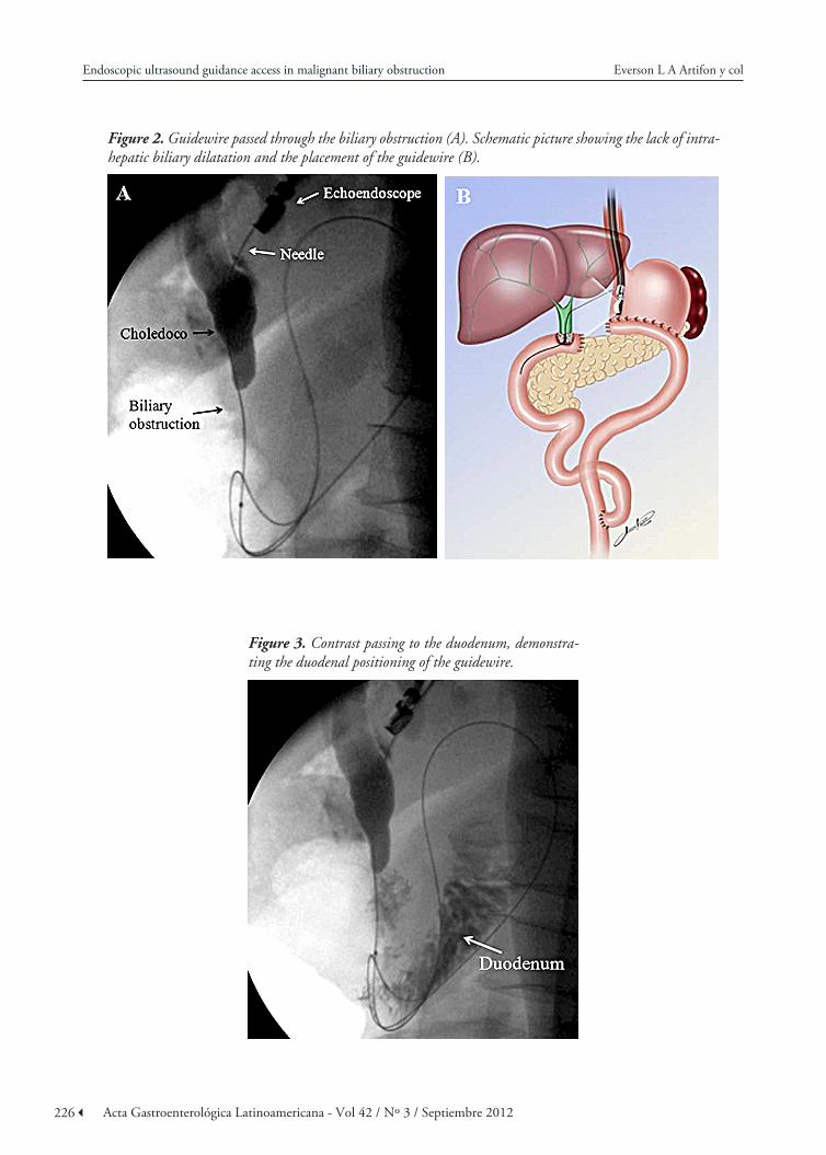
The common bile duct (CBD) was visualized using a linear echoendoscope (GF-UCT140AL5, Olympus, Japan). The dilated CBD was punctured by an extrahepatic approach with a 19 gauge FNA needle (EUSN-19-T, Cook Endoscopy) through the distal part of the gastric remnant-stump in one at-tempt. Bile was aspirated and contrast was injected to obtain the biliary opacification. A dilated CBD with an abrupt stop in its distal part was observed (Figure 1). A 0.035-inch guidewire was passed into the biliary tree, through the stenosis. The needle was withdrawn, maintaining the position of the guidewi-re, and a standard catheter was passed over the gui-dewire (Figure 2). Contrast was injected to demons-trate the duodenal position of the guidewire and to show the length of the stenosis (Figure 3). Then, a partially covered SEMS (PC-SEMS) (10 mm x 60 mm, Wallflex, Boston Scientific) was passed over the guidewire, with its distal end in the duodenum for internal drainage. The proximal end of the PC-SEMS was placed in the CBD (Figure 4). Adequate contrast drainage was observed (Figure 5). The total procedure lasted 35 minutes. There were no early or delayed complications and the procedure was effec-tive, relieving jaundice at one week and after a one month follow-up (Table 1).
Figure 1. Endoscopic ultrasound guided extrahepatic biliary puncture: cholangiography showing abrupt stop in the distal CBD.
226 Acta Gastroenterológica Latinoamericana - Vol 42 / Nº 3 / Septiembre 2012
Endoscopic ultrasound guidance access in malignant biliary obstruction Everson L A Artifon y col
Figure 2. Guidewire passed through the biliary obstruction (A). Schematic picture showing the lack of intra-hepatic biliary dilatation and the placement of the guidewire (B).
Figure 3. Contrast passing to the duodenum, demonstra-ting the duodenal positioning of the guidewire.

227Acta Gastroenterológica Latinoamericana - Vol 42 / Nº 3 / Septiembre 2012
Endoscopic ultrasound guidance access in malignant biliary obstruction Everson L A Artifon y col
Figure 4. Radiologic image demonstrating the closed self-expandable metallic stent (SEMS) with its distal end in the duodenum (A). SEMS partially opened (B).
Figure 5. Radiologic image of the metal stent (A). Schematic picture demonstrating the final positioning of the self-expandable metallic stent (SEMS) (B).

228 Acta Gastroenterológica Latinoamericana - Vol 42 / Nº 3 / Septiembre 2012
Endoscopic ultrasound guidance access in malignant biliary obstruction Everson L A Artifon y col
Discussion
In patients with biliary obstruction, common alternatives when standard ERCP biliary draina-ge fails include PTBD or surgical intervention.2,3,5 EUS guided biliary access for minimally invasive ap-proach to biliary obstruction has gained increasing importance in the palliation of biliary diseases. EUS-rendezvous to obtain bile duct access was first per-formed by Mallery et al in 2004, but requires an en-doscopically accessible biliary orifice.10 Transmural drainage includes EUS guided choledochoduodeo-nostomy,2,11-15 EUS-guided hepaticogastrostomy,16-19
and EUS-guided choledocoantrostomy.20
The percutaneous transhepatic SEMS placement was first described by Irving et al in 1989.21 It is a well-established technique with technical and clini-cal success of 90% and 77% to 98%, respectively. However, it has complication rates ranging from 8% to 30%, including biliary fistula, cholangitis, perito-nitis, sepsis, hematoma and liver abscesses.22-24 The EUS-guided transhepatic anterograde SEMS place-ment for biliary obstruction in a single session was described by Nguyen-Tang et al in 2010.25
In our case, we performed a variation of the EUS-guided transhepatic anterograde SEMS place-ment. As the biliary orifice was not accessible becau-se of the prior surgery and there was no intrahepa-tic biliary dilation, a percutaneous and EUS-guided transhepatic approach could be difficult. The visua-lization of the CBD from the gastric remnant-stump allowed the alternative of an extrahepatic approach. This modification could be performed because of the long gastric remnant-stump and the adherences in the region of the CDB puncture that apparently brought security to the procedure.
The complications with EUS guided biliary dra-inage like stent migration, bile leakage, pneumope-ritoneum and cholangitis were not observed in this case after a one month folow-up.14,19 This case illus-trates an alternative EUS-BD for operated digestive
tract. However, the extrahepatic approach should be considered only in very selected cases.
References
1. Artifon EL, Chaves DM, Ishioka S, Souza TF, Matuguma SE, Sakai P. Echoguided hepatico-gastrostomy: a case re-port. Clinics (Sao Paulo) 2007;62:799-802.
2. Ang TL, Teo EK, Fock KM. EUS-guided transduodenal biliary drainage in unresectable pancreatic cancer with obs-tructive jaundice. JOP 2007;8:438-443.
3. Artifon EL, Takada J, Okawa L, Moura EG, Sakai P. EUS-guided choledochoduodenostomy for biliary drai-nage in unresectable pancreatic cancer: a case series. JOP 2010;11:597-600.
4. Burmester E, Niehaus J, Leineweber T, Huetteroth T. EUS-cholangio-drainage of the bile duct: report of 4 cases. Gastrointest Endosc 2003;57:246-251.
5. Kahaleh M, Hernandez AJ, Tokar J, Adams RB, Shami VM, Yeaton P. Interventional EUS-guided cholangiography: evaluation of a technique in evolution. Gastrointest Endosc 2006;64:52-59.
6. Covey AM, Brown KT. Percutaneous transhepatic biliary drainage. Tech Vasc Interv Radiol 2008;11:14-20.
7. Smith AC, Dowsett JF, Russell RC, Hatfield AR, Cotton PB. Randomised trial of endoscopic stenting versus surgi-cal bypass in malignant low bile duct obstruction. Lancet 1994;344:1655-1660.
8. Lai EC, Mok FP, Tan ES, Lo CM, Fan ST, You KT, Wong J. Endoscopic biliary drainage for severe acute cholangitis. N Engl J Med 1992;326:1582-1586.
9. Pessa ME, Hawkins IF, Vogel SB. The treatment of acute cholangitis: percutaneous transhepatic biliary drainage be-fore definitive therapy. Ann Surg 1987;72:389-392.
10. Mallery S, Matlock J, Freeman ML. EUS-guided rendez-vous drainage of obstructed biliary and pancreatic ducts: Report of 6 cases. Gastrointest Endosc 2004;59:100-107.
11. Giovannini M, Moutardier V, Pesenti C, Bories E, Lelong B, Delpero JR. Endoscopic ultrasound-guided bilioduode-nal anastomosis: a new technique for biliary drainage. En-doscopy 2001;33:898-900.
12. Yamao K, Bhatia V, Mizuno N, Sawaki A, Ishikawa H, Ta-jika M, Hoki N, Shimizu Y, Ashida R, Fukami N. EUS-guided choledochoduodenostomy for palliative biliary dra-inage in patients with malignant biliary obstruction: results of long-term follow-up. Endoscopy 2008;40:340-342.
13. Hanada K, Iiboshi T, Ishii Y. Endoscopic ultrasound-gui-ded choledochoduodenostomy for palliative biliary draina-ge in cases with inoperable pancreas head carcinoma. Dig Endosc 2009;21 (Suppl 1):S75-S78.
14. Itoi T, Yamao K, EUS 2008 Working Group. EUS 2008 Working Group document: evaluation of EUS-guided cho-ledochoduodenostomy (with video). Gastrointest Endosc 2009;69 (Suppl 2):S8-S12.
15. Park do H, Koo JE, Oh J, Lee YH, Moon SH, Lee SS, Seo DW, Lee SK, Kim MH. EUS-guided biliary drainage with one-step placement of a fully covered metal stent for malig-nant biliary obstruction: a prospective feasibility study. Am J Gastroenterol 2009;104:2168-2174.
Table 1. Baseline characteristics and follow-up.

229Acta Gastroenterológica Latinoamericana - Vol 42 / Nº 3 / Septiembre 2012
Endoscopic ultrasound guidance access in malignant biliary obstruction Everson L A Artifon y col
16. Giovannini M, Dotti M, Bories E, Moutardier V, Pesenti C, Danisi C, Delpero JR. Hepaticogastrostomy by echo-en-doscopy as a palliative treatment in a patient with metastatic biliary obstruction. Endoscopy 2003;35:1076-1078.
17. Bories E, Pesenti C, Caillol F, Lopes C, Giovannini M. Transgastric endoscopic ultrasonography-guided biliary dra-inage: results of a pilot study. Endoscopy 2007;39:287-291.
18. Will U, Thieme A, Fueldner F, Gerlach R, Wanzar I, Meyer F. Treatment of biliary obstruction in selected patients by endoscopic ultrasonography (EUS)-guided transluminal bi-liary drainage. Endoscopy 2007;39:292-295.
19. Savides TJ, Varadarajulu S, Palazzo L, EUS 2008 Working Group. EUS 2008 Working Group document: evaluation of EUS-guided hepaticogastrostomy. Gastrointest Endosc 2009;69 (Suppl 2):S3-S7.
20. Artifon LA, Okawa L, Takada J, Gupta K, Moura EGH, Sakai P. EUS-guided choledochoantrostomy: an alternative for biliary drainage in unresectable pancreatic cancer with duodenal invasion. Gastroint Endosc 2011;73:1317-1320.
21. Irving JD, Adam A, Dick R, Dondelinger RF, Lunderquist A, Roche A. Gianturco expandable metallic biliary stents: results of a European clinical trial. Radiology 1989;172:321-326.
22. Stoker J, Laméris JS. Complications of percutaneously inser-ted biliary Wallstents. J Vasc Interv Radiol 1993;4:767-772.
23. Becker CD, Glättli A, Maibach R, Baer HU. Percutaneous palliation of malignant obstructive jaundice with the Walls-tent endoprosthesis: follow-up and reintervention in pa-tients with hilar and non-hilar obstruction. J Vasc Interv Radiol 1993;4:597-604.
24. Lee MJ, Dawson SL, Mueller PR, Krebs TL, Saini S, Hahn PF. Palliation of malignant bile duct obstruction with meta-llic biliary endoprostheses: technique, results, and complica-tions. J Vasc Interv Radiol 1992;3:665-671.
25. Nguyen-Tang T, Binmoeller KF, Sanchez-Yague A, Shah JN. Endoscopic ultrasound (EUS)-guided transhepatic anterograde self-expandable metal stent (SEMS) place-ment across malignant biliary obstruction. Endoscopy 2010;42:232-236.