Estrogen-Androgen Paradox Is Atherosclerotic Cardiovascular ...

Case Studies: Atherosclerotic Case Studies: Atherosclerotic
Heart Disease and ArrhythmiasHeart Disease and Arrhythmias
Clifford Hale, MD FACP DBIMClifford Hale, MD FACP DBIM
Daniel Zamarripa, MDDaniel Zamarripa, MD
Sensitivity / Specificity for CAD generally defined as >70% stenosis of at least one vessel
SensitivitySensitivity
Stress EKG 68%
Stress Echo 86%
Nuclear Stress Test 91%
EBCT 93%
CCTA 93%
Cardiac Cath 99%
SpecificitySpecificity
Stress EKGStress EKG 74%
Stress Echo 77%
Nuclear Stress Test 80%
EBCT 89%
CCTA 96%
Cardiac Cath 99%
Sensitivity = ability to detect disease when it is Sensitivity = ability to detect disease when it is present . present .
Specificity = ability to correctly exclude disease when it is abSpecificity = ability to correctly exclude disease when it is absentsent..
Predictive value influenced by prePredictive value influenced by pre--test prevalence or probability.test prevalence or probability.

Sensitivity Comparison of Testing Modalities
Based Upon Extent of Disease
• 61 year-old man
– Treated hypertension and hyperlipidemia: well-controlled
– ECG: left bundle branch block (LBBB)
– Screening calcium scan (EBCT) done 2009
• Total score 547 (>75 percentile for age)
• LM 25, LAD 155, RCA 264, Cx 103
– Exercise testing
• Exercise duration 12.5 METS
• Nuclear stress: “fixed septal defect due to bundle branch block”
• Echo stress: “normal ejection fraction and wall motion”
– This year he presented to an ER with chest pain; he ruled out for MI but was taken to cath due to his high calcium score.
• Cath: 30% proximal RCA; luminal irregularities in the LAD and LCA.
Slide 4
Case 1Case 1

Newly Acquired LBBB Newly Acquired LBBB
in Community Populationin Community PopulationFramingham Study PopulationFramingham Study Population
• Eighteen years of observation; 55 new LBBBs in 5,209 people
• Average age of onset 62
• Most LBBBs occurred with HTN, CAD, and cardiac enlargement
• 48% developed CAD or CHF at or after onset
• Within 10 years 50% died from cardiovascular disease
• LBBB contributed independently to increased risk of cardiovascular death
Annals of Internal Medicine 1979 90;(3):303
LBBB
No LBBB
Ten years
50% CV mortality
Ave. onset age 62
7,392 Men From a General Population7,392 Men From a General Population
BBB at Baseline and 28BBB at Baseline and 28--Year FollowYear Follow--upupCoronary Death and Sudden DeathCoronary Death and Sudden Death
HR LBBB all cause mortality 1.84HR LBBB all cause mortality 1.84
European Heart Journal 2005; 26:2300European Heart Journal 2005; 26:2300--23062306
Sudden deathCoronary Deaths

LBBB in Patients with Chronic CADLBBB in Patients with Chronic CAD
• 15,609 with CAD had coronary angiogram and ventriculography
• 522 had BBB
• BBB did not correlate with location of coronary artery stenosis or LV wall wall abnormality
• 4.9 year follow-up; 2,386 died
• Those with LBBB had a 5X mortality risk
• Those with RBBB had 2X mortality risk
• Cox regression showed LBBB, but not RBBB, is a strong predictor of mortality in this population
J Am Coll Cardiol 1987;10:73-80
Exercise Induced LBBBExercise Induced LBBB
• 17,277 exercise tests• Follow-up 3.7 years• 70 Exercise induced LBBB• 25 patients with cardiac events
-17 with exercise induced LBBB-8 in control group
• 7 deaths-5 with exercise induced LBBB-2 in control group
J Cardiovasc Electrophysiol 2009; 20:781

Left Anterior Hemiblock and MortalityLeft Anterior Hemiblock and Mortality
JACC 2005;46(5):858-63
LAHB
LAHB
LAHB
LAHB
• 61 year-old man
– Treated hypertension and hyperlipidemia: well-controlled
– ECG: left bundle branch block (LBBB)
– Screening calcium scan (EBCT) done 2009
• Total score 547 (>75 percentile for age)
• LM 25, LAD 155, RCA 264, Cx 103
– Exercise testing
• Exercise duration 12.5 METS
• Nuclear stress: “fixed septal defect due to bundle branch block”
• Echo stress: “normal ejection fraction and wall motion”
– This year he presented to an ER with chest pain; he ruled out for MI but was taken to cath due to his high calcium score.
• Cath: 30% proximal RCA; luminal irregularities in the LAD and LCA.
Slide 10
Case 1Case 1

Cumulative Survival by Coronary Calcium ScoreCumulative Survival by Coronary Calcium Score
Time to FollowTime to Follow--up (Years)up (Years)
0 (n=11,044)
1-10 (n=3,567)
11-100 (n=5,032)
101-299 (n=2,616)
300-399 (n=561)
400-699 (n=955)
700-999 (n=514)
1,000+ (n=964)
Cum
ula
tive
Surv
ival
0.0 2.0 4.0 6.0 8.0 10.0 12.0
0.70
0.75
0.80
0.85
0.90
0.95
1.00
LongLong--Term Prognosis Associated with Coronary Term Prognosis Associated with Coronary Calcification: OutcomesCalcification: Outcomes
JACC 2007; 49: 1860-70
0.0 2.0 4.0 6.0 8.0 10.0 12.0
0.70
0.75
0.80
0.85
0.90
0.95
1.00
Time to FollowTime to Follow--up (Years)up (Years)
Cum
ula
tive
Cum
ula
tive
Surv
ival
Surv
ival 0 Vessel (n=24,340)
1 Vessel (n=596)
2 Vessel (n=143)
3 Vessel (n=28)
Left Main (n=146)
Cumulative Cumulative Survival by the Coronary Calcium Extent in the Survival by the Coronary Calcium Extent in the Number of Vascular TerritoriesNumber of Vascular Territories
Long-Term Prognosis Associated with Coronary Calcification: Outcomes
JACC 2007

• 61 year-old man
– Treated hypertension and hyperlipidemia: well-controlled
– ECG: left bundle branch block (LBBB)
– Screening calcium scan (EBCT) done 2009
• Total score 547 (>75 percentile for age)
• LM 25, LAD 155, RCA 264, Cx 103
– Exercise testing
• Exercise duration 12.5 METS
• Nuclear stress: “fixed septal defect due to bundle branch block”
• Stress echo: “normal ejection fraction and wall motion”
– This year he presented to an ER with chest pain; he ruled out for MI but was taken to cath due to his high calcium score.
• Cath: 30% proximal RCA; luminal irregularities in the LAD and LCA.
Slide 13
Case 1Case 1
Duke Treadmill ScoreDuke Treadmill Score
• (Bruce exercise minutes) minus (5 x maximal ST segment deviation in mm) minus (4 x exercise angina) where
– 0 = none; 1 = non-limiting; 2 = limiting
• Low risk – score > +5
� (97% five-year survival)
• Moderate risk – score from -10 to + 4
� (31% have 3-vessel or LM disease with 90% five year survival)
• High Risk – score <-11
� (74% have 3-vessel or LM disease with 65% five year survival)

For example, a 60-year-old man with a 3-MET capacity has 40% of the age-expected exercise capacity for sedentary men and 30% of that for active men. *
* This statement was approved by the American Heart Association Science
Advisory and Coordinating Committee in June 2001.
Estimating Expected Exercise Capacity for AgeEstimating Expected Exercise Capacity for Age
A useful equation to estimate A useful equation to estimate
expectedexpected METs for an active personMETs for an active person
18 18 –– (age x 0.15) = Expected METs(age x 0.15) = Expected METs
Case 1 expectedCase 1 expected
18 18 –– (61 x 0.15) = 8.85 METs(61 x 0.15) = 8.85 METs
Case 1 achieved = 12.5 METsCase 1 achieved = 12.5 METs
Myers J et al. N Engl J Med 2002;346:793-801.
Exercise Capacity and AllExercise Capacity and All--Cause MortalityCause Mortality

Chronotropic InsufficiencyChronotropic Insufficiency
Unable to achieve 85%
predicted max HR off beta
blockers
Adjusted relative risk of cardiac event 2.2
HeartHeart--Rate Rate Recovery Recovery
One One Minute after Minute after Peak Peak ExerciseExercise..
Cole CR et al. N Engl J Med 1999;341:1351-1357.
2428 patients without known heart
disease
An abnormal value for the recovery of
heart rate defined as a reduction of
≤12 beats per minute (bpm) from the
heart rate at peak exercise
Circles represent the relative risk of death
within six years for each of the quintiles as
compared with the fifth quintile which had the lowest risk of death
Red lines represent the 95 percent
confidence interval.
relative risk of death
within six years
2428 patients without
known heart disease

Mortality Risk in Normotensive Individuals
with Hypertensive Response to Exercise
6578 asymptomatic individuals (74 percent without hypertension at baseline) who underwent submaximal Bruce treadmill tests and were followed for 20 years
Among individuals with baseline BP <140/90 mmHg,
Bruce stage 2 BP >180/90 compared to ≤180/90
mmHg was associated with a significant increase in
risk of cardiovascular death after adjustment for rest BP and other risk factors (adjusted hazard ratio for systolic 1.96, 95% CI 1.40-2.74 and for diastolic 1.48,
95% CI 1.06-2.06).
Weiss S A et al. Circulation 2010;121:2109-2116
• 61 year-old man
– Treated hypertension and hyperlipidemia: well-controlled
– ECG: left bundle branch block (LBBB)
– Screening calcium scan (EBCT) done 2009
• Total score 547 (>75 percentile for age)
• LM 25, LAD 155, RCA 264, Cx 103
– Exercise testing
• Exercise duration 12.5 METS
• Nuclear stress: “fixed septal defect due to bundle branch block”
• Echo stress: “normal ejection fraction and wall motion”
– This year he presented to an ER with chest pain; he ruled out for MI but was taken to cath due to his high calcium score.
• Cath: 30% proximal RCA; luminal irregularities in the LAD and LCA.
Slide 20
Case 1Case 1

JACC 2011
Adjusted for CAD risk factors, the
presence of any nonobstructive plaque
was associated with higher mortality
with the highest risk among those
exhibiting nonobstructive CAD in 3
vessels
JACC 2011
Higher mortality for non obstructive CAD
was observed even among patients with
low 10-year Framingham risk
(3.4%, p=0.0001) as
well as those with no
traditional, medically
treatable CAD risk factors, including
diabetes mellitus,
hypertension, and
dyslipidemia
(6.7%,p=0.0001)
Case 1: Relative Mortality Risk?
• 100 %
• 150 %
• 200 %
• 250 %
• 300 %
• 300 %+
61 year-old manTreated hypertension and hyperlipidemia: well-controlledECG: left bundle branch block (LBBB)Screening calcium scan (EBCT) done 2009
Total score 547 (>75 percentile for age)LM 25, LAD 155, RCA 264, Cx 103
Exercise testing
Exercise duration 12.5 METSNuclear stress: “fixed septal defect due to bundle branch block”Echo stress: “normal ejection fraction and wall motion”
This year he presented to an ER with chest pain; he ruled out for MI but was taken to cath due to his high calcium score.
Cath: 30% proximal RCA; luminal irregularities in the LAD and LCA.
Relative
Mortality
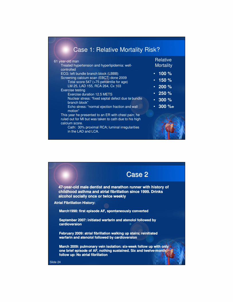
Atrial Atrial Fibrillation History: Fibrillation History:
March1998: first episode AF, spontaneously converted March1998: first episode AF, spontaneously converted
September 2007: initiated warfarin and atenolol followed by September 2007: initiated warfarin and atenolol followed by
cardioversioncardioversion
February 2009: atrial fibrillation February 2009: atrial fibrillation walking up walking up stairs; reinitiated stairs; reinitiated
warfarin warfarin and and atenolol followed by cardioversionatenolol followed by cardioversion
March 2009: pulmonary vein isolationMarch 2009: pulmonary vein isolation: : sixsix--week week follow up follow up with only with only
one brief episode of AF, nothing sustained. Six and one brief episode of AF, nothing sustained. Six and twelvetwelve--month month
follow up: No follow up: No atrial fibrillationatrial fibrillation
Case 2Case 2
Slide 24
4747--yearyear--old male dentist and marathon runner with history of old male dentist and marathon runner with history of
childhood asthma and atrial fibrillation since 1999. Drinks childhood asthma and atrial fibrillation since 1999. Drinks
alcohol socially once or twice weeklyalcohol socially once or twice weekly
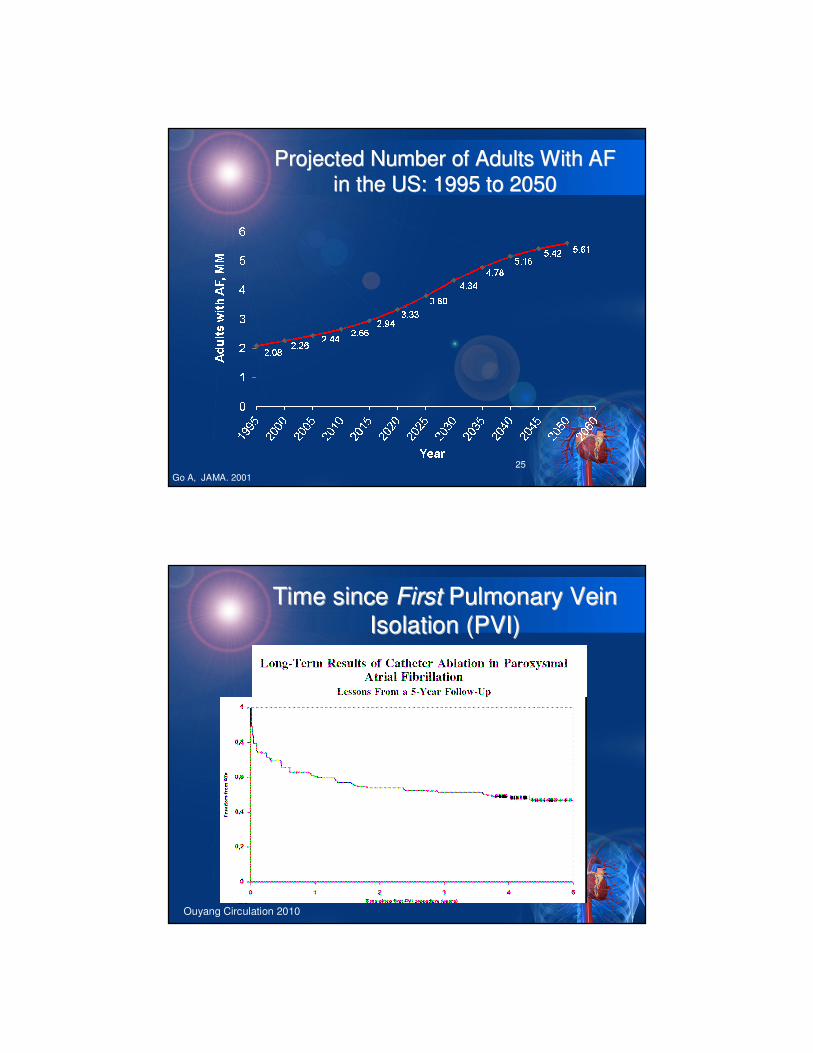
25
Go A, JAMA. 2001
Projected Number of Adults With AF Projected Number of Adults With AF
in the US: 1995 to 2050in the US: 1995 to 2050
26
Time since Time since FirstFirst Pulmonary Vein Pulmonary Vein
Isolation (PVI) Isolation (PVI)
Ouyang Circulation 2010

27Ouyang Circulation 2010
Time since Time since LastLast Pulmonary Pulmonary
Vein Vein Isolation (PVI) Isolation (PVI)
Stroke in AFStroke in AF
• Stroke in AF is often severe and results in long-term
disability or death. Approximately every fifth stroke is due to AF; furthermore, undiagnosed ‘silent AF’ is a likely cause of
some ‘cryptogenic’ strokes. Paroxysmal AF carries the
same stroke risk as permanent or persistent AF
• Cognitive dysfunction, including vascular dementia, may be
related to AF. Small observational studies suggest that asymptomatic embolic events may contribute to cognitive
dysfunction in AF patients in the absence of an overt stroke.

Case 2: Relative Mortality Risk?
• 100 %
• 150 %
• 200 %
• 250 %
• 300 %
• 300 %+
Atrial Fibrillation History: Atrial Fibrillation History:
March1998: first episode AF, spontaneously converted March1998: first episode AF, spontaneously converted
September 2007: initiated warfarin and atenolol September 2007: initiated warfarin and atenolol
followed by cardioversionfollowed by cardioversion
February 2009: atrial fibrillation walking up stairs; February 2009: atrial fibrillation walking up stairs;
reinitiated warfarin and atenolol followed by reinitiated warfarin and atenolol followed by
cardioversioncardioversion
March 2009: pulmonary vein isolation: sixMarch 2009: pulmonary vein isolation: six--week follow week follow
up with only one brief episode of AF, nothing up with only one brief episode of AF, nothing
sustained. Six and twelvesustained. Six and twelve--month follow up: No month follow up: No
atrial fibrillationatrial fibrillation
4747--yearyear--old male dentist and marathon runner with old male dentist and marathon runner with
history of childhood asthma and atrial fibrillation history of childhood asthma and atrial fibrillation
since 1999. Drinks alcohol socially once or twice since 1999. Drinks alcohol socially once or twice
weeklyweekly
Relative
Mortality
7575--yearyear--old male exold male ex--smoker with exemplary cardiovascular risk factors smoker with exemplary cardiovascular risk factors
evaluated in 2006 (age 69) for dyspnea and a positive stress tesevaluated in 2006 (age 69) for dyspnea and a positive stress testt
2006 SPECT: Symptom2006 SPECT: Symptom--limited stress ECG showed 3 mm downsloping limited stress ECG showed 3 mm downsloping
ST depression at 8.2 METs. ST depression at 8.2 METs.
Resting EF 55%; stress EF 60%; TID ratio 1.2; 4% reversible defeResting EF 55%; stress EF 60%; TID ratio 1.2; 4% reversible defect RCA ct RCA
distributiondistribution
CT Angiogram: Ca++ score 598 (82 %CT Angiogram: Ca++ score 598 (82 %--tile). Prox LAD 25tile). Prox LAD 25--49%; 149%; 1stst diag. diag.
30% ostial; prox. RCA 130% ostial; prox. RCA 1--24%; mid24%; mid--RCA 25RCA 25--49%, and ostial PDA 149%, and ostial PDA 1--24%24%
2010 SPECT : Symptom2010 SPECT : Symptom--limited stress to 7.7 METs with 3mm ST limited stress to 7.7 METs with 3mm ST
depression. New 12% reversible defect RCA distribution. EF 58%depression. New 12% reversible defect RCA distribution. EF 58%
2010 CTA: calcium score 770 (872010 CTA: calcium score 770 (87thth percentile). LAD score 495. RCA, 192percentile). LAD score 495. RCA, 192
Case 3Case 3

Myocardial Perfusion Imaging (MPI)Myocardial Perfusion Imaging (MPI)SPECT, Radionuclide Scan, Nuclear ScanningSPECT, Radionuclide Scan, Nuclear Scanning
Myocardial Perfusion Imaging with Myocardial Perfusion Imaging with
Exercise or Pharmacologic StressExercise or Pharmacologic Stress
Indications for Indications for
Pharmacologic StressPharmacologic Stress
Unable to exercise
Aortic stenosis
LBBB
Pacemaker
Recent MI
Severe HTN
Imaging agentsImaging agents
Thallium-201 (K+)
Tc-99m sestamibi (Ca++)
(e,g,. Cardiolite)
Technitium tetrofosmin
(e.g., Myoview)
Technitium teboroxime (e.g.,Cardiotec)
Pharmacologic StressPharmacologic Stress
Vasodilators (Vasodilators (““stealersstealers””))
Adenosine and analogs
Dipyridamole
Inotrope/Chronotrope
Dobutamine

Transient Ischemic Dilation Ratio (TID)Average ventricular size after stress compared with rest
JACC 2003; 42:1818-1825
TID ratios > 1.22 with exercise or > TID ratios > 1.22 with exercise or >
1.36 with pharmacologic stress 1.36 with pharmacologic stress
suggest extensive CAD, even in suggest extensive CAD, even in
presence of normal MPI.presence of normal MPI.
(71% sensitivity and 95% specificity)(71% sensitivity and 95% specificity)
JACC 1996;27:1612-1620 J Nucl Cardiol 1999;6:397-405
Death and MI
Revascularization
Quartiles TID
Normal SPECT
7575--yearyear--old male exold male ex--smoker with exemplary cardiovascular risk factors smoker with exemplary cardiovascular risk factors
evaluated in 2006 (age 69) for dyspnea and a positive stress tesevaluated in 2006 (age 69) for dyspnea and a positive stress testt
2006 SPECT: Symptom2006 SPECT: Symptom--limited stress ECG showed 3 mm downsloping limited stress ECG showed 3 mm downsloping
ST depression at 8.2 METs. ST depression at 8.2 METs.
Resting EF 55%; stress EF 60%; TID ratio 1.2; 4% reversible defeResting EF 55%; stress EF 60%; TID ratio 1.2; 4% reversible defect RCA ct RCA
distributiondistribution
CT Angiogram: Ca++ score 598 (82CT Angiogram: Ca++ score 598 (82ndnd percentile). Prox LAD 25percentile). Prox LAD 25--49%; 149%; 1stst
diag. 30% ostial; prox. RCA 1diag. 30% ostial; prox. RCA 1--24%; mid24%; mid--RCA 25RCA 25--49%, and ostial PDA 149%, and ostial PDA 1--
24%24%
2010 SPECT : Symptom2010 SPECT : Symptom--limited stress to 7.7 METs with 3mm ST limited stress to 7.7 METs with 3mm ST
depression. New 12% reversible defect RCA distribution. EF 58%depression. New 12% reversible defect RCA distribution. EF 58%
2010 CTA: calcium score 770 (872010 CTA: calcium score 770 (87thth percentile). LAD score 495. RCA, 192percentile). LAD score 495. RCA, 192
Case 3Case 3

Silent Lesions (<50%)Relationship Between Calcium Score and the Severity of Coronary Artery
Stenosis
Slide 35
Newer Anatomic Imaging
3 D reconstruction of the heart and blood
vessels developed from data acquired by a
CT technology that allows for increased
spatial and temporal resolution.
Rapid x-ray of the heart allowing
for the detection of calcium buildup
in the coronaries.
Density x brightness=Agaston score
Calcium score CCTA
Slide 36

CTA Will Replace Diagnostic Invasive Angiography
• Accurate
• Prognostic
value
Sensitivity Specificity Negative
Protective
Value
Per segment 91% 96% 98%
Per patient 96% 90% 99%
486 suspected acute coronary syndrome
patients in ER:
84% discharged home after normal CT30 days: no events1 year: no MI
Ann Emerg Med 2009
Slide 37
Case 3 Relative Mortality Risk?Case 3 Relative Mortality Risk?
• 100 %
• 150 %
• 200 %
• 250 %
• 300 %
• 300 %+
7575--yearyear--old male exold male ex--smoker with exemplary cardiovascular risk smoker with exemplary cardiovascular risk
factors evaluated in 2006 (age 69) for dyspnea and a positive stfactors evaluated in 2006 (age 69) for dyspnea and a positive stress ress
testtest
2006 SPECT: Symptom2006 SPECT: Symptom--limited stress ECG showed 3 mm limited stress ECG showed 3 mm
downsloping ST depression at 8.2 METs. downsloping ST depression at 8.2 METs.
Resting EF 55%; stress EF 60%; TID ratio 1.2; 4% reversible defeResting EF 55%; stress EF 60%; TID ratio 1.2; 4% reversible defect ct
RCA distributionRCA distribution
CT Angiogram: Ca++ score 598 (82 %CT Angiogram: Ca++ score 598 (82 %--tile). Prox LAD 25tile). Prox LAD 25--49%; 149%; 1stst
diag. 30% ostial; prox. RCA 1diag. 30% ostial; prox. RCA 1--24%; mid24%; mid--RCA 25RCA 25--49%, and ostial 49%, and ostial
PDA 1PDA 1--24%24%
2010 SPECT : Symptom2010 SPECT : Symptom--limited stress to 7.7 METs with 3mm ST limited stress to 7.7 METs with 3mm ST
depression. New 12% reversible defect RCA distribution. EF 58%depression. New 12% reversible defect RCA distribution. EF 58%
2010 CTA: calcium score 770 (872010 CTA: calcium score 770 (87thth percentile). LAD score 495. RCA, percentile). LAD score 495. RCA,
192192
RelativeMortality

6262--yearyear--old old male male investment advisor with history of an investment advisor with history of an ““abnormal ECGabnormal ECG”” since since
1988 (age 38) and echocardiographic evidence of an MI in 1996 (a1988 (age 38) and echocardiographic evidence of an MI in 1996 (age 46).ge 46).
55’’88”” 167 lbs (BMI 25) BP 132/75 with an exemplary risk factor profil167 lbs (BMI 25) BP 132/75 with an exemplary risk factor profile. Good e. Good
medical records depict excellent care and good health otherwise.medical records depict excellent care and good health otherwise.
March 2011 SPECT 8.0 METs (2006: 10 METs): March 2011 SPECT 8.0 METs (2006: 10 METs):
ECG: positive 2mm ST depression five minutes into exercise resolECG: positive 2mm ST depression five minutes into exercise resolving two ving two
minutes into recoveryminutes into recovery
SPECT: moderate to severe fixed perfusion defect anterior wall, SPECT: moderate to severe fixed perfusion defect anterior wall, post stress post stress
ejection fraction 37% (post stress EF 2006 51%)ejection fraction 37% (post stress EF 2006 51%)
Echocardiogram : LVID 6.0, LA 3.6, Septum 1.0, PW 1.2, resEchocardiogram : LVID 6.0, LA 3.6, Septum 1.0, PW 1.2, resting EF 50%, ting EF 50%,
and mild anteroseptal hypokinesisand mild anteroseptal hypokinesis
ECG: QS V1ECG: QS V1--V3; IVCD (QRS 0.14)V3; IVCD (QRS 0.14)
June 2012 SPECT (10 METs): ECG and SPECT unchanged except stressJune 2012 SPECT (10 METs): ECG and SPECT unchanged except stress EF EF
back to 51%back to 51%
Case 4Case 4
Slide 39


Left Ventricular Ejection Fraction
(EDV-ESV)/EDV
Modality• M-mode echocardiography
• 2-D echocardiography
• 3-D echocardiography
• MRI*
• CT
• Nuclear Cardiac Imaging
– SPECT
– MUGA* (RGV, RNA RNCA ERNA)
Sources of Error• Gating and rhythm abnormalities
• Identifying the endocardial border
• Detecting end-systole and end-diastole
• Software algorithm variations
• Geometric assumptions
• Image planes
• Regional wall motion variations
• Acoustic windows
• Anatomic variations- Modified Quinones (planar)
- Modified Simpson (biplane method of disks)*
*Recommended by the American Society of Echocardiography
* Most reliable non-invasive methods
Comparing LVEF by Comparing LVEF by
Echo, MPI (SPECT), and MRIEcho, MPI (SPECT), and MRI
• Echo M-Mode cube method 39 + 16%
• Echo M-Mode Teichholz method 29 + 15%
• Echo 2-D Simpsons Biplane 31 + 10%
• Radionuclide Ventriculography 24 + 9%
• Cardiovascular MR 30 + 11%
Eur Heart J 2000; 21, 1387-1396
52 patients with chronic stable heart failure

Exercise Ejection FractionExercise Ejection Fraction
6262--yearyear--old male investment advisor with history of an abnormal ECG sincold male investment advisor with history of an abnormal ECG since 1988 e 1988
(age 38) and echocardiographic evidence of an MI in 1996 (age 46(age 38) and echocardiographic evidence of an MI in 1996 (age 46).).
55’’88”” 167 lbs (BMI 25) BP 132/75 with an exemplary risk factor profil167 lbs (BMI 25) BP 132/75 with an exemplary risk factor profile. Good e. Good
medical records depict excellent care and good health otherwise.medical records depict excellent care and good health otherwise.
March 2011 SPECT 8.0 METs (2006: 10 METs): March 2011 SPECT 8.0 METs (2006: 10 METs):
ECG: positive 2mm ST depression five minutes into exercise resolECG: positive 2mm ST depression five minutes into exercise resolving two ving two
minutes into recoveryminutes into recovery
SPECT: moderate to severe fixed perfusion defect anterior wall, SPECT: moderate to severe fixed perfusion defect anterior wall, post stress post stress
ejection fraction 37% (post stress EF 2006 51%)ejection fraction 37% (post stress EF 2006 51%)
Echocardiogram : LVID 6.0, LA 3.6, Septum 1.0, PW 1.2, resting EEchocardiogram : LVID 6.0, LA 3.6, Septum 1.0, PW 1.2, resting EF 50%, and F 50%, and
mild anteroseptal hypokinesismild anteroseptal hypokinesis
ECG: QS V1ECG: QS V1--V3; IVCD (QRS 0.14)V3; IVCD (QRS 0.14)
June 2012 SPECT (10 METs): ECG and SPECT unchanged except stressJune 2012 SPECT (10 METs): ECG and SPECT unchanged except stress EF EF
back to 51%back to 51%
Case 4Case 4
Slide 46

NonNon--BBB IVCDBBB IVCD
10,899 subjects general population 30 years QRS > 0.11
Circulation: Arrhythmia and Electrophysiology 2011; 4:704-710
HR AllHR All--Cause DeathCause Death HR Cardiac DeathHR Cardiac Death HR Arrhythmic DeathHR Arrhythmic Death
QRS > 0.11 1.48 1.94 2.14
QRS > 0.11
without BBB Patternwithout BBB Pattern2.01 2.53 3.11
Case 4: Relative Mortality Risk?
• 100 %
• 150 %
• 200 %
• 250 %
• 300 %
• 300 %+
6262--yearyear--old male investment advisor with history of an abnormal old male investment advisor with history of an abnormal
ECG since 1988 (age 38) and echocardiographic evidence of an ECG since 1988 (age 38) and echocardiographic evidence of an
MI in 1996 (age 46).MI in 1996 (age 46).
55’’88”” 167 lbs (BMI 25) BP 132/75 with an exemplary risk factor 167 lbs (BMI 25) BP 132/75 with an exemplary risk factor
profile. Good medical records depict excellent care and good profile. Good medical records depict excellent care and good
health otherwise.health otherwise.
March 2011 SPECT 8.0 METs (2006: 10 METs): March 2011 SPECT 8.0 METs (2006: 10 METs):
ECG: positive 2mm ST depression five minutes into exercise ECG: positive 2mm ST depression five minutes into exercise
resolving two minutes into recoveryresolving two minutes into recovery
SPECT: moderate to severe fixed perfusion defect anterior wall, SPECT: moderate to severe fixed perfusion defect anterior wall,
post stress ejection fraction 37% (post stress EF 2006 51%)post stress ejection fraction 37% (post stress EF 2006 51%)
Echocardiogram : LVID 6.0, LA 3.6, Septum 1.0, PW 1.2, Echocardiogram : LVID 6.0, LA 3.6, Septum 1.0, PW 1.2,
resting EF 50%, and mild anteroseptal hypokinesisresting EF 50%, and mild anteroseptal hypokinesis
ECG: QS V1ECG: QS V1--V3; IVCD (QRS 0.14)V3; IVCD (QRS 0.14)
June 2012 SPECT (10 METs): ECG and SPECT unchanged except June 2012 SPECT (10 METs): ECG and SPECT unchanged except
stress EF back to 51%stress EF back to 51%
RelativeMortality

6464--yearyear--old physically active dentist. Due old physically active dentist. Due to palpitations he had to palpitations he had an echocardiogram in December an echocardiogram in December 20042004 that revealed that revealed a bicuspid a bicuspid
aaortic valveortic valve, , aortic aortic root root 3.8 3.8 cmcm, moderate , moderate LVH and LVH and mild mild AI. AI.
He went to different cardiologist in He went to different cardiologist in March March 20062006. Again an . Again an
echocardiogram showed a bicuspid aortic valveechocardiogram showed a bicuspid aortic valve, , an aortic root of an aortic root of
4.4 cm4.4 cm, mild , mild AI, AI, and an EF of 55and an EF of 55--6060%. %.
In March In March 20082008 his echo showed his aortic root at his echo showed his aortic root at 4.2 cm4.2 cm, his left , his left
atrial dimension of 3.5 cm, a LVID of 6.1, an EF of 57%, and atrial dimension of 3.5 cm, a LVID of 6.1, an EF of 57%, and
moderate moderate AI. AI. A CT of the chest to r/o a thoracic aneurysm A CT of the chest to r/o a thoracic aneurysm
showed the aortic root at showed the aortic root at 4.74.7..
Case 5Case 5
Slide 49
Bicuspid Aortic Bicuspid Aortic
ValveValve
• Male > female (2:1)
• Usually picked up between age 40 and 60
• Associated with congenital aortic and proximal coronary
artery problems (Marfan’s; Ehlers Danlos)
• About half are associated with widened and expanding aortic
root and ascending aorta due to (cystic medial
degeneration).
• Increased risk of aortic aneurysm and dissection (5-9 x)
• Nearly all require surgery during lifetime

Bicuspid Aortic Valve and Bicuspid Aortic Valve and
Cystic Medial Degeneration Cystic Medial Degeneration
Retrospective Look at Aortic Valve Retrospective Look at Aortic Valve
ReplacementsReplacements
• 7% replaced before age 50 with 2/3 bicuspid
• 40% between age 50 and 70 with 2/3 bicuspid
• 50%+ after age 70 with 40% bicuspid valves
• Risk rises with aortic insufficiency, aortic stenosis, and
enlarged aortic root, especially > 45mm.
Nearly all bicuspid valves require surgery during lifetime
Aortic valve replacements

• 100 %
• 150 %
• 200 %
• 250 %
• 300 %
• 300 %+
Case 5: Relative Mortality RiskCase 5: Relative Mortality Risk
6464--yearyear--old physically active dentist. Due to palpitations he old physically active dentist. Due to palpitations he
had an echocardiogram in December 2004 that revealed a had an echocardiogram in December 2004 that revealed a
bicuspid aortic valve, aortic root 3.8 cm, moderate LVH and bicuspid aortic valve, aortic root 3.8 cm, moderate LVH and
mild AI. mild AI.
He went to different cardiologist in March 2006. Again an He went to different cardiologist in March 2006. Again an
echocardiogram showed a bicuspid aortic valve, an aortic echocardiogram showed a bicuspid aortic valve, an aortic
root of 4.4 cm, mild AI, and an EF of 55root of 4.4 cm, mild AI, and an EF of 55--60%. 60%.
In March 2008 his echo showed his aortic root at In March 2008 his echo showed his aortic root at 4.2 4.2 cm, cm,
his left atrial dimension of 3.5 cm, a LVID of 6.1, an EF of his left atrial dimension of 3.5 cm, a LVID of 6.1, an EF of
57%, and moderate AI. 57%, and moderate AI. A CT of the chest to r/o a thoracic A CT of the chest to r/o a thoracic
aneurysm showed the aortic root at 4.7 cm.aneurysm showed the aortic root at 4.7 cm.
Relative
Mortality
7575--yearyear--old retired old retired school school teacher with controlled teacher with controlled
hypertension. As part of the physical exam she had hypertension. As part of the physical exam she had
an echocardiogram that led to a chest CT. an echocardiogram that led to a chest CT.
Chest CT December 2009Chest CT December 2009
Aortic root 4.1 cmAortic root 4.1 cm
Ascending aorta is prominent measuring 3.9 cm in Ascending aorta is prominent measuring 3.9 cm in
the midthe mid--ascending segment.ascending segment.
Proximal Aortic Arch 3.9 cm Proximal Aortic Arch 3.9 cm
Descending thoracic aorta 2.7 cmDescending thoracic aorta 2.7 cm
Case 6Case 6
Slide 54

Same retired 75-year-old school teacher two years later:
Chest CT December 2011:
Study described as “unchanged” from December 2009
Aortic root 4.2 cm (was 4.1 cm 2009)
Ascending aorta 4.1 cm in the mid-ascending segment (was
3.9 cm in 2009)
Proximal Aortic Arch 4.2 cm (was. 3.9 cm in 2009)
Descending thoracic aorta 3.2 cm (was 2.7 cm in 2009)
No evidence of dissection.
Case 6 . . . Cont.Case 6 . . . Cont.
Slide 55
The 5The 5--year risk of rupture as a function of aneurysm year risk of rupture as a function of aneurysm
size at recognition was:size at recognition was:
•• 0% for aneurysms less than 4 cm in diameter0% for aneurysms less than 4 cm in diameter
•• 16% (95% CI, 4%16% (95% CI, 4%--28%) for those 4 to 5.9 cm, and28%) for those 4 to 5.9 cm, and
•• 31% (95% CI, 5%31% (95% CI, 5%--56%) for aneurysms 6 cm or more. 56%) for aneurysms 6 cm or more.
FiveFive--year Risk of Rupture year Risk of Rupture
Thoracic AneurysmThoracic Aneurysm
JAMA 1998 280;22:1926

Normal Diameter Normal Diameter and and Upper Limit Upper Limit of of Ascending Ascending and and
Descending Aorta Related Descending Aorta Related to to AgeAge
WRITING GROUP MEMBERS et al. Circulation 2010;121:e266-e369
Our 75Our 75--yearyear--oldold
2009 to 20112009 to 2011
Aortic root 4.1 Aortic root 4.1 –– 4.24.2
Ascending 3.9 Ascending 3.9 –– 4.1 4.1
Descending 2.7 Descending 2.7 –– 3.2 3.2
Average Growth RateAscending thoracic: 0.07 cm/yrDescending thoracic: 0.19 cm/yr
Ann Thorac Surg 2002;74:S1877-80
Mean Mean aortic diameters (in cm) at various aortic diameters (in cm) at various
levels measured by helical CT in 70 adults. levels measured by helical CT in 70 adults.
WRITING GROUP MEMBERS et al. Circulation
2010;121:e266-e369
Average Growth RateAscending thoracic: 0.07 cm/yrDescending thoracic: 0.19 cm/yr
Male
Aortic sinus: 3.63 to 3.91 cm
Ascending aorta: 2.86 cm
Mid-descending aorta: 2.39 to 2.98 cm
At diaphragm: 2.43 to 2.69 cm
Female
Aortic root: 3.5 to 3.72 cm
Ascending aorta: 2.86 cm
Mid-descending aorta: 2.45 to 2.64 cm
At diaphragm: 2.40 to 2.44 cm

Case 6: Relative Mortality Risk?Case 6: Relative Mortality Risk?
• 100 %
• 150 %
• 200 %
• 250 %
• 300 %
• 300 %+
75-year-old retired school teacher with controlled hypertension. As part of the physical exam she had an echocardiogram that led to a chest CT.
Chest CT December 2009Aortic root 4.1 cmAscending aorta is prominent measuring 3.9 cm in the mid-ascending segment.
Proximal Aortic Arch 3.9 cm Descending thoracic aorta 2.7 cm
Chest CT December 2011:Study described as “unchanged” from December 2009 Aortic root 4.2 cm (was 4.1 cm 2009)Ascending aorta 4.1 cm in the mid-ascending segment (was 3.9 cm in 2009)Proximal Aortic Arch 4.2 cm (was. 3.9 cm in 2009)Descending thoracic aorta 3.2 cm (was 2.7 cm in 2009)No evidence of dissection.
Relative
Mortality
65-year-old mechanical engineer with statin-treated hyperlipidemia and well-controlled hypertension. Height 72” and weight 215 pounds. BMI 28.5 BSA 2.175 m2
In May 2010 due to non-specific abdominal pain he had an abdominal ultrasound that showed an abdominal aorta of 4.3 cm
(a) Scenario
May 2012 abdominal ultrasound reveals AAA 4.3 cm
(b) Scenario
Same as (a) except that AAA size is 4.6 cm
Case 7Case 7
Slide 60

Abdominal Aortic AneurysmAbdominal Aortic Aneurysm
• A focal dilatation with at least a 50% increase over the
normal diameter, 3 cm for the abdominal aorta.
• Highly correlated with atherosclerosis, endothelial
dysfunction, and the inflammatory mediators that
mediate endothelial dysfunction.
• Rupture risk is related to diameter, rate of expansion,
and gender.
Lederle, F. A. Ann Intern Med 2003;139:516-522
Prevalence of abdominal aortic aneurysm 4.0 cm or larger in men by age and smoking history
Cardiovascular Risk Factors Aggravate Cardiovascular Risk Factors Aggravate
Aortic AneurysmsAortic Aneurysms
Aortic aneurysms are highly Aortic aneurysms are highly
correlated with atherosclerosis correlated with atherosclerosis
and aggravated by factors that and aggravated by factors that
accelerate atherosclerosis, accelerate atherosclerosis,
especially tobacco smoking.especially tobacco smoking.

Annual Risk of Rupture of Annual Risk of Rupture of
Abdominal Aortic AneurysmAbdominal Aortic Aneurysm
• < 4.0 cm; zero risk
• 4.0 to 4.9 cm 0.5 to 5%
• 5.0 to 5.9 cm 3-15%
• 6.0 to 6.9 cm 10-20%
• 7.0 to 7.9 cm 20 to 40%
• > 8.0 cm 30 to 50%
UpToDate: Natural History and Management of Abdominal Aortic AneurysmAuthors: Mohler and Fairman
Literature review through August 2012
Last updated June 16, 2011
Abdominal Aneurysm Abdominal Aneurysm Growth Rates by Diameter
• 2.8 to 3.9 cm diameter average growth rate 0.19 cm per year
• 4.0 to 4.5 cm diameter average growth rate 0.27 cm per year
• 4.6 to 8.5 cm diameter average growth rate 0.35 cm per year
Growth rates are variable (some don’t grow), so serial measurements are necessary.
Growth is more rapid in smokers
UpToDate: Natural History and Management of Abdominal Aortic Aneurysm
Authors: Mohler and Fairman
Literature review through August 2012
Last updated June 16, 2011

Ann Thorac Surg 2006;81:169-177 © 2006
Risk of Rupture Relative to Risk of Rupture Relative to
Body Surface Area (Body Surface Area (““Indexed RiskIndexed Risk””))
Abdominal Aortic Rupture RatesAbdominal Aortic Rupture Rates

N ENGL J MED Vol 358, January 31, 2008
Endovascular Repair of AAAEndovascular Repair of AAA
At present, EVR accounts for >
60% of all repairs
Operative mortality associated with
EVR only 1/3 that of 0pen repair
Early benefit of EVR lost in the
longer term
After 4 years, aneurysm-related mortality
significantly higher in EVR
Risk extends out at least 8 years Re-intervention
after EVR remains a substantial risk after 4 years
Case 7: Relative Mortality Risk?
• 100 %
• 150 %
• 200 %
• 250 %
• 300 %
• 300 %+
65-year-old mechanical engineer with statin-treated hyperlipidemia and well-controlled hypertension. In May 2010 due to non-specific abdominal pain he had an abdominal ultrasound that showed an abdominal aorta of 4.3 cm
(a) ScenarioMay 2012 abdominal ultrasound reveals AAA 4.3 cm
(b) ScenarioSame as (a) except that AAA size is 4.6 cm
RelativeMortality

45-year old male
Background:
Family History of premature CAD with MI & death in 50s: paternal grandfather, maternal grandfather, maternal aunt, maternal uncle.
Treated and well controlled hyperlipidemia. Borderline normal BP, 5'9" 179#, asymptomatic and runs for exercise.
Had EBCT and "positive" calcium score, but we do not have results. This was followed by a CCTA for "coronary artery disease" on 7/6/09
ETT 7/20/09: 12:00, 13 METS, HR to 172 and reported as EKG negative and normal Echocardiogram response to exercise.
Case 8Case 8
Slide 69
Coronary Artery Calcium Scores at Various Framingham Risk Score Thresholds
Slide 70
Same 45Same 45--yearyear--old maleold male
CTA 2009: CTA 2009:
There There is diffuse calcified plaque throughout the proximal and is diffuse calcified plaque throughout the proximal and
midmid--LAD with two potential areas of flow limiting stenosis. LAD with two potential areas of flow limiting stenosis.
Mild calcified plaque is present in the proximal and midMild calcified plaque is present in the proximal and mid--left left
circumflex artery as well as the proximal and mid RCA without circumflex artery as well as the proximal and mid RCA without
significant coronary artery stenosissignificant coronary artery stenosis
Case 8 . . . Cont.Case 8 . . . Cont.
Slide 71
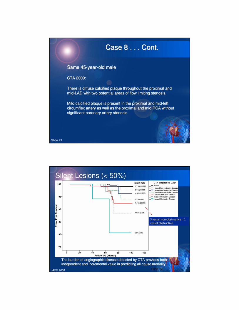
Silent Lesions (< 50%)
The burden of angiographic disease detected by CTA provides bothThe burden of angiographic disease detected by CTA provides both
independent and incremental value in predicting allindependent and incremental value in predicting all--cause mortalitycause mortality
JACC 2008
3 vessel 3 vessel nonnon--obstructive obstructive = 1 = 1
vessel vessel obstructiveobstructive
Slide 72

Silent Lesions (< 50%)
10,14610,146
8,1148,114
5,5945,594
Normal:Normal: RefRef
NonNon--ObstructiveObstructive HR 1.6 (1.2 HR 1.6 (1.2 –– 2.2)2.2)
Obstructive:Obstructive: HR 2.6 (1.9 HR 2.6 (1.9 –– 3.5)3.5)
24,775 patients suspected CAD; age 57 +/24,775 patients suspected CAD; age 57 +/-- 13 13
years 54% maleyears 54% male
Follow up: 2.3 +/Follow up: 2.3 +/-- 1.1 years; 404 deaths1.1 years; 404 deaths
CONFIRM JACC 2011
Coronary Coronary
Angiography Angiography
PrognosisPrognosis
Slide 73
Case 8: Relative Mortality Risk?Case 8: Relative Mortality Risk?
• 100 %
• 150 %
• 200 %
• 250 %
• 300 %
• 300 %+
45 y/o with family history of premature CAD with MI & death in 50s: paternal grandfather, maternal grandfather, maternal aunt, maternal uncle.
Treated and well controlled hyperlipidemia. Borderline normal BP, 5'9" 179#, asymptomatic and runs for exercise.
Had EBCT and "positive" calcium score, but we do not have results. This was followed by a CCTA for "coronary artery disease" on 7/6/09
ETT 7/20/09: 12:00, 13 METS, HR to 172 and reported as EKG negative and normal Echocardiogram response to exercise.
CTA 2009: diffuse calcified plaque throughout the proximal and mid-LAD with two potential areas of flow limiting stenosis.
Mild calcified plaque is present in the proximal and mid-left circumflex artery as well as the proximal and mid RCA without significant coronary artery stenosis
RelativeMortality

6060--yearyear--old female with hypertension and chronic microalbuminuria old female with hypertension and chronic microalbuminuria
with negative with negative renal workup. Also sleep renal workup. Also sleep apnea apnea surgerysurgery
EBCT EBCT February 2008 February 2008 ZeroZero
Echocardiogram Echocardiogram 2008 2008 normalnormal
Cooper Cooper Clinic stress Clinic stress test test February 2008: negative; February 2008: negative; no PVCs (report)no PVCs (report)
Jan 2011 stress test: rsr' pattern in V1Jan 2011 stress test: rsr' pattern in V1--2; 2; negative for ischemia no negative for ischemia no
PVCs PVCs ((Balke Protocol)Balke Protocol)
Dec Dec 2011 2011 -- Stress test done as part of insurance physical Stress test done as part of insurance physical
Case 9Case 9
Slide 75
Case 9
Slide 76

Case 9Case 9
Slide 77
And… The Medical Director Worst Nightmare
“Benign” Arrhythmias that kill

Case 9: Relative Mortality Risk?
• 100 %
• 150 %
• 200 %
• 250 %
• 300 %
• 300 %+
6060--yearyear--old female with hypertension and chronic old female with hypertension and chronic
microalbuminuria microalbuminuria with negative with negative renal workup. Also sleep renal workup. Also sleep
apnea apnea surgerysurgery
EBCT EBCT February 2008 February 2008 ZeroZero
Echocardiogram Echocardiogram 2008 2008 normalnormal
Cooper Cooper Clinic stress Clinic stress test test February 2008: negative; February 2008: negative; no PVCs no PVCs
(report)(report)
Jan 2011 stress test: rsr' pattern in V1Jan 2011 stress test: rsr' pattern in V1--2; 2; negative for negative for
ischemia no PVCs ischemia no PVCs ((Balke Protocol)Balke Protocol)
Dec Dec 2011 2011 -- Stress test done as part of insurance physical Stress test done as part of insurance physical
Relative
Mortality
61 61 yearyear--old male, 5old male, 5’’66”” 272 272 pounds, with stable angina. Diabetes ten years treated pounds, with stable angina. Diabetes ten years treated
with insulin (HbA1c with insulin (HbA1c between 7.5 between 7.5 –– 88%). In 2007 right common femoral artery %). In 2007 right common femoral artery
stented for PAD.stented for PAD.
2010 : Angina 2010 : Angina with positive stress test with positive stress test
New Cath: New Cath:
RCA: RCA: 100% 100% obstructedobstructed
LAD: proximal stent is patent LAD: proximal stent is patent
First diagonal: moderate ostial obstruction First diagonal: moderate ostial obstruction
Circumflex: proximal stent patent Circumflex: proximal stent patent
OM1 50 %; OM2 OM1 50 %; OM2 99% 99%
““The The angina may angina may be possibly due to his chronic total be possibly due to his chronic total occlusion occlusion ––
insufficient insufficient collateral's to RCAcollateral's to RCA””. Treated with long acting nitrates. Treated with long acting nitrates
Case 10Case 10
Slide 80

December 2011: leg ulceration (Severe PAD?)December 2011: leg ulceration (Severe PAD?)
APS: last office visit March 2012 (one page)APS: last office visit March 2012 (one page)
““The patient has recent recurrence of chest painThe patient has recent recurrence of chest pain””
SPECT Study SPECT Study –– 7 minutes modified Bruce protocol 7 minutes modified Bruce protocol
(METS ?)(METS ?)
Resting EF 45% Resting EF 45% -- no significant wall motion no significant wall motion
abnormalityabnormality
No evidence of exercise induced ischemiaNo evidence of exercise induced ischemia
Case 10 . . . Cont.Case 10 . . . Cont.
Slide 81
ARTS II: Event free survival ARTS II: Event free survival At one year, there was no difference in event-free survival between the ARTS II SES
group and the ARTS I CABG group. However, the ARTS II group showed
significantly higher rates of survival free from cardiac death, MI, and reintervention
than the ARTS I bare metal stent group. The groups were not significantly different
in the primary endpoint of survival free from MACCE.
p = <0.001p = 0.003 p = 0.46

ARTS II: MACCE at one year
TCT 2004TCT 2004
Overall MACCE at 1 year
ACC 2005ACC 2005
PTCA Fallacy #1
Fallacy
“Doc said I had a 90% blockage. Thank
goodness he fixed it in time and saved my life”
FactsFacts
•• There is no such thing as a 90% stenosisThere is no such thing as a 90% stenosis
•• Even if there were, in most cases Even if there were, in most cases PTCA PTCA is not is not
a lifea life--saving interventionsaving intervention
The COURAGE ParadigmThe COURAGE Paradigm

Fallacy
“Mr. Jones had severe 2-vessel disease but really didn’t
want a bypass operation, so I stented both vessels”
FactsFacts
•• PTCA PTCA is often the first step on the road to CABGis often the first step on the road to CABG
•• If a patient really wants to avoid CABG at all If a patient really wants to avoid CABG at all
costs, medical therapy is the way to gocosts, medical therapy is the way to go
PTCA Fallacy #2PTCA Fallacy #2
The COURAGE ParadigmThe COURAGE Paradigm
PTCA Fallacy #3
Fallacy
“For most patients with multi-vessel disease, PTCA can
provide comparable long-term survival benefits and quality of life as bypass surgery”
FactsFacts
•• The randomized trials of PCI vs. CABG have The randomized trials of PCI vs. CABG have
included only highly selected patientsincluded only highly selected patients
•• Observational data still suggest improved Observational data still suggest improved
survival with CABG in severe multivessel survival with CABG in severe multivessel
diseasediseaseThe COURAGE ParadigmThe COURAGE Paradigm
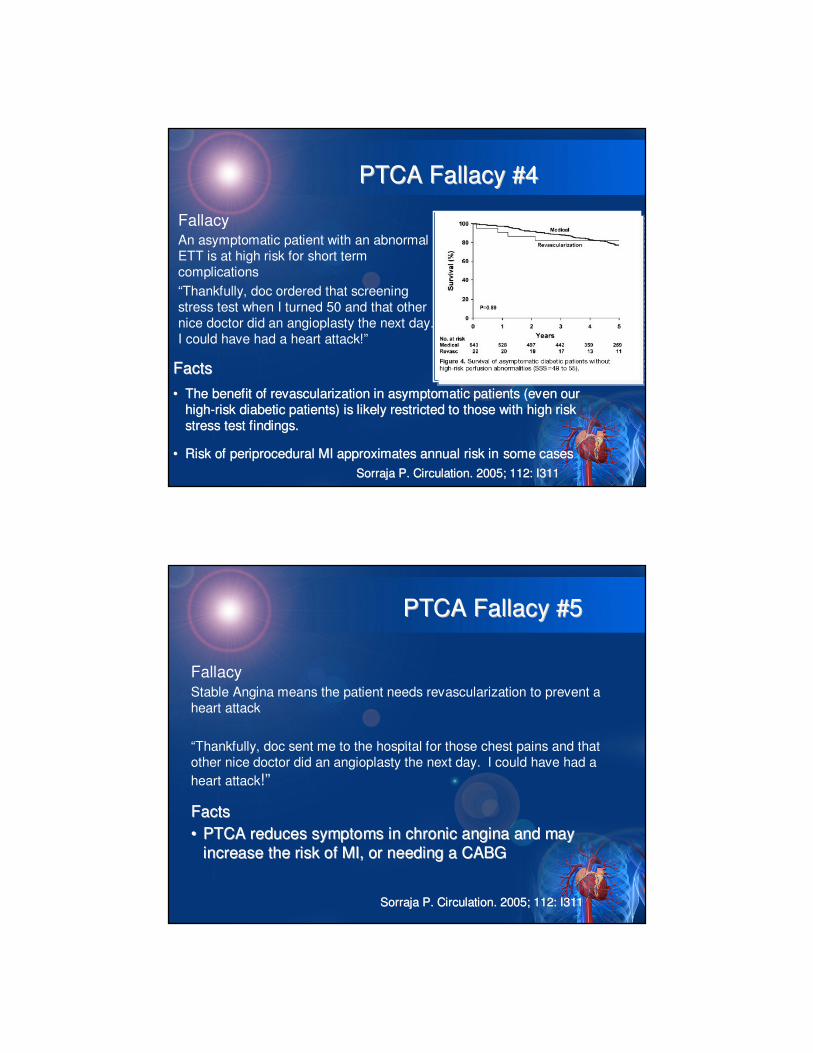
PTCA Fallacy #4PTCA Fallacy #4
FallacyAn asymptomatic patient with an abnormal ETT is at high risk for short term complications
“Thankfully, doc ordered that screening stress test when I turned 50 and that other nice doctor did an angioplasty the next day. I could have had a heart attack!”
FactsFacts
•• The benefit of revascularization in asymptomatic patients (even The benefit of revascularization in asymptomatic patients (even our our
highhigh--risk diabetic patients) is likely restricted to those with high risk diabetic patients) is likely restricted to those with high risk risk
stress test findings.stress test findings.
•• Risk of periprocedural MI approximates annual risk inRisk of periprocedural MI approximates annual risk in some casessome cases
Sorraja P. Circulation. 2005; 112: I311Sorraja P. Circulation. 2005; 112: I311
PTCA Fallacy #5PTCA Fallacy #5
FallacyStable Angina means the patient needs revascularization to prevent a heart attack
“Thankfully, doc sent me to the hospital for those chest pains and that other nice doctor did an angioplasty the next day. I could have had a
heart attack!”
FactsFacts
•• PTCA PTCA reduces symptoms in chronic angina and may reduces symptoms in chronic angina and may
increase the risk of MI, or needing a CABGincrease the risk of MI, or needing a CABG
Sorraja P. Circulation. 2005; 112: I311Sorraja P. Circulation. 2005; 112: I311
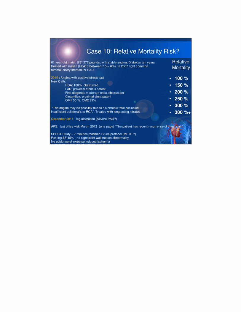
Case 10: Relative Mortality Risk?
• 100 %
• 150 %
• 200 %
• 250 %
• 300 %
• 300 %+
61 year-old male, 5’6” 272 pounds, with stable angina. Diabetes ten years
treated with insulin (HbA1c between 7.5 – 8%). In 2007 right common
femoral artery stented for PAD.
2010 : Angina with positive stress test
New Cath:
RCA: 100% obstructed
LAD: proximal stent is patent
First diagonal: moderate ostial obstruction
Circumflex: proximal stent patent
OM1 50 %; OM2 99%
“The angina may be possibly due to his chronic total occlusion –
insufficient collateral's to RCA”. Treated with long acting nitrates
December 2011: leg ulceration (Severe PAD?)
APS: last office visit March 2012 (one page) “The patient has recent recurrence of chest pain”
SPECT Study – 7 minutes modified Bruce protocol (METS ?)
Resting EF 45% - no significant wall motion abnormality
No evidence of exercise induced ischemia
Relative
Mortality