Case Report Synchronous Fibrolamellar Hepatocellular...
6
Case Report Synchronous Fibrolamellar Hepatocellular Carcinoma and Auricular Myxoma Yessica M. González-Cantú, 1 Cristina Rodriguez-Padilla, 1 Martha Lilia Tena-Suck, 2 Alberto García de la Fuente, 1 Rosa María Mejía-Bañuelos, 3 Raymundo Díaz Mendoza, 1 Samuel Quintanilla-Garza, 4 and Yolaester Batisda-Acuña 5 1 Laboratorio de Inmunolog´ ıa y Virolog´ ıa, Facultad de Ciencias Biol´ ogicas, Universidad Aut´ onoma de Nuevo Le´ on, Avenida Manuel L. Barrag´ an S/N, Ciudad Universitaria, 66451 San Nicol´ as de los Garza, NL, Mexico 2 Departamento de Neuropatolog´ ıa, Instituto Nacional de Neurolog´ ıa y Neurocirug´ ıa, Manuel Velasco Su´ arez, Avenida Insurgentes Sur 3877, Colonia La Fama, 14269 Delegaci´ on Tlalpan, DF, Mexico 3 Hospital General de Zona 17, Instituto Mexicano del Seguro Social, Fortunato Lozano y Roble Colonia Benito Ju´ arez, 64420 Monterrey, NL, Mexico 4 Unidad de Medicina Familiar No. 73, Instituto Mexicano del Seguro Social, Pablo de Mej´ ıa No. 526, Colonia Zona Centro, 25000 Saltillo, COAH, Mexico 5 Hospital General Regional No. 1, Instituto Mexicano del Seguro Social, Bosques de los Olivos 101, 61301 Morelia, MICH, Mexico Correspondence should be addressed to Yessica M. Gonz´ alez-Cant´ u; [email protected] Received 24 July 2015; Accepted 3 September 2015 Academic Editor: Tibor Tot Copyright © 2015 Yessica M. Gonz´ alez-Cant´ u et al. is is an open access article distributed under the Creative Commons Attribution License, which permits unrestricted use, distribution, and reproduction in any medium, provided the original work is properly cited. Synchronic occurrence of benign and malignant tumors is extremely rare. Fibrolamellar hepatocellular carcinoma represents 1% to 2% of all hepatocarcinomas, while myxomas represent about half of all the cases of primary tumors of the heart. We present the case of a 53-year-old woman with a leſt atrial myxoma that was surgically removed. Several weeks later, the patient returned to the hospital with abdominal pain. CT scan showed a mass in the leſt lobe of the liver that was resected and diagnosed as fibrolamellar hepatocellular carcinoma. As of this writing, the patient is healthy. 1. Introduction Hepatocellular carcinoma (HCC) represents 80% to 90% of all liver cancers [1]. According to the International Classifica- tion of Disease for Oncology, HCC is categorized as HCC-not otherwise specified (HCC-NOS), scirrhous HCC, spindle cell variant HCC, clear cell HCC, pleomorphic HCC, and fibro- lamellar HCC (FLHCC) [2]. FLHCC is a relatively rare form of HCC that affects mostly young people and is not associated with any diseases of the liver such as cirrhosis or hepatitis virus infection [3]. FLHCC is less aggressive than HCC [4]. Primary cardiac tumors are rare, representing 0.0017% to 0.19% of autopsy cases [5]. Myxomas represent about 50% of all cardiac tumors [6] and are located mostly in the leſt atrium (leſt-right ratio = 4 : 1) [5]. Most cardiac myxomas are sporadic, although a small percentage of them are familial and sometimes attributable to Carney syndrome. Here we describe the case of a 53-year-old woman who was diagnosed with fibrolamellar hepatocellular carcinoma synchronous with leſt atrial myxoma. 2. Case Presentation A 53-year-old female with no known medical conditions reported the start of symptoms 4 months before consultation. e patient reported palpitations that exacerbated by cardio- vascular exercise and attenuated with rest. e cardiologist requested an ECG and diagnosed sinus tachycardia. Subse- quently, a transthoracic echocardiography was performed, and a mass dependent on the leſt atrium compatible with Hindawi Publishing Corporation Case Reports in Pathology Volume 2015, Article ID 241708, 5 pages http://dx.doi.org/10.1155/2015/241708
Transcript of Case Report Synchronous Fibrolamellar Hepatocellular...
Case ReportSynchronous Fibrolamellar Hepatocellular Carcinoma andAuricular Myxoma
Yessica M. González-Cantú,1 Cristina Rodriguez-Padilla,1 Martha Lilia Tena-Suck,2
Alberto García de la Fuente,1 Rosa María Mejía-Bañuelos,3 Raymundo Díaz Mendoza,1
Samuel Quintanilla-Garza,4 and Yolaester Batisda-Acuña5
1Laboratorio de Inmunologıa y Virologıa, Facultad de Ciencias Biologicas, Universidad Autonoma de Nuevo Leon,Avenida Manuel L. Barragan S/N, Ciudad Universitaria, 66451 San Nicolas de los Garza, NL, Mexico2Departamento de Neuropatologıa, Instituto Nacional de Neurologıa y Neurocirugıa, Manuel Velasco Suarez,Avenida Insurgentes Sur 3877, Colonia La Fama, 14269 Delegacion Tlalpan, DF, Mexico3Hospital General de Zona 17, Instituto Mexicano del Seguro Social, Fortunato Lozano y Roble Colonia Benito Juarez,64420 Monterrey, NL, Mexico4Unidad de Medicina Familiar No. 73, Instituto Mexicano del Seguro Social, Pablo de Mejıa No. 526, Colonia Zona Centro,25000 Saltillo, COAH, Mexico5Hospital General Regional No. 1, Instituto Mexicano del Seguro Social, Bosques de los Olivos 101, 61301 Morelia, MICH, Mexico
Correspondence should be addressed to Yessica M. Gonzalez-Cantu; [email protected]
Received 24 July 2015; Accepted 3 September 2015
Academic Editor: Tibor Tot
Copyright © 2015 Yessica M. Gonzalez-Cantu et al. This is an open access article distributed under the Creative CommonsAttribution License, which permits unrestricted use, distribution, and reproduction in any medium, provided the original work isproperly cited.
Synchronic occurrence of benign and malignant tumors is extremely rare. Fibrolamellar hepatocellular carcinoma represents 1%to 2% of all hepatocarcinomas, while myxomas represent about half of all the cases of primary tumors of the heart. We present thecase of a 53-year-old woman with a left atrial myxoma that was surgically removed. Several weeks later, the patient returned to thehospital with abdominal pain. CT scan showed a mass in the left lobe of the liver that was resected and diagnosed as fibrolamellarhepatocellular carcinoma. As of this writing, the patient is healthy.
1. Introduction
Hepatocellular carcinoma (HCC) represents 80% to 90% ofall liver cancers [1]. According to the International Classifica-tion ofDisease forOncology, HCC is categorized asHCC-nototherwise specified (HCC-NOS), scirrhousHCC, spindle cellvariant HCC, clear cell HCC, pleomorphic HCC, and fibro-lamellar HCC (FLHCC) [2]. FLHCC is a relatively rare formofHCC that affectsmostly young people and is not associatedwith any diseases of the liver such as cirrhosis or hepatitisvirus infection [3]. FLHCC is less aggressive than HCC [4].
Primary cardiac tumors are rare, representing 0.0017% to0.19% of autopsy cases [5]. Myxomas represent about 50%of all cardiac tumors [6] and are located mostly in the leftatrium (left-right ratio = 4 : 1) [5]. Most cardiac myxomas are
sporadic, although a small percentage of them are familialand sometimes attributable to Carney syndrome.
Here we describe the case of a 53-year-old woman whowas diagnosed with fibrolamellar hepatocellular carcinomasynchronous with left atrial myxoma.
2. Case Presentation
A 53-year-old female with no known medical conditionsreported the start of symptoms 4months before consultation.The patient reported palpitations that exacerbated by cardio-vascular exercise and attenuated with rest. The cardiologistrequested an ECG and diagnosed sinus tachycardia. Subse-quently, a transthoracic echocardiography was performed,and a mass dependent on the left atrium compatible with
Hindawi Publishing CorporationCase Reports in PathologyVolume 2015, Article ID 241708, 5 pageshttp://dx.doi.org/10.1155/2015/241708

2 Case Reports in Pathology
(a) (b)
(c) (d)
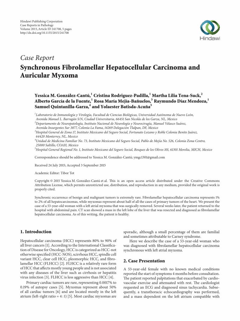
Figure 1: (a) Macroscopic image of the left auricular myxoma. (b) shows fusiform hyperchromatic cells immersed in a myxoid matrix andthe cardiac muscle (H and E stain, 50x). (c) Groups of fusiform cells immersed in a myxoid matrix surrounded by abundant inflammatorycells (H and E stain, 100x). (d) Fusiform cells under higher magnification (H and E stain, 400x).
auricular myxoma was detected. The patient was scheduledfor surgery, a median sternotomy for a transseptal approachto the right atrium was performed, and the patient begantreatment with IMMUNEPOTENT CRP (bovine dialyzableleukocyte extract) which was administered orally to promotepostsurgical recovery.
The patient underwent surgery without complicationsand was discharged from the hospital. The mass was diag-nosed by pathology as left atrial myxoma with muralthrombus (Figure 1). Three months after discharge from thehospital, the patient presented epigastric abdominal pain ofmoderate intensity that irradiated to the mesogastrium andright upper quadrant, which was accompanied by postpran-dial pain, nausea, and constipation.The patient was admittedto the hospital to evaluate her abdominal pain. Her heart ratewas 100 bpm and her blood pressure was 130/80mmHg; alllaboratory tests were normal.
A CT scan of the abdomenwas performed, which showed8 × 8 × 7.3 cm mass in the left lobe of the liver (Figure 2(a)).The lesion was hypervascular and had central calcificationand a tendency toward necrosis (Figures 2(b) and 2(c)). Thepatient underwent surgery for tumor resection.
After postoperative analysis from pathology, the diag-nosis was of a 9 cm, Grade III moderately differentiatedfibrolamellar hepatocellular carcinoma with clean resectionborders (AJCC, T2, N0, Mx) (Figures 3(a)–3(d)). The patientis currently asymptomatic and receives annual check-ups.
3. Discussion
Multiple primary malignant neoplasms (MPMN) are definedas a diagnosis of two or more independent primary malig-nancies of different histologies/origins in an individual [7].They are classified as MPMN according to the followingcriteria: tumors must be malignant, they must be located indifferent organs or tissues, and the possibility of metastasismust be ruled out completely. The incidence rate of MPNMvaries from 0.7% to 11.7% [8]. The association of benign andmalignant tumors, in absence of underlying genetic diseases,is extremely rare [9].
Fibrolamellar hepatocellular carcinoma and cardiacmyx-oma are a rare presentation of synchronous tumors. Ochoaet al. (2011) described one case of hepatocarcinoma syn-chronous with atrial myxoma [9]. There at least two cases

Case Reports in Pathology 3
(a)
(b)
(c)
Figure 2: Images from the abdominal CT. (a) and (b) show the 8 × 8 × 7.3 cm tumor located in segment IV of the left lobe of the liver thatlaterally displaces the gallbladder. The lesion has defined lobular borders that show a heterogeneous peripheral areas and a hypodense centerwith calcification. (c) shows changes in the sternum with presence of surgical material.
(a) (b)
(c) (d)
Figure 3: (a) Macroscopic image of the liver that shows a 9 × 7 cm, central yellowish lesion with diffuse, infiltrating borders with a whitishcentral lesion. (b) shows neoplastic cells separated into nodules by connective tissue (H and E stain, 50x). (c) Neoplastic cells with loss of thenucleus-cytoplasm relationship (H and E stain, 100x). (d) Image that shows the cellular and nuclear pleomorphism in greater detail (H and Estain, 400x).

4 Case Reports in Pathology
of metastasis mimicking atrial myxoma in hepatocellularcarcinoma [1, 10]. Fibrolamellar hepatocellular carcinoma(FLHCC) usually appears in younger patients (median age of25 years) [11], although recent studies show two distinct peaksof incidence, the first at 10 to 30 years of age and the second at70 to 79 years of age [2, 4]. At the moment of diagnosis, ourpatient was 53 years old, which is inconsistent with the datareported in the literature.
FLHCC is usually asymptomatic, and most patients visita doctor when they note the presence of an abdominal mass.Those patients with symptomatology report vague symptomssuch as weight loss and abdominal pain or discomfort [3].Imaging studies, such as ultrasound, MRI, and CT scans, arean integral part of FLHCC diagnosis [4]. Our patient wasdiagnosed with FLHCC during a CT scan performed a fewweeks after surgery to remove myxoma.
Liver function tests for patients with FLHCC are usuallynormal, although sometimes alpha-fetoprotein and amino-transferases show slightly increased levels [3, 12].
FHLCC prefers the left lobe of the liver, and, grossly, itis usually yellow to pale tan and firm, with an average sizeof 6–19 cm at its longest diameter. About 75% of tumorspresent a central scar; some tumors present foci of necrosis,hemorrhage, and calcification [3, 11].
Histopathologically, the tumor is composed of large po-lygonal cells with large vesicular nuclei containing marginal-ized chromatin and prominent nucleoli with eosinophilic(oncocytic) cytoplasm surrounded by distinctly lamellarstroma. Calcifications and pale bodies are seen in more than50% of cases [11, 13]. Stainable copper is found in 75% ofall FLHCC cases [12]. Immunohistochemical markers forFHLCC are CK7, EMA, mCEA, CA19-9, EpCAM, and CD68[11, 13]. Whenever possible, complete surgical resection is thepreferred treatment for FLHCC [4].
Myxomas represent 50% of the very rare cases of primarycardiac tumors. The most frequent location for myxomas isthe left atrium, and they usually originate from the endo-cardium of the atrial septum [14, 15]. Generally, myxomasappear in women aged 30 to 70 years. Clinically, myxomasare characterized by dyspnea, syncope, arrhythmias, edema,hemoptysis, and sudden death. Some of the systemic charac-teristics of myxoma include embolic phenomena in 30% to40% of patients. Approximately 10% to 15% of patients areasymptomatic and the myxoma is found accidentally dur-ing imaging studies [16]. Transthoracic or transesophagealechocardiography andMRI are the most common diagnostictools for neoplasms of the heart [14]. In our case, the patientreported tachycardia, which led to the discovery of themyxoma by means of transesophageal echocardiography.
Surgery is the election treatment for myxoma; it must beperformed without delay because of the high risk of suddendeath from thromboembolism or valvular obstruction; resec-tion of the tumor must be total [14, 17].
Macroscopically, myxomas are pedunculated and gelati-nous, and their surface can be smooth, villous, or friable and,in some cases, they show areas of hemorrhage.Their diameterranges from 1 to 15 cm, and theirweight ranges between 15 and180 grams [5, 18].
Histologically, myxomas are made up of stellate andpolyhedral (lepidic) cells.These tumor cells have small roundor oval nuclei and variable amounts of cytoplasm. Myxomacells can appear singly or in small clusters or chords, andwhen grouped around small spaces they can simulate glan-dular structures; they rarely show mitotic activity. Myxomashave abundant, fibrillary, slightly basophilic, mucoid matrix[19]. These tumors may form large and small fibrin-plateletthrombi, which may be an additional source of emboli [19].
Myxomas show a diffuse positive reaction for theimmunohistochemical marker Vimentin and focal expres-sion of CD34, CD68, and SMA [20]. They also have variablepositivity for cytokeratin, S100, factor VIII, and Calretinin[5, 20].
The IMMUNEPOTENT CRP (bovine dialyzable leuko-cyte extract) is a mixture of low molecular weight substancesand is a drug capable of modulating the immune response.It has been used for years in clinical trials of multiple typesof diseases such as cancer, toxic shock, and postoperativerecovery, as in the clinical case presented here, increasing thequality of life of the patients [21, 22].
Synchronous tumors are rare and the coexistence ofmalignant and benign tumors is rarer still. Additionally,the association of fibrolamellar hepatocellular carcinomaand auricular myxoma is scarcely reported in the literature.In our case, our patient was treated in accordance withthe established surgical procedures, received oral doses ofIMMUNEPOTENT CRP, and, as of this writing, is com-pletely asymptomatic.
Conflict of Interests
The authors declare that there is no conflict of interestsregarding the publication of this paper.
Acknowledgments
The authors wish to thank the Laboratorio de Inmunologıay Virologıa (Facultad de Ciencias Biologicas, UniversidadAutonoma de Nuevo Leon) for the economic support. Theyalso thank MSc. Alejandra Arreola-Triana for her editorialwork on this paper.
References
[1] A. M. Anwar, Y. F. M. Nosir, M. A. R. Chamsi-Pasha, A. Ajam,andH. Chamsi-Pasha, “Right atrial metastasis mimickingmyx-oma in advanced hepatocellular carcinoma,” Echocardiography,vol. 27, no. 1, pp. 80–83, 2010.
[2] T. Eggert, K. A. McGlynn, A. Duffy, M. P. Manns, T. F. Greten,and S. F. Altekruse, “Fibrolamellar hepatocellular carcinoma inthe USA, 2000–2010: a detailed report on frequency, treatmentand outcome based on the surveillance, epidemiology, and endResults database,” United European Gastroenterology Journal,vol. 1, no. 5, pp. 351–357, 2013.
[3] P. Herman, A. L. Chagas, M. V. Perini et al., “Surgical treatmentof fibrolamellar hepatocellular carcinoma: an underestimatedmalignant tumor?” Hepatobiliary & Pancreatic Diseases Inter-national, vol. 13, no. 6, pp. 618–621, 2014.

Case Reports in Pathology 5
[4] I. I. P. Lim, B. A. Farber, and M. P. LaQuaglia, “Advancesin fibrolamellar hepatocellular carcinoma: a review,” EuropeanJournal of Pediatric Surgery, vol. 24, no. 6, pp. 461–466, 2014.
[5] Y. Wang and F. E. Sharkey, “Myxoma of the small bowel ina 47-year-old woman with a left atrial myxoma,” Archives ofPathology and Laboratory Medicine, vol. 127, no. 4, pp. 481–484,2003.
[6] N. Barbetakis, T. Xenikakis, A. Efstathiou, G. Samanidis, C.Tsilikas, and I. Fessatidis, “Synchronous left ventricular myx-oma andmalignant fibrous histiocytoma: simultaneous surgicalmanagement,” Hellenic Journal of Cardiology, vol. 49, no. 5, pp.371–373, 2008.
[7] M. Fernandez-Ruiz, J.-M. Guerra-Vales, F.-J. Castelbon-Fernandez, J. Llenas-Garcıa, L. Caurcel-Dıaz, and F. Colina-Ruizdelgado, “Multiple primary malignancies in Spanishpatients with hepatocellular carcinoma: analysis of a hospital-based tumor registry,” Journal of Gastroenterology andHepatology, vol. 24, no. 8, pp. 1424–1430, 2009.
[8] S. J. Oh, D. S. Bae, and B. J. Suh, “Synchronous triple primarycancers occurring in the stomach, kidney, and thyroid,” Annalsof Surgical Treatment and Research, vol. 88, no. 6, pp. 345–348,2015.
[9] M. L. Ochoa, A. G. C. Gordillo, and M. M. Perez, “Carcinomahepatocelular sincronico con mixoma auricular. Informe de uncaso,” Investigacion Clınica, vol. 52, no. 2, 2011.
[10] E. Maffei, T. Arcadi, and F. Cademartiri, “Hepatocellular car-cinoma mimicking an atrial mixoma,” European Heart Journal,vol. 35, no. 13, p. 876, 2014.
[11] D. Castro-Villabon, L. E. Barrera-Herrera, P. A. Rodrıguez-Urrego et al., “Hepatocellular carcinomawith both fibrolamellarand classical components: an unusual morphological pattern,”Case Reports in Pathology, vol. 2015, Article ID 609780, 5 pages,2015.
[12] S. Kakar, L. J. Burgart, K. P. Batts, J. Garcia, D. Jain, and L. D.Ferrell, “Clinicopathologic features and survival in fibrolamellarcarcinoma: comparison with conventional hepatocellular carci-noma with and without cirrhosis,” Modern Pathology, vol. 18,no. 11, pp. 1417–1423, 2005.
[13] V. B. Weeda, M. Murawski, A. J. McCabe et al., “Fibrolamellarvariant of hepatocellular carcinoma does not have a bettersurvival than conventional hepatocellular carcinoma—resultsand treatment recommendations from the Childhood LiverTumour Strategy Group (SIOPEL) experience,” European Jour-nal of Cancer, vol. 49, no. 12, pp. 2698–2704, 2013.
[14] K. Durgut, N. Gormus, M. Ozulku, U. Ozergin, and C. Ozpinar,“Clinical features and surgical treatment of cardiac myxoma:report of 18 cases,” Asian Cardiovascular and Thoracic Annals,vol. 10, no. 2, pp. 111–114, 2002.
[15] P. Becker R, A. Ramırez M, R. Zalaquett S et al., “Mixomacardiaco: caracterizacion clınica, metodos diagnosticos y resul-tados alejados del tratamiento quirurgico durante tres decadasde experiencia,” RevistaMedica de Chile, vol. 136, no. 3, pp. 287–295, 2008.
[16] S.Mehra, Y. Parviz, andA. Al-Mohammad, “Left atrial myxomawith concurrent saddle pulmonary embolism and duke’s Bcolorectal adenocarcinoma,” Journal of Medical Cases, 2013.
[17] G. Samanidis, K. Perreas, P. Kalogris et al., “Surgical treatmentof primary intracardiac myxoma: 19 years of experience,”Interactive Cardiovascular and Thoracic Surgery, vol. 13, no. 6,pp. 597–600, 2011.
[18] R. Cohen, G. Singh, D. Mena, C. A. Garcia, P. Loarte, andB. Mirrer, “Atrial myxoma: a case presentation and review,”Cardiology Research, vol. 3, no. 1, pp. 41–44, 2012.
[19] L. E. Wold and J. T. Lie, “Cardiac myxomas: a clinicopathologicprofile,” The American Journal of Pathology, vol. 101, no. 1, pp.219–240, 1980.
[20] J.-G. Wang, Y.-J. Li, H. Liu, N.-N. Li, J. Zhao, and X.-M. Xing,“Clinicopathologic analysis of cardiac myxomas: Seven years’experience with 61 patients,” Journal of Thoracic Disease, vol. 4,no. 3, pp. 272–283, 2012.
[21] M. A. Franco-Molina, “Anti-inflammatory and antioxidanteffects of IMMUNEPOTENT CRP in Lipopolysaccharide(LPS)-stimulated human macrophages,” African Journal ofMicrobiology Research, vol. 5, no. 22, 2011.
[22] H. H. Lara, L. I. Turrent, E. N. Garza-Trevino, R. Tamez-Guerra, and C. Rodriguez-Padilla, “Clinical and immunolog-ical assessment in breast cancer patients receiving anticancertherapy and bovine dialyzable leukocyte extract as an adjuvant,”Experimental and Therapeutic Medicine, vol. 1, no. 3, pp. 425–431, 2010.

Submit your manuscripts athttp://www.hindawi.com
Stem CellsInternational
Hindawi Publishing Corporationhttp://www.hindawi.com Volume 2014
Hindawi Publishing Corporationhttp://www.hindawi.com Volume 2014
MEDIATORSINFLAMMATION
of
Hindawi Publishing Corporationhttp://www.hindawi.com Volume 2014
Behavioural Neurology
EndocrinologyInternational Journal of
Hindawi Publishing Corporationhttp://www.hindawi.com Volume 2014
Hindawi Publishing Corporationhttp://www.hindawi.com Volume 2014
Disease Markers
Hindawi Publishing Corporationhttp://www.hindawi.com Volume 2014
BioMed Research International
OncologyJournal of
Hindawi Publishing Corporationhttp://www.hindawi.com Volume 2014
Hindawi Publishing Corporationhttp://www.hindawi.com Volume 2014
Oxidative Medicine and Cellular Longevity
Hindawi Publishing Corporationhttp://www.hindawi.com Volume 2014
PPAR Research
The Scientific World JournalHindawi Publishing Corporation http://www.hindawi.com Volume 2014
Immunology ResearchHindawi Publishing Corporationhttp://www.hindawi.com Volume 2014
Journal of
ObesityJournal of
Hindawi Publishing Corporationhttp://www.hindawi.com Volume 2014
Hindawi Publishing Corporationhttp://www.hindawi.com Volume 2014
Computational and Mathematical Methods in Medicine
OphthalmologyJournal of
Hindawi Publishing Corporationhttp://www.hindawi.com Volume 2014
Diabetes ResearchJournal of
Hindawi Publishing Corporationhttp://www.hindawi.com Volume 2014
Hindawi Publishing Corporationhttp://www.hindawi.com Volume 2014
Research and TreatmentAIDS
Hindawi Publishing Corporationhttp://www.hindawi.com Volume 2014
Gastroenterology Research and Practice
Hindawi Publishing Corporationhttp://www.hindawi.com Volume 2014
Parkinson’s Disease
Evidence-Based Complementary and Alternative Medicine
Volume 2014Hindawi Publishing Corporationhttp://www.hindawi.com