Case report SPONTANEOUS PNEUMOTHORAX IN GERMAN …
6
Bulgarian Journal of Veterinary Medicine, 2020 ONLINE FIRST ISSN 1311-1477; DOI: 10.15547/bjvm.2303 Case report SPONTANEOUS PNEUMOTHORAX IN GERMAN SHORTHAIR POINTER DUE TO PENETRATION OF A GRASS AWN V. S. NEDEV 1 & I. I. KALKANOV 2 1 Department of Veterinary Surgery, 2 Department of General and Clinical Pathology, Faculty of Veterinary Medicine, Trakia University, Stara Zagora, Bulgaria Summary Nedev, V. S. & I. I. Kalkanov, 2020. Spontaneous pneumothorax in German Shorthair Pointer due to penetration of a grass awn. Bulg. J. Vet. Med. (online first). This case report describes an incident of pneumothorax in 4-month old male German Shorthaired Pointer caused by a grass awn. The patient was brought to the Small Animal Clinic of the Faculty of Veterinary Medicine at Trakia University, Stara Zagora, Bulgaria with respiratory distress and anxi- ety. Tachypnea, cyanotic mucous membranes and tachycardia were observed during physical exami- nation. Abnormal collection of air in the pleural space between the lung and the chest was observed radiographically. A diagnosis of primary spontaneous closed pneumothorax was made based on these findings. Later pathoanatomical findings showed the presence of the grass awn that had penetrated through the skin. Key words: closed, grass awn, dog pneumothorax Pneumothorax is an emergency condition in both dogs and cats. It occurs when free air or gas is accumulated within the tho- racic cavity (Dinev & Simeonova, 2007). According to the etiology, pneumothorax can be divided into: spontaneous, trau- matic or iatrogenic (Pawloski & Broad- dus, 2010). Also, it can be classified as open, closed and tension, according to the pathophysiology. Spontaneous tension pneumothorax is defined as a condition in which a flap of tissue acts as a one-way valve and air accumulated in the pleural space during inspiration cannot be ex- pelled during expiration (Monnet, 2003). Etiology of spontaneous pneumothorax in animals include: bullous emphysema, pulmonary blebs, pulmonary neoplasia, dirofilariasis, Paragonimus spp., tape- worm infestation, Aelurostrongylus ab- strusus, bacterial and viral pneumonia, pulmonary abscesses, and parasitic granu- lomas (Pawloski & Broaddus, 2010). Mi- gration of aspirated foreign bodies is an unusual cause for pneumothorax. Occa- sionally, grass awns may migrate via the trachea to the lungs and after perforating them could reach the diaphragm (Hunt et al., 2004), but there are no reports about
Transcript of Case report SPONTANEOUS PNEUMOTHORAX IN GERMAN …

Bulgarian Journal of Veterinary Medicine, 2020 ONLINE FIRST ISSN 1311-1477; DOI: 10.15547/bjvm.2303
Case report
SPONTANEOUS PNEUMOTHORAX IN GERMAN SHORTHAIR POINTER DUE TO PENETRATION OF A GRASS AWN
V. S. NEDEV1 & I. I. KALKANOV2
1Department of Veterinary Surgery, 2Department of General and Clinical Pathology, Faculty of Veterinary Medicine, Trakia University, Stara Zagora, Bulgaria
Summary
Nedev, V. S. & I. I. Kalkanov, 2020. Spontaneous pneumothorax in German Shorthair Pointer due to penetration of a grass awn. Bulg. J. Vet. Med. (online first). This case report describes an incident of pneumothorax in 4-month old male German Shorthaired Pointer caused by a grass awn. The patient was brought to the Small Animal Clinic of the Faculty of Veterinary Medicine at Trakia University, Stara Zagora, Bulgaria with respiratory distress and anxi-ety. Tachypnea, cyanotic mucous membranes and tachycardia were observed during physical exami-nation. Abnormal collection of air in the pleural space between the lung and the chest was observed radiographically. A diagnosis of primary spontaneous closed pneumothorax was made based on these findings. Later pathoanatomical findings showed the presence of the grass awn that had penetrated through the skin.
Key words: closed, grass awn, dog pneumothorax
Pneumothorax is an emergency condition in both dogs and cats. It occurs when free air or gas is accumulated within the tho-racic cavity (Dinev & Simeonova, 2007). According to the etiology, pneumothorax can be divided into: spontaneous, trau-matic or iatrogenic (Pawloski & Broad-dus, 2010). Also, it can be classified as open, closed and tension, according to the pathophysiology. Spontaneous tension pneumothorax is defined as a condition in which a flap of tissue acts as a one-way valve and air accumulated in the pleural space during inspiration cannot be ex-pelled during expiration (Monnet, 2003).
Etiology of spontaneous pneumothorax in animals include: bullous emphysema, pulmonary blebs, pulmonary neoplasia, dirofilariasis, Paragonimus spp., tape-worm infestation, Aelurostrongylus ab-strusus, bacterial and viral pneumonia, pulmonary abscesses, and parasitic granu-lomas (Pawloski & Broaddus, 2010). Mi-gration of aspirated foreign bodies is an unusual cause for pneumothorax. Occa-sionally, grass awns may migrate via the trachea to the lungs and after perforating them could reach the diaphragm (Hunt et al., 2004), but there are no reports about
Spontaneous pneumothorax in German Shorthair Pointer due to penetration of a grass awn
BJVM, ××, No × 2
grass awn penetrating through the skin and causing pneumothorax.
The purpose of this case report was to present and discuss a rare case of pneu-mothorax in a dog, caused by unusual migration of a grass awn in the thoracic cavity.
Case presentation
A 4-month old male German Shorthaired Pointer was referred to the Small Animal Clinic in the Faculty of Veterinary Medi-cine, Trakia University, Stara Zagora, Bulgaria. The dog’s history revealed pro-gressive worsening of health manifested by dyspnoea, decreased appetite and anxi-ety. A treatment with antibiotics, NSAIDs, vitamin C was prescribed by a private veterinarian, which improved the dog’s condition for a while, but the dog’s condi-tion worsened again in the morning of referral. The animal was trained in fields full of widely growing grasses.
When admitted to the clinic, the dog has a body temperature 39.3 oC, pulse rate of 180 bpm and showed tachypnea, ab-
dominal breathing and cyanotic mucous membranes. The dog was directed for an X-ray examination. Dorsoventral and late-ral recumbent thoracic radiographs were taken. During the X-ray exam, the condi-tion of the dog worsened and cardiopul-monary resuscitation (CPR) was started. The outcome of CPR was lethal. To reveal the cause of death, a pathoanatomical exa-mination was done.
The clinical examination did not re-veal any evidence of penetration through the skin since no scars or wounds were detected.
The lateral and dorsoventral radio-graphs (Fig. 1) revealed an air-filled chest cavity. The lungs were withdrawn from the thoracic wall, with increased density due to collapse and atelectasis. The heart silhouette was elevated in dorsoventral direction and the dome of the diaphragm was displaced in caudal direction.
The body of the dog was investigated according to the standard protocol of au-topsy. The macroscopic changes estab-lished by inspection comprised strong
Fig. 1. X-ray findings. Black arrow shows the caudal displacement of diaphragm’s dome. White arrow points at the elevated heart silhouette and lungs atelectasis. White stars show air-filled thoracic cavity.
V. S. Nedev & I. I. Kalkanov
BJVM, ××, No × 3
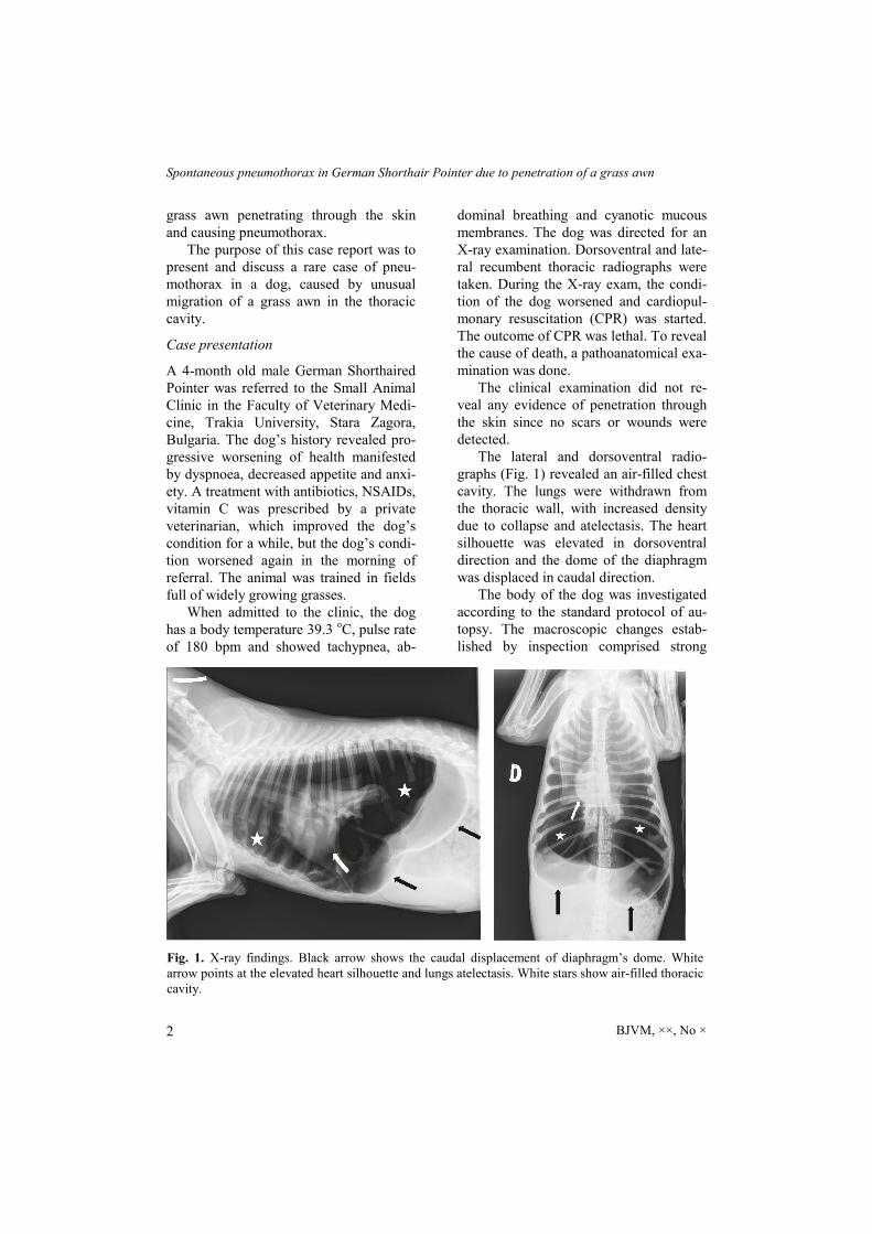
swelling of the body. Visible mucous membranes of conjunctivae and oral cavi-ty were cyanotic. After abdominal dissec-tion, diffuse hyperaemia on the surface of the abdominal organs was observed. The diaphragm was depressed as a result of the increased intrathoracic pressure in the cavity (Fig. 2). Because of the caudal dis-placement of the diaphragm, the liver,
stomach and intestines were pushed cau-dally. The liver had a congestive, brittle consistency, with gallbladder overflow. Mesenteric blood vessels were overfilled and mesenteric lymph nodes had reacted. Medulla of kidneys was hyperaemic.
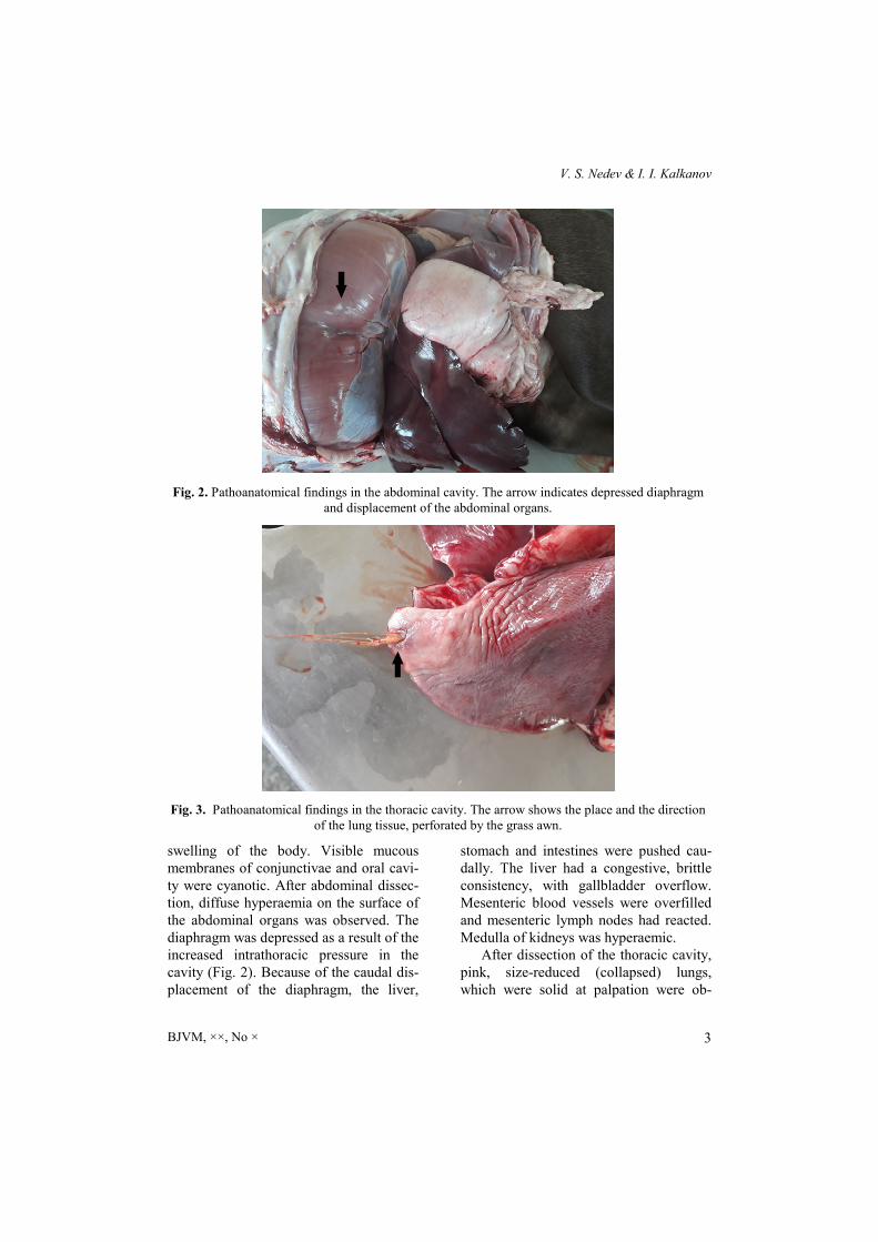
After dissection of the thoracic cavity, pink, size-reduced (collapsed) lungs, which were solid at palpation were ob-
Fig. 2. Pathoanatomical findings in the abdominal cavity. The arrow indicates depressed diaphragm and displacement of the abdominal organs.
Fig. 3. Pathoanatomical findings in the thoracic cavity. The arrow shows the place and the direction of the lung tissue, perforated by the grass awn.
Spontaneous pneumothorax in German Shorthair Pointer due to penetration of a grass awn
BJVM, ××, No × 4
served (Fig. 3). There was also a scanty amount of white foamy fluid. At the apical part of the right lung, the foreign body was ascertained. The foreign body was a grass awn, embedded in the lung tissue. In the 4th intercostal area, the inlet of the grass awn, that caused the pneumothorax, was detected.
Spontaneous pneumothorax is defined as a closed pneumothorax in which air accumulates in the pleural space in the absence of either a traumatic or iatrogenic cause (Pawloski & Broaddus, 2010). This type of pneumothorax has several differ-ent etiologies. It is classified as primary with no obvious clinical evidence of pul-monary damage and secondary, which has an obvious clinical evidence of pulmonary disease (Kramek & Caywood, 1987). Cli-nical signs of spontaneous pneumothorax include acute onset of dyspnoea, tachyp-noea, tachycardia, hypotension, cyanosis, decreased respiratory sounds and hyper-resonance on auscultation. Occasionally, depression, lethargy, anorexia, pyrexia and cough might be noted, depending on the underlying etiology of the spontaneous pne-umothorax (Radlinsky & Fossum, 2019). In our case, the same symptoms were pre-sent.
The continuous influx of air into the pleural cavity increases the intrapleural pressure. This makes the chest barrel-shaped and fixed in maximal extension, causing hypoventilation and compression of the major vessels (MacPhail & Fossum, 2019). The result is decreased venous return, cardiopulmonary collapse and death. The complaints in our case were probably a result of movement of the for-eign body. Rapid worsening of the dog’s condition in the last hours might be attrib-uted to the complete rupture of the foreign body capsule in the lung.
The most common cause of spontane-ous pneumothorax in the dogs is lacera-tion of pulmonary blebs or bullae (Lips-comb et al., 2003). In our case, the reason of the respiratory distress was a foreign body: grass awn. The hunting and working dogs are predisposed to retain the grass awn in the long hair coats, because of in-creased exposure to plants in the field while exercising and can aspirate it while breathing with open-mouth (Hopper et al., 2004). Grass awns have a tendency to migrate through the trachea and bronchi into the periphery of the lung parenchyma in an antegrade direction because of their sharp backward pointing barbs (Lotti & Niebauer, 1992). The history and clinical examination did not reveal any evidence of penetration through the skin since scars or wounds were not detected, although the lack of a wound does not rule out skin penetration.
Most dogs with foreign body aspi-ration develop clinical signs within hours to 15 days (Lotti & Niebauer, 1992). The duration of clinical signs in dogs with spontaneous pneumothorax is reported to range from 0 to 28 days (Puerto et al., 2002). The history of our patient revealed that it has been trained in fields with widely growing grasses and the foreign body was found to penetrate through the thoracic wall at necropsy. The caudal and the accessory lung lobes are the most commonly involved following inhalation of grass awns (Schultz & Zwingenberger, 2008). In the present case, the caudal lobe was also affected.
Clinical management is most difficult in patients with intrathoracic or sublumbar grass awns (Brennan & Ihrke, 1983). Out-come following surgery for spontaneous pneumothorax is more favourable than when nonsurgical means are used (Puerto et al., 2002). The migratory nature and
V. S. Nedev & I. I. Kalkanov
BJVM, ××, No × 5
small size of the grass awns makes local-isation and complete resection by explora-tory surgery difficult (Aronson & Grego-ry, 1995). The grass awn may have caused disease in the thorax, but also may have migrated to the abdomen by the time of surgery (Schultz & Zwingenberger, 2008).
Diagnosis of spontaneous pneumo-thorax is based on the clinical signs and the results of diagnostic imaging. Tho-racic radiography might show elevation of the cardiac silhouette from the sternum, increased lung lobe opacity due to atelec-tasis, retraction of the lung edges from the thoracic wall, flattened diaphragm and mediastinal shift (Tsioli et al., 2014). Ra-diographic diagnosis of pneumothorax requires differentiation between intrapul-monary air and free air in the pleural space (Monnet, 2003). It is important to remember that elevation of the cardiac silhouette away from the sternum may not be seen if the animal is in a standing posi-tion and horizontal beam radiography is performed (Pawloski & Broaddus, 2010). Differential diagnoses for X-ray findings include ruptured pulmonary neoplasm, infectious/inflammatory etiologies for ruptured pulmonary abscess or granuloma, trauma, and cysts (Puerto et al., 2002).
In conclusion, this is a rare case of pneumothorax due to a grass awn. Most cases in literature described penetration of the foreign body through the mouth or the nose, but there is no report about an inci-dent with grass awn penetrating through the skin.
REFERENCES
Aronson, L. R. & C. R. Gregory, 1995. Infec-tious pericardial effusion in five dogs. Vet-erinary Surgery, 24, 402–407.
Brennan, K. E. & P. J. Ihrke, 1983. Grass awn migration in dogs and cats: A retrospective
study of 182 cases. Journal of the Ameri-can Veterinary Medical Association, 182, 12011204.
Dinev, D. & G. Simeonova, 2007. Emergency Veterinary Medicine, 1st edn, Stara Zagora (BG).
Hopper, B. J., N. V. Lester, P. J. Irwin, C. E. Eger & J. L. Richardson, 2004. Imaging diagnosis: Pneumothorax and focal perito-nitis in a dog due to migration of an in-haled grass awn. Veterinary Radiolo-gy & Ultrasound, 45, 136–136.
Hunt, G. B., A. Worth & A. Marchevsky, 2004. Migration of wooden skewer foreign bodies from the gastrointestinal tract in eight dogs. The Journal of Small Animal Practice, 45, 362–367.
Kramek, B. A. & D. D. Caywood, 1987. Pneumothorax. The Veterinary Clinics of North America Small Animal Practice, 17, 285300.
Lipscomb, V. J., R. J. Hardie & R. R. Du-bielzig, 2003. Spontaneous pneumothorax caused by pulmonary blebs and bullae in 12 dogs. Journal of the American Animal Hospital Association, 39, 435445.
Lotti, U. & G. W. Niebauer, 1992. Tracheo-bronchial foreign bodies of plant origin in 153 hunting dogs. Compendium on Con-tinuing Education for the Practising Vet-erinarian, 14, 900–904.
MacPhail, C. & T. W. Fossum, 2019. Surgery of the lower respiratory system: Lungs and thoracic wall. In: Small Animal Surgery, 5th edn, ed T. Fossum, Elsevier, Phila-delphia, pp. 884916.
Monnet, E., 2003. Pleura and pleural space. In: Textbook of Small Animal Surgery, 3rd edn, ed D. Slatter, Saunders, pp. 387–405.
Pawloski, D & K. Broaddus, 2010. Pneumo-thorax: A Review. Journal of the Ameri-can Animal Hospital Association, 46, 385397.
Puerto, D. A., D. J. Brockman, C. Lindquist, K. Drobatz, 2002. Surgical and nonsurgi-cal management of and selected risk fac-tors for spontaneous pneumothorax in
Spontaneous pneumothorax in German Shorthair Pointer due to penetration of a grass awn
BJVM, ××, No × 6
dogs: 64 cases (1986-1999). Journal of the American Veterinary Medical Association, 220, 1670–1674.
Radlinsky, M. G. & T. W. Fossum, 2019. Sur-gery of the digestive system. In: Small Animal Surgery, 5th edn, ed T. Fossum, El-sevier, Philadelphia,. 5th edn, ed T. Fos-sum, Elsevier, Philadelphia, pp. 331512.
Schultz, R. M. & A. Zwingenberger, 2008. Radiographic, computed tomographic, and ultrasonographic findings with migrating intrathoracic grass awns in dogs and cats. Veterinary Radiology & Ultrasound, 49, 249255.
Tsioli, V., A. Limberis, D. Pardali & A. Galatos, 2014. Tension pneumothorax secondary to a grass awn in a dog. Veteri-nary Record Case Reports, 2, doi: 10.1136/vetreccr-2013-000039.
Paper received 04.11.2019; accepted for publication 20.01.2020
Correspondence: Vladi Nedev, DVM, PhD Department of Veterinary Surgery, Faculty of Veterinary Medicine, Trakia University, 6000 Stara Zagora, Bulgaria e-mail: [email protected]