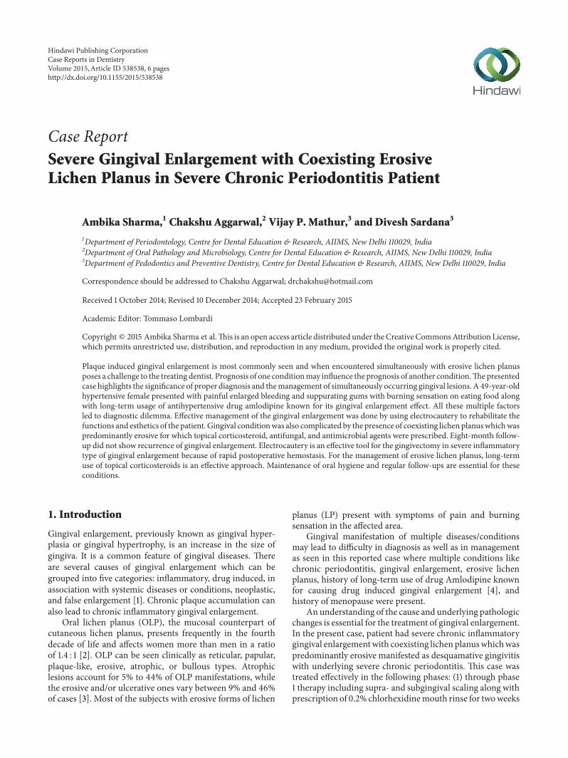
Case Report Severe Gingival Enlargement with Coexisting...
7
Case Report Severe Gingival Enlargement with Coexisting Erosive Lichen Planus in Severe Chronic Periodontitis Patient Ambika Sharma, 1 Chakshu Aggarwal, 2 Vijay P. Mathur, 3 and Divesh Sardana 3 1 Department of Periodontology, Centre for Dental Education & Research, AIIMS, New Delhi 110029, India 2 Department of Oral Pathology and Microbiology, Centre for Dental Education & Research, AIIMS, New Delhi 110029, India 3 Department of Pedodontics and Preventive Dentistry, Centre for Dental Education & Research, AIIMS, New Delhi 110029, India Correspondence should be addressed to Chakshu Aggarwal; [email protected] Received 1 October 2014; Revised 10 December 2014; Accepted 23 February 2015 Academic Editor: Tommaso Lombardi Copyright © 2015 Ambika Sharma et al. is is an open access article distributed under the Creative Commons Attribution License, which permits unrestricted use, distribution, and reproduction in any medium, provided the original work is properly cited. Plaque induced gingival enlargement is most commonly seen and when encountered simultaneously with erosive lichen planus poses a challenge to the treating dentist. Prognosis of one condition may influence the prognosis of another condition. e presented case highlights the significance of proper diagnosis and the management of simultaneously occurring gingival lesions. A 49-year-old hypertensive female presented with painful enlarged bleeding and suppurating gums with burning sensation on eating food along with long-term usage of antihypertensive drug amlodipine known for its gingival enlargement effect. All these multiple factors led to diagnostic dilemma. Effective management of the gingival enlargement was done by using electrocautery to rehabilitate the functions and esthetics of the patient. Gingival condition was also complicated by the presence of coexisting lichen planus which was predominantly erosive for which topical corticosteroid, antifungal, and antimicrobial agents were prescribed. Eight-month follow- up did not show recurrence of gingival enlargement. Electrocautery is an effective tool for the gingivectomy in severe inflammatory type of gingival enlargement because of rapid postoperative hemostasis. For the management of erosive lichen planus, long-term use of topical corticosteroids is an effective approach. Maintenance of oral hygiene and regular follow-ups are essential for these conditions. 1. Introduction Gingival enlargement, previously known as gingival hyper- plasia or gingival hypertrophy, is an increase in the size of gingiva. It is a common feature of gingival diseases. ere are several causes of gingival enlargement which can be grouped into five categories: inflammatory, drug induced, in association with systemic diseases or conditions, neoplastic, and false enlargement [1]. Chronic plaque accumulation can also lead to chronic inflammatory gingival enlargement. Oral lichen planus (OLP), the mucosal counterpart of cutaneous lichen planus, presents frequently in the fourth decade of life and affects women more than men in a ratio of 1.4 : 1 [2]. OLP can be seen clinically as reticular, papular, plaque-like, erosive, atrophic, or bullous types. Atrophic lesions account for 5% to 44% of OLP manifestations, while the erosive and/or ulcerative ones vary between 9% and 46% of cases [3]. Most of the subjects with erosive forms of lichen planus (LP) present with symptoms of pain and burning sensation in the affected area. Gingival manifestation of multiple diseases/conditions may lead to difficulty in diagnosis as well as in management as seen in this reported case where multiple conditions like chronic periodontitis, gingival enlargement, erosive lichen planus, history of long-term use of drug Amlodipine known for causing drug induced gingival enlargement [4], and history of menopause were present. An understanding of the cause and underlying pathologic changes is essential for the treatment of gingival enlargement. In the present case, patient had severe chronic inflammatory gingival enlargement with coexisting lichen planus which was predominantly erosive manifested as desquamative gingivitis with underlying severe chronic periodontitis. is case was treated effectively in the following phases: (1) through phase 1 therapy including supra- and subgingival scaling along with prescription of 0.2% chlorhexidine mouth rinse for two weeks Hindawi Publishing Corporation Case Reports in Dentistry Volume 2015, Article ID 538538, 6 pages http://dx.doi.org/10.1155/2015/538538
Transcript of Case Report Severe Gingival Enlargement with Coexisting...
Case ReportSevere Gingival Enlargement with Coexisting ErosiveLichen Planus in Severe Chronic Periodontitis Patient
Ambika Sharma,1 Chakshu Aggarwal,2 Vijay P. Mathur,3 and Divesh Sardana3
1Department of Periodontology, Centre for Dental Education & Research, AIIMS, New Delhi 110029, India2Department of Oral Pathology and Microbiology, Centre for Dental Education & Research, AIIMS, New Delhi 110029, India3Department of Pedodontics and Preventive Dentistry, Centre for Dental Education & Research, AIIMS, New Delhi 110029, India
Correspondence should be addressed to Chakshu Aggarwal; [email protected]
Received 1 October 2014; Revised 10 December 2014; Accepted 23 February 2015
Academic Editor: Tommaso Lombardi
Copyright © 2015 Ambika Sharma et al.This is an open access article distributed under theCreative CommonsAttribution License,which permits unrestricted use, distribution, and reproduction in any medium, provided the original work is properly cited.
Plaque induced gingival enlargement is most commonly seen and when encountered simultaneously with erosive lichen planusposes a challenge to the treating dentist. Prognosis of one conditionmay influence the prognosis of another condition.Thepresentedcase highlights the significance of proper diagnosis and themanagement of simultaneously occurring gingival lesions. A 49-year-oldhypertensive female presented with painful enlarged bleeding and suppurating gums with burning sensation on eating food alongwith long-term usage of antihypertensive drug amlodipine known for its gingival enlargement effect. All these multiple factorsled to diagnostic dilemma. Effective management of the gingival enlargement was done by using electrocautery to rehabilitate thefunctions and esthetics of the patient. Gingival conditionwas also complicated by the presence of coexisting lichen planuswhichwaspredominantly erosive for which topical corticosteroid, antifungal, and antimicrobial agents were prescribed. Eight-month follow-up did not show recurrence of gingival enlargement. Electrocautery is an effective tool for the gingivectomy in severe inflammatorytype of gingival enlargement because of rapid postoperative hemostasis. For the management of erosive lichen planus, long-termuse of topical corticosteroids is an effective approach. Maintenance of oral hygiene and regular follow-ups are essential for theseconditions.
1. Introduction
Gingival enlargement, previously known as gingival hyper-plasia or gingival hypertrophy, is an increase in the size ofgingiva. It is a common feature of gingival diseases. Thereare several causes of gingival enlargement which can begrouped into five categories: inflammatory, drug induced, inassociation with systemic diseases or conditions, neoplastic,and false enlargement [1]. Chronic plaque accumulation canalso lead to chronic inflammatory gingival enlargement.
Oral lichen planus (OLP), the mucosal counterpart ofcutaneous lichen planus, presents frequently in the fourthdecade of life and affects women more than men in a ratioof 1.4 : 1 [2]. OLP can be seen clinically as reticular, papular,plaque-like, erosive, atrophic, or bullous types. Atrophiclesions account for 5% to 44% of OLP manifestations, whilethe erosive and/or ulcerative ones vary between 9% and 46%of cases [3]. Most of the subjects with erosive forms of lichen
planus (LP) present with symptoms of pain and burningsensation in the affected area.
Gingival manifestation of multiple diseases/conditionsmay lead to difficulty in diagnosis as well as in managementas seen in this reported case where multiple conditions likechronic periodontitis, gingival enlargement, erosive lichenplanus, history of long-term use of drug Amlodipine knownfor causing drug induced gingival enlargement [4], andhistory of menopause were present.
An understanding of the cause and underlying pathologicchanges is essential for the treatment of gingival enlargement.In the present case, patient had severe chronic inflammatorygingival enlargementwith coexisting lichen planuswhichwaspredominantly erosive manifested as desquamative gingivitiswith underlying severe chronic periodontitis. This case wastreated effectively in the following phases: (1) through phase1 therapy including supra- and subgingival scaling along withprescription of 0.2% chlorhexidinemouth rinse for twoweeks
Hindawi Publishing CorporationCase Reports in DentistryVolume 2015, Article ID 538538, 6 pageshttp://dx.doi.org/10.1155/2015/538538
2 Case Reports in Dentistry
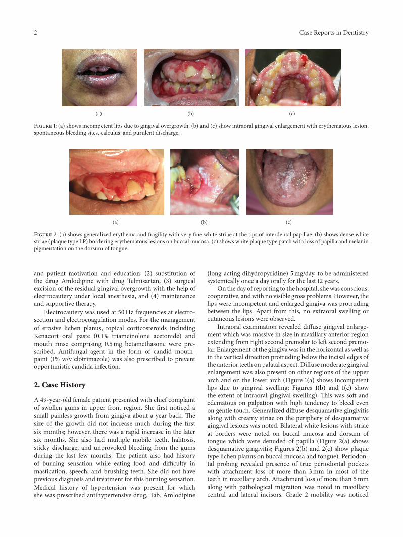
(a) (b) (c)
Figure 1: (a) shows incompetent lips due to gingival overgrowth. (b) and (c) show intraoral gingival enlargement with erythematous lesion,spontaneous bleeding sites, calculus, and purulent discharge.
(a) (b) (c)
Figure 2: (a) shows generalized erythema and fragility with very fine white striae at the tips of interdental papillae. (b) shows dense whitestriae (plaque type LP) bordering erythematous lesions on buccal mucosa. (c) shows white plaque type patch with loss of papilla and melaninpigmentation on the dorsum of tongue.
and patient motivation and education, (2) substitution ofthe drug Amlodipine with drug Telmisartan, (3) surgicalexcision of the residual gingival overgrowth with the help ofelectrocautery under local anesthesia, and (4) maintenanceand supportive therapy.
Electrocautery was used at 50Hz frequencies at electro-section and electrocoagulation modes. For the managementof erosive lichen planus, topical corticosteroids includingKenacort oral paste (0.1% triamcinolone acetonide) andmouth rinse comprising 0.5mg betamethasone were pre-scribed. Antifungal agent in the form of candid mouth-paint (1% w/v clotrimazole) was also prescribed to preventopportunistic candida infection.
2. Case History
A 49-year-old female patient presented with chief complaintof swollen gums in upper front region. She first noticed asmall painless growth from gingiva about a year back. Thesize of the growth did not increase much during the firstsix months; however, there was a rapid increase in the latersix months. She also had multiple mobile teeth, halitosis,sticky discharge, and unprovoked bleeding from the gumsduring the last few months. The patient also had historyof burning sensation while eating food and difficulty inmastication, speech, and brushing teeth. She did not haveprevious diagnosis and treatment for this burning sensation.Medical history of hypertension was present for whichshe was prescribed antihypertensive drug, Tab. Amlodipine
(long-acting dihydropyridine) 5mg/day, to be administeredsystemically once a day orally for the last 12 years.
On the day of reporting to the hospital, shewas conscious,cooperative, andwith no visible gross problems.However, thelips were incompetent and enlarged gingiva was protrudingbetween the lips. Apart from this, no extraoral swelling orcutaneous lesions were observed.
Intraoral examination revealed diffuse gingival enlarge-ment which was massive in size in maxillary anterior regionextending from right second premolar to left second premo-lar. Enlargement of the gingiva was in the horizontal as well asin the vertical direction protruding below the incisal edges ofthe anterior teeth on palatal aspect. Diffusemoderate gingivalenlargement was also present on other regions of the upperarch and on the lower arch (Figure 1(a) shows incompetentlips due to gingival swelling; Figures 1(b) and 1(c) showthe extent of intraoral gingival swelling). This was soft andedematous on palpation with high tendency to bleed evenon gentle touch. Generalized diffuse desquamative gingivitisalong with creamy striae on the periphery of desquamativegingival lesions was noted. Bilateral white lesions with striaeat borders were noted on buccal mucosa and dorsum oftongue which were denuded of papilla (Figure 2(a) showsdesquamative gingivitis; Figures 2(b) and 2(c) show plaquetype lichen planus on buccal mucosa and tongue). Periodon-tal probing revealed presence of true periodontal pocketswith attachment loss of more than 3mm in most of theteeth in maxillary arch. Attachment loss of more than 5mmalong with pathological migration was noted in maxillarycentral and lateral incisors. Grade 2 mobility was noticed
Case Reports in Dentistry 3
(a) (b)
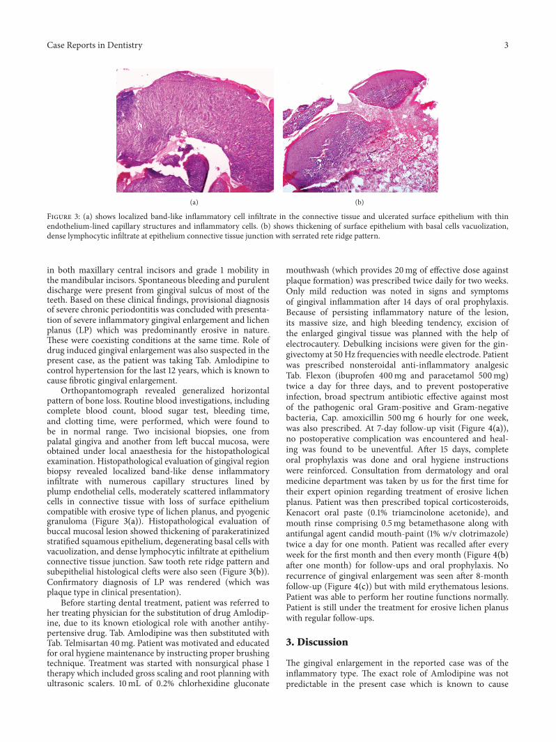
Figure 3: (a) shows localized band-like inflammatory cell infiltrate in the connective tissue and ulcerated surface epithelium with thinendothelium-lined capillary structures and inflammatory cells. (b) shows thickening of surface epithelium with basal cells vacuolization,dense lymphocytic infiltrate at epithelium connective tissue junction with serrated rete ridge pattern.
in both maxillary central incisors and grade 1 mobility inthe mandibular incisors. Spontaneous bleeding and purulentdischarge were present from gingival sulcus of most of theteeth. Based on these clinical findings, provisional diagnosisof severe chronic periodontitis was concluded with presenta-tion of severe inflammatory gingival enlargement and lichenplanus (LP) which was predominantly erosive in nature.These were coexisting conditions at the same time. Role ofdrug induced gingival enlargement was also suspected in thepresent case, as the patient was taking Tab. Amlodipine tocontrol hypertension for the last 12 years, which is known tocause fibrotic gingival enlargement.
Orthopantomograph revealed generalized horizontalpattern of bone loss. Routine blood investigations, includingcomplete blood count, blood sugar test, bleeding time,and clotting time, were performed, which were found tobe in normal range. Two incisional biopsies, one frompalatal gingiva and another from left buccal mucosa, wereobtained under local anaesthesia for the histopathologicalexamination. Histopathological evaluation of gingival regionbiopsy revealed localized band-like dense inflammatoryinfiltrate with numerous capillary structures lined byplump endothelial cells, moderately scattered inflammatorycells in connective tissue with loss of surface epitheliumcompatible with erosive type of lichen planus, and pyogenicgranuloma (Figure 3(a)). Histopathological evaluation ofbuccal mucosal lesion showed thickening of parakeratinizedstratified squamous epithelium, degenerating basal cells withvacuolization, and dense lymphocytic infiltrate at epitheliumconnective tissue junction. Saw tooth rete ridge pattern andsubepithelial histological clefts were also seen (Figure 3(b)).Confirmatory diagnosis of LP was rendered (which wasplaque type in clinical presentation).
Before starting dental treatment, patient was referred toher treating physician for the substitution of drug Amlodip-ine, due to its known etiological role with another antihy-pertensive drug. Tab. Amlodipine was then substituted withTab. Telmisartan 40mg. Patient was motivated and educatedfor oral hygiene maintenance by instructing proper brushingtechnique. Treatment was started with nonsurgical phase 1therapy which included gross scaling and root planning withultrasonic scalers. 10mL of 0.2% chlorhexidine gluconate
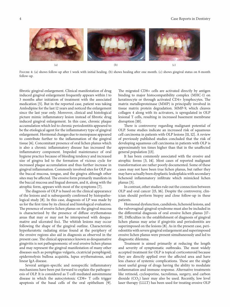
mouthwash (which provides 20mg of effective dose againstplaque formation) was prescribed twice daily for two weeks.Only mild reduction was noted in signs and symptomsof gingival inflammation after 14 days of oral prophylaxis.Because of persisting inflammatory nature of the lesion,its massive size, and high bleeding tendency, excision ofthe enlarged gingival tissue was planned with the help ofelectrocautery. Debulking incisions were given for the gin-givectomy at 50Hz frequencies with needle electrode. Patientwas prescribed nonsteroidal anti-inflammatory analgesicTab. Flexon (ibuprofen 400mg and paracetamol 500mg)twice a day for three days, and to prevent postoperativeinfection, broad spectrum antibiotic effective against mostof the pathogenic oral Gram-positive and Gram-negativebacteria, Cap. amoxicillin 500mg 6 hourly for one week,was also prescribed. At 7-day follow-up visit (Figure 4(a)),no postoperative complication was encountered and heal-ing was found to be uneventful. After 15 days, completeoral prophylaxis was done and oral hygiene instructionswere reinforced. Consultation from dermatology and oralmedicine department was taken by us for the first time fortheir expert opinion regarding treatment of erosive lichenplanus. Patient was then prescribed topical corticosteroids,Kenacort oral paste (0.1% triamcinolone acetonide), andmouth rinse comprising 0.5mg betamethasone along withantifungal agent candid mouth-paint (1% w/v clotrimazole)twice a day for one month. Patient was recalled after everyweek for the first month and then every month (Figure 4(b)after one month) for follow-ups and oral prophylaxis. Norecurrence of gingival enlargement was seen after 8-monthfollow-up (Figure 4(c)) but with mild erythematous lesions.Patient was able to perform her routine functions normally.Patient is still under the treatment for erosive lichen planuswith regular follow-ups.
3. Discussion
The gingival enlargement in the reported case was of theinflammatory type. The exact role of Amlodipine was notpredictable in the present case which is known to cause
4 Case Reports in Dentistry
(a) (b) (c)
Figure 4: (a) shows follow-up after 1 week with initial healing. (b) shows healing after one month. (c) shows gingival status on 8-monthfollow-up.
fibrotic gingival enlargement. Clinical manifestation of druginduced gingival enlargement frequently appears within 1 to3 months after initiation of treatment with the associatedmedication [5]. But in the reported case, patient was takingAmlodipine for the last 12 years and noticed the enlargementsince the last year only. Moreover, clinical and histologicalpicture mimic inflammatory lesion instead of fibrotic druginduced gingival enlargement. In this case, chronic plaqueaccumulation which led to chronic periodontitis appeared tobe the etiological agent for the inflammatory type of gingivalenlargement. Hormonal changes due tomenopause appearedto contribute further to the inflammation of the gingivaltissue [6]. Concomitant presence of oral lichen planus whichis also a chronic inflammatory disease has increased theinflammatory component. Impeded maintenance of oralhygiene practice because of bleeding tendency and increasedsize of gingiva led to the formation of vicious cycle forincreased plaque accumulation and thus further increase ingingival inflammation. Commonly involved sites for OLP arethe buccal mucosa, tongue, and the gingiva although othersites may be affected.The erosive form primarily manifests inthe buccal mucosa and lingual dorsum, and it, along with theatrophic form, appears with most of the symptoms [7].
The diagnosis of OLP is based on the clinical appearanceof the lesions and is subsequently confirmed by histopatho-logical study [8]. In this case, diagnosis of LP was made byus for the first time by its clinical and histological evaluation.The presence of erosive lichen planus on the gingival mucosais characterized by the presence of diffuse erythematousareas that may or may not be interspersed with desqua-mative and ulcerated foci. The whitish lesions may occurfollowing the shape of the gingival outline. Characteristichyperkeratotic radiating striae found at the periphery ofthe erosive regions also aid in diagnosis as observed in thepresent case.The clinical appearance known as desquamativegingivitis is not pathognomonic of oral erosive lichen planusand may represent the gingival manifestation of many otherdiseases such as pemphigus vulgaris, cicatricial pemphigoid,epidermolysis bullosa acquisita, lupus erythematosus, andlinear IgA disease.
Several antigen-specific and nonspecific inflammatorymechanisms have been put forward to explain the pathogen-esis of OLP. It is considered as T-cell-mediated autoimmunedisease in which the autocytotoxic CD8+ T cells triggerapoptosis of the basal cells of the oral epithelium [9].
The migrated CD8+ cells are activated directly by antigenbinding to major histocompatibility complex (MHC-1) onkeratinocyte or through activated CD4+ lymphocytes. Thematrix metalloproteinase (MMP) is principally involved intissue matrix protein degradation. MMP-9, which cleavescollagen 4 along with its activators, is upregulated in OLPlesional T cells, resulting in increased basement membranedisruption [10].
There is controversy regarding malignant potential ofOLP. Some studies indicate an increased risk of squamouscell carcinoma in patients with OLP lesions [11, 12]. A reviewof previously published studies concluded that the risk ofdeveloping squamous cell carcinoma in patients with OLP isapproximately ten times higher than that in the unaffectedgeneral population [13].
It has been commonly associated with the erosive andatrophic forms [3, 14]. Most cases of reported malignanttransformation are rather poorly documented. Some of thesecases may not have been true lichen planus, but rather theymay have actually been dysplastic leukoplakiawith secondarylichenoid inflammatory infiltrate which mimicked lichenplanus [5].
In contrast, other studies rule out the connection betweenOLP and oral cancer [15, 16]. Despite the controversy, clin-ician should perform biopsy and close follow-up in thesepatients.
Hormonal dysfunction, candidosis, lichenoid lesions, andthe vulvovaginal-gingival syndromemust also be included inthe differential diagnosis of oral erosive lichen planus [17–19]. Difficulties in the establishment of diagnosis of gingivallichen planus may arise if gingivitis and periodontitis aresuperimposed on the lesions [8]. As in the present case, peri-odontitis with severe gingival enlargement and superimposederosive lichen planus were present simultaneously and led todiagnostic dilemma.
Treatment is aimed primarily at reducing the lengthand severity of symptomatic outbreaks. The most widelyaccepted treatment for OLP is topical corticosteroid becausethey are directly applied over the affected area and haveless chance of systemic complications. These are the singlemost useful group of drugs having the ability to modulateinflammation and immune response. Alternative treatmentslike retinoid, cyclosporine, tacrolimus, surgery, and carbondioxide (CO
2) laser may also be used. Recently, low level
laser therapy (LLLT) has been used for treating erosive OLP

Case Reports in Dentistry 5
with minimal side effects [20–22]. Treatment choice mayvary from patient to patient depending on their symptomatichistory, severity of lesions, and systemic condition of thepatient. Excellent oral hygiene is also required to reduce theseverity of the symptoms, but it can be difficult for patientsto achieve high levels of hygiene during periods of activedisease. Currently, there is no definitive cure for OLP. Unlikecutaneous lichen planus, which in most cases progressesby short-term outbreaks that almost always respond wellto treatment or even regress after a few months, OLP ischaracterized by its chronicity, persistence with periods ofexacerbation and quiescence, and resistance to therapy.
In the present case, topical corticosteroids, antifungalagents, and antimicrobial mouth rinse were prescribed.Kenacort oral paste (0.1% triamcinolone acetonide) andmouth rinse prepared by dissolving Tab. Betnesol 0.5mg(betamethasone) in 10mL of water were prescribed afterconsultation with oral diagnostician. Since prolonged useof corticosteroids can cause opportunistic infection likeoral candidiasis or thrush, antifungal agent as Candid gel(clotrimazole 1% w/v) was also prescribed to the patient fortwice daily application. Patient was motivated and educatedfor maintaining good oral hygiene.
For patientswith gingival overgrowth, themodification oftissue topography by surgical recontouring or gingivectomymay be undertaken to create amaintainable oral environment[17]. In the present case, treatment was largely limited to themaintenance of an improved level of oral hygiene by doingoral prophylaxis at every recall visit and surgical removal ofthe overgrowth tissues. Since the lesion was predominantlyinflammatory in nature, electrocautery was used for thegingivectomy procedure. Electrocautery permits an adequatecontouring of the tissue and controls hemorrhage [23, 24].Patient was relieved symptomatically for OLP but not curedcompletely but was able to perform the routine functions andis aesthetically and psychologically satisfied.
4. Conclusion
Proper diagnosis and management are essential when mul-tiple diseases or conditions manifest in the oral cavity asseen in the present case. Electrocautery is effective meansof gingivectomy in severely inflamed, massive sized gingivalenlargement with marked tendency for bleeding.
Topical steroids are safer and effective means for the pal-liative treatment of erosive lichen planus. Routine follow-upwith maintenance of good oral hygiene is also an importantpart of management of gingival lesions.
Conflict of Interests
The authors declare that there is no conflict of interestsregarding the publication of this paper.
References
[1] M. G. Newman, H. Takie, P. R. Klokkevold, and F. A. Carranza,“Gingival enlargement,” in Carranza's Clinical Periodontology,p. 373, Saunders, 10th edition, 2007.
[2] P. B. Sugerman, N. W. Savage, L. J. Walsh et al., “The pathogen-esis of oral lichen planus,” Critical Reviews in Oral Biology andMedicine, vol. 13, no. 4, pp. 350–365, 2002.
[3] C. Scully, M. Beyli, M. C. Ferreiro et al., “Update on oral lichenplanus: etiopathogenesis and management,” Critical Reviews inOral Biology and Medicine, vol. 9, no. 1, pp. 86–122, 1998.
[4] R. A. Seymour, J. S. Ellis, J. M. Thomason, S. Monkman, and J.R. Idle, “Amlodipine-induced gingival overgrowth,” Journal ofClinical Periodontology, vol. 21, no. 4, pp. 281–283, 1994.
[5] S. J. Meraw and P. J. Sheridan, “Medically induced gingivalhyperlasia,”MayoClinic Proceedings, vol. 73, pp. 1196–1199, 1996.
[6] A. N. Haas, C. K. Rosing, R. V. Oppermann, J. M. Albandar, andC. Susin, “Association among menopause, hormone replace-ment therapy, and periodontal attachment loss in SouthernBrazilian women,” Journal of Periodontology, vol. 80, no. 9, pp.1380–1387, 2009.
[7] D. Eisen, M. Carrozzo, J.-V. B. Sebastian, and K. Thongprasom,“Number V Oral lichen planus: clinical features and manage-ment,” Oral Diseases, vol. 11, no. 6, pp. 338–349, 2005.
[8] I. R. Rubira and J. P. Gobetti, “Gingival lichen planus: a diagnos-tic problem,”The Journal of theMichiganDental Association, vol.76, no. 2, pp. 44–48, 1994.
[9] L. R. Eversole, “Immunopathogenesis of oral lichen planus andrecurrent aphthous stomatitis,” Seminars in CutaneousMedicineand Surgery, vol. 16, no. 4, pp. 284–294, 1997.
[10] X. J. Zhou, P. B. Sugerman,N.W. Savage, andL. J.Walsh, “Matrixmetalloproteinases and their inhibitors in oral lichen planus,”Journal of Cutaneous Pathology, vol. 28, no. 2, pp. 72–82, 2001.
[11] N. A. Barnard, C. Scully, J.W. Eveson, S. Cunningham, and S. R.Porter, “Oral cancer development in patients with oral lichenplanus,” Journal of Oral Pathology and Medicine, vol. 22, no. 9,pp. 421–424, 1993.
[12] S. Silverman Jr., “Oral lichen planus: a potentially premalignantlesion,” Journal of Oral and Maxillofacial Surgery, vol. 58, no. 11,pp. 1286–1288, 2000.
[13] M.Drangsholt, E. L. Truelove, T.H.Morton Jr., and J. B. Epstein,“A man with a 30-year history of oral lesions,” Journal ofEvidence-Based Dental Practice, vol. 1, no. 2, pp. 123–135, 2001.
[14] S. Silverman Jr. and S. Bahl, “Oral lichen planus update: clinicalcharacteristics, treatment responses, and malignant transfor-mation,” The American Journal of Dentistry, vol. 10, no. 6, pp.259–263, 1997.
[15] E. Eisenberg and D. J. Krutchkoff, “Lichenoid lesions of oralmucosa: diagnostic criteria and their importance in the allegedrelationship to oral cancer,”Oral SurgeryOralMedicine andOralPathology, vol. 73, no. 6, pp. 699–704, 1992.
[16] M. A. Ramer, A. Altchek, L. Deligdisch, R. Phelps, A. Mon-tazem, and P. M. Buonocore, “Lichen planus and the vulvovag-inal-gingival syndrome,” Journal of Periodontology, vol. 74, no.9, pp. 1385–1393, 2003.
[17] B. L. Pihlstrom, “Prevention and treatment of Dilantin-associ-ated gingival enlargement,” Compendium, vol. 6, no. 14, supple-ment, pp. S506–S510, 1990.
[18] B. W. Neville, D. D. Damm, C. M. Allen, and J. E. Bouquot,Oral & Maxillofacial Pathology, W.B. Saunders, Philadelphia,Pa, USA, 1995.
[19] W.-Y. Yih, T. Maier, F. J. Kratochvil, and M. B. Zieper, “Analysisof desquamative gingivitis using direct immunofluorescence inconjunction with histology,” Journal of Periodontology, vol. 69,no. 6, pp. 678–685, 1998.

6 Case Reports in Dentistry
[20] T. Passeron,W. Zakaria, N. Ostovari, F.Mantoux, J. P. H. Lacour,and J. P. Ortonne, “Treatment of erosive oral lichen planus bythe 308 nm excimer laser,” Lasers in Surgery and Medicine, vol.34, no. 3, p. 205, 2004.
[21] M. Trehan and C. R. Taylor, “Low-dose excimer 308-nm laserfor the treatment of oral lichen planus,”Archives of Dermatology,vol. 140, no. 4, pp. 415–420, 2004.
[22] K. Kollner, M. Wimmershoff, M. Landthaler, and U. Hohen-leutner, “Treatment of oral lichen planus with the 308-nmUVBexcimer laser-Early preliminary results in eight patients,” Lasersin Surgery and Medicine, vol. 33, no. 3, pp. 158–160, 2003.
[23] I. Glickman and L. R. Imber, “Comparison of gingival resectionwith electrosurgery and periodontal knives: a biometric andhistologic study,” Journal of Periodontology, vol. 41, no. 3, pp.142–148, 1970.
[24] M. J. Oringer, “Electrosurgery for definitive conservative mod-ern periodontal therapy,” Dental Clinics of North America, vol.13, no. 1, pp. 53–73, 1969.

Submit your manuscripts athttp://www.hindawi.com
Hindawi Publishing Corporationhttp://www.hindawi.com Volume 2014
Oral OncologyJournal of
DentistryInternational Journal of
Hindawi Publishing Corporationhttp://www.hindawi.com Volume 2014
Hindawi Publishing Corporationhttp://www.hindawi.com Volume 2014
International Journal of
Biomaterials
Hindawi Publishing Corporationhttp://www.hindawi.com Volume 2014
BioMed Research International
Hindawi Publishing Corporationhttp://www.hindawi.com Volume 2014
Case Reports in Dentistry
Hindawi Publishing Corporationhttp://www.hindawi.com Volume 2014
Oral ImplantsJournal of
Hindawi Publishing Corporationhttp://www.hindawi.com Volume 2014
Anesthesiology Research and Practice
Hindawi Publishing Corporationhttp://www.hindawi.com Volume 2014
Radiology Research and Practice
Environmental and Public Health
Journal of
Hindawi Publishing Corporationhttp://www.hindawi.com Volume 2014
The Scientific World JournalHindawi Publishing Corporation http://www.hindawi.com Volume 2014
Hindawi Publishing Corporationhttp://www.hindawi.com Volume 2014
Dental SurgeryJournal of
Drug DeliveryJournal of
Hindawi Publishing Corporationhttp://www.hindawi.com Volume 2014
Hindawi Publishing Corporationhttp://www.hindawi.com Volume 2014
Oral DiseasesJournal of
Hindawi Publishing Corporationhttp://www.hindawi.com Volume 2014
Computational and Mathematical Methods in Medicine
ScientificaHindawi Publishing Corporationhttp://www.hindawi.com Volume 2014
PainResearch and TreatmentHindawi Publishing Corporationhttp://www.hindawi.com Volume 2014
Preventive MedicineAdvances in
Hindawi Publishing Corporationhttp://www.hindawi.com Volume 2014
EndocrinologyInternational Journal of
Hindawi Publishing Corporationhttp://www.hindawi.com Volume 2014
Hindawi Publishing Corporationhttp://www.hindawi.com Volume 2014
OrthopedicsAdvances in