Intrauterine insemination versus fallopian tube sperm perfusion in non tubal infertility
International Journal of Reproductive BioMedicineVolume 17, Issue no. 8, https://doi.org/10.18502/ijrm.v17i8.4825Production and Hosting by Knowledge E
Case Report
Secondary infertility due to intrauterine fetalbone retention: A case report and review ofthe literatureAtossa Mahdavi 1 M.D., Sasan Kazemian2 M.D., Emad Koohestani3 M.D.1Department of Obstetrics, Gynecology and Infertility, Shariati Hospital, Tehran University ofMedical Sciences, Tehran, Iran.2Shahed University, Tehran, Iran.3Tehran University of Medical Science, Tehran, Iran.
Corresponding Author:
Atossa Mahdavi;
Department of Infertility,
Shariati Hospital, North
Karegar Street, Tehran, Iran.
Postal code: 1411713135
Tel: (+98) 21-88008810
Email:
Received 16 December 2018
Revised 18 February 2019
Accepted 30 January 2019
Production and Hosting by
Knowledge Ecc© Atossa Mahdavi et al. This
article is distributed under the
terms of the Creative
Commons Attribution License,
which permits unrestricted use
and redistribution provided
that the original author and
source are credited.
Editor-in-Cheif
Aflatoonian Abbas M.D.
AbstractBackground: Intrauterine retention of fetal bone fragments is a rare condition that couldhappen after abortion (especially illegal abortion). It can cause secondary infertility asbon fragments can work as an intrauterine contraceptive device.
Case: A 25-year-old Iranian woman was referred to Shariati Hospital due to infer-tility. During infertility work up to normal semen analysis, adequate ovarian reservewith regular ovulatory cycles was documented. An ultrasound scan revealed focalechogenic shadowing lesions inside the uterine cavity. Hysteroscopy was conductedand many intrauterine bone fragments were revealed. Six months after hysteroscopicremoval of fetal bones, the patient became pregnant and delivered a healthy and termbaby.Conclusion: Intrauterine fetal bone retention is a scarce event that happensafter pregnancy termination due to the incomplete evacuation of fetal tissues.It can cause dysfunctional uterine bleeding, menorrhagia, dysmenorrhea, pelvicpain, abnormal vaginal discharge, and secondary infertility. The detection of theproblem and the removal of the remained bones by hysteroscopy have madepossible to treat the patient safely and restore normal uterine function and femalefertility.
Key words: Bone, Infertility, Hysteroscopy, Pregnancy, Abortion.
How to cite this article: Mahdavi A, Kazemian S, Koohestani E. “Secondary infertility due to intrauterine fetal bone retention: A case report andreview of the literature” Int J Reprod BioMed 2019; 17: 591–594. https://doi.org/10.18502/ijrm.v17i8.4825 Page 591
International Journal of Reproductive BioMedicine Mahdavi et al.
1. Introduction
Retention of fetal bone fragments in the uterus
is a rare condition that usually happens after
pregnancy termination in the second or third
trimesters of pregnancy (1, 2). Retained fetal
fragments may cause pelvic pain, vaginal
discharge, abnormal uterine bleeding, and
secondary infertility. The true incidence is not
known, but it is important to be recognized and
managed as soon as possible because it is a
potentially treatable cause of secondary infertility.
These bony fragments work as an intrauterine
contraceptive device (IUCD) and stimulate
prostaglandin secretion by endometrial tissue
causing secondary infertility (3, 4). Hysteroscopy as
a diagnostic and curative method to remove these
bony parts can restore fertility in most of these
patients (1–5).
In this case report, we present a patient with
secondary infertility who did reveal her previous
illegal abortion only after hysteroscopy removal of
the pieces of fetal bone.
2. Case Presentation
A 25-yr-old Iranian infertile woman was referred
to the Shariati Hospital affiliated to the Tehran
University of Medical Sciences. She was a healthy
young lady with the problem of 1.5 yr of
infertility. She mentioned regular menstrual cycles
with normal volume and duration of bleeding
without any pain during or between the menses.
She didn’t report any history of abortion or even
pregnancy before in her first visit. A routine
gynecological examination didn’t reveal any
pathologic finding. Semen analysis of her husband
was normal. Her lab tests and hysterosalpingo-
graphy were normal. An ultrasound scan revealed
focal echogenic shadowing lesions inside the
uterine cavity.
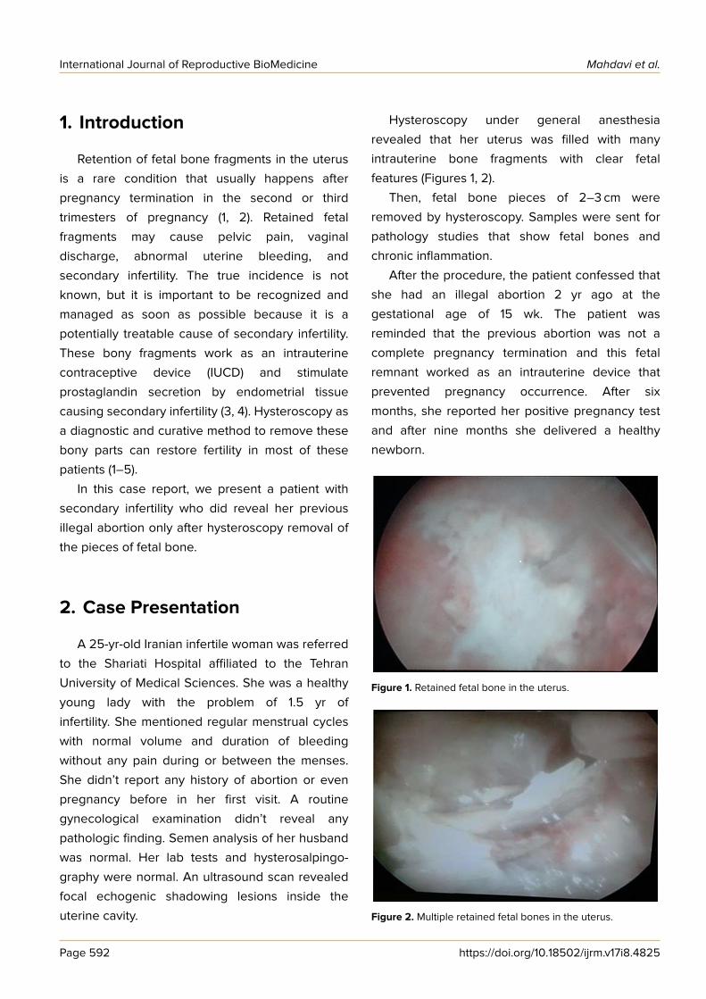
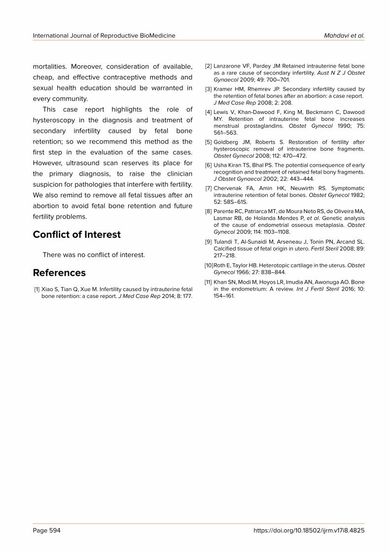
Hysteroscopy under general anesthesia
revealed that her uterus was filled with many
intrauterine bone fragments with clear fetal
features (Figures 1, 2).
Then, fetal bone pieces of 2–3 cm were
removed by hysteroscopy. Samples were sent for
pathology studies that show fetal bones and
chronic inflammation.
After the procedure, the patient confessed that
she had an illegal abortion 2 yr ago at the
gestational age of 15 wk. The patient was
reminded that the previous abortion was not a
complete pregnancy termination and this fetal
remnant worked as an intrauterine device that
prevented pregnancy occurrence. After six
months, she reported her positive pregnancy test
and after nine months she delivered a healthy
newborn.
Figure 1. Retained fetal bone in the uterus.
Figure 2. Multiple retained fetal bones in the uterus.
Page 592 https://doi.org/10.18502/ijrm.v17i8.4825

International Journal of Reproductive BioMedicine Intrauterine fetal bone retention
2.1. Ethical consideration
Written informed consent was obtained from
patient.
3. Discussion
Intrauterine fetal bone retention is a scarce
event. It happens after an induced pregnancy
termination especially during second and third
trimesters due to the incomplete evacuation of
fetal tissues (1, 2, 6). Bone fragments cause some
symptoms such as dysfunctional uterine bleeding,
menorrhagia, dysmenorrhea, pelvic pain, abnormal
vaginal discharge, and infertility (7). Although,
according to genetic analysis, bone formation
caused by osseous metaplasia due to inflamm-
ation, has been reported in eight women with
histories of previous abortion (8). This bone
formation can interfere with normal uterine
function and can work as a contraceptive resulting
in secondary infertility or abortion. There are
recent reports of calcified tissue of fetal origin in
utero (9), with the oldest article about the
exogenous foreign body in uterus returning back
to 1966 (10). In a systematic review published in
2016, infertility with a frequency of 56% was the
most common presenting symptom and only 5% of
the patients were asymptomatic (11). Irregular
bleeding, pain symptoms, and infection were
reported in 20%, 12%, and 6.5% of patients,
respectively. Interestingly, the interval between the
last pregnancy and the time of presentation or
incidental diagnosis ranged from 1 to 40 (median of
5 yr) (11).
In this case, fetal bones remained from previous
abortion that was performed 24 months ago. As
we know any external object may cause
gynecological symptoms such as dysmenorrhea,
menorrhagia, pelvic pain, vaginal discharge, but in
this case despite bone retention there was not any
gynecologic complaint. The patient was only
suffering from secondary infertility despite being a
potentially fertile woman. The patient’s denial
about previous elective abortion because of legal
and religious issues did not reveal the reason for
infertility during the work up. It was revealed only
after hysteroscopy as a valuable tool to diagnose
and manage the patient safely. This case highlights
the importance of precise history-taking during
patient visits, especially in our crowded public
clinics. Also, this case elucidates the importance of
awareness and vigilance about illegal abortions
especially in countries where abortion is not legally
permitted. However, in this case, hysteroscopy was
a valuable tool to manage the patient safely. The
removal of the remained bones restored fertility.
Recent progressions in hysteroscopy technique
have made it possible to detect and remove bones
easily and with least side effects.
An interesting finding from this report is the
ability to become pregnant soon after the
operation. Our patient became pregnant six
months after hysteroscopy and immediately after
her decision to become pregnant. It is in
accordance with Khan et al.’s finding that
spontaneous pregnancy or symptom relief were
remarkable (11). But they reminded about the
possibility of increased spontaneous miscarriage
due to the remnants of osseous material in the
endometrium. Fortunately, our patient delivered a
healthy term baby.
Medical abortion is not a legal procedure in
most Middle Eastern countries including Iran.
Some women with an unwanted pregnancy are
forced to perform it in the poor hygienic setting
(known as backstreet abortion) by inexperienced
or non-qualified people, sometimes even by the
mother herself (called self-induced). Also, later
pregnancy termination in these regions increases
the probability of incomplete evacuation, resulting
in more mortality and morbidity complications.
Increased public awareness besides revisions in
some rules can help to prevent the morbidities and
https://doi.org/10.18502/ijrm.v17i8.4825 Page 593
International Journal of Reproductive BioMedicine Mahdavi et al.
mortalities. Moreover, consideration of available,
cheap, and effective contraceptive methods and
sexual health education should be warranted in
every community.
This case report highlights the role of
hysteroscopy in the diagnosis and treatment of
secondary infertility caused by fetal bone
retention; so we recommend this method as the
first step in the evaluation of the same cases.
However, ultrasound scan reserves its place for
the primary diagnosis, to raise the clinician
suspicion for pathologies that interfere with fertility.
We also remind to remove all fetal tissues after an
abortion to avoid fetal bone retention and future
fertility problems.
Conflict of Interest
There was no conflict of interest.
References[1] Xiao S, Tian Q, Xue M. Infertility caused by intrauterine fetal
bone retention: a case report. J Med Case Rep 2014; 8: 177.
[2] Lanzarone VF, Pardey JM Retained intrauterine fetal boneas a rare cause of secondary infertility. Aust N Z J ObstetGynaecol 2009; 49: 700–701.
[3] Kramer HM, Rhemrev JP. Secondary infertility caused bythe retention of fetal bones after an abortion: a case report.J Med Case Rep 2008; 2: 208.
[4] Lewis V, Khan-Dawood F, King M, Beckmann C, DawoodMY. Retention of intrauterine fetal bone increasesmenstrual prostaglandins. Obstet Gynecol 1990; 75:561–563.
[5] Goldberg JM, Roberts S. Restoration of fertility afterhysteroscopic removal of intrauterine bone fragments.Obstet Gynecol 2008; 112: 470–472.
[6] Usha Kiran TS, Bhal PS. The potential consequence of earlyrecognition and treatment of retained fetal bony fragments.J Obstet Gynaecol 2002; 22: 443–444.
[7] Chervenak FA, Amin HK, Neuwirth RS. Symptomaticintrauterine retention of fetal bones. Obstet Gynecol 1982;52: 58S–61S.
[8] Parente RC, Patriarca MT, de Moura Neto RS, de Oliveira MA,Lasmar RB, de Holanda Mendes P, et al. Genetic analysisof the cause of endometrial osseous metaplasia. ObstetGynecol 2009; 114: 1103–1108.
[9] Tulandi T, Al-Sunaidi M, Arseneau J, Tonin PN, Arcand SL.Calcified tissue of fetal origin in utero. Fertil Steril 2008; 89:217–218.
[10]Roth E, Taylor HB. Heterotopic cartilage in the uterus.ObstetGynecol 1966; 27: 838–844.
[11] Khan SN, Modi M, Hoyos LR, Imudia AN, Awonuga AO. Bonein the endometrium: A review. Int J Fertil Steril 2016; 10:154–161.
Page 594 https://doi.org/10.18502/ijrm.v17i8.4825


![Early intrauterine development of mixed giant … · Early intrauterine development of mixed giant ... but with intrauterine death at 29 weeks [5]. Fetal . Early intrauterine development](https://static.fdocuments.net/doc/165x107/5b63022f7f8b9ade588b8aac/early-intrauterine-development-of-mixed-giant-early-intrauterine-development.jpg)