Case Report Pulmonary gangliocytic paraganglioma: a case ... · Pulmonary gangliocytic...
6
Int J Clin Exp Pathol 2014;7(1):432-437 www.ijcep.com /ISSN:1936-2625/IJCEP1311011 Case Report Pulmonary gangliocytic paraganglioma: a case report and review of the literature Dongliang Lin 1 , Yanjiao Hu 1 , Xiaoming Xing 1 , Li Ding 2 , Hui Liu 1 , Yujun Li 1 , Fenggang Xiang 1 1 Department of Pathology, The Affiliated Hospital of Medical College, Qingdao University, Qingdao, China; 2 Depart- ment of Prevention Care, The Affiliated Hospital of Medical College, Qingdao University, Qingdao, China Received November 6, 2013; Accepted December 5, 2013; Epub December 15, 2013; Published January 1, 2014 Abstract: Gangliocytic paraganglioma (GP) is a rare histologic type of neuroendocrine tumors. We report a case of pulmonary GP in a 29-year-old male presenting with an asymptomatic endobronchial nodule. Grossly, the tumor showed a 4.0x3.8x3.5 cm well-defined nodule with yellowish cut surface. Microscopically, the tumor was composed of three distinct cellular types: epithelioid cells, ganglion-like cells and spindle cells. Meanwhile, transitional cells, having morphologic features between ganglion-like and epithelioid cells, were also presented. The epithelioid cells arranged in various morphologic architectures, including Zellballen, papillary, cystic and microcystic pattern. The epithelioid cells were positive for AE1/AE3, CAM 5.2, chromogranin A and synaptophysin. Ganglion-like cells showed immunoreactivity for chromogranin A and synaptophysin. A few ganglion-like cells were also positive for AE1/AE3 and/or CAM 5.2. The spindle cells were positive for S-100 protein and neurofilament. The transitional cells showed a similar immunohistochemical profile to the epithelioid cells. The authors believe stem cell theory is a reasonable explanation for the origin of GP. GP probably originate from some kind of mucosa associated stem cell which can differentiate into diverse cellular lineages. Keywords: Gangliocytic paraganglioma, bronchial, histomorphology, immunohistochemistry, histogenesis Introduction GP is a unique neuroendocrine tumor that dif- fers from other neuroendocrine tumors. It was initially reported as ganglioneuroma by Dahl et al in 1957 [1] and first named GP by Kepes et al in 1971 [2]. The exact origin of GP remains unclear till now. It occurs almost exclusively in the second portion of the duodenum, especial- ly in periampullary region. Most cases in duode- num showed the epithelioid cells were positive for pancreatic polypeptide [3]. Therefore, some researchers consider GP as a hamartoma developing in misplaced embryonic pancreatic tissue [4-6]. However, GP has also been described in other locations including jejunum [7], esophagus [8], stomach [6], pancreas [9], appendix [10], nasopharynx [11] and lung [12- 15]. Herein we present an additional case of pulmonary GP which displays diverse histologi- cal structures. Meanwhile, we would also com- ment on the origin of GP. Case report A 29-year-old asymptomatic male was admit- ted to our hospital because of a pulmonary mass detected by chest computerized tomo- graphic (CT) scan during his annual physical examination. The CT scan revealed a 38 mm endobronchial mass almost completely obst- ructing the bronchial lumen of right lower lobe (Figure 1). From the CT scan, suspicious paren- chymal involvement of adjacent right middle and lower lobes was demonstrated. Broncho- scopy displayed a mass partly occluding the bronchus. Bronchoscopic biopsy was reported as carcinoid because of positive staining of AE1/AE3, synaptophysin and chromogranin A in epithelioid cells. Subsequent lobectomy of the right middle and lower lobes with the dissection of the mediastinal lymph node was performed. Grossly, the tumor showed a 4.0x3.8x3.5 cm well-defined nodule with yellowish cut surface. Histological examination of the tumor revealed an apparently well-defined but nonencapsulat- ed neoplasm which did not involve the lung parenchyma. The tumor consisted of three dis- tinct cellular elements. The first, uniform epi- thelioid cells had round to oval-shaped nuclei with granular chromatin, inconspicuous nucleo- li, and eosinophilic to clear cytoplasm. They
Transcript of Case Report Pulmonary gangliocytic paraganglioma: a case ... · Pulmonary gangliocytic...
Int J Clin Exp Pathol 2014;7(1):432-437www.ijcep.com /ISSN:1936-2625/IJCEP1311011
Case ReportPulmonary gangliocytic paraganglioma: a case report and review of the literature
Dongliang Lin1, Yanjiao Hu1, Xiaoming Xing1, Li Ding2, Hui Liu1, Yujun Li1, Fenggang Xiang1
1Department of Pathology, The Affiliated Hospital of Medical College, Qingdao University, Qingdao, China; 2Depart-ment of Prevention Care, The Affiliated Hospital of Medical College, Qingdao University, Qingdao, China
Received November 6, 2013; Accepted December 5, 2013; Epub December 15, 2013; Published January 1, 2014
Abstract: Gangliocytic paraganglioma (GP) is a rare histologic type of neuroendocrine tumors. We report a case of pulmonary GP in a 29-year-old male presenting with an asymptomatic endobronchial nodule. Grossly, the tumor showed a 4.0x3.8x3.5 cm well-defined nodule with yellowish cut surface. Microscopically, the tumor was composed of three distinct cellular types: epithelioid cells, ganglion-like cells and spindle cells. Meanwhile, transitional cells, having morphologic features between ganglion-like and epithelioid cells, were also presented. The epithelioid cells arranged in various morphologic architectures, including Zellballen, papillary, cystic and microcystic pattern. The epithelioid cells were positive for AE1/AE3, CAM 5.2, chromogranin A and synaptophysin. Ganglion-like cells showed immunoreactivity for chromogranin A and synaptophysin. A few ganglion-like cells were also positive for AE1/AE3 and/or CAM 5.2. The spindle cells were positive for S-100 protein and neurofilament. The transitional cells showed a similar immunohistochemical profile to the epithelioid cells. The authors believe stem cell theory is a reasonable explanation for the origin of GP. GP probably originate from some kind of mucosa associated stem cell which can differentiate into diverse cellular lineages.
Keywords: Gangliocytic paraganglioma, bronchial, histomorphology, immunohistochemistry, histogenesis
Introduction
GP is a unique neuroendocrine tumor that dif-fers from other neuroendocrine tumors. It was initially reported as ganglioneuroma by Dahl et al in 1957 [1] and first named GP by Kepes et al in 1971 [2]. The exact origin of GP remains unclear till now. It occurs almost exclusively in the second portion of the duodenum, especial-ly in periampullary region. Most cases in duode-num showed the epithelioid cells were positive for pancreatic polypeptide [3]. Therefore, some researchers consider GP as a hamartoma developing in misplaced embryonic pancreatic tissue [4-6]. However, GP has also been described in other locations including jejunum [7], esophagus [8], stomach [6], pancreas [9], appendix [10], nasopharynx [11] and lung [12-15]. Herein we present an additional case of pulmonary GP which displays diverse histologi-cal structures. Meanwhile, we would also com-ment on the origin of GP.
Case report
A 29-year-old asymptomatic male was admit-ted to our hospital because of a pulmonary
mass detected by chest computerized tomo-graphic (CT) scan during his annual physical examination. The CT scan revealed a 38 mm endobronchial mass almost completely obst- ructing the bronchial lumen of right lower lobe (Figure 1). From the CT scan, suspicious paren-chymal involvement of adjacent right middle and lower lobes was demonstrated. Broncho- scopy displayed a mass partly occluding the bronchus. Bronchoscopic biopsy was reported as carcinoid because of positive staining of AE1/AE3, synaptophysin and chromogranin A in epithelioid cells. Subsequent lobectomy of the right middle and lower lobes with the dissection of the mediastinal lymph node was performed.
Grossly, the tumor showed a 4.0x3.8x3.5 cm well-defined nodule with yellowish cut surface. Histological examination of the tumor revealed an apparently well-defined but nonencapsulat-ed neoplasm which did not involve the lung parenchyma. The tumor consisted of three dis-tinct cellular elements. The first, uniform epi-thelioid cells had round to oval-shaped nuclei with granular chromatin, inconspicuous nucleo-li, and eosinophilic to clear cytoplasm. They

Pulmonary gangliocytic paraganglioma
433 Int J Clin Exp Pathol 2014;7(1):432-437
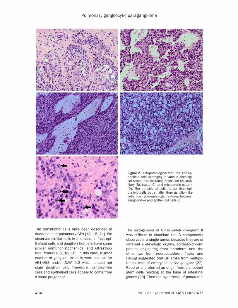
arranged in various histological structures, including Zellballen (Figure 2A), papillary (Figure 2B), cystic (Figure 2C), and microcystic pattern (Figure 2D). The second, large gangli-on-like cells were characterized by abundant amphophilic cytoplasm, large round-shaped nuclei, and prominent nucleoli. They were dis-persed within the nests of epithelioid cells and the background filled with spindle cells. The third, spindle cells were located in the periph-ery of the epithelioid cells. Additionally, transi-tional cells, having morphologic features between ganglion-like and epithelioid cells, were also presented (Figure 2E). They were larger than epithelioid cells but smaller than ganglion-like cells. Cellular atypia, mitotic activ-ity and necrosis were not detected. Regional lymph nodes were not invaded.
Immunohistochemically, all of the components showed immunoreactivity for neuron specific enolase. The epithelioid cells were positive for AE1/AE3 (Figure 3A), CAM 5.2, chromogranin A (Figure 3B) and synaptophysin (Figure 3C), but negative for neurofilament. A number of these cells were weakly positive for S-100 protein. Ganglion-like cells showed immunoreactivity for chromogranin A and synaptophysin. They were negative for neurofilament. Some of gan-glion-like cells were also reactive for S-100 pro-tein. Interestingly, we observed a few AE1/AE3 and/or CAM 5.2 positive ganglion-like cells (Figure 3D). Besides neuron specific enolase, the spindle cells were positive for S-100 protein (Figure 3E) and neurofilament, but negative for other markers. The transitional cells showed a similar immunohistochemical profile to the epi-thelioid cells.
Discussion
GP is a rare epithelioid tumor with distinctive feature that differs from other neuroendocrine tumors. It spans all age groups (15-84) with an median of 52.3 years, occurs with a slight male predominance (M:F ratio, 114:76) [3], The most common location of GPs is duodenum (90%), especially in periampullary region [3]. Two of the most common symptoms of duodenal GP were gastrointestinal bleeding and abdominal pain [3]. Recently, some extra-duodenal cases have also been described. To our knowledge, excluding the present case, there have been 4 cases of pulmonary GP reported in the English literature (Table 1). All of the 5 cases occurred in adults, and they appear to affect middle-aged and old male more commonly. Of the 5 cases, 4 were located in bronchus. This data indicated bronchus may be a common location of pulmonary GP. The initial clinical manifesta-tion of pulmonary GP could be chest pain, pneumonia or Cushing’s syndrome [12-14]. In the current case, the patient was asymptoma- tic.
Microscopically, GP has three characteristic components: epithelioid cells, ganglion-like cells, and spindle cells. The portion of this three components varies widely from case to case [6, 16], which pose a challenge for pathologists to make a definite diagnosis especially in a small biopsy. We can assume, if this tumor display a predominance of one or two of above compo-nents, various differential diagnoses, including carcinoid, paraganglioma, carcinoma, schwan-noma or ganglioneuroma, should be consid-ered. In this case, the epithelioid cells arranged in various histological structures, including Zellballen, papillary, cystic and microcystic pat-tern. The Zellballen structure showed variable nest like carcinoid or paraganglioma. The papil-lary structure was first mentioned by Burke and Helwig [6]. This structure brought us to think of pulmonary sclerosing haemangioma which is almost exclusively located in lung parenchyma. Of the 100 cases of pulmonary sclerosing hem-angioma reported by Devouassoux-Shishebo- ran et al, only one was described as an endo-bronchial polyp [17]. However, pulmonary GP appears not to involve adjacent lung parenchy-ma [14]. The cystic pattern has not been men-tioned in the literature. However, some authors had described pseudoglandular structures which seemed to have some similarity to the microcystic structure we described [18-20].
Figure 1. Chest CT scan showing an endobronchial mass almost completely obstructing the bronchial lu-men of right lower lobe.
Pulmonary gangliocytic paraganglioma
434 Int J Clin Exp Pathol 2014;7(1):432-437
The transitional cells have been described in duodenal and pulmonary GPs [13, 18, 21]. We observed similar cells in this case. In fact, epi-thelioid cells and ganglion-like cells have some similar immunohistochemical and ultrastruc-tural features [5, 16, 18]. In this case, a small number of ganglion-like cells were positive for AE1/AE3 and/or CAM 5.2 which should not stain ganglion cell. Therefore, ganglion-like cells and epithelioid cells appear to come from a same progenitor.
The histogenesis of GP is widely divergent. It was difficult to elucidate the 3 components observed in a single tumor, because they are of different embryologic origins, epithelioid com-ponent originating from entoderm and the other two from neuroectoderm. Taylor and Helwig suggested that GP arose from multipo-tential cells of embryonic celiac ganglion [22]. Reed et al preferred an origin from pluripotent stem cells residing at the base of intestinal glands [23]. Then the hypothesis of pancreatic
Figure 2. Histopathological features. The ep-ithelioid cells arranging in various histologi-cal structures, including Zellballen (A), pap-illary (B), cystic (C), and microcystic pattern (D). The transitional cells, larger than epi-thelioid cells but smaller than ganglion-like cells, having morphologic features between ganglion-like and epithelioid cells (E).

Pulmonary gangliocytic paraganglioma
435 Int J Clin Exp Pathol 2014;7(1):432-437
origin was popular. In consideration of the con-sistent location in the duodenum and immuno-reactivity for pancreatic polypeptide, Perrone et al suggested this lesion represented a hamar-tomatous process derived from the ventral pri-mordium of the pancreas [5]. However, there are several problems the pancreatic hypothesis can not solve. First, it can not provide a reason-able explanation for those extra-duodenal cases. Secondly, pancreatic polypeptide is not a specific marker for pancreatic neuroendo-crine tumor. Carcinoids of the rectum, colon,
lung and bile duct also showed reactivity for pancreatic polypeptide [24-26]. Thirdly, althou- gh GP is composed of endodermal and ectoder-mal tissues, the cases with lymph node or dis-tant metastasis argue against the theory of hamartoma [3, 27]. Recently, Ogata et al. reported a case of duodenal GP with glandular component [28] which belongs to endodermal tissue. This unique case delivered a powerful support to stem cell origin but a rebuttal to the pancreatic hypothesis. Reviewing all the sites involved in the literature, we can find GP tends
Figure 3. Immunohistochemical staining of GP. The epithelioid cells were positive for AE1/AE3 (A), chromogranin A (B) and synap-tophysin (C). A few CAM 5.2 positive ganglion-like cells were also identified (D). The spindle cells were positive for S-100 protein (E).

Pulmonary gangliocytic paraganglioma
436 Int J Clin Exp Pathol 2014;7(1):432-437
to a mucosa associated tumor. Therefore GP probably originates from some kind of mucosa associated stem cell which can differentiate into diverse cellular lineages. Considering the transitional cells and the similarity between ganglion-like cells and epithelioid cells, we speculate ganglion-like cells and epithelioid cells come from a common cellular lineage.
Disclosure of conflict of interest
The authors have no conflicts of interest relat-ed to this article to declare.
Address correspondence to: Dr. Fenggang Xiang, Department of Pathology, The Affiliated Hospital of Medical College, Qingdao University, 16 Jiangsu Road, Qingdao 266003, China. Tel: +86 532 8291 9353; Fax: +86 532 8291 9600; E-mail: [email protected]
References
[1] Dahl EV, Waugh JM, Dahlin DC. Gastrointesti-nal ganglioneuromas; brief review with report of a duodenal ganglioneuroma. Am J Pathol 1957; 33: 953-65.
[2] Kepes JJ, Zacharias DL. Gangliocytic paragan-gliomas of the duodenum. A report of two cas-es with light and electron microscopic exami-nation. Cancer 1971; 27: 61-7.
[3] Okubo Y, Wakayama M, Nemoto T, Kitahara K, Nakayama H, Shibuya K, Yokose T, Yamada M, Shimodaira K, Sasai D, Ishiwatari T, Tsuchiya M, Hiruta N. Literature survey on epidemiology and pathology of gangliocytic paraganglioma. BMC Cancer 2011; 11: 187.
[4] Guarda LA, Ordonez NG, del Junco GW, Luna MA. Gangliocytic paraganglioma of the duode-num: an immunocytochemical study. Am J Gastroenterol 1983; 78: 794-8.
[5] Perrone T, Sibley RK, Rosai J. Duodenal gan-gliocytic paraganglioma. An immunohisto-chemical and ultrastructural study and a hy-pothesis concerning its origin. Am J Surg Pathol 1985; 9: 31-41.
[6] Burke AP, Helwig EB. Gangliocytic paraganglio-ma. Am J Clin Pathol 1989; 92: 1-9.
[7] Aung W, Gallagher HJ, Joyce WP, Hayes DB, Leader M. Gastrointestinal haemorrhage from a jejunal gangliocytic paraganglioma. J Clin Pathol 1995; 48: 84-5.
[8] Weinrach DM, Wang KL, Blum MG, Yeldandi AV, Laskin WB. Multifocal presentation of gan-gliocytic paraganglioma in the mediastinum and esophagus. Hum Pathol 2004; 35: 1288-91.
[9] Tomic S, Warner T. Pancreatic somatostatin-secreting gangliocytic paraganglioma with lymph node metastases. Am J Gastroenterol 1996; 91: 607-8.
[10] van Eeden S, Offerhaus GJ, Peterse HL, Dinge-mans KP, Blaauwgeers HL. Gangliocytic para-ganglioma of the appendix. Histopathology 2000; 36: 47-9.
[11] Sinkre P, Lindberg G, Albores-Saavedra J. Na-sopharyngeal gangliocytic paraganglioma. Arch Pathol Lab Med 2001; 125: 1098-100.
[12] Hironaka M, Fukayama M, Takayashiki N, Saito K, Sohara Y, Funata N. Pulmonary gangliocytic paraganglioma: case report and comparative immunohistochemical study of related neuro-endocrine neoplasms. Am J Surg Pathol 2001; 25: 688-93.
[13] Kee AR, Forrest CH, Brennan BA, Papadimitri-ou JM, Glancy RJ. Gangliocytic paraganglioma of the bronchus: a case report with follow-up and ultrastructural assessment. Am J Surg Pathol 2003; 27: 1380-5.
[14] Palau MA, Merino MJ, Quezado M. Corticotro-pin-producing pulmonary gangliocytic para-ganglioma associated with Cushing’s syn-drome. Hum Pathol 2006; 37: 623-6.
Table 1. Clinical feature of pulmonary gangliocytic paragangliomaCase no.
Sex/Age (yr)
Size (mm) Location Clinical
presentation Treatment Follow-up
1 [12] M/75 16 Bifurcation of the right middle and lower lobe bronchus
Right anterior chest pain
Lobectomy of the right middle and lower lobes with dissection of the mediastinal
NA
2 [13] F/54 9 Right lower lobe bronchus Right lower lobe pneumonia
Bronchoscopic resection Recurrent was confirmed 6 months later
Further bronchoscopic and laser therapy
No evidence of tumor additional 4 months later
3 [14] M/55 8 Left inferior pulmonary ligament
Cushing’s syndrome
NA NA
4 [15] M/61 NA Right lower lobe bronchus NA Lobectomy of the right lower lobe NA
Present case
M/29 40 Right lower lobe bronchus Asymptomatic Lobectomy of the right middle and lower lobes with dissection of the mediastinal
No evidence of tumor 7 months later
F, female; M, male; NA, not available.

Pulmonary gangliocytic paraganglioma
437 Int J Clin Exp Pathol 2014;7(1):432-437
[15] Gucer H, Mete O. Endobronchial Gangliocytic Paraganglioma: Not All Keratin-Positive Endo-bronchial Neuroendocrine Neoplasms are Pul-monary Carcinoids. Endocr Pathol 2013; [Epub ahead of print].
[16] Hamid QA, Bishop AE, Rode J, Dhillon AP, Rosenberg BF, Reed RJ, Sibley RK, Polak JM. Duodenal gangliocytic paragangliomas: a study of 10 cases with immunocytochemical neuroendocrine markers. Hum Pathol 1986; 17: 1151-7.
[17] Devouassoux-Shisheboran M, Hayashi T, Lin-noila RI, Koss MN, Travis WD. A clinicopatho-logic study of 100 cases of pulmonary scleros-ing hemangioma with immunohistochemical studies: TTF-1 is expressed in both round and surface cells, suggesting an origin from primi-tive respiratory epithelium. Am J Surg Pathol 2000; 24: 906-16.
[18] Inai K, Kobuke T, Yonehara S, Tokuoka S. Duo-denal gangliocytic paraganglioma with lymph node metastasis in a 17-year-old boy. Cancer 1989; 63: 2540-5.
[19] Barbareschi M, Frigo B, Aldovini D, Leonardi E, Cristina S, Falleni M. Duodenal gangliocytic paraganglioma. Report of a case and review of the literature. Virchows Arch A Pathol Anat His-topathol 1989; 416: 81-9.
[20] Dookhan DB, Miettinen M, Finkel G, Gibas Z. Recurrent duodenal gangliocytic paraganglio-ma with lymph node metastases. Histopathol-ogy 1993; 22: 399-401.
[21] Scheithauer BW, Nora FE, LeChago J, Wick MR, Crawford BG, Weiland LH, Carney JA. Duodenal gangliocytic paraganglioma. Clinicopathologic and immunocytochemical study of 11 cases. Am J Clin Pathol 1986; 86: 559-65.
[22] Taylor HB, Helwig EB. Benign nonchromaffin paragangliomas of the duodenum. Virchows Arch Pathol Anat Physiol Klin Med 1962; 335: 356-66.
[23] Reed RJ, Caroca PJ Jr, Harkin JC. Gangliocytic paraganglioma. Am J Surg Pathol 1977; 1: 207-16.
[24] Federspiel BH, Burke AP, Sobin LH, Shekitka KM. Rectal and colonic carcinoids. A clinico-pathologic study of 84 cases. Cancer 1990; 65: 135-40.
[25] Ishida T, Yokoyama H, Sugio K, Kaneko S, Sug-imachi K, Hara N, Ohta M. Carcinoid tumor of the lung: clinicopathological and immunohisto-chemical studies. Eur J Surg Oncol 1992; 18: 180-7.
[26] Maitra A, Krueger JE, Tascilar M, Offerhaus GJ, Angeles-Angeles A, Klimstra DS, Hruban RH, Albores-Saavedra J. Carcinoid tumors of the extrahepatic bile ducts: a study of seven cas-es. Am J Surg Pathol 2000; 24: 1501-10.
[27] Rowsell C, Coburn N, Chetty R. Gangliocytic paraganglioma: a rare case with metastases of all 3 elements to liver and lymph nodes. Ann Diagn Pathol 2011; 15: 467-71.
[28] Ogata S, Horio T, Sugiura Y, Aiko S, Aida S. Duo-denal gangliocytic paraganglioma with region-al lymph node metastasis and a glandular component. Pathol Int 2011; 61: 104-7.