CASE REPORT PELVI-URETERIC JUNCTION · PDF fileinfection like pylonephritis, hydronephrosis,...
5
ABSTRACT PELVI-URETERIC JUNCTION OBSTRUCTION SECONDARY TO BILATERAL ABERRANT VESSELS AND RENAL STONES – CASE REPORT. 1 2 1 3 1 1 Yauba MS, Tella U, Akuhwa TR, Sadiq AA, Ibrahim H, Imam R 1 2 Department of Paediatrics, Department of Surgery, 3 Department of Anaesthesia and Intensive Care, University of Maiduguri Teaching Hospital, Maiduguri, Borno State, Nigeria. Correspondence and reprint request to: Dr Saad Mohammed Yauba, Department of Paediatrics, University of Maiduguri Teaching Hospital, Maiduguri, Borno State, Nigeria eMail:- [email protected] Keywords: Aberrant vessels, children, obstructive uropathy, Pelvi-ureteric junction, renal stones. Background: Obstructive uropathy is one of the causes of morbidity and mortality in children because it may lead to renal dysfunction. Pelvi-ureteric junction (PUJ) obstruction secondary to concomitant aberrant vessels and renal stones is uncommon. Method: We describe an adolescent patient with a bilateral obstructive uropathy secondary to renal stones and bilateral aberrant blood vessels crossing the lower poles of the kidneys. Conclusion: Co-existence of aberrant crossing blood vessels and renal stones as a cause of obstructive uropathy in children is rare and when left untreated for a prolonged period of time can lead to chronic kidney disease. CASE REPORT INTRODUCTION Pelvi-ureteric junction (PUJ) obstruction is defined as a functional or anatomic obstruction to outflow of urine from the renal pelvis to the ureter, leading to deterioration of the function 1 of the affected kidney. Pelvi-ureteric junction obstruction is one of the common causes of obstructive uropathy which can be congenital 2 or acquired. The obstruction may be due to lower polar aberrant crossing vessel just 3 compressing the PUJ. The condition is commonly unilateral but bilateral PUJ 3 obstruction may occur. Significant urinary obstruction may result in tubular dilation, 4 glomerulosclerosis, inflammation and fibrosis. Late presentation is associated with complication such as recurrent urinary tract infection like pylonephritis, hydronephrosis, renal stones, pyonephrosis, impaired renal 5 function or complete loss of renal unit. CASE PRESENTATION B.M.M, an eleven year old primary two boy, presented with two months history of left flank pain, three-week history of body swelling and two days history of haematuria. The left flank pain was colicky, non-radiating and associated with crying during painful episodes. There was associated vomiting and facial puffiness, worst in the morning but subsides in the afternoon. Two days prior to presentation, he developed haematuria, convulsions and reduced urinary output. There was no history of increased urinary frequency, dysuria, pyuria, lithouria or fever. No preceding history of pharyngitis or pyoderma and no history of similar illness in Page 33 2014;(8)1: 33-37 Kanem Journal of Medical Sciences
Transcript of CASE REPORT PELVI-URETERIC JUNCTION · PDF fileinfection like pylonephritis, hydronephrosis,...
ABSTRACT
PELVI-URETERIC JUNCTION OBSTRUCTION SECONDARY TO BILATERAL ABERRANT VESSELS AND RENAL STONES – CASE REPORT.
1 2 1 3 1 1Yauba MS, Tella U, Akuhwa TR, Sadiq AA, Ibrahim H, Imam R
1 2Department of Paediatrics, Department of Surgery,
3Department of Anaesthesia and Intensive Care,
University of Maiduguri Teaching Hospital, Maiduguri, Borno State, Nigeria.
Correspondence and reprint request to: Dr Saad Mohammed Yauba, Department of Paediatrics, University of Maiduguri Teaching Hospital,
Maiduguri, Borno State, Nigeria eMail:- [email protected]
Keywords: Aberrant vessels, children, obstructive uropathy, Pelvi-ureteric junction, renal stones.
Background: Obstructive uropathy is one of the causes of morbidity and mortality in children because it may lead to renal dysfunction. Pelvi-ureteric junction (PUJ) obstruction secondary to concomitant aberrant vessels and renal stones is uncommon. Method: We describe an adolescent patient with a bilateral obstructive uropathy secondary to renal stones and bilateral aberrant blood vessels crossing the lower poles of the kidneys.Conclusion: Co-existence of aberrant crossing blood vessels and renal stones as a cause of obstructive uropathy in children is rare and when left untreated for a prolonged period of time can lead to chronic kidney disease.
CASE REPORT
INTRODUCTIONPelvi-ureteric junction (PUJ) obstruction is defined as a functional or anatomic obstruction to outflow of urine from the renal pelvis to the ureter, leading to deterioration of the function
1 of the affected kidney. Pelvi-ureteric junction obstruction is one of the common causes of obstructive uropathy which can be congenital
2 or acquired. The obstruction may be due to lower polar aberrant crossing vessel just
3compressing the PUJ. The condition is commonly unilateral but bilateral PUJ
3obstruction may occur. Significant urinary obstruction may result in tubular dilation,
4 glomerulosclerosis, inflammation and fibrosis.Late presentation is associated with complication such as recurrent urinary tract infection like pylonephritis, hydronephrosis,
renal stones, pyonephrosis, impaired renal 5 function or complete loss of renal unit.
CASE PRESENTATIONB.M.M, an eleven year old primary two boy, presented with two months history of left flank pain, three-week history of body swelling and two days history of haematuria. The left flank pain was colicky, non-radiating and associated with crying during painful episodes. There was associated vomiting and facial puffiness, worst in the morning but subsides in the afternoon. Two days prior to presentation, he developed haematuria, convulsions and reduced urinary output. There was no history of increased urinary frequency, dysuria, pyuria, lithouria or fever. No preceding history of pharyngitis or pyoderma and no history of similar illness in
Page 33 2014;(8)1: 33-37 Kanem Journal of Medical Sciences
CASE REPORT Yauba MS et al
the past. He is not a known sickle cell anaemia, diabetic or hypertensive patient. He is first of his mother's four children in a monogamous family setting of low socio-economic status.
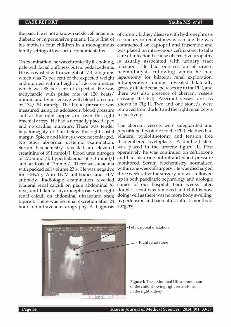
On examination, he was chronically ill-looking, pale with facial puffiness but no pedal oedema. He was wasted with a weight of 27.4 kilograms which was 76 per cent of the expected weight and stunted with a height of 126 centimetres which was 88 per cent of expected. He was tachycardic with pulse rate of 120 beats/ minute and hypertensive with blood pressure of 134/ 84 mmHg. The blood pressure was measured using an adolescent blood pressure cuff at the right upper arm over the right brachial artery. He had a normally placed apex and no cardiac murmurs. There was tender hepatomegaly of 4cm below the right costal margin. Spleen and kidneys were not enlarged. No other abnormal systemic examination. Serum biochemistry revealed an elevated creatinine of 691 mmol/l, blood urea nitrogen of 27.5mmol/l, hyperkalaemia of 7.3 mmol/l and acidosis of 17mmol/l. There was anaemia with packed cell volume 23%. He was negative for HBsAg, Anti HCV antibodies and HIV antibody. Radiologic examination revealed bilateral renal calculi on plain abdominal X-rays, and bilateral hydronephrosis with right renal calculi on abdominal ultrasound scan; figure I. There was no renal excretion after 24 hours on intravenous urography. A diagnosis
of chronic kidney disease with hydronephrosis secondary to renal stones was made. He was commenced on captopril and frusemide and was placed on intravenous ceftriaxone, to take care of infection because obstructive uropathy is usually associated with urinary tract infection.. He had one session of urgent haemodialysis following which he had laparotomy for bilateral renal exploration. Intraoperative findings revealed bilaterally grossly dilated renal pelvises up to the PUJ, and there was also presence of aberrant vessels crossing the PUJ. Aberrant vessels are ass shown in Fig II. Two and one stone/s were removed from the left and the right renal pelvis respectively.
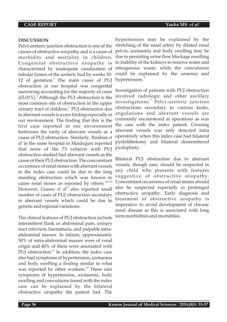
The aberrant vessels were safeguarded and repositioned posterior to the PUJ. He then had bilateral pyelolithotomy and tension free dismembered pyeloplasty. A double-J stent was placed in the ureters; figure III. Post operatively he was continued on ceftriaxone and had his urine output and blood pressure monitored. Serum biochemistry normalised within one week of surgery. He was discharged three weeks after the surgery and was followed up in both paediatric nephrology and urologic clinics of our hospital. Four weeks later, double-J stent was removed and child is now doing well as there was no more body swelling, hypertension and haematuria after 7 months of surgery.
Pelvicalyceal dilatation
Right renal stone
Figure I: The abdominal Ultra sound scan of the child showing right renal stones in the right kidney
Page 34 2014;(8)1: 33-37 Kanem Journal of Medical Sciences
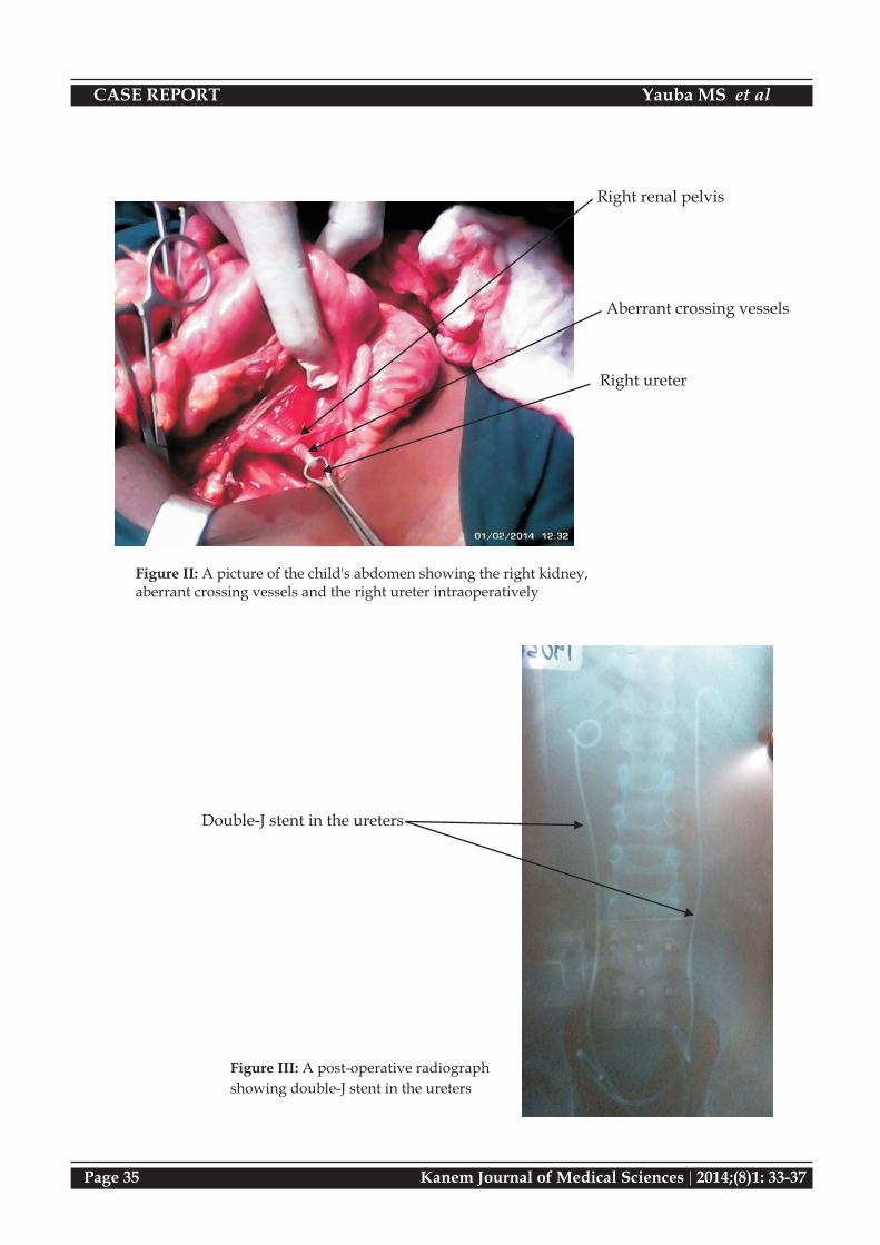
Right renal pelvis
Aberrant crossing vessels
Right ureter
Figure II: A picture of the child's abdomen showing the right kidney, aberrant crossing vessels and the right ureter intraoperatively
Double-J stent in the ureters
Figure III: A post-operative radiograph
showing double-J stent in the ureters
CASE REPORT Yauba MS et al
Page 35 2014;(8)1: 33-37 Kanem Journal of Medical Sciences
DISCUSSIONPelvi-ureteric junction obstruction is one of the causes of obstructive uropathy and is a cause of morbidity and mortality in children. Congeni ta l obs t ruc t ive uropathy i s characterised by inadequate canalization of tubular lumen of the ureteric bud by weeks 10-
6 12 of gestation. The main cause of PUJ obstruction in our hospital was congenital narrowing accounting for the majority of cases
2 (63.01%). Although the PUJ obstruction is the most common site of obstruction in the upper
7 urinary tract of children, PUJ obstruction due to aberrant vessels is a rare finding especially in our environment. The finding that this is the first case reported in our environment buttresses the rarity of aberrant vessels as a cause of PUJ obstruction. Similarly, Ibrahim et
2 al in the same hospital in Maiduguri reported that none of the 73 subjects with PUJ obstruction studied had aberrant vessels as the cause of their PUJ obstruction. The concomitant occurrence of renal stones with aberrant vessels in the index case could be due to the long standing obstruction which was known to
5,8,9,10 cause renal stones as reported by others.11 However, Grasso et al also reported small
number of cases of PUJ obstruction secondary to aberrant vessels which could be due to genetic and regional variations.
The clinical features of PUJ obstruction include intermittent flank or abdominal pain, urinary tract infection, haematuria, and palpable intra-abdominal masses. In infants, approximately 50% of intra-abdominal masses were of renal origin and 40% of them were associated with
12PUJ obstruction. In addition, the index case also had symptoms of hypertension, azotaemia and body swelling a finding similar to what
1,8 was reported by other workers. These rare symptoms of hypertension, azotaemia, body swelling and convulsions found with the index case can be explained by the bilateral obstructive uropathy the patient had. The
hypertension may be explained by the stretching of the renal artery by dilated renal pelvis; azotaemia and body swelling may be due to persisting urine flow blockage resulting in inability of the kidneys to remove water and nitrogenous waste; while the convulsions could be explained by the uraemia and
8hypertension.
Investigation of patients with PUJ obstruction involved radiologic and other ancillary
7 investigations. Pelvi-ureteric junction obstructions secondary to various kinks, angulations and aberrant vessels are commonly encountered at operations as was the case with the index patient. Crossing aberrant vessels was only detected intra operatively when this index case had bilateral pyelolithotomy and bilateral dismembered pyeloplasty.
Bilateral PUJ obstruction due to aberrant vessels, though rare, should be suspected in any child who presents with features suggest ive of obstruct ive uropathy. Concomitant occurrence of renal stones should also be suspected especially in prolonged obstructive uropathy. Early diagnosis and treatment of obstructive uropathy is imperative to avoid development of chronic renal disease as this is associated with long term morbidities and mortalities.
CASE REPORT Yauba MS et al
Page 36 2014;(8)1: 33-37 Kanem Journal of Medical Sciences
REFERENCES1. Pradhan AA, Sood R, Madhusoodanan
P, Sandhu AS, Gupta SK, Kumar A. Endopyelotomy - a Minimally Invasive Surgical Option for Pelvi-ureteric Junction Obstruction: a Study Of 34 Cases. MJAFI 2003; 59 : 320-323.
2. Ibrahim AG, Hamidu I, Waziri AM. Pelvi-ureteric junction obstruction as seen in University of Maiduguri Teaching Hospital : A six year experience. BOMJ 2013; 10: 39-43
3. Munir MI, Jafari AA, Sulhani GM, Javed SH, Ahmed A, Iqbal Z. Crossing Vessels as a Cause of Pelvi-Ureteric Junction (PUJ) Obstruction. A.P.M.C 2012; 6 (2): 126-130
4. Steinhardt GF, Ramon G, Salinas-Madrigal L. Glomerulosclerosis in obstructive uropathy. J Urol 1988; 140: 1316-8.
5. P a r k J M , B l o o m D A . T h e pathophysiology of uretero-pelvic junction obstruction. UrolClin North AM 1998; 25: 161-169
6. O s t l i n g J . T h e g e n e s i s o f hydronephrosis: Particularly with r e g a r d t o t h e c h a n g e s a t t h e ureteropelvic junction. Acta Chir Scand 1942; 8: 72-76.
7. Braga LHP, Liard A, Bachy B, et al. Ureteropelvic junction obstruction in children: Two variants of the same congenital anomaly? IntBraz J Urol 2003; 29: 528-534.
8. Vaos G. Pelvi-ureteric junction obstruction. In: Sakilaris G (ed). Essentials in Pediatric Urology. Research Signpost: Kerala, India 2012: 125-138.
9. G o n z á l e z , R , S c h i m k e , C M . Ureteropelvic junction obstruction in infants and children. Pediatr Clin North Am 2001; 48: 1505-1509
10. M.K. Hanna, R.D. Jeffs, J.M. Sturgess et a l . , U r e t e r a l s t r u c t u r e a n d ultrastructure. Part II. Congenital ureteropelvic junction obstruction and primary obstructive megaureter. J Urol 1976; 116: 725–730
11. Grasso M, Caruso RP, Philips CK. Obstruction in the adult population: Are crossing vessels significant? Rev Urol 2001; 3: 42-51.
12. Thomas DFM. Prenatal diagnosis: what do we know of long-term outcomes? Jpediatr Urol 2010; 6: 204-211
CASE REPORT Yauba MS et al
Page 37 2014;(8)1: 33-37 Kanem Journal of Medical Sciences