CASE REPORT Open Access Lateral sacral meningocele … · CASE REPORT Open Access Lateral sacral...
6
CASE REPORT Open Access Lateral sacral meningocele presenting as a gluteal mass: a case report Afsoun Seddighi 1* , Amir Saied Seddighi 2 Abstract Introduction: Lateral meningocele is a very rare disorder. It has been reported in patients with neurofibromatosis or Marfan’s syndrome. Previous reports have described lateral meningoceles in the thoracic or cervical region. Lateral meningocele in the sacral area was reported in the literature only once. Case presentation: We describe a 3.5-year-old Iranian girl who presented with a lateral gluteal mass. Neuroimaging and intra-operative evaluation showed that the mass was a lateral sacral meningocele with spinal communication through the iliac bone. We also present a review of the literature about this entity. Conclusions: Although lateral meningoceles especially in the sacral region are rare disorders, their possibility should always be considered in young patients presenting with a paravertebral or gluteal mass. Introduction A meningocele is an outpouching of leptomeninges through a developmental defect in the dura. The arches of the vertebrae at one or more levels are involved with protruded meningeal sac covered with only a layer of skin[1]. Lateral meningoceles are extensions of the dura and arachnoid through an enlarged neural foramen. These often occur in the setting of Marfan syndrome or neuro- fibromatosis type 1 but may also be seen as isolated anomalies. Although they occur in the thoracic or some- times in the cervical region, localization at the sacral spine is very infrequent [2]. Case presentation We present the case of a 3.5-year-old Iranian girl who was referred to us from pediatric surgery clinic because of a gluteal mass. Her parents informed us that the swelling of the right buttock of their daughter had been present since her birth. The size of the mass had been increasing gradually. They did not mention any bowel or bladder dysfunctions. A local physician had aspirated the cyst twice, 8 and 4 months prior to this presenta- tion. Clear fluid had been aspirated each time, bur the mass would always reappear after several weeks. Unfortunately the results of the chemical analysis of the fluid were not available. A physical examination of our patient showed a soft, 3 × 4-cm fluctuant and non-pulsatile mass over her glu- teal region. It was completely covered with normal skin without any vascular or hairy stigmata. The size of this mass did not increase with crying or coughing. Her transillumination test results were positive. Her midline was normal, with no sinus tract or swelling (Figure 1). Results of her sensory, rectal tone and lower limbs motor exams were within normal limits. No abnormal skin lesions or skeletal deformities were found. The cur- vature of her spinal column appeared normal. Results of her routine laboratory investigations were also normal. An anteroposterior X-ray examination of her lumbosacral region showed spina bifida of the L5 and S1 vertebrae (Figure 2). An ultrasound scan showed a very well-defined purely cystic, oblong lesion measuring 3.7 × 4.5 × 2.2 cm in size in our patient’s right upper gluteal region under the gluteal muscles. There was no evidence of internal echoes or solid component or septae within the sac. We planned to perform myelography but unfortunately found it impossible because of our patient’s allergic sen- sitivity to the contrast agent (omnipaque). A lumosacral magnetic resonance imaging (MRI) showed a well- defined cystic mass measuring 36 × 21 × 45 mm in her right buttock. The cystic mass appeared as a low signal * Correspondence: [email protected] 1 Department of Neurosurgery, Rajaie Hospital, Padegan Street, Qazvin, Iran Seddighi and Seddighi Journal of Medical Case Reports 2010, 4:81 http://www.jmedicalcasereports.com/content/4/1/81 JOURNAL OF MEDICAL CASE REPORTS © 2010 Seddighi and Seddighi; licensee BioMed Central Ltd. This is an Open Access article distributed under the terms of the Creative Commons Attribution License (http://creativecommons.org/licenses/by/2.0), which permits unrestricted use, distribution, and reproduction in any medium, provided the original work is properly cited.
Transcript of CASE REPORT Open Access Lateral sacral meningocele … · CASE REPORT Open Access Lateral sacral...

CASE REPORT Open Access
Lateral sacral meningocele presenting as agluteal mass: a case reportAfsoun Seddighi1*, Amir Saied Seddighi2
Abstract
Introduction: Lateral meningocele is a very rare disorder. It has been reported in patients with neurofibromatosisor Marfan’s syndrome. Previous reports have described lateral meningoceles in the thoracic or cervical region.Lateral meningocele in the sacral area was reported in the literature only once.
Case presentation: We describe a 3.5-year-old Iranian girl who presented with a lateral gluteal mass.Neuroimaging and intra-operative evaluation showed that the mass was a lateral sacral meningocele with spinalcommunication through the iliac bone. We also present a review of the literature about this entity.
Conclusions: Although lateral meningoceles especially in the sacral region are rare disorders, their possibilityshould always be considered in young patients presenting with a paravertebral or gluteal mass.
IntroductionA meningocele is an outpouching of leptomeningesthrough a developmental defect in the dura. The archesof the vertebrae at one or more levels are involved withprotruded meningeal sac covered with only a layer ofskin[1].Lateral meningoceles are extensions of the dura and
arachnoid through an enlarged neural foramen. Theseoften occur in the setting of Marfan syndrome or neuro-fibromatosis type 1 but may also be seen as isolatedanomalies. Although they occur in the thoracic or some-times in the cervical region, localization at the sacralspine is very infrequent [2].
Case presentationWe present the case of a 3.5-year-old Iranian girl whowas referred to us from pediatric surgery clinic becauseof a gluteal mass. Her parents informed us that theswelling of the right buttock of their daughter had beenpresent since her birth. The size of the mass had beenincreasing gradually. They did not mention any bowelor bladder dysfunctions. A local physician had aspiratedthe cyst twice, 8 and 4 months prior to this presenta-tion. Clear fluid had been aspirated each time, bur themass would always reappear after several weeks.
Unfortunately the results of the chemical analysis of thefluid were not available.A physical examination of our patient showed a soft,
3 × 4-cm fluctuant and non-pulsatile mass over her glu-teal region. It was completely covered with normal skinwithout any vascular or hairy stigmata. The size of thismass did not increase with crying or coughing. Hertransillumination test results were positive. Her midlinewas normal, with no sinus tract or swelling (Figure 1).Results of her sensory, rectal tone and lower limbs
motor exams were within normal limits. No abnormalskin lesions or skeletal deformities were found. The cur-vature of her spinal column appeared normal.Results of her routine laboratory investigations were
also normal. An anteroposterior X-ray examination ofher lumbosacral region showed spina bifida of the L5and S1 vertebrae (Figure 2).An ultrasound scan showed a very well-defined purely
cystic, oblong lesion measuring 3.7 × 4.5 × 2.2 cm insize in our patient’s right upper gluteal region under thegluteal muscles. There was no evidence of internalechoes or solid component or septae within the sac. Weplanned to perform myelography but unfortunatelyfound it impossible because of our patient’s allergic sen-sitivity to the contrast agent (omnipaque). A lumosacralmagnetic resonance imaging (MRI) showed a well-defined cystic mass measuring 36 × 21 × 45 mm in herright buttock. The cystic mass appeared as a low signal* Correspondence: [email protected]
1Department of Neurosurgery, Rajaie Hospital, Padegan Street, Qazvin, Iran
Seddighi and Seddighi Journal of Medical Case Reports 2010, 4:81http://www.jmedicalcasereports.com/content/4/1/81 JOURNAL OF MEDICAL
CASE REPORTS
© 2010 Seddighi and Seddighi; licensee BioMed Central Ltd. This is an Open Access article distributed under the terms of the CreativeCommons Attribution License (http://creativecommons.org/licenses/by/2.0), which permits unrestricted use, distribution, andreproduction in any medium, provided the original work is properly cited.
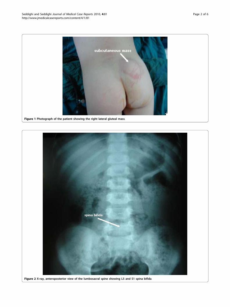
Figure 1 Photograph of the patient showing the right lateral gluteal mass.
Figure 2 X-ray, anteroposterior view of the lumbosacral spine showing L5 and S1 spina bifida.
Seddighi and Seddighi Journal of Medical Case Reports 2010, 4:81http://www.jmedicalcasereports.com/content/4/1/81
Page 2 of 6
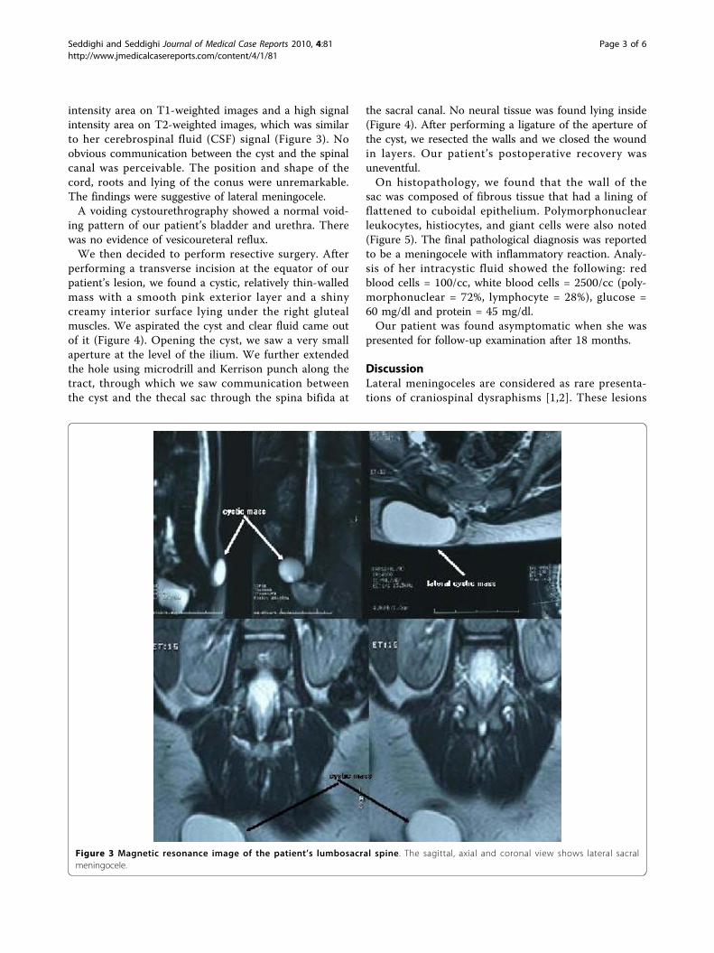
intensity area on T1-weighted images and a high signalintensity area on T2-weighted images, which was similarto her cerebrospinal fluid (CSF) signal (Figure 3). Noobvious communication between the cyst and the spinalcanal was perceivable. The position and shape of thecord, roots and lying of the conus were unremarkable.The findings were suggestive of lateral meningocele.A voiding cystourethrography showed a normal void-
ing pattern of our patient’s bladder and urethra. Therewas no evidence of vesicoureteral reflux.We then decided to perform resective surgery. After
performing a transverse incision at the equator of ourpatient’s lesion, we found a cystic, relatively thin-walledmass with a smooth pink exterior layer and a shinycreamy interior surface lying under the right glutealmuscles. We aspirated the cyst and clear fluid came outof it (Figure 4). Opening the cyst, we saw a very smallaperture at the level of the ilium. We further extendedthe hole using microdrill and Kerrison punch along thetract, through which we saw communication betweenthe cyst and the thecal sac through the spina bifida at
the sacral canal. No neural tissue was found lying inside(Figure 4). After performing a ligature of the aperture ofthe cyst, we resected the walls and we closed the woundin layers. Our patient’s postoperative recovery wasuneventful.On histopathology, we found that the wall of the
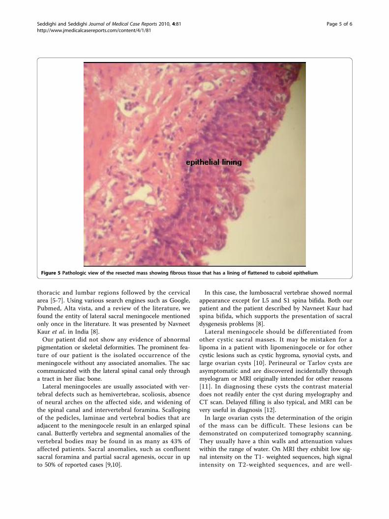
sac was composed of fibrous tissue that had a lining offlattened to cuboidal epithelium. Polymorphonuclearleukocytes, histiocytes, and giant cells were also noted(Figure 5). The final pathological diagnosis was reportedto be a meningocele with inflammatory reaction. Analy-sis of her intracystic fluid showed the following: redblood cells = 100/cc, white blood cells = 2500/cc (poly-morphonuclear = 72%, lymphocyte = 28%), glucose =60 mg/dl and protein = 45 mg/dl.Our patient was found asymptomatic when she was
presented for follow-up examination after 18 months.
DiscussionLateral meningoceles are considered as rare presenta-tions of craniospinal dysraphisms [1,2]. These lesions
Figure 3 Magnetic resonance image of the patient’s lumbosacral spine. The sagittal, axial and coronal view shows lateral sacralmeningocele.
Seddighi and Seddighi Journal of Medical Case Reports 2010, 4:81http://www.jmedicalcasereports.com/content/4/1/81
Page 3 of 6

were first described by Lehman in a patient with otherskeletal findings and distinctive craniofacial features. Hereported a 14-year-old girl with generalized osteosclero-sis, distinctive craniofacial features, and multiple lateralthoracic meningoceles [3]. Subsequently, more patientswith the so-called lateral meningocele syndrome (LMS)have been reported.The existence of an affected mother and daughter
supports the hypothesis that LMS is a dominant
disorder affecting primarily the connective tissue [4].Lateral meningoceles commonly present during thefourth and the fifth decades of life. Neurofibromatosistype 1 is present in approximately 85% of patientswith lateral thoracic meningoceles. Meanwhile, theposition of the cord with respect to the meningocelesac is variable.The incidence of lateral meningoceles was reported to
be 0.3% [4]. Lateral meningoceles are reported in the
Figure 4 An intra-operative view of the right gluteal lesion showing cystic mass containing cerebrospinal fluid.
Seddighi and Seddighi Journal of Medical Case Reports 2010, 4:81http://www.jmedicalcasereports.com/content/4/1/81
Page 4 of 6
thoracic and lumbar regions followed by the cervicalarea [5-7]. Using various search engines such as Google,Pubmed, Alta vista, and a review of the literature, wefound the entity of lateral sacral meningocele mentionedonly once in the literature. It was presented by NavneetKaur et al. in India [8].Our patient did not show any evidence of abnormal
pigmentation or skeletal deformities. The prominent fea-ture of our patient is the isolated occurrence of themeningocele without any associated anomalies. The saccommunicated with the lateral spinal canal only througha tract in her iliac bone.Lateral meningoceles are usually associated with ver-
tebral defects such as hemivertebrae, scoliosis, absenceof neural arches on the affected side, and widening ofthe spinal canal and intervertebral foramina. Scallopingof the pedicles, laminae and vertebral bodies that areadjacent to the meningocele result in an enlarged spinalcanal. Butterfly vertebra and segmental anomalies of thevertebral bodies may be found in as many as 43% ofaffected patients. Sacral anomalies, such as confluentsacral foramina and partial sacral agenesis, occur in upto 50% of reported cases [9,10].
In this case, the lumbosacral vertebrae showed normalappearance except for L5 and S1 spina bifida. Both ourpatient and the patient described by Navneet Kaur hadspina bifida, which supports the presentation of sacraldysgenesis problems [8].Lateral meningocele should be differentiated from
other cystic sacral masses. It may be mistaken for alipoma in a patient with lipomeningocele or for othercystic lesions such as cystic hygroma, synovial cysts, andlarge ovarian cysts [10]. Perineural or Tarlov cysts areasymptomatic and are discovered incidentally throughmyelogram or MRI originally intended for other reasons[11]. In diagnosing these cysts the contrast materialdoes not readily enter the cyst during myelography andCT scan. Delayed filling is also typical, and MRI can bevery useful in diagnosis [12].In large ovarian cysts the determination of the origin
of the mass can be difficult. These lesions can bedemonstrated on computerized tomography scanning.They usually have a thin walls and attenuation valueswithin the range of water. On MRI they exhibit low sig-nal intensity on the T1- weighted sequences, high signalintensity on T2-weighted sequences, and are well-
Figure 5 Pathologic view of the resected mass showing fibrous tissue that has a lining of flattened to cuboid epithelium.
Seddighi and Seddighi Journal of Medical Case Reports 2010, 4:81http://www.jmedicalcasereports.com/content/4/1/81
Page 5 of 6
circumscribed with a thin wall that may enhance aftercontrast administration on T1-weighted images [13].
ConclusionAlthough lateral meningocele especially in the sacralregion is rare, its possibility should always be consideredin patients presenting with a paravertebral or glutealmass. The occurrence of a neurological deficit or thepresence of a spinal defect should make one suspiciousof the presence of an unusually located meningocele.Drainage through needle aspiration or by incision maytransform it into a cerebrospinal fluid fistula. Performingadequate imaging studies such as CT myelography andMRI, therefore, are very helpful to avoid mistakes andensure correct diagnosis.
ConsentWritten informed consent was obtained from thepatient’s next-of-kin for publication of this case reportand any accompanying images. A copy of the writtenconsent is available for review by the Editor-in-Chief ofthis journal.
AbbreviationsMRI: magnetic resonance imaging; CT: computerized tomography; RBC: redblood cell; WBC: white blood cell; PMN: polymorphonuclear; LMS: LateralMeningocele Syndrome.
Author details1Department of Neurosurgery, Rajaie Hospital, Padegan Street, Qazvin, Iran.2Department of Neurosurgery, Shohada Hospital, Tajrish Square, Tehran, Iran.
Authors’ contributionsAS analyzed and interpreted the patient data regarding the lateral sacralmeningocele and performed the surgery. ASS aided in the surgery,performed the review of literature, and was a major contributor in writingthe manuscript. Both authors read and approved the final manuscript.
Competing interestsThe authors declare that they have no competing interests.
Received: 7 January 2009 Accepted: 5 March 2010Published: 5 March 2010
References1. Shetty DS, Lakhkar BN: Lateral sacral lipomeningomyelocele: a rare
anomaly. Neurol India 2002, 50:204-206.2. Erkulvrawtr S, El Gammal T, Hawkins J: Intrathoracic meningoceles and
neurofibromatosis. Arch Neurol 1979, 36:557-559.3. Chen KM, Bird L, Barnes P, Barth R, Hudgins L: Lateral meningocele
syndrome: vertical transmission and expansion of the phenotype. Am JMed Gen 2004, 2:115-121.
4. Gripp KW, Scott CI Jr, Hughes HE, Wallerstein R, Nicholson L, States L,Bason LD, Kaplan P, Zderic SA, Duhaime AC, Miller F, Magnusson MR,Zackai EH: Lateral meningocele syndrome: three new patients andreview of the literature. Am J Med Genet 1997, 70:229-239.
5. Sharma V, Mohanty S, Singh DR: Uncommon craniospinal dysraphism. AnnAcad Med Sing 1996, 25:602-608.
6. Shore RM, Chun RW, Strother CM: Lateral cervical meningocele. ClinPediatr (Phila) 1982, 21:430-433.
7. Gocer AI, Tuna M, Gezercan Y: Multiple anterolateral cervicalmeningoceles associated with neurofibromatosis. Neurosurg Rev 1999,22:124-126.
8. Kaur N, Chandra Mishra S, Vijayaragvan P, Minocha VR: Lateral sacralmeningomyelocele as a gluteal swelling: an unusual presentation.J Indian Med Assoc 2005, 103(10):554-556.
9. Philip N, Andrac L, Moncla A, Sigaudy S, Zanon N, Lena G, Choux M:Multiple lateral meningoceles, distinctive facies and skeletal anomalies: anew case of Lehman syndrome. Clin Dysmorph 1995, 4:347-351.
10. Heckly A, Carsin B, Poulain P: Diagnosis-related pitfall of a lateral sacralcyst. J Neurosurg Spine 2005, 2:72-74.
11. Al-Qahtani S: Tarlov’s cyst. Annals of Saudi Medicine 1998, 18(1):49-51.12. Paulsen RD, Call GA, Murtagh FR: Prevalence and percutaneous drainage
of cysts of the sacral nerve root sheet (Tarlov cyst). Am J Neuroradiol1994, 15:293-299.
13. Occhipinti KA, Frankel SD, Hricak H: The ovary: computed tomographyand magnetic resonance imaging. Radiol Clin North Am 1993,31:1115-1132.
doi:10.1186/1752-1947-4-81Cite this article as: Seddighi and Seddighi: Lateral sacral meningocelepresenting as a gluteal mass: a case report. Journal of Medical CaseReports 2010 4:81.
Submit your next manuscript to BioMed Centraland take full advantage of:
• Convenient online submission
• Thorough peer review
• No space constraints or color figure charges
• Immediate publication on acceptance
• Inclusion in PubMed, CAS, Scopus and Google Scholar
• Research which is freely available for redistribution
Submit your manuscript at www.biomedcentral.com/submit
Seddighi and Seddighi Journal of Medical Case Reports 2010, 4:81http://www.jmedicalcasereports.com/content/4/1/81
Page 6 of 6