Case Report Natal Teeth: A Case Report and...
5
Case Report Natal Teeth: A Case Report and Reappraisal Ghadah A. Malki, Emad A. Al-Badawi, and Mohammad A. Dahlan North Jeddah Specialty Dental Center, Jeddah 23532, Saudi Arabia Correspondence should be addressed to Ghadah A. Malki; [email protected] Received 6 August 2014; Accepted 5 January 2015 Academic Editor: Alberto C. B. Delbem Copyright © 2015 Ghadah A. Malki et al. is is an open access article distributed under the Creative Commons Attribution License, which permits unrestricted use, distribution, and reproduction in any medium, provided the original work is properly cited. e presence of teeth at birth (natal teeth) or within a month aſter delivery (neonatal teeth) is a rare condition. Natal and neonatal teeth are conditions of significant importance to pediatric dentists and pediatricians. is report discusses a case in which a five- day-old infant required extraction of a mobile mandibular natal tooth to avoid the risk of aspiration and interference with feeding. Also, a review of the literature was conducted to discuss the etiology, clinical features, complications, and management of natal and neonatal teeth. 1. Introduction Normal eruption of primary teeth begins with the eruption of mandibular incisors at around 6 months of age [1]. Pre- maturely erupted primary teeth are referred to as congenital teeth, predeciduous teeth, fetal teeth, and dentitia praecox [2, 3]. Massler and Savaral [4] defined tooth/teeth present at birth as “natal teeth” and those erupting during the first month of life as “neonatal teeth.” e difference between “early eruption” and “premature eruption” of natal and neonatal teeth is that “early eruption” occurs due to endocrine system changes while “prema- ture eruption” is considered a pathological phenomenon as incomplete root formation causing the tooth to exfoliate in a short time period [5]. Natal and neonatal teeth are commonly present in the mandibular incisor region with a 66% predilection for females [6]. e prevalence of natal teeth has been inves- tigated by several studies and different ranges have been reported from 1 : 716 to 1 : 3500 live births [3, 6–8]. Most of natal and neonatal teeth are considered early erupting teeth of the normal deciduous dentition [6] and the reported incidence of supernumerary teeth ranges from 1 to 10%. e exact etiology is not known. Several sources suggest a possible hereditary component [4, 6, 9, 10]. An autosomal dominant gene was suggested which was substantiated by a report of a family of 5 siblings who were born with natal teeth [8, 11]. In a study that was conducted on Alaskan Tlingit Indi- ans, the prevalence of natal or neonatal teeth was 9% of their newborns; interestingly enough 62% of the newborns rela- tives were also affected [10]. Furthermore, a positive family history of 7 out of 38 cases of natal and neonatal teeth was found by Kates et al [6]. Environmental factors, particularly polychlorinated biphenyls, appear to increase the incidence of natal teeth [12– 15]. ese exposed children usually display other accompa- nying symptoms, such as dystrophic fingernails and hyper- pigmentation. Natal teeth have been associated with a number of devel- opmental abnormalities and various syndromes, including cleſt lip and palate, Pfeiffer, Ellis-van Creveld (chondroect- odermal dysplasia), Rubinstein-Taybi, steatocystoma multi- plex, pachyonychia congenita (Jadassohn-Lewandowsky), cyclopia, Hallermann-Streiff (Mandibulo-oculo-facial dys- cephaly with hypotrichosis), Pierre-Robin, Wiedeman- Rautenstrauch (neonatal progeria), Pallister-Hall, ectodermal dysplasia, craniofacial dysostosis, multiple adrenogenital, Sotos, steatocystoma, epidermolysis bullosa simplex, and Walker-Warburg syndrome [1, 7, 16–24]. It has been proposed that early erupting primary teeth could be due to abnormal location of the developing tooth germ in relation to the alveolar bone [25]. It was also suggested that this could be the result of hereditary influences Hindawi Publishing Corporation Case Reports in Dentistry Volume 2015, Article ID 147580, 4 pages http://dx.doi.org/10.1155/2015/147580
Transcript of Case Report Natal Teeth: A Case Report and...
Case ReportNatal Teeth: A Case Report and Reappraisal
Ghadah A. Malki, Emad A. Al-Badawi, and Mohammad A. Dahlan
North Jeddah Specialty Dental Center, Jeddah 23532, Saudi Arabia
Correspondence should be addressed to Ghadah A. Malki; [email protected]
Received 6 August 2014; Accepted 5 January 2015
Academic Editor: Alberto C. B. Delbem
Copyright © 2015 Ghadah A. Malki et al. This is an open access article distributed under the Creative Commons AttributionLicense, which permits unrestricted use, distribution, and reproduction in any medium, provided the original work is properlycited.
The presence of teeth at birth (natal teeth) or within a month after delivery (neonatal teeth) is a rare condition. Natal and neonatalteeth are conditions of significant importance to pediatric dentists and pediatricians. This report discusses a case in which a five-day-old infant required extraction of a mobile mandibular natal tooth to avoid the risk of aspiration and interference with feeding.Also, a review of the literature was conducted to discuss the etiology, clinical features, complications, and management of natal andneonatal teeth.
1. Introduction
Normal eruption of primary teeth begins with the eruptionof mandibular incisors at around 6 months of age [1]. Pre-maturely erupted primary teeth are referred to as congenitalteeth, predeciduous teeth, fetal teeth, and dentitia praecox[2, 3]. Massler and Savaral [4] defined tooth/teeth presentat birth as “natal teeth” and those erupting during the firstmonth of life as “neonatal teeth.”
The difference between “early eruption” and “prematureeruption” of natal and neonatal teeth is that “early eruption”occurs due to endocrine system changes while “prema-ture eruption” is considered a pathological phenomenon asincomplete root formation causing the tooth to exfoliate in ashort time period [5].
Natal and neonatal teeth are commonly present in themandibular incisor region with a 66% predilection forfemales [6]. The prevalence of natal teeth has been inves-tigated by several studies and different ranges have beenreported from 1 : 716 to 1 : 3500 live births [3, 6–8].
Most of natal and neonatal teeth are considered earlyerupting teeth of the normal deciduous dentition [6] and thereported incidence of supernumerary teeth ranges from 1 to10%.
The exact etiology is not known. Several sources suggesta possible hereditary component [4, 6, 9, 10]. An autosomaldominant gene was suggested which was substantiated by a
report of a family of 5 siblings who were born with natal teeth[8, 11]. In a study that was conducted on Alaskan Tlingit Indi-ans, the prevalence of natal or neonatal teeth was 9% of theirnewborns; interestingly enough 62% of the newborns rela-tives were also affected [10]. Furthermore, a positive familyhistory of 7 out of 38 cases of natal and neonatal teeth wasfound by Kates et al [6].
Environmental factors, particularly polychlorinatedbiphenyls, appear to increase the incidence of natal teeth [12–15]. These exposed children usually display other accompa-nying symptoms, such as dystrophic fingernails and hyper-pigmentation.
Natal teeth have been associated with a number of devel-opmental abnormalities and various syndromes, includingcleft lip and palate, Pfeiffer, Ellis-van Creveld (chondroect-odermal dysplasia), Rubinstein-Taybi, steatocystoma multi-plex, pachyonychia congenita (Jadassohn-Lewandowsky),cyclopia, Hallermann-Streiff (Mandibulo-oculo-facial dys-cephaly with hypotrichosis), Pierre-Robin, Wiedeman-Rautenstrauch (neonatal progeria), Pallister-Hall, ectodermaldysplasia, craniofacial dysostosis, multiple adrenogenital,Sotos, steatocystoma, epidermolysis bullosa simplex, andWalker-Warburg syndrome [1, 7, 16–24].
It has been proposed that early erupting primary teethcould be due to abnormal location of the developing toothgerm in relation to the alveolar bone [25]. It was alsosuggested that this could be the result of hereditary influences
Hindawi Publishing CorporationCase Reports in DentistryVolume 2015, Article ID 147580, 4 pageshttp://dx.doi.org/10.1155/2015/147580

2 Case Reports in Dentistry
[9]. However, in most cases the pathogenetic factors areimpossible to identify. Therefore, careful evaluation of theseinfants is highly recommended.
Natal teethmanagement is dependent on a number of fac-tors. If the natal tooth is supernumerary, then the treatmentof choice is extraction. When the tooth/teeth are excessivelymobile, extraction is indicated owing to the risk of exfoliationand swallowing or aspiration. However, when reviewing theliterature, no reported cases of aspiration of natal or neonatalteeth were found. In one study, only 38% of natal andneonatal teeth exfoliated in the first year of life [26]. Whennatal teeth are only slightly mobile, they often stabilize soonafter eruption. The most common complaint of natal andneonatal teethwas found to be trauma to the tongue on the tipor ventral surface, a complication referred to as Riga-Fedesyndrome [4]. It occurs in 6–10% of cases of natal teeth [27,28]. It was also suggested that this ulceration could be due tothe fact that the tongue in infants lies immediately betweenthe alveolar ridges [4, 29].
2. Case Report
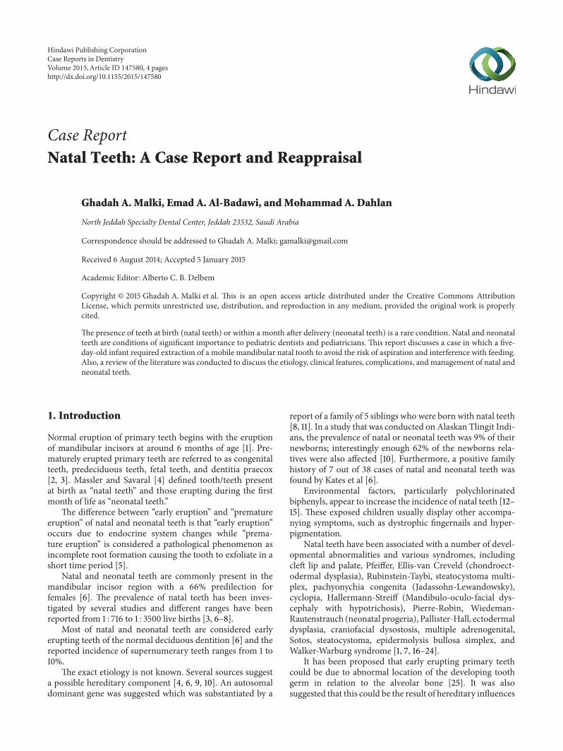
A five-day-old female infant was referred to the pediatricdental clinic, her mother was concerned about the presenceof a tooth in the lower jaw since birth, and she complained ofsoreness during breastfeeding her child. Medical history wasnoncontributory. Extraoral examination showed a symmet-rical face with no lymphadenopathy. Intraoral examinationrevealed a crown of a tooth in the mandibular anteriorregion, small in size, whitish opaque in color (Figure 1), andexhibiting grade II mobility.The lips, gingivae palate, tongue,floor of themouth, and buccal mucosa were clinically normalin appearance and there was no ulceration on the ventral sur-face of the tongue. A diagnosis of natal tooth was made basedon the clinical presentation and confirmed by a periapicalradiograph (Figure 2).
After discussing the treatment options with the mother, itwas decided that extraction was the best treatment since themother was very concerned about the soreness of her breastand she felt she could not continue breastfeeding her child.Before extracting the natal tooth, a pediatricianwas consultedand recommended that vitamin K (0.5–1.0mg) to be givenintramuscularly prior to the extraction to prevent potentialhemorrhage.Thenatal toothwas then extracted under topicalanesthesia and gentle curettage was performed to the extrac-tion socket. The procedure was well tolerated by the infant.The extracted tooth had a crown but was lacking a root. Thepatient was reevaluated five days after extraction and at threemonths.
3. Discussion
The etiology of natal and neonatal teeth remains unde-termined; however it was suggested to be related to var-ious factors, including superficial position of the toothgerm, increased eruption rate due to pyretic incidents, hor-monal stimulation, developmental abnormalities, syndromes,
Figure 1: A 5-day-old female infant with partially erupting nataltooth in the anterior mandibular area, exhibiting grade II mobility.
Figure 2: Periapical radiograph showing the natal tooth in themandibular anterior area.
heredity, and osteoblastic activity within the germ zonerelated to the remodeling phenomenon [1, 7, 8, 16–24, 30, 31].
Natal and neonatal teeth could be either conical or ofnormal shape and size. They usually have an opaque yellow-brownish color. The dimensions of the crowns of these teethare smaller compared to primary teeth that have eruptednormally [1, 8, 23, 24].
Clinically, natal and neonatal teeth can be classifiedaccording to their degree of maturity: (1) a mature natal orneonatal tooth is nearly or fully developed and has moder-ately good prognosis and (2) an immature natal or neonataltooth is incomplete or having a substandard structure with apoor prognosis [8, 32].
Hebling et al. [33] suggested another clinical classificationin their case report according to tooth morphology duringeruption into the oral cavity: (1) shell-shaped crown thatis poorly fixed to the alveolus by gingival tissue with rootabsence, (2) solid crown that is poorly fixed to the alveolus bygingival tissue with little or no root, (3) eruption of the incisalmargin of the crown through the gingival tissues, and (4)gingival edema with palpable but unerupted tooth.
Case Reports in Dentistry 3
The enamel in natal and neonatal teeth is normal forthe age of the children; however, once the teeth eruptprematurely, the uncalcified enamel matrix wears off due toincomplete mineralization leading to teeth becoming yellow-brown in color and continuous breakdown of enamel [6].Furthermore, the increased mobility leads to dentin andcementum cervical changes and possible ensuing of Her-twig’s sheath degeneration preventing root formation [34].Several histological findings have demonstrated that, albeitnormal structure of natal and neonatal teeth enamel, themineralization process of enamel is interrupted by earlyeruption. Hence, the enamel is described as hypomineralizedor dysplastic and is prone to discoloration and wear [30, 35–38].
Histological data on natal and neonatal teeth have alsofound that varying degrees of hypoplastic enamel cover thecrowns of these teeth. The enamel thickness for natal teethis 300mm and for neonatal teeth it is 135mm, whereas innormal primary teeth the enamel layer is between 1000 and1200mm [30].The dentinal area did not reveal any significantdifferences compared to normal primary teeth; however someSEM studies of these teeth have shown large interglobularspaces with abnormal cell inclusions.
The correct diagnosis of natal and neonatal teeth isimportant so as to determine if these teeth are supernumeraryor normal dentition. Bohn nodules and dental lamina cystare additional oral manifestations that may be confused withthese dental conditions; but they can be differentiated byradiographic examination.
Several factors should be considered before a treatmentplan is decided: (1) degree of mobility and implantation, (2)convenience during suckling, (3) interference with breast-feeding, and (4) if the tooth is supernumerary or is part ofthe normal dentition.
If these erupted teeth are diagnosed as part of the normaldentition, maintenance in the mouth is considered theprimary treatment option except if they become a source ofinjury to the baby. If they are implanted well, these teethshould be left in the arch and only removed when they inter-fere with feeding or when they are extremely mobile with arisk of aspiration. Indications for removal include risk of dis-location, subsequent aspiration, and traumatic injury to thebaby’s tongue and/or the maternal breast [29].
According to some investigators, the detection of Riga-Fede disease is an indication for natal/neonatal toothremoval; however, others do not recommend removal sincean acute incisal margin can be relieved by smoothing [24].Tomizawa et al. [39] reported that the treatment of Riga-Fede disease by layering the incisal edge with any photopoly-merizable resin, which is facilitated in rapid healing of theulcers. Having said that, most of these teeth exhibit evidenceof hypomineralization and therefore limited surface area ofenamel is available for resin bonding. Given these factorscombined with the difficulties adequate bonding procedurefrom access to proper moisture control and then enamel sur-face etching renders resin retention uncertain. In addition, ifthe restoration fails, there is a risk that the composite resincould also be swallowed.
Natal/neonatal teeth that show mobility of more than1mmare indicated for extraction; this is due to the probabilityof aspirating or ingesting natal teeth. Another reason for theremoval of the natal/neonatal tooth is to alleviate feeding dif-ficulties or complications like Riga- Fede disease. If extractionis the treatment of choice, it can be deferred till the child is10 days of age or more and has appropriate blood levels ofvitamin K.This ten-day waiting period is to allow the normalflora of the intestine to become established to produce vita-min K, an essential factor for prothrombin production in theliver [1, 8, 37]. Since parenteral vitamin K prevents a lifethreatening haemorrhagic disease of the newborn, the Amer-icanAcademy of Pediatrics recommends that all newborns begiven a single intramuscular dose of 0.5 to 1mg of vitamin K[40]. If it is not possible to delay the extraction, a consultationwith the pediatrician should be initiated, so they can assess ifthere is a need to administer vitamin K, if the newborn didnot receive vitamin K immediately after birth.
Once extraction is performed, it is essential to remove theunderlying dental papilla and Hertwig’s epithelial root sheathduring the extraction of natal tooth/teeth to prevent thedevelopment of root structure that could continue if thesestructures are left in situ.
4. Conclusions
Natal and neonatal teeth are rare occurrences in the oralcavity and proper evaluation and diagnosis are crucial toprovide the best treatment option. Pediatricians are usuallythe first to detect these teeth and early consultation withthe dentist can prevent complications. The decision to main-tain or remove these teeth should be assessed in each caseindependently. Radiographic examination is an essentialdiagnostic tool. Thus far, no studies confirmed the cause andeffect relationship with any of the proposed theories so far.The etiology of natal and neonatal teeth still required furtherinvestigations.
Conflict of Interests
The authors confirm that they have no conflict of interestsconcerning the publication of this paper.
References
[1] A. K. C. Leung and W. L. M. Robson, “Natal teeth: a review,”Journal of the National Medical Association, vol. 98, no. 2, pp.226–228, 2006.
[2] M. B. Portela, L. Damasceno, and L. G. Primo, “Unusual case ofmultiple natal teeth,” Journal of Clinical Pediatric Dentistry, vol.29, no. 1, pp. 37–39, 2004.
[3] J. Zhu and D. King, “Natal and neonatal teeth,”ASDC Journal ofDentistry for Children, vol. 62, no. 2, pp. 123–128, 1995.
[4] M. Massler and B. S. Savara, “Natal and neonatal teeth; a reviewof twenty-four cases reported in the literature,” The Journal ofPediatrics, vol. 36, no. 3, pp. 349–359, 1950.
[5] H. Fauconnier and L. Gerardy, “Precocious or prematuredentition,” Archives de Stomatologie, vol. 8, no. 2, pp. 84–88,1953.

4 Case Reports in Dentistry
[6] G. A. Kates, H. L. Needleman, and L. B. Holmes, “Natal andneonatal teeth: a clinical study,” The Journal of the AmericanDental Association, vol. 109, no. 3, pp. 441–443, 1984.
[7] M.H. Chow, “Natal and neonatal teeth,” Journal of the AmericanDental Association, vol. 100, no. 2, pp. 215–216, 1980.
[8] R. F. Cunha, F. A. C. Boer, D. D. Torriani, andW. T. G. Frossard,“Natal and neonatal teeth: review of the literature,” PediatricDentistry, vol. 23, no. 2, pp. 158–162, 2001.
[9] E. Hals, “Natal and neonatal teeth: histologic investigations intwo brothers,” Oral Surgery, Oral Medicine, Oral Pathology, vol.10, no. 5, pp. 509–521, 1957.
[10] J. T. Mayhall, “Natal and neonatal teeth among the TlingetIndians,” Journal of Dental Research, vol. 46, no. 4, pp. 748–749,1967.
[11] H. W. Hyatt Sr., “Natal teeth. Its occurrence in five siblings,”Clinical Pediatrics, vol. 4, pp. 46–48, 1965.
[12] W. J. Rogan, “PCBs and cola-colored babies: Japan, 1968, andTaiwan, 1979,” Teratology, vol. 26, no. 3, pp. 259–261, 1982.
[13] R. W. Miller, “Congenital PCB poisoning: a reevaluation,”Environmental Health Perspectives, vol. 60, pp. 211–214, 1985.
[14] B. C. Gladen, J. S. Taylor, Y. C. Wu, N. B. Ragan, W. J. Rogan,and C. C. Hsu, “Dermatological findings in children exposedtransplacentally to heat-degraded polychlorinated biphenyls inTaiwan,” The British Journal of Dermatology, vol. 122, no. 6, pp.799–808, 1990.
[15] S. Alaluusua, H. Kiviranta, A. Leppaniemi et al., “Natal andneonatal teeth in relation to environmental toxicants,” PediatricResearch, vol. 52, no. 5, pp. 652–655, 2002.
[16] S. Darwish, K. A. Sastry, and A. Ruprecht, “Natal teeth, bifidtongue and deaf mutism,” Journal of Oral Medicine, vol. 42, no.1, pp. 49–56, 1987.
[17] A. Feinstein, J. Friedman, andM. Schewach-Millet, “Pachyony-chia congenita,” Journal of the American Academy of Dermatol-ogy, vol. 19, no. 4, pp. 705–711, 1988.
[18] D. J. Harris, K. W. Ashcraft, E. C. Beatty, T. M. Holder, and J. C.Leonidas, “Natal teeth, patent ductus arteriosus and intestinalpseudo-obstruction: a lethal syndrome in the newborn,” Clini-cal Genetics, vol. 9, no. 5, pp. 479–482, 1976.
[19] N. M. King and A. M. P. Lee, “Natal teeth and steatocystomamultiplex: a newly recognized syndrome,” Journal of Craniofa-cial Genetics andDevelopmental Biology, vol. 7, no. 3, pp. 311–317,1987.
[20] A. K. C. Leung, “Natal teeth,”The American Journal of Diseasesof Children, vol. 140, no. 3, pp. 249–251, 1986.
[21] M. Ohishi, E. Murakami, T. Haita, T. Naruse, M. Sugino,and H. Inomata, “Hallermann-Streiff syndrome and its oralimplications,” ASDC Journal of Dentistry for Children, vol. 53,no. 1, pp. 32–37, 1986.
[22] M. P. Alvarez, P. V. Crespi, and A. L. Shanske, “Natal molars inPfeiffer syndrome type 3: a case report,” The Journal of ClinicalPediatric Dentistry, vol. 18, no. 1, pp. 21–24, 1993.
[23] S. Mhaske, M. B. Yuwanati, A. Mhaske, R. Ragavendra, K.Kamath, and S. Saawarn, “Natal and neonatal teeth: an overviewof the literature,” ISRN Pediatrics, vol. 2013, Article ID 956269,11 pages, 2013.
[24] R. S. Rao and S. V. Mathad, “Natal teeth: case report and reviewof literature,” Journal of Oral and Maxillofacial Pathology, vol.13, no. 1, pp. 41–46, 2009.
[25] N. N. Nik-Hussein, “Natal and neonatal teeth,” The Journal ofPedodontics, vol. 14, no. 2, pp. 110–112, 1990.
[26] G. Bjuggren, “Premature eruption in the primary dentition—aclinical and radiological study,” Svensk Tandlakare Tidskrift, vol.66, no. 4, pp. 343–355, 1973.
[27] H. S. Chawla, “Management of natal/neonatal/early infancyteeth,” Journal of the Indian Society of Pedodontics andPreventiveDentistry, vol. 11, no. 1, pp. 33–36, 1993.
[28] E. W. To, “A study of natal teeth in Hong Kong Chinese,”International Journal of Paediatric Dentistry, vol. 1, no. 2, pp. 73–76, 1991.
[29] S. Buchanan and C. R. Jenkins, “Riga-Fedes syndrome: natal orneonatal teeth associated with tongue ulceration. Case report,”Australian Dental Journal, vol. 42, no. 4, pp. 225–227, 1997.
[30] L. Bigeard, J. Hemmerle, and J. I. Sommermater, “Clinical andultrastructural study of the natal tooth: enamel and dentinassessments,” ASDC Journal of Dentistry for Children, vol. 63,no. 1, pp. 23–31, 1996.
[31] A. K. Leung, “Management of natal teeth,” The Journal of theAmerican Dental Association, vol. 114, no. 6, article 762, 1987.
[32] J. D. Spouge and W. H. Feasby, “Erupted teeth in the newborn,”Oral Surgery, Oral Medicine, Oral Pathology, vol. 22, no. 2, pp.198–208, 1966.
[33] J. Hebling, A. Zuanon, and D. Vianna, “Dente Natal—a case ofnatal teeth,” Odontologia Clinica, vol. 7, pp. 37–40, 1997.
[34] J. C. Southam, “The structure of natal and neonatal teeth,” TheDental Practitioner and Dental Record, vol. 18, no. 12, pp. 423–427, 1968.
[35] R. A. Anderson, “Natal and neonatal teeth: histologic inves-tigation of two black females,” ASDC Journal of Dentistry forChildren, vol. 49, no. 4, pp. 300–303, 1982.
[36] D. S. Berman and L. M. Silverstone, “Natal and neonatal teeth.A clinical and histological study,”BritishDental Journal, vol. 139,no. 9, pp. 361–364, 1975.
[37] M. Rusmah, “Natal and neonatal teeth: a clinical and histologi-cal study,”The Journal of Clinical Pediatric Dentistry, vol. 15, no.4, pp. 251–253, 1991.
[38] M. Uzamis, S. Olmez, H. Ozturk, and H. Celik, “Clinical andultrastructural study of natal and neonatal teeth,”The Journal ofClinical Pediatric Dentistry, vol. 23, no. 3, pp. 173–177, 1999.
[39] M. Tomizawa, Y. Yamada, K. Tonouchi, H. Watanabe, and T.Noda, “Treatment of Riga-Fede’s disease by resin-coverage ofthe incisal edges and seven cases of natal and neonatal teeth,”Shoni Shikagaku Zasshi, vol. 27, no. 1, pp. 182–190, 1989.
[40] Z. A. Bhutta, G. L. Darmstadt, B. S. Hasan, and R. A. Haws,“Community-based interventions for improving perinatal andneonatal health outcomes in developing countries: a review ofthe evidence,” Pediatrics, vol. 115, no. 2, supplement, pp. 519–617,2005.

Submit your manuscripts athttp://www.hindawi.com
Hindawi Publishing Corporationhttp://www.hindawi.com Volume 2014
Oral OncologyJournal of
DentistryInternational Journal of
Hindawi Publishing Corporationhttp://www.hindawi.com Volume 2014
Hindawi Publishing Corporationhttp://www.hindawi.com Volume 2014
International Journal of
Biomaterials
Hindawi Publishing Corporationhttp://www.hindawi.com Volume 2014
BioMed Research International
Hindawi Publishing Corporationhttp://www.hindawi.com Volume 2014
Case Reports in Dentistry
Hindawi Publishing Corporationhttp://www.hindawi.com Volume 2014
Oral ImplantsJournal of
Hindawi Publishing Corporationhttp://www.hindawi.com Volume 2014
Anesthesiology Research and Practice
Hindawi Publishing Corporationhttp://www.hindawi.com Volume 2014
Radiology Research and Practice
Environmental and Public Health
Journal of
Hindawi Publishing Corporationhttp://www.hindawi.com Volume 2014
The Scientific World JournalHindawi Publishing Corporation http://www.hindawi.com Volume 2014
Hindawi Publishing Corporationhttp://www.hindawi.com Volume 2014
Dental SurgeryJournal of
Drug DeliveryJournal of
Hindawi Publishing Corporationhttp://www.hindawi.com Volume 2014
Hindawi Publishing Corporationhttp://www.hindawi.com Volume 2014
Oral DiseasesJournal of
Hindawi Publishing Corporationhttp://www.hindawi.com Volume 2014
Computational and Mathematical Methods in Medicine
ScientificaHindawi Publishing Corporationhttp://www.hindawi.com Volume 2014
PainResearch and TreatmentHindawi Publishing Corporationhttp://www.hindawi.com Volume 2014
Preventive MedicineAdvances in
Hindawi Publishing Corporationhttp://www.hindawi.com Volume 2014
EndocrinologyInternational Journal of
Hindawi Publishing Corporationhttp://www.hindawi.com Volume 2014
Hindawi Publishing Corporationhttp://www.hindawi.com Volume 2014
OrthopedicsAdvances in