Case Report Multifocal Tubercular Osteomyelitis with...
5
Case Report Multifocal Tubercular Osteomyelitis with Tubercular Breast Abscess: An Atypical Presentation of Tuberculosis Mita Bar, 1 Tuhin Santra, 1 Pradipta Guha, 1 Neha Agrawal, 1 Apu Adhikary, 2 Anirban Das, 3 and Chanchal Mahapatra 4 1 Department of Medicine, Calcutta National Medical College & Hospital, Kolkata, India 2 Department of Medicine, North Bengal Medical College & Hospital, Sushrutanagar, India 3 Department of Medicine, Canning Subdivisional Hospital, Canning, India 4 Department of Medicine, KIMS Medical College & Hospital, Amalapuram, India Correspondence should be addressed to Tuhin Santra; [email protected] Received 22 February 2015; Revised 14 April 2015; Accepted 22 April 2015 Academic Editor: Wirongrong Chierakul Copyright © 2015 Mita Bar et al. is is an open access article distributed under the Creative Commons Attribution License, which permits unrestricted use, distribution, and reproduction in any medium, provided the original work is properly cited. Tuberculosis of spine is common in a developing country like India. However, involvement of spine at multiple levels along with involvement of rib and tubercular breast abscess in an immunocompetent patient without any pulmonary involvement is extremely rare. Here we report a case of 53-year-old immunocompetent lady who presented with quadriparesis and MRI (magnetic resonance imaging) of spine revealed multiple lesions involving cervical, thoracic, lumbar, and sacral region without any involvement of intervertebral disc. On detailed examination she was found to have a lump in right breast. Fine needle aspiration cytology of both paravertebral collection and breast lump revealed presence of acid fast bacilli. She was put on antitubercular drug for one year and she responded well to therapy. 1. Introduction Tuberculosis of spine and rib and tubercular breast abscess are the form of extrapulmonary tuberculosis. Tuberculosis of bone occurs in 10% of patients with extrapulmonary TB (tuberculosis) [1] and spinal TB is the commonest form of skeletal TB [2]. Lower thoracic and lumbar vertebrae are the commonest sites for spinal TB followed by middle thoracic and cervical vertebrae [2]. Usually two or more contiguous vertebrae are involved due to hematogenous spread through one intervertebral artery feeding two adjacent vertebrae [3]. Typically intervertebral disc space along with adjacent vertebral bodies is involved. Cases of spinal TB with intervertebral disc sparing have been reported [4]. However, extensive involvement of all spinal level with extraspinal involvement is extremely rare [5–7]. Also, very few cases of skeletal TB involve ribs [8]. Breast tissue along with skeletal muscles and spleen is relatively resistant to tuberculosis infection [9]. Tubercu- losis of breast is very rare even in countries where TB is endemic. Prevalence of breast TB in India varies between 0.64 and 3.59% [10, 11]. Pregnant and lactating women are more susceptible to develop breast TB due to increased vascularity with dilated ducts and it is relatively uncommon in older women [12]. e condition may mimic pyogenic breast abscess or carcinoma of breast both clinically and radiologically [13]. It is mainly of two types, primary and secondary. Primary form is very rare. Secondary form is seen more frequently and main routes of spread are hematoge- nous, retrograde spread from axillary lymph nodes and direct extension from lung, pleura, mediastinum, costa, and sternum [14, 15]. Presence of multifocal tubercular osteomyelitis and breast TB may cause diagnostic difficulty as the condition mimics malignancy and radiological investigation oſten fails to differ- entiate between malignancy and infection. Diagnosis needs to be confirmed by histological examination. 2. Case Report A 53-year-old nondiabetic and nonhypertensive female patient who was having an insidious onset and gradually Hindawi Publishing Corporation Case Reports in Infectious Diseases Volume 2015, Article ID 629141, 4 pages http://dx.doi.org/10.1155/2015/629141
Transcript of Case Report Multifocal Tubercular Osteomyelitis with...
Case ReportMultifocal Tubercular Osteomyelitis with Tubercular BreastAbscess: An Atypical Presentation of Tuberculosis
Mita Bar,1 Tuhin Santra,1 Pradipta Guha,1 Neha Agrawal,1
Apu Adhikary,2 Anirban Das,3 and Chanchal Mahapatra4
1Department of Medicine, Calcutta National Medical College & Hospital, Kolkata, India2Department of Medicine, North Bengal Medical College & Hospital, Sushrutanagar, India3Department of Medicine, Canning Subdivisional Hospital, Canning, India4Department of Medicine, KIMS Medical College & Hospital, Amalapuram, India
Correspondence should be addressed to Tuhin Santra; [email protected]
Received 22 February 2015; Revised 14 April 2015; Accepted 22 April 2015
Academic Editor: Wirongrong Chierakul
Copyright © 2015 Mita Bar et al.This is an open access article distributed under the Creative Commons Attribution License, whichpermits unrestricted use, distribution, and reproduction in any medium, provided the original work is properly cited.
Tuberculosis of spine is common in a developing country like India. However, involvement of spine at multiple levels along withinvolvement of rib and tubercular breast abscess in an immunocompetent patient without any pulmonary involvement is extremelyrare. Here we report a case of 53-year-old immunocompetent lady who presented with quadriparesis andMRI (magnetic resonanceimaging) of spine revealed multiple lesions involving cervical, thoracic, lumbar, and sacral region without any involvement ofintervertebral disc. On detailed examination she was found to have a lump in right breast. Fine needle aspiration cytology of bothparavertebral collection and breast lump revealed presence of acid fast bacilli. She was put on antitubercular drug for one year andshe responded well to therapy.
1. Introduction
Tuberculosis of spine and rib and tubercular breast abscessare the form of extrapulmonary tuberculosis. Tuberculosisof bone occurs in 10% of patients with extrapulmonary TB(tuberculosis) [1] and spinal TB is the commonest formof skeletal TB [2]. Lower thoracic and lumbar vertebraeare the commonest sites for spinal TB followed by middlethoracic and cervical vertebrae [2]. Usually two or morecontiguous vertebrae are involved due to hematogenousspread through one intervertebral artery feeding two adjacentvertebrae [3]. Typically intervertebral disc space along withadjacent vertebral bodies is involved. Cases of spinal TB withintervertebral disc sparing have been reported [4]. However,extensive involvement of all spinal level with extraspinalinvolvement is extremely rare [5–7]. Also, very few cases ofskeletal TB involve ribs [8].
Breast tissue along with skeletal muscles and spleen isrelatively resistant to tuberculosis infection [9]. Tubercu-losis of breast is very rare even in countries where TB isendemic. Prevalence of breast TB in India varies between
0.64 and 3.59% [10, 11]. Pregnant and lactating women aremore susceptible to develop breast TB due to increasedvascularity with dilated ducts and it is relatively uncommonin older women [12]. The condition may mimic pyogenicbreast abscess or carcinoma of breast both clinically andradiologically [13]. It is mainly of two types, primary andsecondary. Primary form is very rare. Secondary form is seenmore frequently and main routes of spread are hematoge-nous, retrograde spread from axillary lymph nodes anddirect extension from lung, pleura, mediastinum, costa, andsternum [14, 15].
Presence ofmultifocal tubercular osteomyelitis and breastTB may cause diagnostic difficulty as the condition mimicsmalignancy and radiological investigation often fails to differ-entiate between malignancy and infection. Diagnosis needsto be confirmed by histological examination.
2. Case Report
A 53-year-old nondiabetic and nonhypertensive femalepatient who was having an insidious onset and gradually
Hindawi Publishing CorporationCase Reports in Infectious DiseasesVolume 2015, Article ID 629141, 4 pageshttp://dx.doi.org/10.1155/2015/629141
2 Case Reports in Infectious Diseases
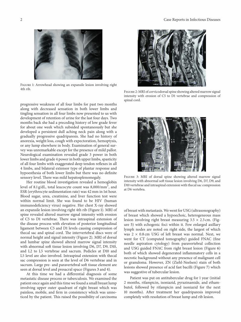
Figure 1: Arrowhead showing an expansile lesion involving right4th rib.
progressive weakness of all four limbs for past two monthsalong with decreased sensation in both lower limbs andtingling sensation in all four limbs now presented to us withdevelopment of retention of urine for the last four days. Twomonths back she had a preceding history of low grade feverfor about one week which subsided spontaneously but shedeveloped a persistent dull aching neck pain along with agradually progressive quadriparesis. She had no history ofanorexia, weight loss, cough with expectoration, hemoptysis,or any lump elsewhere in body. Examination of general sur-vey was unremarkable except for the presence of mild pallor.Neurological examination revealed grade 3 power in bothlower limbs and grade 4 power in both upper limbs, spasticityof all four limbs with exaggerated deep tendon reflexes in all4 limbs, and bilateral extensor type of plantar response andhypoesthesia of both lower limbs but there was no definitesensory level. There was mild hepatosplenomegaly.
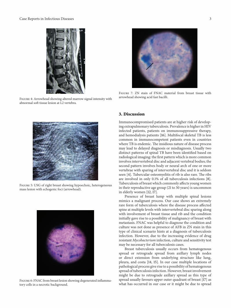
Her routine blood investigation revealed a hemoglobinlevel of 8.1 g/dL, total leucocyte count was 8,000/mm3, andESR (erythrocyte sedimentation rate) was 42mm in 1st hour.Blood sugar, urea, creatinine, and liver function test werewithin normal limit. She was found to be HIV (humanimmunodeficiency virus) negative. Her chest X-ray showedan expansile lesion involving right 4th rib (Figure 1). MRI ofspine revealed altered marrow signal intensity with erosionof C5 to D1 vertebrae. There was intraspinal extension ofthe disease process with elevation of posterior longitudinalligament between C5 and D1 levels causing compression ofthecal sac and spinal cord. The intervertebral discs were ofnormal height and signal intensity (Figure 2). MRI of dorsaland lumbar spine showed altered marrow signal intensitywith abnormal soft tissue lesion involving D6, D7, D9, D10,and L2 to L5 vertebrae and sacrum. Pedicles at D10 andL5 level are also involved. Intraspinal extension with thecalsac compression is seen at the level of D6 vertebrae and insacrum. Large pre- and paravertebral soft tissue element areseen at dorsal level and presacral space (Figures 3 and 4).
At this time we had a differential diagnosis of somemetastatic disease process or tuberculosis. We examined thepatient once again and this timewe found a small breast lumpinvolving upper outer quadrant of right breast which waspainless, mobile, and firm in consistency which was unno-ticed by the patient. This raised the possibility of carcinoma
Figure 2:MRI of cervicodorsal spine showing alteredmarrow signalintensity with erosion of C5 to D1 vertebrae and compression ofspinal cord.
Figure 3: MRI of dorsal spine showing altered marrow signalintensity with abnormal soft tissue lesion involving D6, D7, D9, andD10 vertebrae and intraspinal extensionwith thecal sac compressionat D6 vertebra.
of breastwithmetastasis.Wewent forUSG (ultrasonography)of breast which showed a hypoechoic, heterogeneous masslesion involving right breast measuring 3.5 × 2.3 cm. (Fig-ure 5) with echogenic foci within it. Few enlarged axillarylymph nodes are noted on right side, the largest of whichwas 2 × 0.8 cm USG of left breast was normal. Next, wewent for CT (computed tomography) guided FNAC (fineneedle aspiration cytology) from paravertebral collectionand USG guided FNAC from right breast lesion (Figure 6)both of which showed degenerated inflammatory cells in anecrotic background without any presence of malignant cellor granuloma. However, ZN (Ziehl-Neelsen) stain of bothlesions showed presence of acid fast bacilli (Figure 7) whichwas suggestive of tubercular lesion.
Patient was put on antitubercular drug for 1 year (initial2 months, rifampicin, isoniazid, pyrazinamide, and etham-butol, followed by rifampicin and isoniazid for the next10 months). After treatment her quadriparesis improvedcompletely with resolution of breast lump and rib lesion.
Case Reports in Infectious Diseases 3
Figure 4: Arrowhead showing altered marrow signal intensity withabnormal soft tissue lesion at L2 vertebra.
Figure 5: USG of right breast showing hypoechoic, heterogeneousmass lesion with echogenic foci (arrowhead).
Figure 6: FNAC from breast lesion showing degenerated inflamma-tory cells in a necrotic background.
Figure 7: ZN stain of FNAC material from breast tissue witharrowhead showing acid fast bacilli.
3. Discussion
Immunocompromised patients are at higher risk of develop-ing extrapulmonary tuberculosis. Prevalence is higher inHIVinfected patients, patients on immunosuppressive therapy,and hemodialysis patients [16]. Multifocal skeletal TB is lesscommon in immunocompetent patients even in countrieswhere TB is endemic.The insidious nature of disease processmay lead to delayed diagnosis or misdiagnosis. Usually twodistinct patterns of spinal TB have been identified based onradiological imaging: the first patternwhich ismore commoninvolves intervertebral disc and adjacent vertebral bodies; thesecond pattern involves body or neural arch of one or morevertebrae with sparing of intervertebral disc and it is seldomseen [4]. Tubercular osteomyelitis of rib is also rare. The ribsare involved in only 0.1% of all tuberculosis infections [8].Tuberculosis of breast which commonly affects youngwomenin their reproductive age group (21 to 30 years) is uncommonin elderly women [12, 17].
Presence of breast lump with multiple spinal lesionsmimics a malignant process. Our case shows an extremelyrare form of tuberculosis where the disease process affectedspine at multiple levels with intervertebral disc sparing alongwith involvement of breast tissue and rib and the conditioninitially gave rise to a possibility of malignancy of breast withmetastasis. FNAC was helpful to diagnose the condition andculture was not done as presence of AFB in ZN stain in thistype of clinical scenario hints at a diagnosis of tuberculosisinfection. However, due to the increasing evidence of drugresistantMycobacterium infection, culture and sensitivity testmay be necessary for all tuberculosis cases.
Breast tuberculosis usually occurs from hematogenousspread or retrograde spread from axillary lymph nodesor direct extension from underlying structure like lung,pleura, and costa [14, 15]. In our case multiple locations ofpathological process give rise to a possibility of hematogenousspread of tuberculosis infection.However, breast involvementmight be due to retrograde axillary spread as this type ofspread usually favours upper outer quadrant of breast [17] aswhat has occurred in our case or it might be due to spread
4 Case Reports in Infectious Diseases
from underlying infected rib both of which are a possiblemechanism.
So far there is only one reported case of dual location ofbreast and spine TB [7] and that was reported from outsideIndia. To the best of our knowledge, this is the first evercase of multifocal spinal and breast tuberculosis along withrib involvement, without any pulmonary involvement in animmunocompetent patient from a country like India whereTB is endemic.
4. Conclusion
A differential diagnosis of tuberculosis should be consideredin mind whenever a patient presents with lesion at multiplespinal levels with intervertebral disc sparing. Presence ofa breast lump should be evaluated carefully. Clinical andradiological findings may not be helpful for proper diagnosisbut may be helpful in assessing extent of disease involvement.Histological examination is necessary for proper diagnosis.
Abbreviations
TB: TuberculosisESR: Erythrocyte sedimentation rateHIV: Human immunodeficiency virusMRI: Magnetic resonance imagingUSG: Ultrasonographycm: Centimetermm: MillimeterCT: Computed tomographyFNAC: Fine needle aspiration cytologyZN: Ziehl-Neelsen.
Vertebral Level
C: CervicalD: DorsalL: Lumbar.
Consent
Patient described in this case report has given her consent forthe publication of case report.
Conflict of Interests
The authors have no conflict of interests or financialdisclosure.
References
[1] S. K. Sharma and A. Mohan, “Extrapulmonary tuberculosis,”Indian Journal of Medical Research, vol. 120, no. 4, pp. 316–353,2004.
[2] S. K. Sharma and A.Mohan, “Skeletal tuberculosis,” in Tubercu-losis, p. 343, 2nd edition, 2009.
[3] A. Kulali, S. Cobanoglu, F. Ozyilmaz, R. G. Fessler, and E. C.Benzel, “Spinal tuberculosis with circumferential involvement
of two noncontiguous isolated vertebral levels: case report,”Neurosurgery, vol. 35, no. 6, pp. 1154–1158, 1994.
[4] E. Pertuiset, J. Beaudreuil, F. Liote et al., “Spinal tuberculosis inadults: a study of 103 cases in a developed country, 1980–1994,”Medicine, vol. 78, no. 5, pp. 309–320, 1999.
[5] E. Emel, F. K. Guzey, D. Guzey, N. S. Bas, B. Sel, and I. Alatas,“Non-contiguous multifocal spinal tuberculosis involving cer-vical, thoracic, lumbar and sacral segments: a case report,”European Spine Journal, vol. 15, no. 6, pp. 1019–1024, 2006.
[6] J. Mathew, P. Tripathy, and S. Grewal, “Epidural tuberculosisinvolving the entire spine,” Neurologia i Neurochirurgia Polska,vol. 43, no. 5, pp. 470–474, 2009.
[7] A. Salem, N. Mnif, M. Karray, L. Kribi, T. Ellouze, and R.Hamza, “Double location of breast and spine tuberculosis. Acase report,” Journal de Gynecologie Obstetrique et Biologie dela Reproduction (Paris), vol. 33, no. 2, pp. 148–150, 2004.
[8] D. Ozol, A. Koktener, and M. E. Uyar, “Active pulmonarytuberculosis with vertebra and rib involvement: case report,”Southern Medical Journal, vol. 99, no. 2, pp. 171–173, 2006.
[9] T. T. Alagaratnam and G. B. Ong, “Tuberculosis of the breast,”British Journal of Surgery, vol. 67, no. 2, pp. 125–126, 1980.
[10] R. S. Dharkar, M. H. Kanhere, N. D. Vaishya, and A. K. Bisarya,“Tuberculosis of the breast,” Journal of the Indian MedicalAssociation, vol. 50, no. 5, pp. 207–209, 1968.
[11] P. Mukerjee, M. George, H. B. Maheshwari, and C. P. Rao,“Tuberculosis of the breast,” Journal of the Indian MedicalAssociation, vol. 62, no. 12, pp. 410–412, 1974.
[12] S. R. Shinde, R. Y. Chandawarkar, and S. P. Deshmukh, “Tuber-culosis of the breast masquerading as carcinoma: a study of 100patients,” World Journal of Surgery, vol. 19, no. 3, pp. 379–381,1995.
[13] K. S. Madhusudhan and S. Gamanagatti, “Primary breasttuberculosis masquerading as carcinoma,” Singapore MedicalJournal, vol. 49, no. 1, pp. e3–e5, 2008.
[14] P. Schnarkowski, D. Schmidt, M. Kessler, and M. F. Reiser,“Tuberculosis of the breast. US, mammographic, and CTfindings,” Journal of Computer Assisted Tomography, vol. 18, no.6, pp. 970–971, 1994.
[15] K. K. Oh, J. H. Kim, and S. H. Kook, “Imaging of tuberculousdisease involving breast,” European Radiology, vol. 8, no. 8, pp.1475–1480, 1998.
[16] H. L. Rieder, D. E. Snider Jr., and G. M. Cauthen, “Extrapul-monary tuberculosis in the United States,” American Review ofRespiratory Disease, vol. 141, no. 2, pp. 347–351, 1990.
[17] H. S. Shukla and S. Kumar, “Benign breast disorders in non-western populations: part II-Benign breast disorders in India,”World Journal of Surgery, vol. 13, no. 6, pp. 746–749, 1989.

Submit your manuscripts athttp://www.hindawi.com
Stem CellsInternational
Hindawi Publishing Corporationhttp://www.hindawi.com Volume 2014
Hindawi Publishing Corporationhttp://www.hindawi.com Volume 2014
MEDIATORSINFLAMMATION
of
Hindawi Publishing Corporationhttp://www.hindawi.com Volume 2014
Behavioural Neurology
EndocrinologyInternational Journal of
Hindawi Publishing Corporationhttp://www.hindawi.com Volume 2014
Hindawi Publishing Corporationhttp://www.hindawi.com Volume 2014
Disease Markers
Hindawi Publishing Corporationhttp://www.hindawi.com Volume 2014
BioMed Research International
OncologyJournal of
Hindawi Publishing Corporationhttp://www.hindawi.com Volume 2014
Hindawi Publishing Corporationhttp://www.hindawi.com Volume 2014
Oxidative Medicine and Cellular Longevity
Hindawi Publishing Corporationhttp://www.hindawi.com Volume 2014
PPAR Research
The Scientific World JournalHindawi Publishing Corporation http://www.hindawi.com Volume 2014
Immunology ResearchHindawi Publishing Corporationhttp://www.hindawi.com Volume 2014
Journal of
ObesityJournal of
Hindawi Publishing Corporationhttp://www.hindawi.com Volume 2014
Hindawi Publishing Corporationhttp://www.hindawi.com Volume 2014
Computational and Mathematical Methods in Medicine
OphthalmologyJournal of
Hindawi Publishing Corporationhttp://www.hindawi.com Volume 2014
Diabetes ResearchJournal of
Hindawi Publishing Corporationhttp://www.hindawi.com Volume 2014
Hindawi Publishing Corporationhttp://www.hindawi.com Volume 2014
Research and TreatmentAIDS
Hindawi Publishing Corporationhttp://www.hindawi.com Volume 2014
Gastroenterology Research and Practice
Hindawi Publishing Corporationhttp://www.hindawi.com Volume 2014
Parkinson’s Disease
Evidence-Based Complementary and Alternative Medicine
Volume 2014Hindawi Publishing Corporationhttp://www.hindawi.com