
Case Report Lumbar chronic subdural hematoma mimicking an...
3
© 2017 Surgical Neurology International | Published by Wolters Kluwer - Medknow Editor: Nancy E. Epstein, MD Winthrop Hospital, Mineola, NY, USA OPEN ACCESS For entire Editorial Board visit : http://www.surgicalneurologyint.com SNI: Spine Case Report Lumbar chronic subdural hematoma mimicking an intradural extramedullary tumor: A case report Hyeun Sung Kim, Nitin Adsul, Yoon Seok Ju, Ki Joon Kim, Sung Ho Choi, Jeong Hoon Kim, Sung Kyun Chung, Jeong‑Hoon Choi, Jee‑Soo Jang, Il‑Tae Jang 1 , Seong‑Hoon Oh 2 , Jae Eun Park 3 , Sol Lee 3 Department of Neurosurgery, Nanoori Suwon Hospital, Suwon, 1 Department of Neurosurgery, Nanoori Hospital, Seoul, 2 Department of Neurosurgery, Nanoori Incheon Hospital, Incheon, 3 Department of Nanoori Medical Research, Nanoori Hospital, Seoul, Korea E‑mail: Hyeun Sung Kim ‑ [email protected]; *Nitin Adsul ‑[email protected];Yoon Seok Ju ‑ [email protected]; Ki Joon Kim ‑ [email protected]; Sung Ho Choi ‑ [email protected]; Jeong Hoon Kim ‑ [email protected]; Sung Kyun Chung ‑ [email protected]; Jeong‑Hoon Choi ‑ [email protected]; Jee‑Soo Jang ‑ [email protected]; Il‑Tae Jang ‑ [email protected]; Seong‑Hoon Oh ‑ [email protected]; Jae Eun Park ‑ [email protected]; Sol Lee ‑ [email protected] *Corresponding author Received: 17 July 17 Accepted: 02 August 17 Published: 26 September 17 Abstract Background: Chronic spinal subdural hematomas are extremely rare with only 28 cases reported in the literature. Nevertheless, they should be considered among the differential diagnoses for spinal intradural/extramedullary lesions. Case Report: A 65‑year‑old male presented with progressive back pain and right S1 radiculopathy. Magnetic resonance imaging scan revealed a right‑sided posterolateral intradural/extramedullary lesion at the L5–S1 level. It was hyperintense on T1 and hypointense on T2‑weighted images; on the short TI inversion recovery sequence it was hyperintense. The lesion was excised through a right L5 hemilaminectomy, and the patient was neurologically intact postoperatively. Histopathology revealed a chronic subdural hematoma. Conclusion: Chronic spinal subdural hematoma can mimic intradural extramedullary spinal tumors even in the absence of trauma and/or coagulopathies. Key Words: Chronic, intradural extramedullary tumor, spinal subdural hematoma INTRODUCTION Spinal subdural hematomas (SDHs) are rare, accounting for only 4.1% of all spinal hemorrhages. [5] There are only 28 cases of spinal subdural hematomas reported in the literature. [1] Most occur in the thoracic and/or thoracolumbar regions. [4] Here, we report a chronic SDH occurring in a 65‑year‑old male at the L5–S1 level mimicking an intradural extramedullary tumor. CASE REPORT Clinical and radiographic presentation In the absence of trauma or a history of coagulopathy, a 65‑year‑old male presented with a progressive right lower extremity L5/S1 radiculopathy. On physical examination, straight leg raising was positive on the right side at 70 degrees and the right Achilles response was absent; there was no sensory or motor deficit. Standing lateral dynamic X‑rays showed a grade 1 listhesis at the L4–L5 level [Figure 1a and b]. Magnetic How to cite this article: Kim HS, Adsul N, Ju YS, Kim KJ, Choi SH, Kim JH, et al. Lumbar chronic subdural hematoma mimicking an intradural extramedullary tumor: A case report. Surg Neurol Int 2017;8:231. http://surgicalneurologyint.com/Lumbar-chronic-subdural-hematoma-mimicking-an- intradural-extramedullary-tumor:-A-case-report/ This is an open access article distributed under the terms of the Creative Commons Attribution-NonCommercial-ShareAlike 3.0 License, which allows others to remix, tweak, and build upon the work non-commercially, as long as the author is credited and the new creations are licensed under the identical terms. For reprints contact: [email protected] Access this article online Website: www.surgicalneurologyint.com DOI: 10.4103/sni.sni_262_17 Quick Response Code:
-
Upload
hoangquynh -
Category
Documents
-
view
213 -
download
0
Transcript of Case Report Lumbar chronic subdural hematoma mimicking an...
© 2017 Surgical Neurology International | Published by Wolters Kluwer - Medknow
Editor:Nancy E. Epstein, MDWinthrop Hospital, Mineola, NY, USA
OPEN ACCESSFor entire Editorial Board visit : http://www.surgicalneurologyint.com
SNI: Spine
Case Report
Lumbar chronic subdural hematoma mimicking an intradural extramedullary tumor: A case reportHyeun Sung Kim, Nitin Adsul, Yoon Seok Ju, Ki Joon Kim, Sung Ho Choi, Jeong Hoon Kim, Sung Kyun Chung, Jeong‑Hoon Choi, Jee‑Soo Jang, Il‑Tae Jang1, Seong‑Hoon Oh2, Jae Eun Park3, Sol Lee3
Department of Neurosurgery, Nanoori Suwon Hospital, Suwon, 1Department of Neurosurgery, Nanoori Hospital, Seoul, 2Department of Neurosurgery, Nanoori Incheon Hospital, Incheon, 3Department of Nanoori Medical Research, Nanoori Hospital, Seoul, Korea
E‑mail: Hyeun Sung Kim ‑ [email protected]; *Nitin Adsul ‑[email protected]; Yoon Seok Ju ‑ [email protected]; Ki Joon Kim ‑ [email protected]; Sung Ho Choi ‑ [email protected]; Jeong Hoon Kim ‑ [email protected]; Sung Kyun Chung ‑ [email protected]; Jeong‑Hoon Choi ‑ [email protected]; Jee‑Soo Jang ‑ [email protected]; Il‑Tae Jang ‑ [email protected]; Seong‑Hoon Oh ‑ [email protected]; Jae Eun Park ‑ [email protected]; Sol Lee ‑ [email protected] *Corresponding author
Received: 17 July 17 Accepted: 02 August 17 Published: 26 September 17
AbstractBackground: Chronic spinal subdural hematomas are extremely rare with only 28 cases reported in the literature. Nevertheless, they should be considered among the differential diagnoses for spinal intradural/extramedullary lesions.Case Report: A 65‑year‑old male presented with progressive back pain and right S1 radiculopathy. Magnetic resonance imaging scan revealed a right‑sided posterolateral intradural/extramedullary lesion at the L5–S1 level. It was hyperintense on T1 and hypointense on T2‑weighted images; on the short TI inversion recovery sequence it was hyperintense. The lesion was excised through a right L5 hemilaminectomy, and the patient was neurologically intact postoperatively. Histopathology revealed a chronic subdural hematoma.Conclusion: Chronic spinal subdural hematoma can mimic intradural extramedullary spinal tumors even in the absence of trauma and/or coagulopathies.
Key Words: Chronic, intradural extramedullary tumor, spinal subdural hematoma
INTRODUCTION
Spinal subdural hematomas (SDHs) are rare, accounting for only 4.1% of all spinal hemorrhages.[5] There are only 28 cases of spinal subdural hematomas reported in the literature.[1] Most occur in the thoracic and/or thoracolumbar regions.[4] Here, we report a chronic SDH occurring in a 65‑year‑old male at the L5–S1 level mimicking an intradural extramedullary tumor.
CASE REPORT
Clinical and radiographic presentationIn the absence of trauma or a history of coagulopathy, a 65‑year‑old male presented with a progressive right lower extremity L5/S1 radiculopathy.
On physical examination, straight leg raising was positive on the right side at 70 degrees and the right Achilles response was absent; there was no sensory or motor deficit.
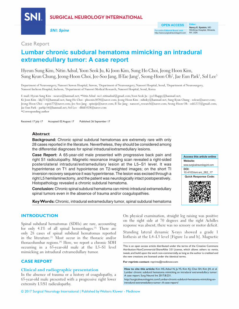
Standing lateral dynamic X‑rays showed a grade 1 listhesis at the L4–L5 level [Figure 1a and b]. Magnetic
How to cite this article: Kim HS, Adsul N, Ju YS, Kim KJ, Choi SH, Kim JH, et al. Lumbar chronic subdural hematoma mimicking an intradural extramedullary tumor: A case report. Surg Neurol Int 2017;8:231.http://surgicalneurologyint.com/Lumbar-chronic-subdural-hematoma-mimicking-an-intradural-extramedullary-tumor:-A-case-report/
This is an open access article distributed under the terms of the Creative Commons Attribution-NonCommercial-ShareAlike 3.0 License, which allows others to remix, tweak, and build upon the work non-commercially, as long as the author is credited and the new creations are licensed under the identical terms.
For reprints contact: [email protected]
Access this article onlineWebsite:www.surgicalneurologyint.comDOI: 10.4103/sni.sni_262_17Quick Response Code:
Surgical Neurology International 2017, 8:231 http://www.surgicalneurologyint.com/content/8/1/231
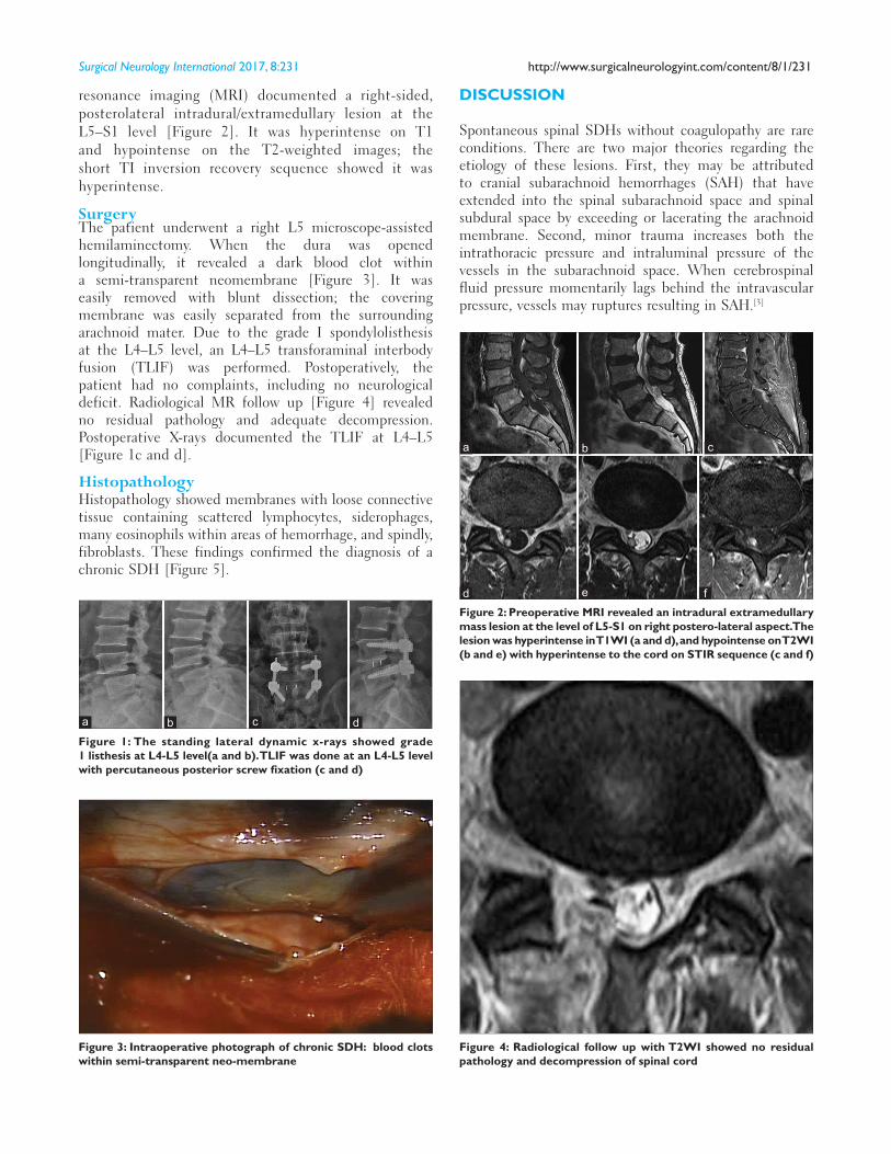
resonance imaging (MRI) documented a right‑sided, posterolateral intradural/extramedullary lesion at the L5–S1 level [Figure 2]. It was hyperintense on T1 and hypointense on the T2‑weighted images; the short TI inversion recovery sequence showed it was hyperintense.
SurgeryThe patient underwent a right L5 microscope‑assisted hemilaminectomy. When the dura was opened longitudinally, it revealed a dark blood clot within a semi‑transparent neomembrane [Figure 3]. It was easily removed with blunt dissection; the covering membrane was easily separated from the surrounding arachnoid mater. Due to the grade I spondylolisthesis at the L4–L5 level, an L4–L5 transforaminal interbody fusion (TLIF) was performed. Postoperatively, the patient had no complaints, including no neurological deficit. Radiological MR follow up [Figure 4] revealed no residual pathology and adequate decompression. Postoperative X‑rays documented the TLIF at L4–L5 [Figure 1c and d].
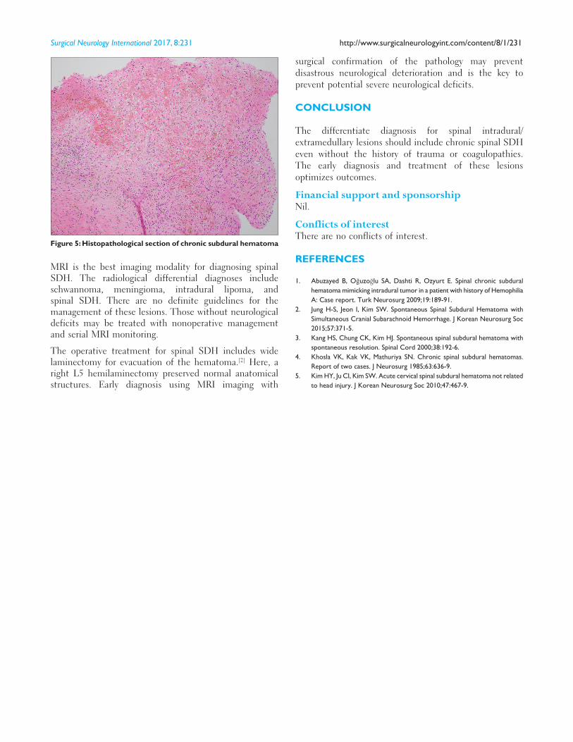
HistopathologyHistopathology showed membranes with loose connective tissue containing scattered lymphocytes, siderophages, many eosinophils within areas of hemorrhage, and spindly, fibroblasts. These findings confirmed the diagnosis of a chronic SDH [Figure 5].
DISCUSSION
Spontaneous spinal SDHs without coagulopathy are rare conditions. There are two major theories regarding the etiology of these lesions. First, they may be attributed to cranial subarachnoid hemorrhages (SAH) that have extended into the spinal subarachnoid space and spinal subdural space by exceeding or lacerating the arachnoid membrane. Second, minor trauma increases both the intrathoracic pressure and intraluminal pressure of the vessels in the subarachnoid space. When cerebrospinal fluid pressure momentarily lags behind the intravascular pressure, vessels may ruptures resulting in SAH.[3]
Figure 3: Intraoperative photograph of chronic SDH: blood clots within semi-transparent neo-membrane
Figure 4: Radiological follow up with T2WI showed no residual pathology and decompression of spinal cord
Figure 1: The standing lateral dynamic x-rays showed grade 1 listhesis at L4-L5 level(a and b). TLIF was done at an L4-L5 level with percutaneous posterior screw fixation (c and d)
dcba
Figure 2: Preoperative MRI revealed an intradural extramedullary mass lesion at the level of L5-S1 on right postero-lateral aspect. The lesion was hyperintense in T1WI (a and d), and hypointense on T2WI (b and e) with hyperintense to the cord on STIR sequence (c and f)
d
cb
f
a
e
Surgical Neurology International 2017, 8:231 http://www.surgicalneurologyint.com/content/8/1/231
MRI is the best imaging modality for diagnosing spinal SDH. The radiological differential diagnoses include schwannoma, meningioma, intradural lipoma, and spinal SDH. There are no definite guidelines for the management of these lesions. Those without neurological deficits may be treated with nonoperative management and serial MRI monitoring.
The operative treatment for spinal SDH includes wide laminectomy for evacuation of the hematoma.[2] Here, a right L5 hemilaminectomy preserved normal anatomical structures. Early diagnosis using MRI imaging with
Figure 5: Histopathological section of chronic subdural hematoma
surgical confirmation of the pathology may prevent disastrous neurological deterioration and is the key to prevent potential severe neurological deficits.
CONCLUSION
The differentiate diagnosis for spinal intradural/extramedullary lesions should include chronic spinal SDH even without the history of trauma or coagulopathies. The early diagnosis and treatment of these lesions optimizes outcomes.
Financial support and sponsorshipNil.
Conflicts of interestThere are no conflicts of interest.
REFERENCES
1. Abuzayed B, Oğuzoğlu SA, Dashti R, Ozyurt E. Spinal chronic subdural hematoma mimicking intradural tumor in a patient with history of Hemophilia A: Case report. Turk Neurosurg 2009;19:189-91.
2. Jung H-S, Jeon I, Kim SW. Spontaneous Spinal Subdural Hematoma with Simultaneous Cranial Subarachnoid Hemorrhage. J Korean Neurosurg Soc 2015;57:371-5.
3. Kang HS, Chung CK, Kim HJ. Spontaneous spinal subdural hematoma with spontaneous resolution. Spinal Cord 2000;38:192-6.
4. Khosla VK, Kak VK, Mathuriya SN. Chronic spinal subdural hematomas. Report of two cases. J Neurosurg 1985;63:636-9.
5. Kim HY, Ju CI, Kim SW. Acute cervical spinal subdural hematoma not related to head injury. J Korean Neurosurg Soc 2010;47:467-9.


![Coordinate cis-[Cr(C2O4)(pm)(OH2)2]+](https://static.fdocuments.net/doc/165x107/5867f4be1a28ab6d408be40b/coordinate-cis-crc2o4pmoh22.jpg)


