Case Report Leprosy Mimicking Common …downloads.hindawi.com/journals/crirh/2014/429698.pdfCase...
6
Case Report Leprosy Mimicking Common Rheumatologic Entities: A Trial for the Clinician in the Era of Biologics Deepak Rath, 1 Shrinath Bhargava, 2 and Bijit Kumar Kundu 1 1 Rheumatology Clinic, Department of Medicine, PGIMER & Dr. RML Hospital, New Delhi 110001, India 2 Department of Dermatology, Dr. RML Hospital, New Delhi 110001, India Correspondence should be addressed to Bijit Kumar Kundu; [email protected] Received 29 August 2014; Accepted 13 October 2014; Published 6 November 2014 Academic Editor: Tsai-Ching Hsu Copyright © 2014 Deepak Rath et al. is is an open access article distributed under the Creative Commons Attribution License, which permits unrestricted use, distribution, and reproduction in any medium, provided the original work is properly cited. Rheumatoid arthritis and seronegative spondyloarthritis, which make up the lion’s share of cases attending a rheumatology clinic, are relatively easy to diagnose. However, when an entity of infective aetiology like leprosy known to be a great mimic of different autoimmune conditions presents with features similar to these, the possibility of it being diagnosed at the outset is very slim indeed. e ease with which the diagnosis of leprosy can be missed assumes sinister proportions as the use of disease modifying agents can have deleterious effects in these patients. In the era of increasing availability and use of biologic disease modifying agents, it is imperative not only to actively rule out the presence of leprosy but also to make it a part of the prebiologic screening of patients in whom biologics are being planned to be administered, especially in leprosy endemic areas. 1. Introduction Affection of skin and nerves by leprosy is quite common and is easily diagnosed. However diagnosing musculoskeletal involvement as being due to leprosy is difficult, given its protean manifestations and the fact that it is a great mimic of many autoimmune conditions. We present two cases in which leprosy presented with features which were the same and were thus initially diagnosed and treated as rheumatoid arthritis (RA) and seronegative spondyloarthritis (SpA), respectively. Only later was the true diagnosis revealed. In areas endemic for leprosy it is imperative to keep in mind the myriad pre- sentations of leprosy including its musculoskeletal manifes- tations, as consequences of misdiagnosis and hence mistreat- ment can be catastrophic, especially in the era of increasing use of biologic disease modifying agents. We conclude that in endemic areas leprosy should be “actively” ruled out and should be made an integral part of prebiologic screening. 2. Case I Mrs. KY, a 35-year-old lady, presented to our rheumatology clinic with the chief complaints of gradual onset slowly progressive polyarthritis affecting the small and large joints of hands in “rheumatoid” distribution, along with nodules over her lower legs on the extensor aspect. e polyarthritis was associated with early morning stiffness which would decrease with activity. Review of the history revealed that she initially had arthritis of wrist followed by knees and ankles a year and half back. e affection of knees and ankles was transient. is was followed a few months later by eruption of nodules on the shins accompanied by feverish feeling. e nodules would resolve aſter a few days leaving behind a hyperpigmented macule. New nodules would subsequently appear and follow the same pattern. Arthritis of the small joints of the hands had developed a few months back, almost a year aſter the affection of the wrist. She had been diagnosed as seronegative RA and treated by a practitioner of alternative medicine, with resolution of symptoms for 3-4 months. She developed intolerance to the medications and had to stop the treatment, but the arthritis and nodules relapsed with a sever- ity more than before. She had presented to the department of medicine of our hospital where she was evaluated initially as a case of polyarthritis with nodules. Her investigations revealed normal haemogram (haemoglobin (Hb): 11.8 gm/dL; total leucocyte count (TLC): 7000/cmm; DC-N79 L35 E6; platelet Hindawi Publishing Corporation Case Reports in Rheumatology Volume 2014, Article ID 429698, 5 pages http://dx.doi.org/10.1155/2014/429698
Transcript of Case Report Leprosy Mimicking Common …downloads.hindawi.com/journals/crirh/2014/429698.pdfCase...

Case ReportLeprosy Mimicking Common Rheumatologic Entities:A Trial for the Clinician in the Era of Biologics
Deepak Rath,1 Shrinath Bhargava,2 and Bijit Kumar Kundu1
1 Rheumatology Clinic, Department of Medicine, PGIMER & Dr. RML Hospital, New Delhi 110001, India2Department of Dermatology, Dr. RML Hospital, New Delhi 110001, India
Correspondence should be addressed to Bijit Kumar Kundu; [email protected]
Received 29 August 2014; Accepted 13 October 2014; Published 6 November 2014
Academic Editor: Tsai-Ching Hsu
Copyright © 2014 Deepak Rath et al. This is an open access article distributed under the Creative Commons Attribution License,which permits unrestricted use, distribution, and reproduction in any medium, provided the original work is properly cited.
Rheumatoid arthritis and seronegative spondyloarthritis, which make up the lion’s share of cases attending a rheumatology clinic,are relatively easy to diagnose. However, when an entity of infective aetiology like leprosy known to be a great mimic of differentautoimmune conditions presents with features similar to these, the possibility of it being diagnosed at the outset is very slim indeed.The ease with which the diagnosis of leprosy can be missed assumes sinister proportions as the use of disease modifying agentscan have deleterious effects in these patients. In the era of increasing availability and use of biologic disease modifying agents, it isimperative not only to actively rule out the presence of leprosy but also to make it a part of the prebiologic screening of patients inwhom biologics are being planned to be administered, especially in leprosy endemic areas.
1. Introduction
Affection of skin and nerves by leprosy is quite commonand is easily diagnosed. However diagnosingmusculoskeletalinvolvement as being due to leprosy is difficult, given itsprotean manifestations and the fact that it is a great mimic ofmany autoimmune conditions.We present two cases inwhichleprosy presentedwith featureswhichwere the same andwerethus initially diagnosed and treated as rheumatoid arthritis(RA) and seronegative spondyloarthritis (SpA), respectively.Only later was the true diagnosis revealed. In areas endemicfor leprosy it is imperative to keep in mind the myriad pre-sentations of leprosy including its musculoskeletal manifes-tations, as consequences of misdiagnosis and hence mistreat-ment can be catastrophic, especially in the era of increasinguse of biologic disease modifying agents. We conclude thatin endemic areas leprosy should be “actively” ruled out andshould be made an integral part of prebiologic screening.
2. Case I
Mrs. KY, a 35-year-old lady, presented to our rheumatologyclinic with the chief complaints of gradual onset slowly
progressive polyarthritis affecting the small and large jointsof hands in “rheumatoid” distribution, along with nodulesover her lower legs on the extensor aspect. The polyarthritiswas associated with early morning stiffness which woulddecrease with activity. Review of the history revealed that sheinitially had arthritis of wrist followed by knees and anklesa year and half back. The affection of knees and ankles wastransient. This was followed a few months later by eruptionof nodules on the shins accompanied by feverish feeling.The nodules would resolve after a few days leaving behind ahyperpigmented macule. New nodules would subsequentlyappear and follow the same pattern. Arthritis of the smalljoints of the hands had developed a few months back, almosta year after the affection of the wrist. She had been diagnosedas seronegative RA and treated by a practitioner of alternativemedicine, with resolution of symptoms for 3-4 months. Shedeveloped intolerance to the medications and had to stop thetreatment, but the arthritis and nodules relapsedwith a sever-ity more than before. She had presented to the department ofmedicine of our hospital where shewas evaluated initially as acase of polyarthritis with nodules. Her investigations revealednormal haemogram (haemoglobin (Hb): 11.8 gm/dL; totalleucocyte count (TLC): 7000/cmm; DC-N79 L35 E6; platelet
Hindawi Publishing CorporationCase Reports in RheumatologyVolume 2014, Article ID 429698, 5 pageshttp://dx.doi.org/10.1155/2014/429698
2 Case Reports in Rheumatology
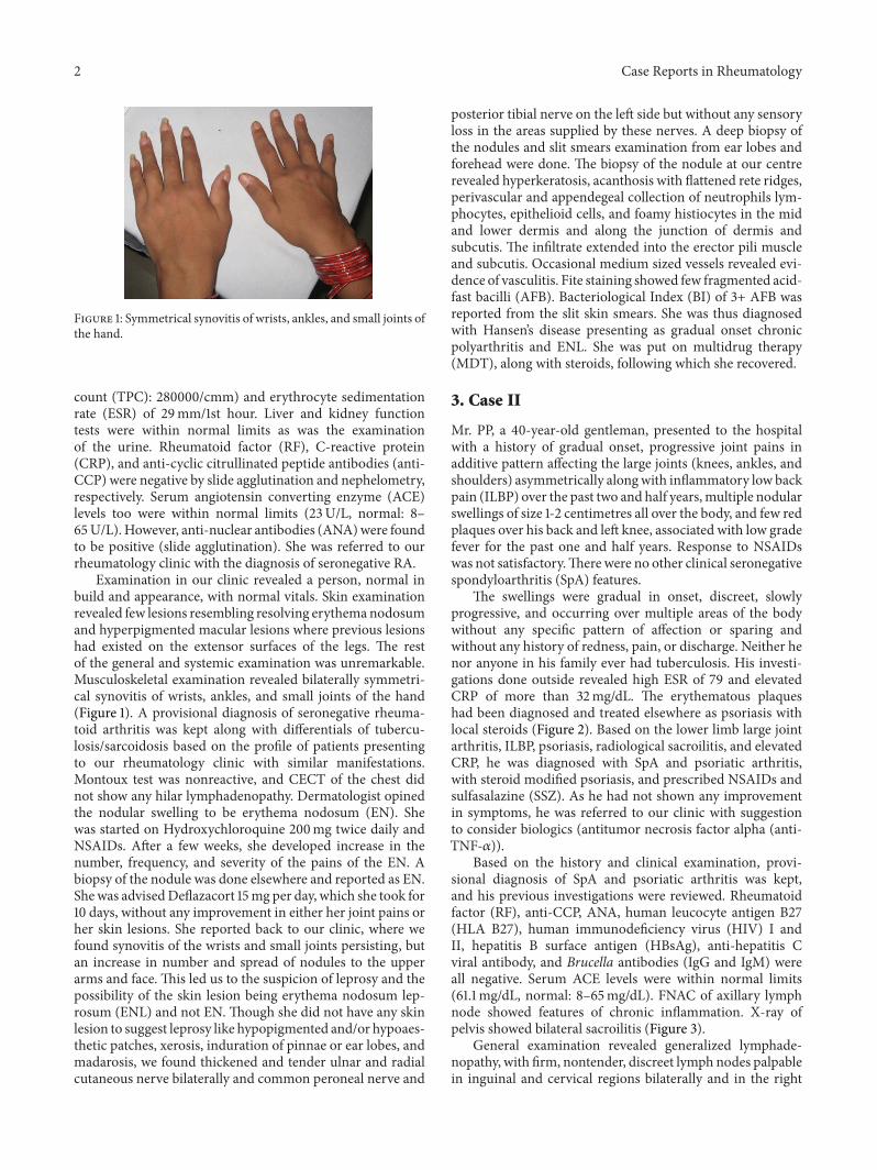
Figure 1: Symmetrical synovitis of wrists, ankles, and small joints ofthe hand.
count (TPC): 280000/cmm) and erythrocyte sedimentationrate (ESR) of 29mm/1st hour. Liver and kidney functiontests were within normal limits as was the examinationof the urine. Rheumatoid factor (RF), C-reactive protein(CRP), and anti-cyclic citrullinated peptide antibodies (anti-CCP) were negative by slide agglutination and nephelometry,respectively. Serum angiotensin converting enzyme (ACE)levels too were within normal limits (23U/L, normal: 8–65U/L). However, anti-nuclear antibodies (ANA)were foundto be positive (slide agglutination). She was referred to ourrheumatology clinic with the diagnosis of seronegative RA.
Examination in our clinic revealed a person, normal inbuild and appearance, with normal vitals. Skin examinationrevealed few lesions resembling resolving erythemanodosumand hyperpigmented macular lesions where previous lesionshad existed on the extensor surfaces of the legs. The restof the general and systemic examination was unremarkable.Musculoskeletal examination revealed bilaterally symmetri-cal synovitis of wrists, ankles, and small joints of the hand(Figure 1). A provisional diagnosis of seronegative rheuma-toid arthritis was kept along with differentials of tubercu-losis/sarcoidosis based on the profile of patients presentingto our rheumatology clinic with similar manifestations.Montoux test was nonreactive, and CECT of the chest didnot show any hilar lymphadenopathy. Dermatologist opinedthe nodular swelling to be erythema nodosum (EN). Shewas started on Hydroxychloroquine 200mg twice daily andNSAIDs. After a few weeks, she developed increase in thenumber, frequency, and severity of the pains of the EN. Abiopsy of the nodule was done elsewhere and reported as EN.Shewas advisedDeflazacort 15mg per day, which she took for10 days, without any improvement in either her joint pains orher skin lesions. She reported back to our clinic, where wefound synovitis of the wrists and small joints persisting, butan increase in number and spread of nodules to the upperarms and face. This led us to the suspicion of leprosy and thepossibility of the skin lesion being erythema nodosum lep-rosum (ENL) and not EN. Though she did not have any skinlesion to suggest leprosy like hypopigmented and/or hypoaes-thetic patches, xerosis, induration of pinnae or ear lobes, andmadarosis, we found thickened and tender ulnar and radialcutaneous nerve bilaterally and common peroneal nerve and
posterior tibial nerve on the left side but without any sensoryloss in the areas supplied by these nerves. A deep biopsy ofthe nodules and slit smears examination from ear lobes andforehead were done. The biopsy of the nodule at our centrerevealed hyperkeratosis, acanthosis with flattened rete ridges,perivascular and appendegeal collection of neutrophils lym-phocytes, epithelioid cells, and foamy histiocytes in the midand lower dermis and along the junction of dermis andsubcutis. The infiltrate extended into the erector pili muscleand subcutis. Occasional medium sized vessels revealed evi-dence of vasculitis. Fite staining showed few fragmented acid-fast bacilli (AFB). Bacteriological Index (BI) of 3+ AFB wasreported from the slit skin smears. She was thus diagnosedwith Hansen’s disease presenting as gradual onset chronicpolyarthritis and ENL. She was put on multidrug therapy(MDT), along with steroids, following which she recovered.
3. Case II
Mr. PP, a 40-year-old gentleman, presented to the hospitalwith a history of gradual onset, progressive joint pains inadditive pattern affecting the large joints (knees, ankles, andshoulders) asymmetrically alongwith inflammatory low backpain (ILBP) over the past two and half years,multiple nodularswellings of size 1-2 centimetres all over the body, and few redplaques over his back and left knee, associated with low gradefever for the past one and half years. Response to NSAIDswas not satisfactory.Therewere no other clinical seronegativespondyloarthritis (SpA) features.
The swellings were gradual in onset, discreet, slowlyprogressive, and occurring over multiple areas of the bodywithout any specific pattern of affection or sparing andwithout any history of redness, pain, or discharge. Neither henor anyone in his family ever had tuberculosis. His investi-gations done outside revealed high ESR of 79 and elevatedCRP of more than 32mg/dL. The erythematous plaqueshad been diagnosed and treated elsewhere as psoriasis withlocal steroids (Figure 2). Based on the lower limb large jointarthritis, ILBP, psoriasis, radiological sacroilitis, and elevatedCRP, he was diagnosed with SpA and psoriatic arthritis,with steroid modified psoriasis, and prescribed NSAIDs andsulfasalazine (SSZ). As he had not shown any improvementin symptoms, he was referred to our clinic with suggestionto consider biologics (antitumor necrosis factor alpha (anti-TNF-𝛼)).
Based on the history and clinical examination, provi-sional diagnosis of SpA and psoriatic arthritis was kept,and his previous investigations were reviewed. Rheumatoidfactor (RF), anti-CCP, ANA, human leucocyte antigen B27(HLA B27), human immunodeficiency virus (HIV) I andII, hepatitis B surface antigen (HBsAg), anti-hepatitis Cviral antibody, and Brucella antibodies (IgG and IgM) wereall negative. Serum ACE levels were within normal limits(61.1mg/dL, normal: 8–65mg/dL). FNAC of axillary lymphnode showed features of chronic inflammation. X-ray ofpelvis showed bilateral sacroilitis (Figure 3).
General examination revealed generalized lymphade-nopathy, with firm, nontender, discreet lymph nodes palpablein inguinal and cervical regions bilaterally and in the right

Case Reports in Rheumatology 3
(a) (b)
Figure 2: (a) Shiny, plaque lesion over left knee diagnosed as steroid modified psoriasis. (b) Erythematous, plaque like lesions over the backthought to be plaque psoriasis.
Figure 3: Sacroilitis.
axillary region with the largest being around 2 cm. Exami-nation of the skin showed multiple nodular lesions over thelegs, trunk, and back. Erythematous plaques were also seenover the back, knees, and shins.Musculoskeletal examinationrevealed asymmetrical synovitis of the wrists, knees, andankles along with tenosynovitis of the feet. The characterof pain suggested a neuritic origin and prompted his neu-rological examination. He was found to have thickened butnontender ulnar and peroneal nerves with evidence of sen-sory deficit in the distribution of the nerves. However otherleprosy features like induration of earlobes and madarosiswere conspicuous by their absence.
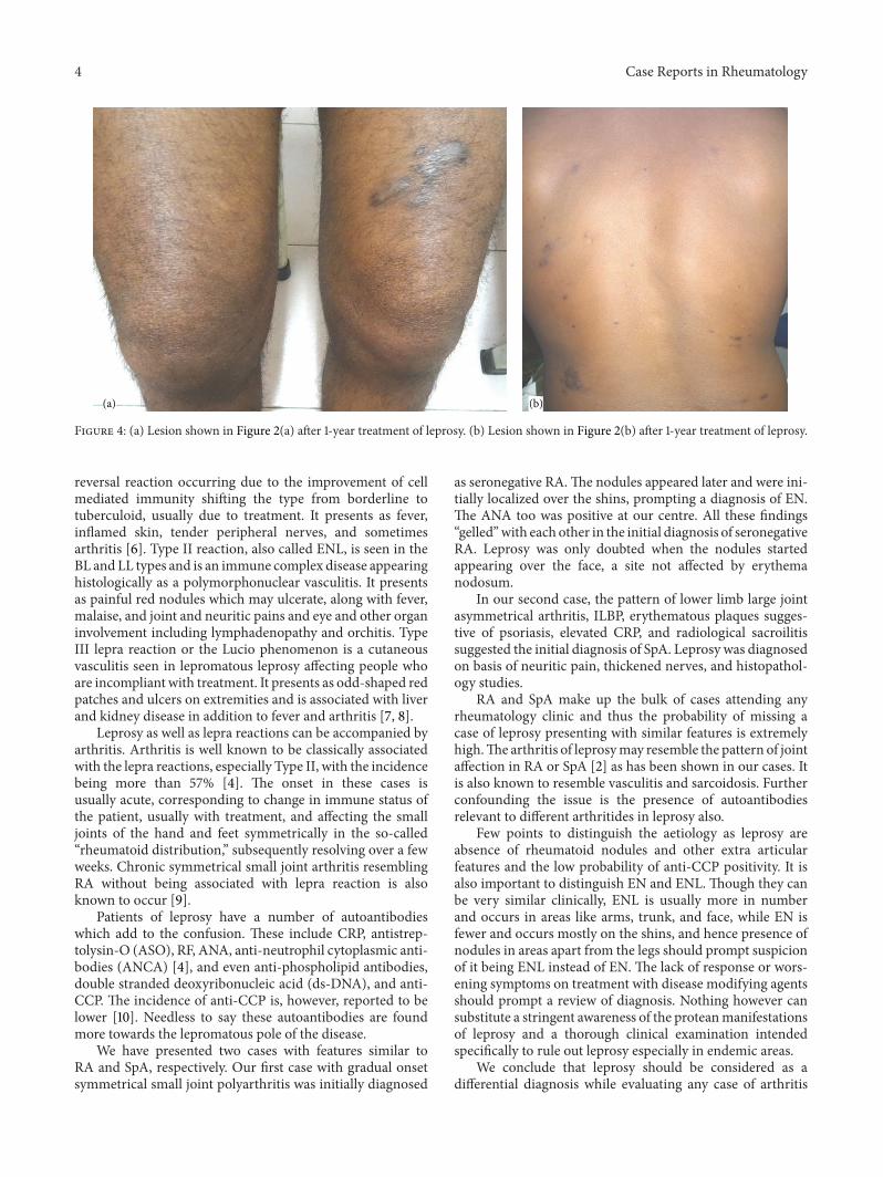
Dermatology review was undertaken and a possibility ofleprosy (BL) with ENL was kept. The BI of the slit skin smearof the patient was reported as 3+. Biopsy of nodules revealednoncaseating granulomas with occasional epithelioid cellsarranged in patches. Multiple acid fast bacilli were visualizedwith Fite stain. He was put onMDT and steroids and showedmarked improvement in his symptoms, both articular anddermatological (Figure 4).
4. Discussion
Leprosy or Hansen’s disease, caused byMycobacterium leprae(M. Leprae), continues to be a public health problem inendemic regions including India, though detection of newcases has decreased from 260063 in 2004 to 127295 in 2011 [1].
Though the classical manifestations affecting the skinand nerves are well known in general, what has come tothe attention in recent times is its “atypical” presentationwith affection of the musculoskeletal system in its variousforms as the primary presentation, with dermatological andneural manifestations occurring later and sometimes not atall. The “atypical” musculoskeletal affection does not includethe classical neuropathic joints of Charcot’s taught to medicalstudents, now rarely seen.
Musculoskeletal affection by leprosy is varied and canmimic RA, SpA, and even vasculitis [2]. The likelihood of itsmisdiagnosis and thus mistreatment, leading to potentiallydisastrous consequences, is high in this scenario. The risk isincreasedmanyfold with the increasing availability and use ofbiologic diseasemodifying agents especially TNF𝛼 inhibitors.Anti-TNF agents cause death of cells expressing TNF anddisrupting granulomas which lead to reactivation of granu-lomatous disease. Though attention has been focused moreon TB, reactivation of leprosy has also been reported [3].
Leprosy can be of five types, namely, tuberculoid (TT),borderline tuberculoid (BT), borderline (BB), borderlinelepromatous (BL), and lepromatous (LL), which correspondto decreasing levels of immunity from TT to LL. Arthritisoccurs in more than half of the cases and is seen in all typesbut most commonly in BL, followed by TT [4]. The Indianclassification has an additional type without skin lesions,called the pure neuritic type, which too can present witharthritis [5].
A characteristic of leprosy is lepra reaction, which can beof three types. Type I can be downgraded with worseningtowards lepromatous features, usually before therapy or the
4 Case Reports in Rheumatology
(a) (b)
Figure 4: (a) Lesion shown in Figure 2(a) after 1-year treatment of leprosy. (b) Lesion shown in Figure 2(b) after 1-year treatment of leprosy.
reversal reaction occurring due to the improvement of cellmediated immunity shifting the type from borderline totuberculoid, usually due to treatment. It presents as fever,inflamed skin, tender peripheral nerves, and sometimesarthritis [6]. Type II reaction, also called ENL, is seen in theBL and LL types and is an immune complex disease appearinghistologically as a polymorphonuclear vasculitis. It presentsas painful red nodules which may ulcerate, along with fever,malaise, and joint and neuritic pains and eye and other organinvolvement including lymphadenopathy and orchitis. TypeIII lepra reaction or the Lucio phenomenon is a cutaneousvasculitis seen in lepromatous leprosy affecting people whoare incompliant with treatment. It presents as odd-shaped redpatches and ulcers on extremities and is associated with liverand kidney disease in addition to fever and arthritis [7, 8].
Leprosy as well as lepra reactions can be accompanied byarthritis. Arthritis is well known to be classically associatedwith the lepra reactions, especially Type II, with the incidencebeing more than 57% [4]. The onset in these cases isusually acute, corresponding to change in immune status ofthe patient, usually with treatment, and affecting the smalljoints of the hand and feet symmetrically in the so-called“rheumatoid distribution,” subsequently resolving over a fewweeks. Chronic symmetrical small joint arthritis resemblingRA without being associated with lepra reaction is alsoknown to occur [9].
Patients of leprosy have a number of autoantibodieswhich add to the confusion. These include CRP, antistrep-tolysin-O (ASO), RF, ANA, anti-neutrophil cytoplasmic anti-bodies (ANCA) [4], and even anti-phospholipid antibodies,double stranded deoxyribonucleic acid (ds-DNA), and anti-CCP. The incidence of anti-CCP is, however, reported to belower [10]. Needless to say these autoantibodies are foundmore towards the lepromatous pole of the disease.
We have presented two cases with features similar toRA and SpA, respectively. Our first case with gradual onsetsymmetrical small joint polyarthritis was initially diagnosed
as seronegative RA.The nodules appeared later and were ini-tially localized over the shins, prompting a diagnosis of EN.The ANA too was positive at our centre. All these findings“gelled”with each other in the initial diagnosis of seronegativeRA. Leprosy was only doubted when the nodules startedappearing over the face, a site not affected by erythemanodosum.
In our second case, the pattern of lower limb large jointasymmetrical arthritis, ILBP, erythematous plaques sugges-tive of psoriasis, elevated CRP, and radiological sacroilitissuggested the initial diagnosis of SpA. Leprosy was diagnosedon basis of neuritic pain, thickened nerves, and histopathol-ogy studies.
RA and SpA make up the bulk of cases attending anyrheumatology clinic and thus the probability of missing acase of leprosy presenting with similar features is extremelyhigh.The arthritis of leprosymay resemble the pattern of jointaffection in RA or SpA [2] as has been shown in our cases. Itis also known to resemble vasculitis and sarcoidosis. Furtherconfounding the issue is the presence of autoantibodiesrelevant to different arthritides in leprosy also.
Few points to distinguish the aetiology as leprosy areabsence of rheumatoid nodules and other extra articularfeatures and the low probability of anti-CCP positivity. It isalso important to distinguish EN and ENL. Though they canbe very similar clinically, ENL is usually more in numberand occurs in areas like arms, trunk, and face, while EN isfewer and occurs mostly on the shins, and hence presence ofnodules in areas apart from the legs should prompt suspicionof it being ENL instead of EN. The lack of response or wors-ening symptoms on treatment with disease modifying agentsshould prompt a review of diagnosis. Nothing however cansubstitute a stringent awareness of the proteanmanifestationsof leprosy and a thorough clinical examination intendedspecifically to rule out leprosy especially in endemic areas.
We conclude that leprosy should be considered as adifferential diagnosis while evaluating any case of arthritis

Case Reports in Rheumatology 5
especially in leprosy endemic areas and should be activelyruled out clinically and if necessary by investigations. Thisassumes utmost importance in view of increasing use of TNF-𝛼 inhibitors and hence should be made an integral part of“biologic screening” prior to institution of biologic therapy.
Conflict of Interests
The authors declare that there is no conflict of interestsregarding the publication of this paper.
References
[1] WHO, “Global leprosy situation, 2012,”Weekly EpidemiologicalRecord, vol. 34, no. 87, pp. 317–328, 2012.
[2] S. Prasad, R. Misra, A. Aggarwal et al., “Leprosy revealed ina rheumatology clinic: a case series,” International Journal ofRheumatic Diseases, vol. 16, no. 2, pp. 129–133, 2013.
[3] J. R. Antonio, R. M. C. Soubhia, V. D. A. Paschoal, C. F.Amarante, andA. R. F. Travolo, “Biological agents: investigationinto leprosy and other infectious diseases before indication,”Anais Brasileiros de Dermatologia, vol. 88, no. 6, supplement 1,pp. 23–25, 2013.
[4] K. Vengadakrishnan, P. K. Saraswat, and P. C. Mathur, “A studyof rheumatological manifestations of leprosy,” Indian Journal ofDermatology, Venereology and Leprology, vol. 70, no. 2, pp. 76–78, 2004.
[5] N. Haroon, V. Agarwal, A. Aggarwal, N. Kumari, N. Krishnani,and R. Misra, “Arthritis as presenting manifestation of pureneuritic leprosy—a rheumatologist’s dilemma,” Rheumatology,vol. 46, no. 4, pp. 653–656, 2007.
[6] C. C. Henriques, B. Lopez, T. Mestre, B. Grima, A. Panarra,and N. Riso, “Leprosy and rheumatoid arthritis: consequenceor association?” BMJ Case Reports, 2012.
[7] S. Salvi and A. Chopra, “Leprosy in a rheumatology setting: achallenging mimic to expose,” Clinical Rheumatology, vol. 32,no. 10, pp. 1557–1563, 2013.
[8] “Leprosy,” 2014, http://www.dermnetnz.org/bacterial/leprosy.html.
[9] S. Chauhan, A. Wakhlu, and V. Agarwal, “Arthritis in leprosy,”Rheumatology, vol. 49, no. 12, pp. 2237–2242, 2010.
[10] A. Chopra, “Rheumatic and other musculoskeletal manifesta-tions and autoantibodies in childhood and adolescent leprosy:significance and relevance,” Journal of Pediatrics, vol. 90, no. 5,pp. 431–436, 2014, 10.1016/j.jped.2014.06.003.

Submit your manuscripts athttp://www.hindawi.com
Stem CellsInternational
Hindawi Publishing Corporationhttp://www.hindawi.com Volume 2014
Hindawi Publishing Corporationhttp://www.hindawi.com Volume 2014
MEDIATORSINFLAMMATION
of
Hindawi Publishing Corporationhttp://www.hindawi.com Volume 2014
Behavioural Neurology
EndocrinologyInternational Journal of
Hindawi Publishing Corporationhttp://www.hindawi.com Volume 2014
Hindawi Publishing Corporationhttp://www.hindawi.com Volume 2014
Disease Markers
Hindawi Publishing Corporationhttp://www.hindawi.com Volume 2014
BioMed Research International
OncologyJournal of
Hindawi Publishing Corporationhttp://www.hindawi.com Volume 2014
Hindawi Publishing Corporationhttp://www.hindawi.com Volume 2014
Oxidative Medicine and Cellular Longevity
Hindawi Publishing Corporationhttp://www.hindawi.com Volume 2014
PPAR Research
The Scientific World JournalHindawi Publishing Corporation http://www.hindawi.com Volume 2014
Immunology ResearchHindawi Publishing Corporationhttp://www.hindawi.com Volume 2014
Journal of
ObesityJournal of
Hindawi Publishing Corporationhttp://www.hindawi.com Volume 2014
Hindawi Publishing Corporationhttp://www.hindawi.com Volume 2014
Computational and Mathematical Methods in Medicine
OphthalmologyJournal of
Hindawi Publishing Corporationhttp://www.hindawi.com Volume 2014
Diabetes ResearchJournal of
Hindawi Publishing Corporationhttp://www.hindawi.com Volume 2014
Hindawi Publishing Corporationhttp://www.hindawi.com Volume 2014
Research and TreatmentAIDS
Hindawi Publishing Corporationhttp://www.hindawi.com Volume 2014
Gastroenterology Research and Practice
Hindawi Publishing Corporationhttp://www.hindawi.com Volume 2014
Parkinson’s Disease
Evidence-Based Complementary and Alternative Medicine
Volume 2014Hindawi Publishing Corporationhttp://www.hindawi.com