Case Report GH-Producing Pituitary Adenoma and Concomitant...
7
Case Report GH-Producing Pituitary Adenoma and Concomitant Rathke’s Cleft Cyst: A Case Report and Short Review Ryota Tamura, 1 Satoshi Takahashi, 1 Katsura Emoto, 2 Hideaki Nagashima, 1 Masahiro Toda, 1 and Kazunari Yoshida 1 1 Department of Neurosurgery, Keio University Hospital, 35 Shinanomachi, Shinjuku-ku, Tokyo 160-8582, Japan 2 Division of Diagnostic Pathology, Keio University Hospital, 35 Shinanomachi, Shinjuku-ku, Tokyo 160-8582, Japan Correspondence should be addressed to Ryota Tamura; [email protected] Received 3 December 2014; Revised 13 March 2015; Accepted 15 March 2015 Academic Editor: Jorge C. Kattah Copyright © 2015 Ryota Tamura et al. is is an open access article distributed under the Creative Commons Attribution License, which permits unrestricted use, distribution, and reproduction in any medium, provided the original work is properly cited. Concomitant pituitary adenoma (PA) and Rathke’s cleſt cyst (RCC) are rare. In some cases, such PA is known to produce pituitary hormones. A 53-year-old man was admitted to our hospital with a diagnosis of lacunar infarction in the leſt basal ganglia. Magnetic resonance imaging (MRI) incidentally showed a suprasellar mass with radiographic features of RCC. When he consulted with a neurosurgical outpatient clinic, acromegaly was suspected based on his appearance. A diagnosis of growth hormone- (GH-) producing PA was confirmed from hormonal examinations and additional MRI. Retrospectively, initial MR images also showed intrasellar mass that is compatible with the diagnosis of PA other than suprasellar RCC. e patient underwent endonasal- endoscopic removal of the PA. Since we judged that the RCC of the patient was asymptomatic, only the PA was completely removed. e postoperative course of the patient was uneventful and GH levels gradually normalized. Only 40 cases of PA with concomitant RCC have been reported to date, including 13 cases of GH-producing PA. In those 13 cases, RCC tended to be located in the sella turcica, and suprasellar RCC like this case appears rare. In a few cases, concomitant RCCs were fenestrated, but GH levels normalized postoperatively as in the cases without RCC fenestration. If radiographic imaging shows typical RCC, and PA is not obvious at first glance, the possibility of concomitant PA still needs to be considered. In terms of treatment, removal of the RCC is not needed to achieve hormone normalization. 1. Introduction e relationship between pituitary adenoma (PA) and Rath- ke’s cleſt cyst (RCC) is controversial. e origin of RCC is generally considered to be derived from remnants of Rathke’s pouch. PA is also formed by proliferation of the anterior wall of Rathke’s pouch. ey have a possibility to be derived from a common ancestry [1]. It has also been thought that they were derived from “transitional” cells between the lining of Rathke’s cleſt and the glandular cells of the anterior pituitary. is theory is based on the fact that PA occasionally contains both elements of fetal Rathke’s pouch and differentiated adenohypophyseal cells [2]. RCC is reported to be found incidentally in 11–33% of postmortem examinations [1], but concomitant PA and RCC are extremely rare. In some cases, such PA is known to produce various pituitary hormones. We report herein a rare case of growth hormone- (GH-) producing PA with concomitant RCC. 2. Clinical Presentation 2.1. Onset and Course. A 53-year-old man had high height from the cradle and also realized his protruding chin. He pre- sented to our hospital with slight paralysis of the right upper extremity. His height was 181 cm that was within the limits of 2 standard deviations. Head MRI showed leſt lacunar infarc- tion of the basal ganglia, as well as a suprasellar mass. e suprasellar mass was hyperintense on T1-weighted MR image (Figure 1(a)) and also isointense on T2-weighted MR image (Figure 1(b)). ese findings for the suprasellar mass were compatible with RCC, and he was referred to the neurosur- gical outpatient clinic for management. At the neurosurgical Hindawi Publishing Corporation Case Reports in Neurological Medicine Volume 2015, Article ID 948025, 6 pages http://dx.doi.org/10.1155/2015/948025
Transcript of Case Report GH-Producing Pituitary Adenoma and Concomitant...
Case ReportGH-Producing Pituitary Adenoma and Concomitant Rathke’sCleft Cyst: A Case Report and Short Review
Ryota Tamura,1 Satoshi Takahashi,1 Katsura Emoto,2 Hideaki Nagashima,1
Masahiro Toda,1 and Kazunari Yoshida1
1Department of Neurosurgery, Keio University Hospital, 35 Shinanomachi, Shinjuku-ku, Tokyo 160-8582, Japan2Division of Diagnostic Pathology, Keio University Hospital, 35 Shinanomachi, Shinjuku-ku, Tokyo 160-8582, Japan
Correspondence should be addressed to Ryota Tamura; [email protected]
Received 3 December 2014; Revised 13 March 2015; Accepted 15 March 2015
Academic Editor: Jorge C. Kattah
Copyright © 2015 Ryota Tamura et al. This is an open access article distributed under the Creative Commons Attribution License,which permits unrestricted use, distribution, and reproduction in any medium, provided the original work is properly cited.
Concomitant pituitary adenoma (PA) and Rathke’s cleft cyst (RCC) are rare. In some cases, such PA is known to produce pituitaryhormones. A 53-year-old man was admitted to our hospital with a diagnosis of lacunar infarction in the left basal ganglia. Magneticresonance imaging (MRI) incidentally showed a suprasellar mass with radiographic features of RCC. When he consulted witha neurosurgical outpatient clinic, acromegaly was suspected based on his appearance. A diagnosis of growth hormone- (GH-)producing PA was confirmed from hormonal examinations and additional MRI. Retrospectively, initial MR images also showedintrasellar mass that is compatible with the diagnosis of PA other than suprasellar RCC. The patient underwent endonasal-endoscopic removal of the PA. Since we judged that the RCC of the patient was asymptomatic, only the PAwas completely removed.The postoperative course of the patient was uneventful and GH levels gradually normalized. Only 40 cases of PA with concomitantRCC have been reported to date, including 13 cases of GH-producing PA. In those 13 cases, RCC tended to be located in thesella turcica, and suprasellar RCC like this case appears rare. In a few cases, concomitant RCCs were fenestrated, but GH levelsnormalized postoperatively as in the cases without RCC fenestration. If radiographic imaging shows typical RCC, and PA is notobvious at first glance, the possibility of concomitant PA still needs to be considered. In terms of treatment, removal of the RCC isnot needed to achieve hormone normalization.
1. Introduction
The relationship between pituitary adenoma (PA) and Rath-ke’s cleft cyst (RCC) is controversial. The origin of RCC isgenerally considered to be derived from remnants of Rathke’spouch. PA is also formed by proliferation of the anterior wallof Rathke’s pouch. They have a possibility to be derived froma common ancestry [1]. It has also been thought that theywere derived from “transitional” cells between the lining ofRathke’s cleft and the glandular cells of the anterior pituitary.This theory is based on the fact that PA occasionally containsboth elements of fetal Rathke’s pouch and differentiatedadenohypophyseal cells [2]. RCC is reported to be foundincidentally in 11–33% of postmortem examinations [1], butconcomitant PA and RCC are extremely rare. In some cases,such PA is known to produce various pituitary hormones.
We report herein a rare case of growth hormone- (GH-)producing PA with concomitant RCC.
2. Clinical Presentation
2.1. Onset and Course. A 53-year-old man had high heightfrom the cradle and also realized his protruding chin. He pre-sented to our hospital with slight paralysis of the right upperextremity. His height was 181 cm that was within the limits of2 standard deviations. Head MRI showed left lacunar infarc-tion of the basal ganglia, as well as a suprasellar mass. Thesuprasellar mass was hyperintense on T1-weightedMR image(Figure 1(a)) and also isointense on T2-weighted MR image(Figure 1(b)). These findings for the suprasellar mass werecompatible with RCC, and he was referred to the neurosur-gical outpatient clinic for management. At the neurosurgical
Hindawi Publishing CorporationCase Reports in Neurological MedicineVolume 2015, Article ID 948025, 6 pageshttp://dx.doi.org/10.1155/2015/948025

2 Case Reports in Neurological Medicine
Rathke’s cleft cyst
(a)
Rathke’s cleft cyst
(b)
Rathke’s cleft cyst
(c)
Rathke’s cleft cyst
Pituitary adenoma
(d)
Figure 1: (a) Sagittal T1-weightedMRI of the head shows a suprasellar, high-intensity mass suspected to represent RCC. PA located below theRCC shows isointensity. (b) Sagittal T2-weightedMRI of the head shows isointense RCC and isointense PA. (c) Contrast-enhanced axial MRIshows nonenhancing RCC. In contrast, the normal pituitary gland shows strong enhancement. (d) Contrast-enhanced coronal MRI showsslight compression of the optic chiasma by RCC. And it shows an intrasellar PA of 9mm in diameter located on the left of normal gland andsuprasellar RCC of 12mm in diameter that compressed stalk to the right side.
outpatient clinic, neurological examination revealed onlyslight right hemiparesis due to lacunar infarction. No visualdisturbance was apparent (Figures 2(a) and 2(b)). Physicalexamination revealed typical features of acromegaly, such assoft tissue swelling visibly resulting in enlargement of thefeet, pronounced brow protrusion, and enlargement of thetongue and teeth spacing. At this time, MRI was reviewedby a neurosurgeon, and an intrasellar mass other than thesuprasellar lesion was identified. The patient underwenthormonal laboratory testing as well as MRI with intravenousinfusion of gadolinium (Gd). Hormonal laboratory testingshowed GH of 8.4 ng/mL, somatomedin C of 607 ng/mL(85∼240 ng/mL), FSH of 7.9mIU/mL, LH of 2.2mIU/mL,testosterone of 4.43 ng/mL, TSH of 2.2 𝜇IU/mL, FT3 of3.2 pg/mL, FT4 of 1.8 ng/mL, serum cortisol of 14.5 𝜇g/dL,and serum prolactin of 17.5 ng/mL.
Pituitary insufficiency is common at presentation of RCC;however pituitary function status was normal in this case [3].
A 75 g oral glucose tolerance test failed to suppress GHto <1 ng/mL. Nadir GH during this test was 8.3 ng⋅mL.In conjunction with the findings from Gd-enhanced MRI,a diagnosis of GH-producing PA was made (Figures 1(c)and 1(d)). At this time, we diagnosed GH-producing PA
with concomitant RCC. Endonasal-endoscopic approach forremoval of the PAwas proposed, with the aim of normalizingGH levels. Before surgery, we also planned to fenestrate theRCC only if this could be achieved without difficulty.
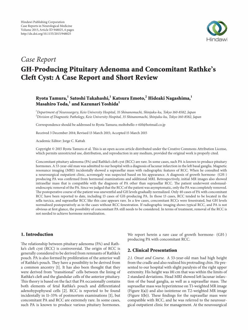
2.2. Operation. Removal of the PA using an endonasal-endoscopic approach was performed. Intraoperatively, themargin between the normal pituitary gland and adenomawasclear (Figure 3(a)). We identified the yellowish adenoma andcompleted gross total removal using suction (Figure 3(b)).We then retracted the pituitary gland and identified the wallof the RCC (Figure 3(c)). We did not aspirate or resect thecyst wall for fear of cerebrospinal fluid (CSF) leakage. Thefloor of the sella turcica was reconstructed using fat tissue(Figure 3(d)). Histologically, the tumor was composed ofmonotonous eosinophilic cells (Figure 4(a)). Positive stainingwas observed with GH immunohistochemistry (Figure 4(b));thus the tumor was diagnosed as GH-producing PA. Postop-erative computed tomography revealed gross total removal ofthe adenoma.
2.3. Postoperative Course. Postoperative course of the patientwas uneventful other than diabetes insipidus, which was

Case Reports in Neurological Medicine 3
Left
(a)
Right
(b)
Figure 2: No visual disturbance was apparent.
Pituitary adenoma
Pituitary gland
(a) (b)
(c) (d)
Figure 3: (a) Neuroendoscope shows the clear margin between normal gland and PA. PA looks soft and yellowish. (b) Neuroendoscopicview after removal of the PA, which was easy to remove. (c) Neuroendoscope shows the wall of the RCC. Resecting the suprasellar RCCwhileretracting normal gland was difficult. (d) The floor of the sella turcica was reconstructed using fat tissue. We did not aspirate or resect thecyst wall.
4 Case Reports in Neurological Medicine
(a) (b)
Figure 4: (a) Photomicrograph shows the tumor is composed of monotonous eosinophilic cells. (H&E stain, original magnification ×400.)(b) Positive staining is observed with GH immunohistochemistry. (GH, original magnification ×400.)
well controlled by medication. GH and somatomedin Clevels gradually improved until 7 days postoperatively. Wedecided to observe the RCC conservatively without addi-tional surgery. The patient was discharged from our hospitalon foot with no neurological sequelae.
Diabetes insipidus was treated by administered medica-tion for 6 months. Pituitary function and visual field werenot disturbed for 6 months. Hormonal laboratory testing12 weeks after the operation showed GH of 0.3 ng/mL andsomatomedin C levels of 131 ng/mL. At the 6-month follow-up time point, he has made satisfactory progress withoutrecurrence.
3. Discussion
A great variety of lesions such as sarcoidosis, intrasellarschwannoma, and gangliocytoma can coexist with pituitaryadenomas and are referred to as collision sellar lesions [4].However, concomitant PA and RCC are relatively rare. Ourreview identified only 40 cases reported to date. In the seriesof PA complicated with RCC, only a small number of caseshave been reported to secrete GH [1, 5–7]. To the best ofour knowledge, only 13 cases of patients with GH-producingPA and concomitant RCC have been reported (Table 1) [1,2, 5–10]. In the present case, PA had been overlooked untilnoted in the neurosurgical outpatient clinic. ComplicatedGH-producing PA was initially suspected from the facialappearance of the patient. The difficulty in diagnosing com-plicated PA was attributable to the location of the RCC. Asuprasellar location of the RCC, as in the present case, is rare,with only 3 of 12 previously reported cases of RCC foundin the suprasellar region [5, 6, 9]. If RCC is located in thesuprasellar lesion, identification of a GH-producing PA isnot easy, because an intrasellar lesion may pass unnoticed insuch cases. In addition, 3 of 4 cases, including the presentcase, involved no visual disturbance, and such patients maynot receive frequent follow-up of the RCC at the outpatientclinic. In cases where both the GH-producing PA and RCCare located in the sella turcica, it is quite possible that onlythe RCC will be noticed, since GH-producing PA tends to
be much smaller than RCC according to previous reports[4, 7, 8, 11]. At the time of diagnosis of RCC on MRI, it maybe useful to perform contrast-enhanced MRI in conjunctionwith hormonal examination to ensure the rare complicationof PA, as in the present case, is not overlooked.
RCC usually appears hyperintense on T1-weighted imag-ing. However, RCC existing concomitant with PA is known topresent with variable intensity on T1-weighted imaging, sinceit tends to be complicated by hemorrhage in the cyst [1, 13].In such cases, enhanced MRI is helpful for diagnosis.
When a nonenhanced cyst is demonstrated in cases ofPA detected by MRI, the possibility of accompanying RCCshould be considered. As for differential diagnosis, RCCusually demonstrates no contrast enhancement of the cystwall as referred to above and displayed no calcification unlikecraniopharyngioma. These radiographic characteristics arehelpful in the differential diagnosis of craniopharyngioma.
As for treatments, there is no need to fenestrate RCCsas far as they are asymptomatic. In reported cases of GH-producing PA and concomitant RCC, fenestration of the RCCand surgical biopsy of the cyst wall were performed for 61% [1,6, 7]. However, GH levels reportedly normalized even in caseswithout surgical fenestration of the RCC. In the present case,we did not perform surgical biopsy of the cyst wall for fear forCSF leakage, andGH levels decreased to within normal rangepostoperatively. It is pretty obvious, but it is not necessaryto remove cyst wall of RCC to normalize GH. However,if the removal of RCC is needed, endoscopic removal isrecommended. Jahangiri et al. said CSF leak did not occurin any of the suprasellar RCCs treated endoscopically, while14% treated microsurgically experienced a CSF leak. Inaddition, compared to microsurgery, endoscopy improvesrate of complete removal and visual outcomes [11].
In the present case, the appearance of the patient sug-gested acromegaly and guided us to a diagnosis of RCCwith concomitant PA. If the PA in this patient had beennonfunctional, correct diagnosis of the concomitant PAwould have been much more difficult. This case offers areminder that although rare, RCC can accompany PA. We

Case Reports in Neurological Medicine 5
Table1:Summaryof
Rathke’scle
ftcystcombinedwith
pituitary
adenom
aprodu
cing
grow
thho
rmon
e.
Authors
Age
(yrs)
Sex
PAsiz
e(m
m)
RCCsiz
e(m
m)
RCClocatio
nRC
CT1WI
RCCT2
WI
RCCGd
PAremoval
RCCremoval
Visualfield
Our
case
53M
912
Suprasellar
High
Iso.
No
Total
No
Normal
Miyagietal.[1]
44M
N/A
N/A
Intrasellar
Low
High
No
Subtotal
Total
Normal
Sumidae
tal.[7]
67F
1822
Intrasellar
Low
High
No
N/A
Removal
Normal
44F
288
Enclo
sed
Low
High
No
Removal
No
Normal
18M
128
Intrasellar
High
High
No
Removal
No
Normal
46M
147
Intrasellar
Low
High
No
Removal
No
Normal
56F
1413
Intrasellar
High
Low
No
Removal
Removal
Normal
48M
158
Intrasellar
High
Low
No
Removal
No
Normal
Nish
ioetal.[6]
44M
N/A
N/A
Suprasellar
Low
High
No
Subtotal
Removal
Normal
35F
N/A
N/A
Suprasellar
Low
High
No
Removal
Removal
Normal
62F
21N/A
Intrasellar
Low
High
No
Removal
Partially
N/A
Lucase
tal.[5]
47F
613
Suprasellar
Low
High
No
Removal
Removal
Binasalfi
elddefect
Ikedae
tal.[12]
50M
N/A
N/A
Enclo
sed
Low
High
No
N/A
N/A
Quadrantano
psia
Azarpira
etal.[9]
50F
N/A
N/A
Intrasellar
N/A
N/A
N/A
Removal
Removal
Bilateraltem
poral
hemiano
psia
F:female,M:m
ale,N/A
:not
available,T1WI:T1-w
eightedim
age,T2
WI:T2
-weightedim
age,Gd:
gado
linium,PA:pitu
itary
adenom
a,RC
C:Ra
thke’scleft
cyst,
TR:totalremoval,u
nc:u
nclassified,w
k:week,and
yr:year.

6 Case Reports in Neurological Medicine
recommend performing contrast-enhanced MRI routinelyon suspicion of RCC.
Conflict of Interests
The authors have no personal financial or institutional inter-est in any of the drugs, materials, or devices described in thispaper.
References
[1] A. Miyagi, M. Iwasaki, T. Shibuya et al., “Pituitary adenomacombined with Rathke’s cleft cyst—case report,” NeurologiaMedico-Chirurgica, vol. 33, no. 9, pp. 643–650, 1993.
[2] B. Ranjith, G. Adam, M. Jordan, R. Timothy, J. Thomas, andW. Gavin, “Symptomatic Rathke’s cleft cyst with a co-existingpituitary tumor; brief review of the literature,” Asian Journal ofNeurosurgery, vol. 8, pp. 183–187, 2013.
[3] R. Trifanescu, V. Stavrinides, P. Plaha et al., “Outcome insurgically treated Rathke’s cleft cysts: Long-term monitoringneeded,” European Journal of Endocrinology, vol. 165, no. 1, pp.33–37, 2011.
[4] K. Maria, K. George, W. Pieter, J. Andre, and S. Andreas,“Collision sellar lesions: experience with eight cases and reviewof the literature,” Pituitary, vol. 13, no. 1, pp. 8–17, 2010.
[5] J. Lucas, D. Kawanaa, E. Richard, G. William, A. Jason, and C.Joseph, “Simultaneous symptomatic Rathke’s cleft cyst and GHsecreting pituitary adenoma: a case report,” Pituitary, vol. 7, no.1, pp. 39–44, 2004.
[6] S. Nishio, J. Mizuno, D. L. Barrow, Y. Takei, and G. T. Tindall,“Pituitary tumors composed of adenohypophysial adenomaand Rathke’s cleft cyst elements: a clinicopathological study,”Neurosurgery, vol. 21, no. 3, pp. 371–377, 1987.
[7] M. Sumida, K. Arita, K. Migita, A. Tominaga, K. Iida, andK. Kurisu, “Concomitant pituitary adenoma and Rathke’s cleftcyst,” Neuroradiology, vol. 43, no. 9, pp. 755–759, 2001.
[8] F. Gessler, V. C. Coon, S. S. Chin, and W. T. Couldwell,“Coexisting rathke cleft cyst and pituitary adenoma presentingwith pituitary apoplexy: report of two cases,” Skull Base Reports,vol. 1, no. 2, pp. 099–104, 2011.
[9] N. Azarpira, S. Pakbaz, S. Torabineghad, J. Musavi, and M.Rakei, “Acromegaly associated withmixed pituitary adenoma—gangliocytoma and rathke’s cleft cyst,” Turkish Neurosurgery,vol. 23, no. 4, pp. 527–530, 2013.
[10] S. J. Noh, J. Y. Ahn, K. S. Lee, and S. H. Kim, “Pituitary adenomaand concomitant Rathke’s cleft cyst,” Acta Neurochirurgica, vol.149, no. 12, pp. 1223–1228, 2007.
[11] A. Jahangiri, M. Potts, S. Kunwar, L. Blevins, I. H. El-Sayed,and M. K. Aghi, “Extended endoscopic endonasal approach forsuprasellar Rathke’s cleft cysts,” Journal of Clinical Neuroscience,vol. 21, no. 5, pp. 779–785, 2014.
[12] H. Ikeda, T. Yoshimoto, andR.Katakura, “A case of Rathke’s cleftcyst within a pituitary adenoma presenting with acromegaly—do ‘transitional cell tumors of the pituitary gland’ really exist?”Acta Neuropathologica, vol. 83, no. 2, pp. 211–215, 1992.
[13] R. W. Vancura, K. M. Jacob, and I. Damjanov, “A 70-year-oldmanwith diplopia, nausea, and vomiting,”Archives of Pathologyand Laboratory Medicine, vol. 130, no. 3, pp. 403–404, 2006.

Submit your manuscripts athttp://www.hindawi.com
Stem CellsInternational
Hindawi Publishing Corporationhttp://www.hindawi.com Volume 2014
Hindawi Publishing Corporationhttp://www.hindawi.com Volume 2014
MEDIATORSINFLAMMATION
of
Hindawi Publishing Corporationhttp://www.hindawi.com Volume 2014
Behavioural Neurology
EndocrinologyInternational Journal of
Hindawi Publishing Corporationhttp://www.hindawi.com Volume 2014
Hindawi Publishing Corporationhttp://www.hindawi.com Volume 2014
Disease Markers
Hindawi Publishing Corporationhttp://www.hindawi.com Volume 2014
BioMed Research International
OncologyJournal of
Hindawi Publishing Corporationhttp://www.hindawi.com Volume 2014
Hindawi Publishing Corporationhttp://www.hindawi.com Volume 2014
Oxidative Medicine and Cellular Longevity
Hindawi Publishing Corporationhttp://www.hindawi.com Volume 2014
PPAR Research
The Scientific World JournalHindawi Publishing Corporation http://www.hindawi.com Volume 2014
Immunology ResearchHindawi Publishing Corporationhttp://www.hindawi.com Volume 2014
Journal of
ObesityJournal of
Hindawi Publishing Corporationhttp://www.hindawi.com Volume 2014
Hindawi Publishing Corporationhttp://www.hindawi.com Volume 2014
Computational and Mathematical Methods in Medicine
OphthalmologyJournal of
Hindawi Publishing Corporationhttp://www.hindawi.com Volume 2014
Diabetes ResearchJournal of
Hindawi Publishing Corporationhttp://www.hindawi.com Volume 2014
Hindawi Publishing Corporationhttp://www.hindawi.com Volume 2014
Research and TreatmentAIDS
Hindawi Publishing Corporationhttp://www.hindawi.com Volume 2014
Gastroenterology Research and Practice
Hindawi Publishing Corporationhttp://www.hindawi.com Volume 2014
Parkinson’s Disease
Evidence-Based Complementary and Alternative Medicine
Volume 2014Hindawi Publishing Corporationhttp://www.hindawi.com


![MR Imaging of Cavernous Sinus Involvement by Pituitary ...€¦ · abnormal pituitary gland and sellar region [13-16], only a few anecdotal cases of CSI by a pituitary adenoma have](https://static.fdocuments.net/doc/165x107/5ed565c51d25941f923a987c/mr-imaging-of-cavernous-sinus-involvement-by-pituitary-abnormal-pituitary-gland.jpg)