Case Report Combined Scleral Flap with Donor Scleral Patch...
5
Case Report Combined Scleral Flap with Donor Scleral Patch Graft for Anterior Tube Placement in Glaucoma Drainage Device Surgery Jea H. Yu, 1,2 Chuck Nguyen, 1,2 Esmeralda Gallemore, 1 and Ron P. Gallemore 1,2 1 Clinical Research, Retina Macula Institute, Torrance, CA, USA 2 University of California, Los Angeles, Los Angeles, CA, USA Correspondence should be addressed to Ron P. Gallemore; [email protected] Received 24 December 2015; Revised 30 June 2016; Accepted 18 July 2016 Academic Editor: Alexander A. Bialasiewicz Copyright © 2016 Jea H. Yu et al. is is an open access article distributed under the Creative Commons Attribution License, which permits unrestricted use, distribution, and reproduction in any medium, provided the original work is properly cited. Purpose. To report a new technique for anterior placement of tubes for glaucoma drainage devices to reduce the risk of tube erosions. Methods. Retrospective review of select cases of Ahmed Valve surgery combined with the novel method of a limbal-based scleral flap covered by a scleral patch graſt to cover the tube at the entrance through the limbus. Intraoperative and postoperative illustrations are shown to highlight the method of tube placement. Results. In this retrospective case series, 3 patients are presented illustrating the technique. Two had neovascular glaucoma and one had primary open-angle glaucoma (POAG). On average, intraocular pressure was reduced from 39 ± 14 mmHg to 15 ± 2 mmHg and the number of glaucoma medications was reduced from 4±1 to 0. Preop- erative and most recent visual acuities were hand-motion (HM) and HM, 20/60 and 20/50, and 20/70 and 20/30, respectively. Conclusion. e combination of a limbal-based scleral flap with scleral patch graſt to cover the tube with glaucoma drainage devices may be an effective means to reduce erosion and protect against endophthalmitis. 1. Introduction Glaucoma Drainage Devices (GDD) (e.g., Ahmed, Baerveldt) are utilized in the management of complex cases of glaucoma, particularly in neovascular and uveitic and other intractable forms [1]. ey provide an attractive alternative to trabeculec- tomy surgery, particularly in younger patients, with a lower risk of endophthalmitis [2, 3]. e TVT study demonstrated higher long-term success for tubes over trabeculectomy, although comparable rates of IOP control were achieved [4]. One disadvantage of GDD is erosion of the tube, particularly when placed through the limbus and into the anterior chamber (AC). Even with the placement of a patch graſt, erosion rates of approximately 8% are reported [5] with an associated incidence of endophthalmitis as high as 2% of patients undergoing GDD surgery [6]. One alternative to this approach is posterior placement of the tube—into the pars plana [7]. is may reduce rates of erosion but not all patients are good candidates for posterior placement and proper posterior placement may require assistance of a retinal surgeon to clear the vitreous and prevent clogging of the tube. Here we report a novel technique for anterior placement of the Seton valve tube utilizing a scleral tunnel combined with a scleral patch graſt. No cases of erosion or dellen formation were reported in our series. 2. Methods We report a retrospective case series of patients undergoing Ahmed Valve placement into the AC combined with vitre- oretinal surgery, when indicated, utilizing a scleral tunnel technique. Figure 1 illustrates an example case. All patients underwent retrobulbar anesthesia and aſter prompt prepa- ration and draping, a 23-gauge infusion cannula was placed in the inferotemporal quadrant 3-4 mm posterior to the limbus. e infusion was attached to the cannula and directly visualized before turning on. Intraocular pressure (IOP) was set at 10–20 mmHg to protect the optic nerve from fluctuations of IOP during the procedure. A conjunctival peritomy was created in the superotemporal quadrant. e limbus was marked at 9:00 and 12:00 for the right eye and 12:00 and 3:00 for the Hindawi Publishing Corporation Case Reports in Ophthalmological Medicine Volume 2016, Article ID 2124581, 4 pages http://dx.doi.org/10.1155/2016/2124581
Transcript of Case Report Combined Scleral Flap with Donor Scleral Patch...
Case ReportCombined Scleral Flap with Donor Scleral Patch Graft forAnterior Tube Placement in Glaucoma Drainage Device Surgery
Jea H Yu12 Chuck Nguyen12 Esmeralda Gallemore1 and Ron P Gallemore12
1Clinical Research Retina Macula Institute Torrance CA USA2University of California Los Angeles Los Angeles CA USA
Correspondence should be addressed to Ron P Gallemore rongallemoremdgmailcom
Received 24 December 2015 Revised 30 June 2016 Accepted 18 July 2016
Academic Editor Alexander A Bialasiewicz
Copyright copy 2016 Jea H Yu et alThis is an open access article distributed under the Creative CommonsAttribution License whichpermits unrestricted use distribution and reproduction in any medium provided the original work is properly cited
Purpose To report a new technique for anterior placement of tubes for glaucoma drainage devices to reduce the risk of tube erosionsMethods Retrospective review of select cases of AhmedValve surgery combinedwith the novelmethod of a limbal-based scleral flapcovered by a scleral patch graft to cover the tube at the entrance through the limbus Intraoperative and postoperative illustrationsare shown to highlight themethodof tube placementResults In this retrospective case series 3 patients are presented illustrating thetechnique Two had neovascular glaucoma and one had primary open-angle glaucoma (POAG) On average intraocular pressurewas reduced from 39 plusmn 14mmHg to 15 plusmn 2mmHg and the number of glaucoma medications was reduced from 4 plusmn 1 to 0 Preop-erative and most recent visual acuities were hand-motion (HM) and HM 2060 and 2050 and 2070 and 2030 respectivelyConclusionThe combination of a limbal-based scleral flap with scleral patch graft to cover the tube with glaucoma drainage devicesmay be an effective means to reduce erosion and protect against endophthalmitis
1 Introduction
GlaucomaDrainageDevices (GDD) (eg Ahmed Baerveldt)are utilized in themanagement of complex cases of glaucomaparticularly in neovascular and uveitic and other intractableforms [1]They provide an attractive alternative to trabeculec-tomy surgery particularly in younger patients with a lowerrisk of endophthalmitis [2 3] The TVT study demonstratedhigher long-term success for tubes over trabeculectomyalthough comparable rates of IOP control were achieved [4]One disadvantage of GDD is erosion of the tube particularlywhen placed through the limbus and into the anteriorchamber (AC) Even with the placement of a patch grafterosion rates of approximately 8 are reported [5] with anassociated incidence of endophthalmitis as high as 2 ofpatients undergoing GDD surgery [6] One alternative tothis approach is posterior placement of the tubemdashinto thepars plana [7] This may reduce rates of erosion but not allpatients are good candidates for posterior placement andproper posterior placementmay require assistance of a retinalsurgeon to clear the vitreous and prevent clogging of the tube
Here we report a novel technique for anterior placement ofthe Seton valve tube utilizing a scleral tunnel combined witha scleral patch graft No cases of erosion or dellen formationwere reported in our series
2 Methods
We report a retrospective case series of patients undergoingAhmed Valve placement into the AC combined with vitre-oretinal surgery when indicated utilizing a scleral tunneltechnique Figure 1 illustrates an example case All patientsunderwent retrobulbar anesthesia and after prompt prepa-ration and draping a 23-gauge infusion cannula was placedin the inferotemporal quadrant 3-4mm posterior to thelimbusThe infusion was attached to the cannula and directlyvisualized before turning on
Intraocular pressure (IOP) was set at 10ndash20mmHg toprotect the optic nerve from fluctuations of IOP duringthe procedure A conjunctival peritomy was created in thesuperotemporal quadrant The limbus was marked at 900and 1200 for the right eye and 1200 and 300 for the
Hindawi Publishing CorporationCase Reports in Ophthalmological MedicineVolume 2016 Article ID 2124581 4 pageshttpdxdoiorg10115520162124581
2 Case Reports in Ophthalmological Medicine
lowast
lowast
lowast lowast
(a)
lowast
(b) (c)
(d) (e)
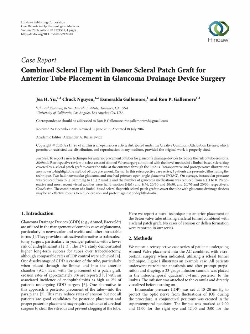
Figure 1 (a) Outline of scleral flap made with crescent blade Note 7-0 vicryl limbal retraction suture and marking of 9001015840 10301015840 and12001015840 positions (lowast) (b) Ahmed Valve sutures in place (lowast) and AC entered and formed with viscoelastic on a 23-gauge needle (c) Tube cutand inserted into AC beneath the flap (d) Human scleral patch graft fixed in place 1-2mm behind limbus 7-0 vicryl suture (e) Conjunctivaclosed with running locking 7-0 vicryl suture
left eye and at the midpoint between these two markingsA 7-0 vicryl stay suture was placed through the limbusat approximately 50 corneal depth and the eye rotatedinferonasally Westcottrsquos scissors were used to bluntly dissectbeneath the subtenons posteriorly and then the lateral andsuperior rectus muscles were localized with muscle hookswhereafter two 7-0 vicryl sutures were preplaced for theAhmedValve between the twomuscles 9mmposterior to thelimbus and 5mm apart The limbal marking points were alsoused to help align the valve which was then positioned andsutured in place We believe precise placement of the valveis critical to minimize strabismus related complications A 5times 5mm limbal-based scleral flap was then created using abent crescent blade (Figure 1(a)) The retraction suture wasreleased and any vitrectomy procedures were then performedat this point When placing the tube in the anterior chambercare is taken to prevent both iris andor corneal touch byplacing a bent 23-gauge needle attached to a viscoelastic-likeProVisc through the surgical limbus parallel to the iris plane(Figure 1(b)) and maintaining the AC with a small amount ofviscoelasticThe goal is to have the AC volume normalizedmdashcountering any aqueous loss that resulted from entering thechamber The tube was cut at a 30-degree angle for ease ofplacement through the ostomy created by the 23-gauge needle(Figure 1(c)) A scleral patch or a piece of tutoplast was thensutured in place over the scleral flap (Figure 1(d)) and theconjunctiva was closed with a running locking 7-0 vicrylsuture (Figure 1(e)) Also we have not found it necessary
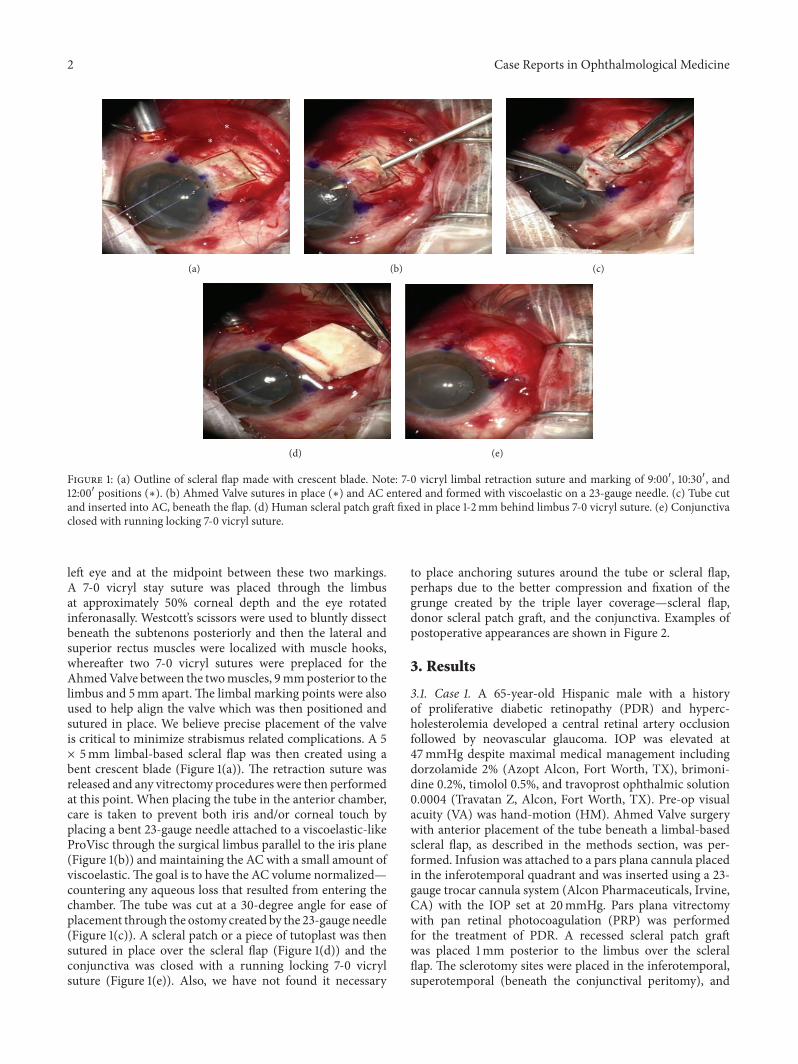
to place anchoring sutures around the tube or scleral flapperhaps due to the better compression and fixation of thegrunge created by the triple layer coveragemdashscleral flapdonor scleral patch graft and the conjunctiva Examples ofpostoperative appearances are shown in Figure 2
3 Results
31 Case 1 A 65-year-old Hispanic male with a historyof proliferative diabetic retinopathy (PDR) and hyperc-holesterolemia developed a central retinal artery occlusionfollowed by neovascular glaucoma IOP was elevated at47mmHg despite maximal medical management includingdorzolamide 2 (Azopt Alcon Fort Worth TX) brimoni-dine 02 timolol 05 and travoprost ophthalmic solution00004 (Travatan Z Alcon Fort Worth TX) Pre-op visualacuity (VA) was hand-motion (HM) Ahmed Valve surgerywith anterior placement of the tube beneath a limbal-basedscleral flap as described in the methods section was per-formed Infusion was attached to a pars plana cannula placedin the inferotemporal quadrant and was inserted using a 23-gauge trocar cannula system (Alcon Pharmaceuticals IrvineCA) with the IOP set at 20mmHg Pars plana vitrectomywith pan retinal photocoagulation (PRP) was performedfor the treatment of PDR A recessed scleral patch graftwas placed 1mm posterior to the limbus over the scleralflap The sclerotomy sites were placed in the inferotemporalsuperotemporal (beneath the conjunctival peritomy) and
Case Reports in Ophthalmological Medicine 3
(a) (b) (c)
Figure 2 (a) 1 day sp surgery (b) 22 days sp surgery (c) 90 days sp surgery
2060
Pre-op
(a)
20200
POD 1
(b)
20200
POD 15 + Bev
(c)
2060
POD 36
(d)
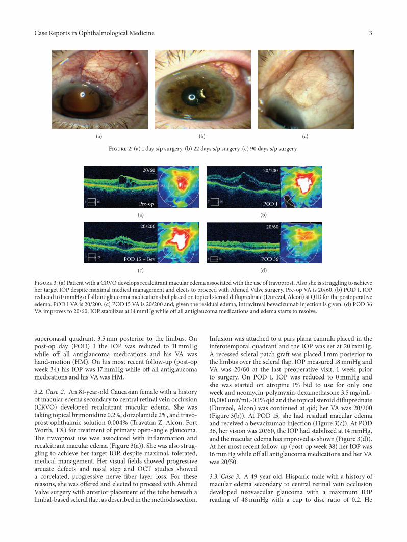
Figure 3 (a) Patient with a CRVOdevelops recalcitrant macular edema associated with the use of travoprost Also she is struggling to achieveher target IOP despite maximal medical management and elects to proceed with Ahmed Valve surgery Pre-op VA is 2060 (b) POD 1 IOPreduced to 0mmHgoff all antiglaucomamedications but placed on topical steroid difluprednate (Durezol Alcon) atQID for the postoperativeedema POD 1 VA is 20200 (c) POD 15 VA is 20200 and given the residual edema intravitreal bevacizumab injection is given (d) POD 36VA improves to 2060 IOP stabilizes at 14mmHg while off all antiglaucoma medications and edema starts to resolve
superonasal quadrant 35mm posterior to the limbus Onpost-op day (POD) 1 the IOP was reduced to 11mmHgwhile off all antiglaucoma medications and his VA washand-motion (HM) On his most recent follow-up (post-opweek 34) his IOP was 17mmHg while off all antiglaucomamedications and his VA was HM
32 Case 2 An 81-year-old Caucasian female with a historyof macular edema secondary to central retinal vein occlusion(CRVO) developed recalcitrant macular edema She wastaking topical brimonidine 02 dorzolamide 2 and travo-prost ophthalmic solution 0004 (Travatan Z Alcon FortWorth TX) for treatment of primary open-angle glaucomaThe travoprost use was associated with inflammation andrecalcitrant macular edema (Figure 3(a)) She was also strug-gling to achieve her target IOP despite maximal toleratedmedical management Her visual fields showed progressivearcuate defects and nasal step and OCT studies showeda correlated progressive nerve fiber layer loss For thesereasons she was offered and elected to proceed with AhmedValve surgery with anterior placement of the tube beneath alimbal-based scleral flap as described in themethods section
Infusion was attached to a pars plana cannula placed in theinferotemporal quadrant and the IOP was set at 20mmHgA recessed scleral patch graft was placed 1mm posterior tothe limbus over the scleral flap IOP measured 18mmHg andVA was 2060 at the last preoperative visit 1 week priorto surgery On POD 1 IOP was reduced to 0mmHg andshe was started on atropine 1 bid to use for only oneweek and neomycin-polymyxin-dexamethasone 35mgmL-10000 unitmL-01 qid and the topical steroid difluprednate(Durezol Alcon) was continued at qid her VA was 20200(Figure 3(b)) At POD 15 she had residual macular edemaand received a bevacizumab injection (Figure 3(c)) At POD36 her vision was 2060 the IOP had stabilized at 14mmHgand themacular edema has improved as shown (Figure 3(d))At her most recent follow-up (post-op week 38) her IOP was16mmHg while off all antiglaucoma medications and her VAwas 2050
33 Case 3 A 49-year-old Hispanic male with a history ofmacular edema secondary to central retinal vein occlusiondeveloped neovascular glaucoma with a maximum IOPreading of 48mmHg with a cup to disc ratio of 02 He
4 Case Reports in Ophthalmological Medicine
was treated with a bevacizumab injection and was startedon maximal medical glaucoma management brimonidine02 dorzolamide 2 timolol 05 bimatoprost ophthalmicsolution 003 (Lumigan Allergan Irvine CA) and Aceta-zolamide 250mg tablets reducing his IOP to 15mmHgAfter two more bevacizumab injections his IOP increasedto 25mmHg while on the same regimen He elected toundergo Ahmed Valve placement with limbal-based scleralflap for tube placement in the AC with a scleral patch graftA pars plana maintainer was utilized during the procedureto keep the IOP at 20mmHg Pars plana vitrectomy withPRP was also performed for treatment of ischemic peripheralretina On POD 1 the patientrsquos IOP was well-controlled at6mmHg and his VA was count fingers at 4 feet while offall antiglaucoma medications At his most recent follow-up (post-op week 28) his IOP was 22mmHg while off allantiglaucoma medications and his VA was 2030
4 Discussion
Glaucoma drainage devices (GDD) are effective for themanagement of complex cases of glaucoma refractory tomedical management Traditionally the tube of the drainagedevice is placed into the AC through the limbus The tube isthen covered with a scleral or other patch graft overlappingthe limbus corneal junction Alternatively a scleral flap maybe used By combining the two techniques complicationsmay be reduced The scleral flap allows more posterior anddeeper entry into the AC reducing the risk of erosion at thelimbus as well as the risk of dellen formation at the limbusThe overlying patch provides further protection from erosionof the entire tube and may be recessed a millimeter fromthe limbus when combined with the scleral flap reducingcosmetic issues of the patch at the limbus as well as the risk ofdellen formation One of the most dreaded complications ofGDD surgery is erosion of the tube through the conjunctivaand rates of 25ndash71 have been reported [8] Subsequentendophthalmitis can occur with loss of vision and even loss ofthe eye [9] To reduce the risk of erosion grafts are placed overthe tube at the corneal limbal junction with a wide range ofmaterial utilized including donor human sclera pericardiumcorneal buttons and fascia lata Erosion still occurs despitethe placement of these graftsThe cause of erosion appears tobe focal compression of the conjunctiva by the elevated tubecombinedwith drying of the tissues caused by the irregularityof the surface at the junction of the cornea and limbus Inorder to create a smoother entry of the tube into the ACandminimize compression of the conjunctiva by the tube wehave utilized a scleral tunnel technique for placement of thetube into the AC This is a simple technique requiring onlyminutes to perform and when combined with the overlyingscleral patch graft has eliminated all cases of tube erosionwithanterior placement of the tube in our practice Scleral flapsalone have been found to have a higher rate or tube erosionthan a scleral patch graft alone [10] and the rates of erosionwith the patch graft are as noted aboveWhilewe have utilizedthis technique only with the placement of an Ahmed Valveit could be used for any drainage device We suggest the useof a scleral tunnel with scleral flap for the placement of all
GDD that are inserted into the AC to reduce the chance ofconjunctival erosion dellen formation and endophthalmitisand to enhance the cosmetic results
Competing Interests
The authors declare that they have no competing interests
Acknowledgments
The authors thank Dr Anne Coleman for her encourage-ment
References
[1] A D Mata S E Burk P A Netland S Baltatzis W ChristenandC S Foster ldquoManagement of uveitic glaucomawithAhmedglaucoma valve implantationrdquo Ophthalmology vol 106 no 11pp 2168ndash2172 1999
[2] G S Ang Z Varga andT Shaarawy ldquoPostoperative infection inpenetrating versus non-penetrating glaucoma surgeryrdquo BritishJournal of Ophthalmology vol 94 no 12 pp 1571ndash1576 2010
[3] J D Stein D Ruiz Jr D Belsky P P Lee and F A SloanldquoLongitudinal rates of postoperative adverse outcomes afterglaucoma surgery among medicare beneficiaries 1994 to 2005rdquoOphthalmology vol 115 no 7 pp 1109e7ndash1116e7 2008
[4] S J Gedde J C Schiffman W J Feuer L W Herndon J DBrandt and D L Budenz ldquoTreatment outcomes in the tubeversus trabeculectomy (TVT) study after five years of follow-uprdquoAmerican Journal of Ophthalmology vol 153 no 5 pp 789ndash803e2 2012
[5] P K Wishart A Choudhary and D Wong ldquoAhmed glaucomavalves in refractory glaucoma a 7-year auditrdquo British Journal ofOphthalmology vol 94 no 9 pp 1174ndash1179 2010
[6] A A Al-Torbak S Al-Shahwan I Al-Jadaan A Al-HommadiandD P Edward ldquoEndophthalmitis associated with the Ahmedglaucoma valve implantrdquo British Journal of Ophthalmology vol89 no 4 pp 454ndash458 2005
[7] J O Wallsh R P Gallemore M Taban C Hu and B ShararehldquoPars plana ahmed valve and vitrectomy in patients withglaucoma associated with posterior segment diseaserdquo Retinavol 33 no 10 pp 2059ndash2068 2013
[8] S A W Low D B Rootman D S Rootman and G ETrope ldquoRepair of eroded glaucoma drainage devices mid-termoutcomesrdquo Journal of Glaucoma vol 21 no 9 pp 619ndash622 2012
[9] J R Safneck ldquoEndophthalmitis a review of recent trendsrdquoSaudi Journal of Ophthalmology vol 26 no 2 pp 181ndash189 2012
[10] J C Das Z Chaudhuri P Sharma and S Bhomaj ldquoThe ahmedglaucoma valve in refractory glaucoma experiences in Indianeyesrdquo Eye vol 19 no 2 pp 183ndash190 2005
Submit your manuscripts athttpwwwhindawicom
Stem CellsInternational
Hindawi Publishing Corporationhttpwwwhindawicom Volume 2014
Hindawi Publishing Corporationhttpwwwhindawicom Volume 2014
MEDIATORSINFLAMMATION
of
Hindawi Publishing Corporationhttpwwwhindawicom Volume 2014
Behavioural Neurology
EndocrinologyInternational Journal of
Hindawi Publishing Corporationhttpwwwhindawicom Volume 2014
Hindawi Publishing Corporationhttpwwwhindawicom Volume 2014
Disease Markers
Hindawi Publishing Corporationhttpwwwhindawicom Volume 2014
BioMed Research International
OncologyJournal of
Hindawi Publishing Corporationhttpwwwhindawicom Volume 2014
Hindawi Publishing Corporationhttpwwwhindawicom Volume 2014
Oxidative Medicine and Cellular Longevity
Hindawi Publishing Corporationhttpwwwhindawicom Volume 2014
PPAR Research
The Scientific World JournalHindawi Publishing Corporation httpwwwhindawicom Volume 2014
Immunology ResearchHindawi Publishing Corporationhttpwwwhindawicom Volume 2014
Journal of
ObesityJournal of
Hindawi Publishing Corporationhttpwwwhindawicom Volume 2014
Hindawi Publishing Corporationhttpwwwhindawicom Volume 2014
Computational and Mathematical Methods in Medicine
OphthalmologyJournal of
Hindawi Publishing Corporationhttpwwwhindawicom Volume 2014
Diabetes ResearchJournal of
Hindawi Publishing Corporationhttpwwwhindawicom Volume 2014
Hindawi Publishing Corporationhttpwwwhindawicom Volume 2014
Research and TreatmentAIDS
Hindawi Publishing Corporationhttpwwwhindawicom Volume 2014
Gastroenterology Research and Practice
Hindawi Publishing Corporationhttpwwwhindawicom Volume 2014
Parkinsonrsquos Disease
Evidence-Based Complementary and Alternative Medicine
Volume 2014Hindawi Publishing Corporationhttpwwwhindawicom
2 Case Reports in Ophthalmological Medicine
lowast
lowast
lowast lowast
(a)
lowast
(b) (c)
(d) (e)
Figure 1 (a) Outline of scleral flap made with crescent blade Note 7-0 vicryl limbal retraction suture and marking of 9001015840 10301015840 and12001015840 positions (lowast) (b) Ahmed Valve sutures in place (lowast) and AC entered and formed with viscoelastic on a 23-gauge needle (c) Tube cutand inserted into AC beneath the flap (d) Human scleral patch graft fixed in place 1-2mm behind limbus 7-0 vicryl suture (e) Conjunctivaclosed with running locking 7-0 vicryl suture
left eye and at the midpoint between these two markingsA 7-0 vicryl stay suture was placed through the limbusat approximately 50 corneal depth and the eye rotatedinferonasally Westcottrsquos scissors were used to bluntly dissectbeneath the subtenons posteriorly and then the lateral andsuperior rectus muscles were localized with muscle hookswhereafter two 7-0 vicryl sutures were preplaced for theAhmedValve between the twomuscles 9mmposterior to thelimbus and 5mm apart The limbal marking points were alsoused to help align the valve which was then positioned andsutured in place We believe precise placement of the valveis critical to minimize strabismus related complications A 5times 5mm limbal-based scleral flap was then created using abent crescent blade (Figure 1(a)) The retraction suture wasreleased and any vitrectomy procedures were then performedat this point When placing the tube in the anterior chambercare is taken to prevent both iris andor corneal touch byplacing a bent 23-gauge needle attached to a viscoelastic-likeProVisc through the surgical limbus parallel to the iris plane(Figure 1(b)) and maintaining the AC with a small amount ofviscoelasticThe goal is to have the AC volume normalizedmdashcountering any aqueous loss that resulted from entering thechamber The tube was cut at a 30-degree angle for ease ofplacement through the ostomy created by the 23-gauge needle(Figure 1(c)) A scleral patch or a piece of tutoplast was thensutured in place over the scleral flap (Figure 1(d)) and theconjunctiva was closed with a running locking 7-0 vicrylsuture (Figure 1(e)) Also we have not found it necessary
to place anchoring sutures around the tube or scleral flapperhaps due to the better compression and fixation of thegrunge created by the triple layer coveragemdashscleral flapdonor scleral patch graft and the conjunctiva Examples ofpostoperative appearances are shown in Figure 2
3 Results
31 Case 1 A 65-year-old Hispanic male with a historyof proliferative diabetic retinopathy (PDR) and hyperc-holesterolemia developed a central retinal artery occlusionfollowed by neovascular glaucoma IOP was elevated at47mmHg despite maximal medical management includingdorzolamide 2 (Azopt Alcon Fort Worth TX) brimoni-dine 02 timolol 05 and travoprost ophthalmic solution00004 (Travatan Z Alcon Fort Worth TX) Pre-op visualacuity (VA) was hand-motion (HM) Ahmed Valve surgerywith anterior placement of the tube beneath a limbal-basedscleral flap as described in the methods section was per-formed Infusion was attached to a pars plana cannula placedin the inferotemporal quadrant and was inserted using a 23-gauge trocar cannula system (Alcon Pharmaceuticals IrvineCA) with the IOP set at 20mmHg Pars plana vitrectomywith pan retinal photocoagulation (PRP) was performedfor the treatment of PDR A recessed scleral patch graftwas placed 1mm posterior to the limbus over the scleralflap The sclerotomy sites were placed in the inferotemporalsuperotemporal (beneath the conjunctival peritomy) and
Case Reports in Ophthalmological Medicine 3
(a) (b) (c)
Figure 2 (a) 1 day sp surgery (b) 22 days sp surgery (c) 90 days sp surgery
2060
Pre-op
(a)
20200
POD 1
(b)
20200
POD 15 + Bev
(c)
2060
POD 36
(d)
Figure 3 (a) Patient with a CRVOdevelops recalcitrant macular edema associated with the use of travoprost Also she is struggling to achieveher target IOP despite maximal medical management and elects to proceed with Ahmed Valve surgery Pre-op VA is 2060 (b) POD 1 IOPreduced to 0mmHgoff all antiglaucomamedications but placed on topical steroid difluprednate (Durezol Alcon) atQID for the postoperativeedema POD 1 VA is 20200 (c) POD 15 VA is 20200 and given the residual edema intravitreal bevacizumab injection is given (d) POD 36VA improves to 2060 IOP stabilizes at 14mmHg while off all antiglaucoma medications and edema starts to resolve
superonasal quadrant 35mm posterior to the limbus Onpost-op day (POD) 1 the IOP was reduced to 11mmHgwhile off all antiglaucoma medications and his VA washand-motion (HM) On his most recent follow-up (post-opweek 34) his IOP was 17mmHg while off all antiglaucomamedications and his VA was HM
32 Case 2 An 81-year-old Caucasian female with a historyof macular edema secondary to central retinal vein occlusion(CRVO) developed recalcitrant macular edema She wastaking topical brimonidine 02 dorzolamide 2 and travo-prost ophthalmic solution 0004 (Travatan Z Alcon FortWorth TX) for treatment of primary open-angle glaucomaThe travoprost use was associated with inflammation andrecalcitrant macular edema (Figure 3(a)) She was also strug-gling to achieve her target IOP despite maximal toleratedmedical management Her visual fields showed progressivearcuate defects and nasal step and OCT studies showeda correlated progressive nerve fiber layer loss For thesereasons she was offered and elected to proceed with AhmedValve surgery with anterior placement of the tube beneath alimbal-based scleral flap as described in themethods section
Infusion was attached to a pars plana cannula placed in theinferotemporal quadrant and the IOP was set at 20mmHgA recessed scleral patch graft was placed 1mm posterior tothe limbus over the scleral flap IOP measured 18mmHg andVA was 2060 at the last preoperative visit 1 week priorto surgery On POD 1 IOP was reduced to 0mmHg andshe was started on atropine 1 bid to use for only oneweek and neomycin-polymyxin-dexamethasone 35mgmL-10000 unitmL-01 qid and the topical steroid difluprednate(Durezol Alcon) was continued at qid her VA was 20200(Figure 3(b)) At POD 15 she had residual macular edemaand received a bevacizumab injection (Figure 3(c)) At POD36 her vision was 2060 the IOP had stabilized at 14mmHgand themacular edema has improved as shown (Figure 3(d))At her most recent follow-up (post-op week 38) her IOP was16mmHg while off all antiglaucoma medications and her VAwas 2050
33 Case 3 A 49-year-old Hispanic male with a history ofmacular edema secondary to central retinal vein occlusiondeveloped neovascular glaucoma with a maximum IOPreading of 48mmHg with a cup to disc ratio of 02 He
4 Case Reports in Ophthalmological Medicine
was treated with a bevacizumab injection and was startedon maximal medical glaucoma management brimonidine02 dorzolamide 2 timolol 05 bimatoprost ophthalmicsolution 003 (Lumigan Allergan Irvine CA) and Aceta-zolamide 250mg tablets reducing his IOP to 15mmHgAfter two more bevacizumab injections his IOP increasedto 25mmHg while on the same regimen He elected toundergo Ahmed Valve placement with limbal-based scleralflap for tube placement in the AC with a scleral patch graftA pars plana maintainer was utilized during the procedureto keep the IOP at 20mmHg Pars plana vitrectomy withPRP was also performed for treatment of ischemic peripheralretina On POD 1 the patientrsquos IOP was well-controlled at6mmHg and his VA was count fingers at 4 feet while offall antiglaucoma medications At his most recent follow-up (post-op week 28) his IOP was 22mmHg while off allantiglaucoma medications and his VA was 2030
4 Discussion
Glaucoma drainage devices (GDD) are effective for themanagement of complex cases of glaucoma refractory tomedical management Traditionally the tube of the drainagedevice is placed into the AC through the limbus The tube isthen covered with a scleral or other patch graft overlappingthe limbus corneal junction Alternatively a scleral flap maybe used By combining the two techniques complicationsmay be reduced The scleral flap allows more posterior anddeeper entry into the AC reducing the risk of erosion at thelimbus as well as the risk of dellen formation at the limbusThe overlying patch provides further protection from erosionof the entire tube and may be recessed a millimeter fromthe limbus when combined with the scleral flap reducingcosmetic issues of the patch at the limbus as well as the risk ofdellen formation One of the most dreaded complications ofGDD surgery is erosion of the tube through the conjunctivaand rates of 25ndash71 have been reported [8] Subsequentendophthalmitis can occur with loss of vision and even loss ofthe eye [9] To reduce the risk of erosion grafts are placed overthe tube at the corneal limbal junction with a wide range ofmaterial utilized including donor human sclera pericardiumcorneal buttons and fascia lata Erosion still occurs despitethe placement of these graftsThe cause of erosion appears tobe focal compression of the conjunctiva by the elevated tubecombinedwith drying of the tissues caused by the irregularityof the surface at the junction of the cornea and limbus Inorder to create a smoother entry of the tube into the ACandminimize compression of the conjunctiva by the tube wehave utilized a scleral tunnel technique for placement of thetube into the AC This is a simple technique requiring onlyminutes to perform and when combined with the overlyingscleral patch graft has eliminated all cases of tube erosionwithanterior placement of the tube in our practice Scleral flapsalone have been found to have a higher rate or tube erosionthan a scleral patch graft alone [10] and the rates of erosionwith the patch graft are as noted aboveWhilewe have utilizedthis technique only with the placement of an Ahmed Valveit could be used for any drainage device We suggest the useof a scleral tunnel with scleral flap for the placement of all
GDD that are inserted into the AC to reduce the chance ofconjunctival erosion dellen formation and endophthalmitisand to enhance the cosmetic results
Competing Interests
The authors declare that they have no competing interests
Acknowledgments
The authors thank Dr Anne Coleman for her encourage-ment
References
[1] A D Mata S E Burk P A Netland S Baltatzis W ChristenandC S Foster ldquoManagement of uveitic glaucomawithAhmedglaucoma valve implantationrdquo Ophthalmology vol 106 no 11pp 2168ndash2172 1999
[2] G S Ang Z Varga andT Shaarawy ldquoPostoperative infection inpenetrating versus non-penetrating glaucoma surgeryrdquo BritishJournal of Ophthalmology vol 94 no 12 pp 1571ndash1576 2010
[3] J D Stein D Ruiz Jr D Belsky P P Lee and F A SloanldquoLongitudinal rates of postoperative adverse outcomes afterglaucoma surgery among medicare beneficiaries 1994 to 2005rdquoOphthalmology vol 115 no 7 pp 1109e7ndash1116e7 2008
[4] S J Gedde J C Schiffman W J Feuer L W Herndon J DBrandt and D L Budenz ldquoTreatment outcomes in the tubeversus trabeculectomy (TVT) study after five years of follow-uprdquoAmerican Journal of Ophthalmology vol 153 no 5 pp 789ndash803e2 2012
[5] P K Wishart A Choudhary and D Wong ldquoAhmed glaucomavalves in refractory glaucoma a 7-year auditrdquo British Journal ofOphthalmology vol 94 no 9 pp 1174ndash1179 2010
[6] A A Al-Torbak S Al-Shahwan I Al-Jadaan A Al-HommadiandD P Edward ldquoEndophthalmitis associated with the Ahmedglaucoma valve implantrdquo British Journal of Ophthalmology vol89 no 4 pp 454ndash458 2005
[7] J O Wallsh R P Gallemore M Taban C Hu and B ShararehldquoPars plana ahmed valve and vitrectomy in patients withglaucoma associated with posterior segment diseaserdquo Retinavol 33 no 10 pp 2059ndash2068 2013
[8] S A W Low D B Rootman D S Rootman and G ETrope ldquoRepair of eroded glaucoma drainage devices mid-termoutcomesrdquo Journal of Glaucoma vol 21 no 9 pp 619ndash622 2012
[9] J R Safneck ldquoEndophthalmitis a review of recent trendsrdquoSaudi Journal of Ophthalmology vol 26 no 2 pp 181ndash189 2012
[10] J C Das Z Chaudhuri P Sharma and S Bhomaj ldquoThe ahmedglaucoma valve in refractory glaucoma experiences in Indianeyesrdquo Eye vol 19 no 2 pp 183ndash190 2005
Submit your manuscripts athttpwwwhindawicom
Stem CellsInternational
Hindawi Publishing Corporationhttpwwwhindawicom Volume 2014
Hindawi Publishing Corporationhttpwwwhindawicom Volume 2014
MEDIATORSINFLAMMATION
of
Hindawi Publishing Corporationhttpwwwhindawicom Volume 2014
Behavioural Neurology
EndocrinologyInternational Journal of
Hindawi Publishing Corporationhttpwwwhindawicom Volume 2014
Hindawi Publishing Corporationhttpwwwhindawicom Volume 2014
Disease Markers
Hindawi Publishing Corporationhttpwwwhindawicom Volume 2014
BioMed Research International
OncologyJournal of
Hindawi Publishing Corporationhttpwwwhindawicom Volume 2014
Hindawi Publishing Corporationhttpwwwhindawicom Volume 2014
Oxidative Medicine and Cellular Longevity
Hindawi Publishing Corporationhttpwwwhindawicom Volume 2014
PPAR Research
The Scientific World JournalHindawi Publishing Corporation httpwwwhindawicom Volume 2014
Immunology ResearchHindawi Publishing Corporationhttpwwwhindawicom Volume 2014
Journal of
ObesityJournal of
Hindawi Publishing Corporationhttpwwwhindawicom Volume 2014
Hindawi Publishing Corporationhttpwwwhindawicom Volume 2014
Computational and Mathematical Methods in Medicine
OphthalmologyJournal of
Hindawi Publishing Corporationhttpwwwhindawicom Volume 2014
Diabetes ResearchJournal of
Hindawi Publishing Corporationhttpwwwhindawicom Volume 2014
Hindawi Publishing Corporationhttpwwwhindawicom Volume 2014
Research and TreatmentAIDS
Hindawi Publishing Corporationhttpwwwhindawicom Volume 2014
Gastroenterology Research and Practice
Hindawi Publishing Corporationhttpwwwhindawicom Volume 2014
Parkinsonrsquos Disease
Evidence-Based Complementary and Alternative Medicine
Volume 2014Hindawi Publishing Corporationhttpwwwhindawicom