USGS & Critical Infrastructure Christopher Terzich, Chair RCCC.

Case Report
Dr. Madalina GreereProf Assoc. Liana Gheorghe MD. PhD
Center for Digestive Diseases & Liver Transplantation Fundeni Clinical Institute,
Bucharest

Clinical history
27-years old women, no smoker, with appendectomy in the pastdiagnosed in 2000 with indeterminate left colitistreated with sulfasalazine for induction and maintaining remissionthe patient discontinue the prescribed treatment no longer after the remission

Clinical history
In January 2004 (4 years later) she was admitted to our department for treatment and close examination of her new flare of symptoms
Clinical presentation: diarrhea (8-10 stools/day with mucus), rectal bleeding, urgent bowel movements, abdominal cramps and pain, fever, fatigue, and weight loss.
Laboratory findings: Hb-11.3, Le-8000, PLT-480000, Fgb-523, PCR-25

Clinical history
Negative coprologic tests
Colonoscopy done on hospital revealed ulcerative lesions extended over a wide area from the sigmoid colon to the cecum with a discontinued pattern of the lesions

Edematous mucosa with disappearance of the vascular pattern, aphtous ulcers interposed with areas
of normal mucosa

Histological findings• Irregular glandular
architecture - shortened glands, of unequal sizes with diffuse inflammation in the lamina propria, and crypt microabscesses, cryptic eroded superficial epithelium
• basal plasmacytosis• muscularis mucosae
infiltrated by inflammatory cells.

Histological findingsCryptic microabcess(eroded gland with exudate in the lumen) and cryptitis (PMN in the glandular epithelium)
Conclusion - colonic lesions of diffuse chronic inflammation, non-granulomatous and transmucosal, without lesions characteristic of CMV infection on fragments examined.

Diagnosis
The endoscopic appearance, clinical and histological findings at this patient are high suggestive for colonic Crohn disease, inflammatory pattern A2L2B1 (Montreal classification), moderately-severe activity (CDAI 320)

Therapeutic approach
Systemic corticotherapy without clinical improvement Remicade 5mg/kgc (in a clinical trial)- induction doses 5mg/kgc at week 0, 2 and 6 without the possibility to continue the biologic maintenance treatment at that moment Spectacular clinical response (clinical remission after 2 weeks)Azathioprine 2.5mg/kgc but
the patient is noncompliant

Follow-up
Between 2005-2009 the patient presents several moderate disease flares with remission after corticotherapy and maintenance treatment with mesalazine 4g/day
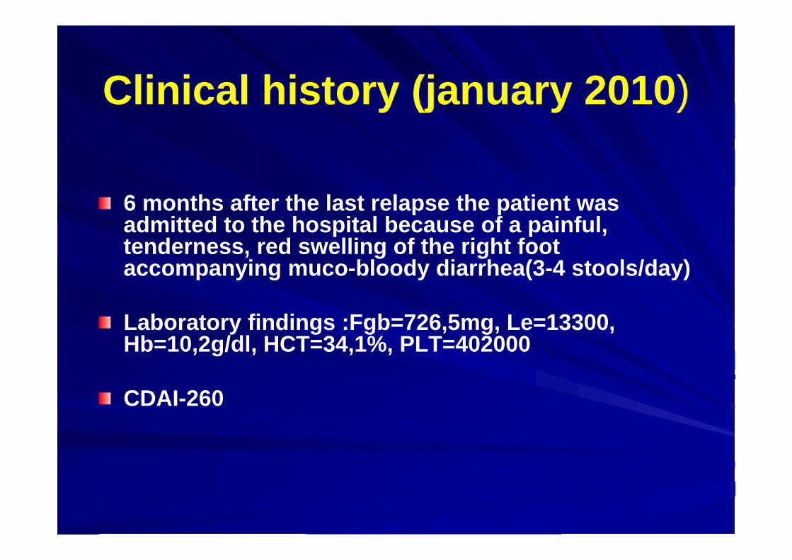
Clinical history (january 2010)
6 months after the last relapse the patient was admitted to the hospital because of a painful, tenderness, red swelling of the right foot accompanying muco-bloody diarrhea(3-4 stools/day)
Laboratory findings :Fgb=726,5mg, Le=13300, Hb=10,2g/dl, HCT=34,1%, PLT=402000
CDAI-260

Clinical and paraclinicalfindings
Clinical examination revealed an erythematous area of the lower right leg with draining pustulesRadiographs of the left ankle showed soft-tissue edema without evidence of osseous involvement.Negative coprocultureCl difficie toxine A/B negative Colonoscopy showed lesions limited to the colon without involvement of the terminal ileum

Multiple ulcers with a mucopurulent base, violaceousundermined border and peripheral erythema


Colonoscopy: ulcers, edema, inflammatory pseudopolyps


Diagnosis The patient has skin lesions mimic those of a pyogenic infection but association with IBD is highly suggestive for a sterile inflammatory process involving neutrophils
The two most common forms of neutrophilicdermatosis are pyoderma gangrenosum (PG) and Sweet's syndrome, each of which may be idiopathic or related to an underlying systemic disease

Skin lesions and IBDPG may precede or follow the diagnosis of an associated IBD, and may or may not parallel the clinical course of the associated diseasePG is one of the most common skin disorders linked to inflammatory bowel diseaseThe proportion of patients with inflammatory bowel disease who develop PG appears to be small In a cohort study of 2402 patients with inflammatory bowel disease, PG was detected in only 0.75 percent of patients
Farhi D, Cosnes J, Zizi N, et al. A cohort study of 2402 patients. Medicine (Baltimore) 2008; 87:281.

The differential features of PG and Sweet's syndrome suggests that the most likely diagnosis in this patient is pyoderma
gangrenosum.
Differential Diagnosis

Clinical question: What treatment do you recommend?
Prednisone 0.75-1mg/kgc
Mesalazine 4g
Azathioprine 2.5mg/kgc
Biological therapy
MTX 25mg/zi

Therapeutic approach
The regimen that we have chosen was:Induction therapy
Maintenance therapy
• Prednison 40mg/day
• Mesalazine 4g
• IFX induction doses (5mg/kgc at 0,2,6 wk)
•AZA 2.5mg/kgc
•Mesalazine 4g
Specific treatment for PG lesions: wound dressing+Diprofos perilesional injections+Dapsone 100mg/day

Pyoderma gangrenosum-Pathogenesis
PG is characterized by neutrophil-predominant infiltrates in the skin. The reason for the development of the inflammatory process that leads to PG remains unclearThe primary factors considered to contribute to the pathogenesis of PG : abnormalities in neutrophil functiongenetic variationsdysregulation of the innate immune
Ahronowitz I, Harp J, Shinkai.Am J Clin Dermatol 2012; 13:191.

Pyoderma gangrenosum-Epidemiology
3 to 10 cases per million people per year
an average age of onset between 40 and 60 years
women are more frequently affected
Ruocco E, Sangiuliano S, Gravina AG, et al. Pyoderma gangrenosum: an updated review. J Eur Acad Dermatol Venereol 2009; 23:1008.

Pyoderma gangrenosum –Clinical Types
Ulcerative (classic) PG – begins as a tender, inflammatory papule, pustule or vesicle that develops on normal-appearing skin or at a site of trauma ; lower extremities and trunk are the most common sites of involvementThe initial inflammatory lesion subsequently expands peripherally and degenerates centrally, leading to ulcer formation. The base of the ulcer is purulent and necrotic, and the depth of the ulcer often extends into subcutaneous fat and occasionally reaches the fascia
Ruocco E, Sangiuliano S, Gravina AG, et al. Pyoderma gangrenosum: an updated review. J Eur Acad Dermatol Venereol 2009; 23:1008.

Pyoderma gangrenosum –Clinical Types
Bullous (atypical) PG – Bullous PG is a less common, superficial variant of PG that is most commonly seen in patients with PG related to hematologic disease Pustular PG – Pustular PG usually occurs in patients with inflammatory bowel disease, and tends to arise during periods of acute exacerbations of bowel disease . Affected patients exhibit the rapid development of painful pustules surrounded by erythema. Concomitant fever and arthralgias are common
Powell FC, Hackett BC, Wallach D. in: Fitzpatrick's Dermatology in General Medicine, 8th ed, Goldsmith LA, Katz SI, Gilchrest BA, et al. (Eds), McGraw-Hill Companies, Inc., 2012. Vol 1, p.371.

Pyoderma gangrenosum –Clinical Types
Vegetative PG –also known as superficial granulomatous pyoderma is a localized, solitary, superficial form of PG that presents as an indolent, mildly painful nodule, plaque, or ulcer. A verrucousquality is often present. The undermined borders and purulent bases of ulcerative PG are absent. The head and neck are the most common sites for vegetative PG

Pyoderma gangrenosum-Treatment
Secondary infection, if present, should be treated. FIRST-LINE THERAPY —Local care — Wounds should be cleansed gently with tepid sterile saline or a mild antiseptic prior to dressing changes Wound dressings that promote a moist wound environment and do not adhere to the wound base are preferred, as they may be beneficial for healing Pathergy (exacerbation of lesions at sites of trauma) can occur in PG. Thus, unnecessary traumatic insults to the wound, such as the use of wet to dry dressings and the application of caustic substances should be avoided
Miller J, Yentzer BA, Clark A, et al. A review and update on new therapies. J Am Acad Dermatol 2010; 62:646.

Pyoderma gangrenosum-Treatment
Surgery — Surgical procedures are considered only in select cases, such as those in which accumulation of necrotic tissue presents a risk for infection or where vital tissues such as tendons or ligaments are exposed in the ulcer bedLocal corticosteroids — are usually applied once or twice daily or/and intralesional corticosteroids injected circumferentially into the ulcer periphery.Local calcineurin inhibitors — topical tacrolimus in concentrations of 0.03% to 0.3% has demonstrated efficacy for PG in multiple case reportsSystemic glucocorticoids / Systemic cyclosporine

Pyoderma gangrenosum-Treatment
SECOND-LINE AND ADJUNCTIVE THERAPIES —Conventional immunosuppressants —Immunosuppressive agents such as mycophenolatemofetil, methotrexate, and azathioprine have been utilized for the treatment of PG. These agents are generally considered to be most beneficial when used as adjunctive or glucocorticoid-sparing agents, rather than as monotherapyDapsone — administered as monotherapy or as a glucocorticoid-sparing age
Ahronowitz I, Harp J, Shinkai.Am J Clin Dermatol 2012; 13:191.

Pyoderma gangrenosum-Treatment


1 month after appropriate therapy:improvement of skin lesionsclinical remission
Follow-up

6 months later…
Reactivation of skin lesions
Clinical remission without mucosal healing at colonoscopy


Treatment and outcomeA wound culture yielded colonies of meticilinoresistent Staphylococcus aures
The patient followed the same treatment for skin lesions with: Diprofos perilesional injections and Dapsone 100mg/day + AB (Vancomycin and FQ iv)
Good outcome through healing skin lesions


January 2012 (2 years later)
Another flare occurs two weeks before the next application of IFX
Clinical presentation: 4-5 stools / day (diurnal and nocturnal) with pathological products, without active skin lesionsLaboratory findings: no anemia, leukocytosis (secondary to corticostherapy), PCR-9.13.Colonoscopy: longitudinal ulcers in the rectum, no other injuries 30cm explorated.IFX level<3

How should we treat at this point?
Shortening the interval between administration of IFX to 4 weeks
Dose escalation of IFX to 10mg/kgc at 8 wk
MTX 25mg/week
Prednisone 0.75-1mg/kgc

What guidelines recommend?
Our therapeutic choice was: IFX 5mg/kgc at 4 weeks

Follow-upFavorable outcome after shortening the interval between administration of IFX to 4 week Clinical and endoscopic remission
Restart the initial IFX regimen (5mg/kgcevery 8 weeks)

January 2013 (one year after the last flare)
Moderate flare, 2 weeks after the last application of IFX:Clinical presentation: 6-7stools/day sometimes with mucus and bloodLaboratory findings : PCR-7, Fgb-500 no anemia, no leukocytosisNegative coproculture, negative Cl. Difficile toxin A/B IFX level<3Colonoscopy shows lesions localized only in the rectum (superificiale ulcers, edematous mucosa and friable mucosa)

Colonoscopy: ulcers, swelling, redness, friable mucosa with spontaneous bleeding

What treatment suggestion do you have in this situation?A new course of prednisone 0.75-1mg/kgc
Budenofalk foam 6 mg 1/day
Increasing the dose of IFX to 10mg/kgc
Shortening the interval between administration of IFX to 4 wk
Salofalk 4g/day

The chosen regimen was:
• IFX 10mg/kgc at 8 wk
•Budenofalk foam 6mg/day
• AZA 2.5mg/kgc
•Salofalk 4 g/day

Follow-upMarch 2013 (after 3 months of IFX dose
escalation)
Favorable outcome under IFX 10 mg/kgc+rectal foam BudenofalkClinical and endoscopic remission which allows dose reduction of IFX to 5mg/kgc

Conclusions
We are facing a young patient with Crohn's disease that associates multiple relapses and extraintestinal manifestations (severe lesions of PG ) which requires biological therapyEffective treatment of the bowel disease in this case results in resolution of the PGOver time, the patient loses the response to biological therapy requiring for IFX dose optimizationA therapeutic goal is to adopt a personalized approach to therapy considering the particular disease severity and its heterogeneity

thatttttttThank you!