Case Report Calcifying epithelial odontogenic tumor ... · Int J Clin Exp Pathol...
7
Int J Clin Exp Pathol 2016;9(5):5733-5739 www.ijcep.com /ISSN:1936-2625/IJCEP0024156 Case Report Calcifying epithelial odontogenic tumor: report of three cases with immunohistochemical study Wonae Lee 1 , Na-Hye Myung 1 , Chul-Hwan Kim 2 1 Department of Pathology, Dankook University College of Medicine, 2 Department of Oral and Maxillofacial Surgery, Dankook University College of Dentistry, Dankook University, Republic of Korea Received January 16, 2016; Accepted March 26, 2016; Epub May 1, 2016; Published May 15, 2016 Abstract: Calcifying epithelial odontogenic tumor (CEOT) is a rare benign odontogenic tumor. Here we report 3 cases of CEOT. All patients were women and the age of the patients ranged from 39-48 years. All cases were located in the maxilla. Histologically, one case was epithelial-predominant and consisted of large irregular cribriform sheets of polygonal epithelial cells, dense eosinophilic amyloid-like globules, and frequent concentric calcifications. The other two cases were amyloid-rich and consisted of scattered small nests of polygonal epithelial cells, abundant stroma with amyloid-like globules, and lacked calcifications. Immunohistochemically, tumor cells in all cases were diffusely strongly positive for AE1/AE3, cytokeratin (CK)5, Cam5.2, CK19, 34βE12, and p63. Tumor cells in two cases were positive for CK7 and in one case, they were positive for CD10. Vimentin was strongly positive in one case, whereas weakly positive in two cases. A variable number of CD1a-positive Langerhans cells were observed among nests of tumor cells in all cases. In summary, CEOT exhibits distinct but various histological and immunohistochemical features. Keywords: Calcifying epithelial odontogenic tumor, amyloid, calcification, immunohistochemistry Introduction Calcifying epithelial odontogenic tumor (CEOT) is a rare odontogenic tumor that accounts for approximately 1% of all odontogenic tumors [1]. It is a benign, slow-growing, locally invasive odontogenic tumor. It generally occurs in patients between 20-60 years of age, with a mean age of diagnosis of 40 [1, 2]. It affects men and women equally. Histologically, CEOT consists of three distinct histological compo- nents: sheets of polyhedral epithelial cells, amyloid deposits, and calcifications [1, 2]. Here we present 3 cases of CEOT with unique immu- nohistochemical findings. Case reports Case 1 A 48-year-old woman complained of a painful swelling in her right cheek that had been pres- ent for 2 months. A panoramic radiograph revealed a mixed radiolucent-radiopaque mass in the right maxillary bone. A facial computed tomography (CT) revealed a large osteolytic mass (4.8×3.3×3.9 cm) with amorphous and stippled calcifications (Figure 1A). The mass was associated with an unerupted tooth. Inci- sional biopsy was performed and the diagnosis of CEOT was made. Subsequently, partial hemi- maxillectomy was performed. Macroscopically, the mass appeared as a nonencapsulated, circumscribed, pale gray, firm solid. Micro- scopically, the tumor had an epithelial-predomi- nant pattern. The tumor consisted of large irreg- ular cribriform sheets of polygonal epithelial cells, abundant extracellular dense eosinophilic amyloid-like globules, and concentric lamellar calcifications called Lisengang rings (Figure 1B). The epithelial cells had abundant eosino- philic cytoplasm, well-developed intercellular bridges, moderate nuclear pleomorphism, occasional binucleation, smudged or vesicular chromatin, inconspicuous or small nucleoli, and no mitotic figures (Figure 1C). The amyloid-like material was stained intensely with Congo red and showed apple-green birefringence when subjected to polarized light (Figure 1D). Tumor
Transcript of Case Report Calcifying epithelial odontogenic tumor ... · Int J Clin Exp Pathol...
Int J Clin Exp Pathol 2016;9(5):5733-5739www.ijcep.com /ISSN:1936-2625/IJCEP0024156
Case ReportCalcifying epithelial odontogenic tumor: report of three cases with immunohistochemical study
Wonae Lee1, Na-Hye Myung1, Chul-Hwan Kim2
1Department of Pathology, Dankook University College of Medicine, 2Department of Oral and Maxillofacial Surgery, Dankook University College of Dentistry, Dankook University, Republic of Korea
Received January 16, 2016; Accepted March 26, 2016; Epub May 1, 2016; Published May 15, 2016
Abstract: Calcifying epithelial odontogenic tumor (CEOT) is a rare benign odontogenic tumor. Here we report 3 cases of CEOT. All patients were women and the age of the patients ranged from 39-48 years. All cases were located in the maxilla. Histologically, one case was epithelial-predominant and consisted of large irregular cribriform sheets of polygonal epithelial cells, dense eosinophilic amyloid-like globules, and frequent concentric calcifications. The other two cases were amyloid-rich and consisted of scattered small nests of polygonal epithelial cells, abundant stroma with amyloid-like globules, and lacked calcifications. Immunohistochemically, tumor cells in all cases were diffusely strongly positive for AE1/AE3, cytokeratin (CK)5, Cam5.2, CK19, 34βE12, and p63. Tumor cells in two cases were positive for CK7 and in one case, they were positive for CD10. Vimentin was strongly positive in one case, whereas weakly positive in two cases. A variable number of CD1a-positive Langerhans cells were observed among nests of tumor cells in all cases. In summary, CEOT exhibits distinct but various histological and immunohistochemical features.
Keywords: Calcifying epithelial odontogenic tumor, amyloid, calcification, immunohistochemistry
Introduction
Calcifying epithelial odontogenic tumor (CEOT) is a rare odontogenic tumor that accounts for approximately 1% of all odontogenic tumors [1]. It is a benign, slow-growing, locally invasive odontogenic tumor. It generally occurs in patients between 20-60 years of age, with a mean age of diagnosis of 40 [1, 2]. It affects men and women equally. Histologically, CEOT consists of three distinct histological compo-nents: sheets of polyhedral epithelial cells, amyloid deposits, and calcifications [1, 2]. Here we present 3 cases of CEOT with unique immu-nohistochemical findings.
Case reports
Case 1
A 48-year-old woman complained of a painful swelling in her right cheek that had been pres-ent for 2 months. A panoramic radiograph revealed a mixed radiolucent-radiopaque mass in the right maxillary bone. A facial computed
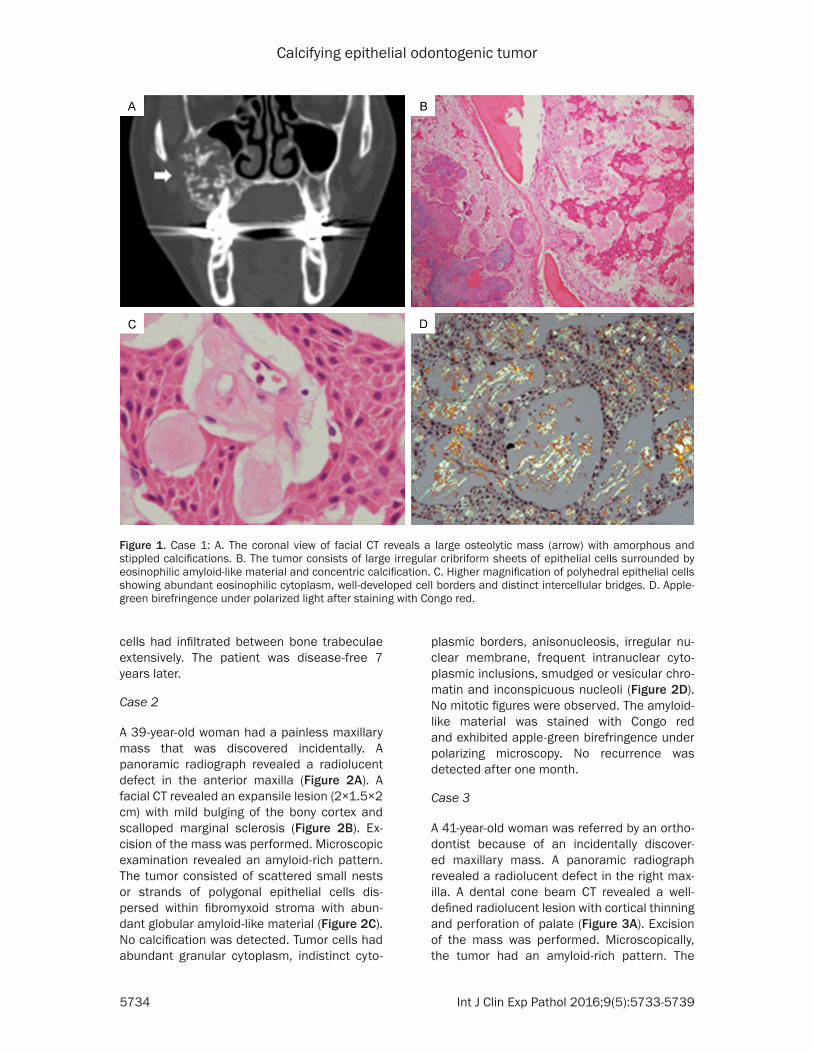
tomography (CT) revealed a large osteolytic mass (4.8×3.3×3.9 cm) with amorphous and stippled calcifications (Figure 1A). The mass was associated with an unerupted tooth. Inci- sional biopsy was performed and the diagnosis of CEOT was made. Subsequently, partial hemi-maxillectomy was performed. Macroscopically, the mass appeared as a nonencapsulated, circumscribed, pale gray, firm solid. Micro- scopically, the tumor had an epithelial-predomi-nant pattern. The tumor consisted of large irreg-ular cribriform sheets of polygonal epithelial cells, abundant extracellular dense eosinophilic amyloid-like globules, and concentric lamellar calcifications called Lisengang rings (Figure 1B). The epithelial cells had abundant eosino-philic cytoplasm, well-developed intercellular bridges, moderate nuclear pleomorphism, occasional binucleation, smudged or vesicular chromatin, inconspicuous or small nucleoli, and no mitotic figures (Figure 1C). The amyloid-like material was stained intensely with Congo red and showed apple-green birefringence when subjected to polarized light (Figure 1D). Tumor
Calcifying epithelial odontogenic tumor
5734 Int J Clin Exp Pathol 2016;9(5):5733-5739
cells had infiltrated between bone trabeculae extensively. The patient was disease-free 7 years later.
Case 2
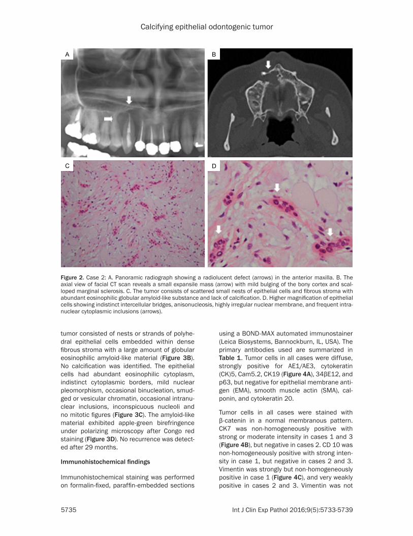
A 39-year-old woman had a painless maxillary mass that was discovered incidentally. A panoramic radiograph revealed a radiolucent defect in the anterior maxilla (Figure 2A). A facial CT revealed an expansile lesion (2×1.5×2 cm) with mild bulging of the bony cortex and scalloped marginal sclerosis (Figure 2B). Ex- cision of the mass was performed. Microscopic examination revealed an amyloid-rich pattern. The tumor consisted of scattered small nests or strands of polygonal epithelial cells dis-persed within fibromyxoid stroma with abun-dant globular amyloid-like material (Figure 2C). No calcification was detected. Tumor cells had abundant granular cytoplasm, indistinct cyto-
plasmic borders, anisonucleosis, irregular nu- clear membrane, frequent intranuclear cyto-plasmic inclusions, smudged or vesicular chro-matin and inconspicuous nucleoli (Figure 2D). No mitotic figures were observed. The amyloid-like material was stained with Congo red and exhibited apple-green birefringence under polarizing microscopy. No recurrence was detected after one month.
Case 3
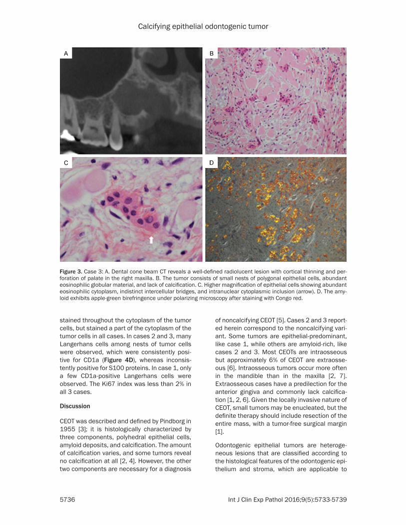
A 41-year-old woman was referred by an ortho-dontist because of an incidentally discover- ed maxillary mass. A panoramic radiograph revealed a radiolucent defect in the right max-illa. A dental cone beam CT revealed a well-defined radiolucent lesion with cortical thinning and perforation of palate (Figure 3A). Excision of the mass was performed. Microscopically, the tumor had an amyloid-rich pattern. The
Figure 1. Case 1: A. The coronal view of facial CT reveals a large osteolytic mass (arrow) with amorphous and stippled calcifications. B. The tumor consists of large irregular cribriform sheets of epithelial cells surrounded by eosinophilic amyloid-like material and concentric calcification. C. Higher magnification of polyhedral epithelial cells showing abundant eosinophilic cytoplasm, well-developed cell borders and distinct intercellular bridges. D. Apple-green birefringence under polarized light after staining with Congo red.
Calcifying epithelial odontogenic tumor
5735 Int J Clin Exp Pathol 2016;9(5):5733-5739
tumor consisted of nests or strands of polyhe-dral epithelial cells embedded within dense fibrous stroma with a large amount of globular eosinophilic amyloid-like material (Figure 3B). No calcification was identified. The epithelial cells had abundant eosinophilic cytoplasm, indistinct cytoplasmic borders, mild nuclear pleomorphism, occasional binucleation, smud- ged or vesicular chromatin, occasional intranu-clear inclusions, inconspicuous nucleoli and no mitotic figures (Figure 3C). The amyloid-like material exhibited apple-green birefringence under polarizing microscopy after Congo red staining (Figure 3D). No recurrence was detect-ed after 29 months.
Immunohistochemical findings
Immunohistochemical staining was performed on formalin-fixed, paraffin-embedded sections
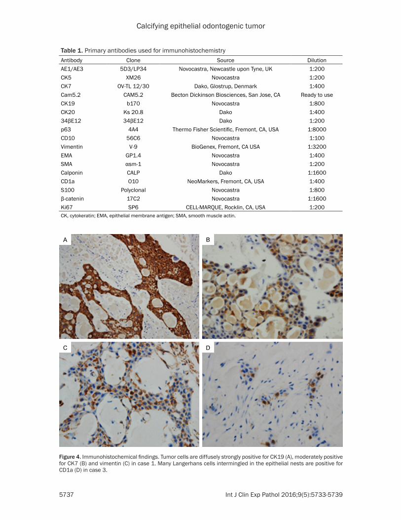
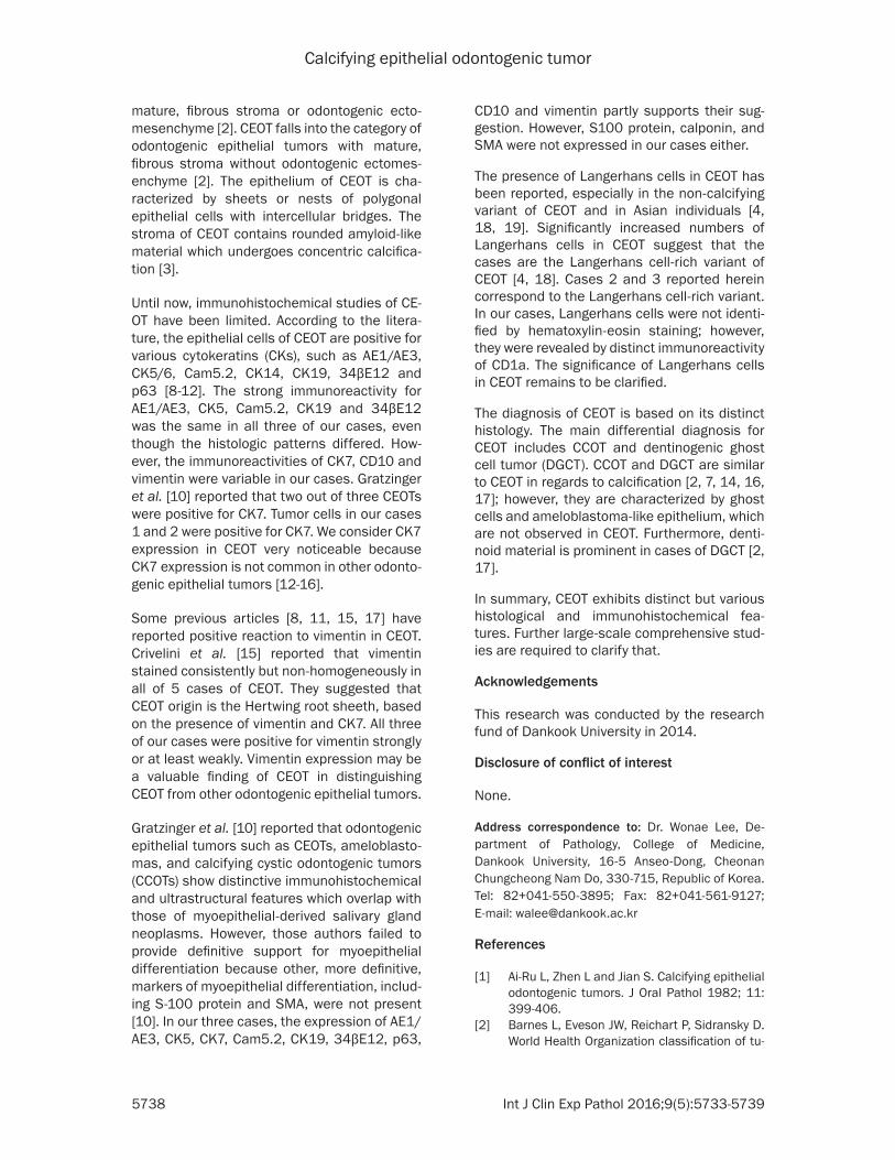
using a BOND-MAX automated immunostainer (Leica Biosystems, Bannockburn, IL, USA). The primary antibodies used are summarized in Table 1. Tumor cells in all cases were diffuse, strongly positive for AE1/AE3, cytokeratin (CK)5, Cam5.2, CK19 (Figure 4A), 34βE12, and p63, but negative for epithelial membrane anti-gen (EMA), smooth muscle actin (SMA), cal-ponin, and cytokeratin 20.
Tumor cells in all cases were stained with β-catenin in a normal membranous pattern. CK7 was non-homogeneously positive with strong or moderate intensity in cases 1 and 3 (Figure 4B), but negative in cases 2. CD 10 was non-homogeneously positive with strong inten-sity in case 1, but negative in cases 2 and 3. Vimentin was strongly but non-homogeneously positive in case 1 (Figure 4C), and very weakly positive in cases 2 and 3. Vimentin was not
Figure 2. Case 2: A. Panoramic radiograph showing a radiolucent defect (arrows) in the anterior maxilla. B. The axial view of facial CT scan reveals a small expansile mass (arrow) with mild bulging of the bony cortex and scal-loped marginal sclerosis. C. The tumor consists of scattered small nests of epithelial cells and fibrous stroma with abundant eosinophilic globular amyloid-like substance and lack of calcification. D. Higher magnification of epithelial cells showing indistinct intercellular bridges, anisonucleosis, highly irregular nuclear membrane, and frequent intra-nuclear cytoplasmic inclusions (arrows).
Calcifying epithelial odontogenic tumor
5736 Int J Clin Exp Pathol 2016;9(5):5733-5739
stained throughout the cytoplasm of the tumor cells, but stained a part of the cytoplasm of the tumor cells in all cases. In cases 2 and 3, many Langerhans cells among nests of tumor cells were observed, which were consistently posi-tive for CD1a (Figure 4D), whereas inconsis-tently positive for S100 proteins. In case 1, only a few CD1a-positive Langerhans cells were observed. The Ki67 index was less than 2% in all 3 cases.
Discussion
CEOT was described and defined by Pindborg in 1955 [3]; it is histologically characterized by three components, polyhedral epithelial cells, amyloid deposits, and calcification. The amount of calcification varies, and some tumors reveal no calcification at all [2, 4]. However, the other two components are necessary for a diagnosis
of noncalcifying CEOT [5]. Cases 2 and 3 report-ed herein correspond to the noncalcifying vari-ant. Some tumors are epithelial-predominant, like case 1, while others are amyloid-rich, like cases 2 and 3. Most CEOTs are intraosseous but approximately 6% of CEOT are extraosse-ous [6]. Intraosseous tumors occur more often in the mandible than in the maxilla [2, 7]. Extraosseous cases have a predilection for the anterior gingiva and commonly lack calcifica-tion [1, 2, 6]. Given the locally invasive nature of CEOT, small tumors may be enucleated, but the definite therapy should include resection of the entire mass, with a tumor-free surgical margin [1].
Odontogenic epithelial tumors are heteroge-neous lesions that are classified according to the histological features of the odontogenic epi-thelium and stroma, which are applicable to
Figure 3. Case 3: A. Dental cone beam CT reveals a well-defined radiolucent lesion with cortical thinning and per-foration of palate in the right maxilla. B. The tumor consists of small nests of polygonal epithelial cells, abundant eosinophilic globular material, and lack of calcification. C. Higher magnification of epithelial cells showing abundant eosinophilic cytoplasm, indistinct intercellular bridges, and intranuclear cytoplasmic inclusion (arrow). D. The amy-loid exhibits apple-green birefringence under polarizing microscopy after staining with Congo red.
Calcifying epithelial odontogenic tumor
5737 Int J Clin Exp Pathol 2016;9(5):5733-5739
Table 1. Primary antibodies used for immunohistochemistry Antibody Clone Source DilutionAE1/AE3 5D3/LP34 Novocastra, Newcastle upon Tyne, UK 1:200CK5 XM26 Novocastra 1:200CK7 OV-TL 12/30 Dako, Glostrup, Denmark 1:400Cam5.2 CAM5.2 Becton Dickinson Biosciences, San Jose, CA Ready to use CK19 b170 Novocastra 1:800CK20 Ks 20.8 Dako 1:40034βE12 34βE12 Dako 1:200p63 4A4 Thermo Fisher Scientific, Fremont, CA, USA 1:8000CD10 56C6 Novocastra 1:100Vimentin V-9 BioGenex, Fremont, CA USA 1:3200EMA GP1.4 Novocastra 1:400SMA αsm-1 Novocastra 1:200Calponin CALP Dako 1:1600CD1a O10 NeoMarkers, Fremont, CA, USA 1:400S100 Polyclonal Novocastra 1:800β-catenin 17C2 Novocastra 1:1600Ki67 SP6 CELL-MARQUE, Rocklin, CA, USA 1:200CK, cytokeratin; EMA, epithelial membrane antigen; SMA, smooth muscle actin.
Figure 4. Immunohistochemical findings. Tumor cells are diffusely strongly positive for CK19 (A), moderately positive for CK7 (B) and vimentin (C) in case 1. Many Langerhans cells intermingled in the epithelial nests are positive for CD1a (D) in case 3.
Calcifying epithelial odontogenic tumor
5738 Int J Clin Exp Pathol 2016;9(5):5733-5739
mature, fibrous stroma or odontogenic ecto-mesenchyme [2]. CEOT falls into the category of odontogenic epithelial tumors with mature, fibrous stroma without odontogenic ectomes-enchyme [2]. The epithelium of CEOT is cha- racterized by sheets or nests of polygonal epithelial cells with intercellular bridges. The stroma of CEOT contains rounded amyloid-like material which undergoes concentric calcifica-tion [3].
Until now, immunohistochemical studies of CE- OT have been limited. According to the litera-ture, the epithelial cells of CEOT are positive for various cytokeratins (CKs), such as AE1/AE3, CK5/6, Cam5.2, CK14, CK19, 34βE12 and p63 [8-12]. The strong immunoreactivity for AE1/AE3, CK5, Cam5.2, CK19 and 34βE12 was the same in all three of our cases, even though the histologic patterns differed. How- ever, the immunoreactivities of CK7, CD10 and vimentin were variable in our cases. Gratzinger et al. [10] reported that two out of three CEOTs were positive for CK7. Tumor cells in our cases 1 and 2 were positive for CK7. We consider CK7 expression in CEOT very noticeable because CK7 expression is not common in other odonto-genic epithelial tumors [12-16].
Some previous articles [8, 11, 15, 17] have reported positive reaction to vimentin in CEOT. Crivelini et al. [15] reported that vimentin stained consistently but non-homogeneously in all of 5 cases of CEOT. They suggested that CEOT origin is the Hertwing root sheeth, based on the presence of vimentin and CK7. All three of our cases were positive for vimentin strongly or at least weakly. Vimentin expression may be a valuable finding of CEOT in distinguishing CEOT from other odontogenic epithelial tumors.
Gratzinger et al. [10] reported that odontogenic epithelial tumors such as CEOTs, ameloblasto-mas, and calcifying cystic odontogenic tumors (CCOTs) show distinctive immunohistochemical and ultrastructural features which overlap with those of myoepithelial-derived salivary gland neoplasms. However, those authors failed to provide definitive support for myoepithelial differentiation because other, more definitive, markers of myoepithelial differentiation, includ-ing S-100 protein and SMA, were not present [10]. In our three cases, the expression of AE1/AE3, CK5, CK7, Cam5.2, CK19, 34βE12, p63,
CD10 and vimentin partly supports their sug-gestion. However, S100 protein, calponin, and SMA were not expressed in our cases either.
The presence of Langerhans cells in CEOT has been reported, especially in the non-calcifying variant of CEOT and in Asian individuals [4, 18, 19]. Significantly increased numbers of Langerhans cells in CEOT suggest that the cases are the Langerhans cell-rich variant of CEOT [4, 18]. Cases 2 and 3 reported herein correspond to the Langerhans cell-rich variant. In our cases, Langerhans cells were not identi-fied by hematoxylin-eosin staining; however, they were revealed by distinct immunoreactivity of CD1a. The significance of Langerhans cells in CEOT remains to be clarified.
The diagnosis of CEOT is based on its distinct histology. The main differential diagnosis for CEOT includes CCOT and dentinogenic ghost cell tumor (DGCT). CCOT and DGCT are similar to CEOT in regards to calcification [2, 7, 14, 16, 17]; however, they are characterized by ghost cells and ameloblastoma-like epithelium, which are not observed in CEOT. Furthermore, denti-noid material is prominent in cases of DGCT [2, 17].
In summary, CEOT exhibits distinct but various histological and immunohistochemical fea-tures. Further large-scale comprehensive stud-ies are required to clarify that.
Acknowledgements
This research was conducted by the research fund of Dankook University in 2014.
Disclosure of conflict of interest
None.
Address correspondence to: Dr. Wonae Lee, De- partment of Pathology, College of Medicine, Dankook University, 16-5 Anseo-Dong, Cheonan Chungcheong Nam Do, 330-715, Republic of Korea. Tel: 82+041-550-3895; Fax: 82+041-561-9127; E-mail: [email protected]
References
[1] Ai-Ru L, Zhen L and Jian S. Calcifying epithelial odontogenic tumors. J Oral Pathol 1982; 11: 399-406.
[2] Barnes L, Eveson JW, Reichart P, Sidransky D. World Health Organization classification of tu-
Calcifying epithelial odontogenic tumor
5739 Int J Clin Exp Pathol 2016;9(5):5733-5739
mours: Pathology and genetics of head and neck tumours. Edited by Barnes L. Lyon: IARC Press; 2005. pp. 284-313.
[3] Pindborg JJ. Calcifying epithelial odontogenic tumors. Acta Pathologica Microbiologica Scandinavica 1956; 38: 71-71.
[4] Takata T, Ogawa I, Miyauchi M, Ijuhin N, Nikai H and Fujita M. Non-calcifying Pindborg tumor with Langerhans cells. J Oral Pathol Med 1993; 22: 378-383.
[5] Philipsen HP and Reichart PA. Calcifying epi-thelial odontogenic tumour: biological profile based on 181 cases from the literature. Oral Oncology 2000; 36: 17-26.
[6] Lanzer M, Gander T, Grätz K, Kruse A and Lübbers HT. Extraosseous calcifying epithelial odontogenic tumour. Oral Surgery 2014; 7: 177-183.
[7] Lee SK and Kim YS. Current concepts and oc-currence of epithelial odontogenic tumors: II. Calcifying epithelial odontogenic tumor versus ghost cell odontogenic tumors derived from calcifying odontogenic cyst. J Pathol Transl Med 2014; 48: 175-187.
[8] Azevedo RS, Mosqueda-Taylor A, Carlos R, Cabral MG, Romañach MJ, Paes de Almeida O and Pires FR. Calcifying epithelial odontogenic tumor (CEOT): a clinicopathologic and immu-nohistochemical study and comparison with dental follicles containing CEOT-like areas. Oral Surg Oral Med Oral Pathol Oral Radiol 2013; 116: 759-768.
[9] Ng KH and Siar CH. A clinicopathological and immunohistochemical study of the calcifying epithelial odontogenic tumour (Pindborg tu-mour) in Malaysians. J Laryngol Otol 1996; 110: 757-762.
[10] Gratzinger D, Salama ME, Poh CF and Rouse RV. Ameloblastoma, calcifying epithelial odon-togenic tumor, and glandular odontogenic cyst show a distinctive immunophenotype with some myoepithelial antigen expression. J Oral Pathol Med 2008; 37: 177-184.
[11] Poomsawat S and Punyasingh J. Calcifying epi-thelial odontogenic tumor: an immunohisto-chemical case study. J Mol Histol 2007; 38: 103-109.
[12] Sherlin HJ, Natesan A, Ram P, Ramani P and Thiruvenkadam C. Immunohistochemical pro-filing of Ameloblastomas using cytokeratin, vi-mentin, smooth muscle actin, CD34 and S100. Ann Maxillofac Surg 2013; 3: 51-57.
[13] Heikinheimo K, Hormia M, Stenman G, Virtanen I and Happonen RP. Patterns of ex-pression of intermediate filaments in amelo-blastoma and human fetal tooth germ. J Oral Pathol Med 1989; 18: 264-273.
[14] Gunhan O, Celasun B, Can C and Finci R. The nature of ghost cells in calcifying odontogenic cyst: an immunohistochemical study. Ann Dent 1993; 52: 30-33.
[15] Crivelini MM, Felipini RC, Coclete GA and Soubhia AMP. Immunoexpression of keratins in the calcifying cystic odontogenic tumor epi-thelium. J Oral Pathol Med 2009; 38: 393-396.
[16] Fregnani ER, Pires FR, Quezada RD, Shih Ie M, Vargas PA and de Almeida OP. Calcifying odon-togenic cyst: clinicopathological features and immunohistochemical profile of 10 cases. J Oral Pathol Med 2003; 32: 163-170.
[17] Sauk JJ, Cocking-Johnson D and Warings M. Identification of basement membrane compo-nents and intermediate filaments in calcifying epithelial odontogenic tumors. J Oral Pathol 1985; 14: 133-140.
[18] Afrogheh A, Schneider J, Mohamed N and Hille J. Calcifying epithelial odontogenic tumour with clear langerhans cells: a novel variant, re-port of a case and review of the literature. Head Neck Pathol 2014; 8: 214-219.
[19] Asano M, Takahashi T, Kusama K, Iwase T, Hori M, Yamanoi H, Tanaka H and Moro I. A variant of calcifying epithelial odontogenic tumor with Langerhans cells. J Oral Pathol Med 1990; 19: 430-434.