Case Analysis Neuroendocrine Carcinomas of the … of this, neuroendocrine carcinomas of the colon J...
9
N euroendocrine cells are diffusely distributed throughout the body and are found in the gastro- intestinal tract, pancreas, lung, thyroid, adrenal gland, and many other organs. The gastrointestinal tract has the largest component of neuroendocrine cells. In spite of this, neuroendocrine carcinomas of the colon J Soc Colon Rectal Surgeon (Taiwan) September 2008 Case Analysis Neuroendocrine Carcinomas of the Colon and Rectum: Result of a 15-year Experience Cheng-Chou Lai 1 Chih-Wei Wang 2 Chung-Rong Changchien 1 Reiping Tang 1 Jy-Ming Chiang 1 Yau-Tong You 1 Pau-Shiu Hsien 1 Wen-Sy Tsai 1 Chien Yuh Yeh 1 Jeng-Yi Wang 3 Jinn-Shiun Chen 1 1 Division of Colon and Rectal Surgery, Department of Surgery, Chang Gung Medical Center, Taipei, Taiwan 2 Department of Pathology, Chang Gung Medical Center, Taipei, Taiwan 3 Division of Colon and Rectal Surgery, Department of Surgery, Chang Gung Memorial Hospital, Chiayi, Taiwan Key Words Neuroendocrine carcinoma; Small cell; Large cell; Adjuvant chemotherapy; Colon and rectum Purpose. The experience of the uncommon malignancy, neuroendocrine carcinomas of the colon and rectum with emphasis on the pathology and clinical characteristics at a single hospital was reviewed. Methods. Of more than 11,000 colon or rectal cancers removed from July 1992 to June 2007 at Chang Gung Medical Center in Taipei, 11 cases diag- nosed as colon or rectal neuroendocrine carcinoma were evaluated. Pa- thology was reviewed by a single pathologist. Medical records were retrospectively reviewed and patients were analyzed in terms of cli- nicopathologic and demographic characteristics including neuroendo- crine type, tumor location, tumor stage, responses to treatment (operative procedure, chemotherapy or radiotherapy), metastases, and survival. Results. Five patients had distant metastasis at the time of diagnosis. Pallia- tive chemotherapy or radiotherapy did not seem to offer a modest improve- ment in survival for these patients, with an overall survival of less than 11 months after diagnosis. Lymph node metastasis was found in 75%, and the distant metastasis in 45% of the 11 patients at the time of diagnosis. Overall survival rates for six-month, one-year, and three-year survival were 73 per- cent, 45 percent, 20 percent, respectively. These findings are in accor- dance with other publications, which demonstrate that the neuroendocrine carcinomas behave aggressively and are associated with worse prognosis than that of conventional adenocarcinomas of the same stage. Unexpect- edly, improved results were found for two stage IIA and IIIB rectal small cell neuroendocrine carcinoma patients administered with adjuvant chemo- therapy treatment with cisplatin and etoposide at our hospital. They were alive without evidence of disease at more than 10 years after treatment. Conclusions. Neuroendocrine malignancies are rare but behave aggres- sively in the colon or rectum, accounting for less than 0.1 percent of all colorectal cancers at our institution. Aggressive adjuvant chemotherapy with cisplatin and etoposide might offer a better chance of long-term sur- vival for patients with stage II and III neuroendocrine carcinomas in the colon and rectum, which deserves further investigation. [J Soc Colon Rectal Surgeon (Taiwan) 2008;19:87-95] Received: July 14, 2008. Accepted: July 31, 2008. Correspondence to: Dr. Jinn-Shiun Chen, Division of Colon and Rectal Surgery, Department of Surgery, Chang Gung Medical Center, No. 5, Fusing St., Gueishan Township, Taoyuan County 333, Taiwan. Tel: +886-3-328-1200 ext. 2101; Fax: +886-3-327-8355; E-mail: [email protected] 87
-
Upload
trankhuong -
Category
Documents
-
view
219 -
download
0
Transcript of Case Analysis Neuroendocrine Carcinomas of the … of this, neuroendocrine carcinomas of the colon J...
Neuroendocrine cells are diffusely distributed
throughout the body and are found in the gastro-
intestinal tract, pancreas, lung, thyroid, adrenal gland,
and many other organs. The gastrointestinal tract has
the largest component of neuroendocrine cells. In
spite of this, neuroendocrine carcinomas of the colon
J Soc Colon Rectal Surgeon (Taiwan) September 2008
Case Analysis
Neuroendocrine Carcinomas of the Colon
and Rectum: Result of a 15-year Experience
Cheng-Chou Lai1
Chih-Wei Wang2
Chung-Rong Changchien1
Reiping Tang1
Jy-Ming Chiang1
Yau-Tong You1
Pau-Shiu Hsien1
Wen-Sy Tsai1
Chien Yuh Yeh1
Jeng-Yi Wang3
Jinn-Shiun Chen1
1Division of Colon and Rectal Surgery,
Department of Surgery, Chang Gung
Medical Center, Taipei, Taiwan2Department of Pathology, Chang Gung
Medical Center, Taipei, Taiwan3Division of Colon and Rectal Surgery,
Department of Surgery, Chang Gung
Memorial Hospital, Chiayi, Taiwan
Key Words
Neuroendocrine carcinoma;
Small cell;
Large cell;
Adjuvant chemotherapy;
Colon and rectum
Purpose. The experience of the uncommon malignancy, neuroendocrinecarcinomas of the colon and rectum with emphasis on the pathology and
clinical characteristics at a single hospital was reviewed.
Methods. Of more than 11,000 colon or rectal cancers removed from July1992 to June 2007 at Chang Gung Medical Center in Taipei, 11 cases diag-nosed as colon or rectal neuroendocrine carcinoma were evaluated. Pa-thology was reviewed by a single pathologist. Medical records wereretrospectively reviewed and patients were analyzed in terms of cli-nicopathologic and demographic characteristics including neuroendo-crine type, tumor location, tumor stage, responses to treatment (operative
procedure, chemotherapy or radiotherapy), metastases, and survival.
Results. Five patients had distant metastasis at the time of diagnosis. Pallia-tive chemotherapy or radiotherapy did not seem to offer a modest improve-ment in survival for these patients, with an overall survival of less than 11months after diagnosis. Lymph node metastasis was found in 75%, and thedistant metastasis in 45% of the 11 patients at the time of diagnosis. Overallsurvival rates for six-month, one-year, and three-year survival were 73 per-cent, 45 percent, 20 percent, respectively. These findings are in accor-dance with other publications, which demonstrate that the neuroendocrinecarcinomas behave aggressively and are associated with worse prognosisthan that of conventional adenocarcinomas of the same stage. Unexpect-edly, improved results were found for two stage IIA and IIIB rectal smallcell neuroendocrine carcinoma patients administered with adjuvant chemo-therapy treatment with cisplatin and etoposide at our hospital. They werealive without evidence of disease at more than 10 years after treatment.
Conclusions. Neuroendocrine malignancies are rare but behave aggres-sively in the colon or rectum, accounting for less than 0.1 percent of allcolorectal cancers at our institution. Aggressive adjuvant chemotherapywith cisplatin and etoposide might offer a better chance of long-term sur-vival for patients with stage II and III neuroendocrine carcinomas in the
colon and rectum, which deserves further investigation.[J Soc Colon Rectal Surgeon (Taiwan) 2008;19:87-95]
Received: July 14, 2008. Accepted: July 31, 2008.
Correspondence to: Dr. Jinn-Shiun Chen, Division of Colon and Rectal Surgery, Department of Surgery, Chang Gung Medical Center,
No. 5, Fusing St., Gueishan Township, Taoyuan County 333, Taiwan. Tel: +886-3-328-1200 ext. 2101; Fax: +886-3-327-8355;
E-mail: [email protected]
87

and rectum are rare entities. Previous publications
demonstrated an incidence of this tumor ranging from
0.1 percent to 3.9 percent of all colorectal malignan-
cies.1 Either the introduction of more sensitive diag-
nostic tools (e.g. immunohistochemical stains) or an
overall increased awareness among physicians con-
tributes largely to the rising incidence of neuroendo-
crine carcinomas.2
Neuroendocrine tumors of the colon and rectum
are divided into two groups: carcinoid tumors with
low-grade atypia and malignancy; and endocrine cell
carcinomas with high-grade atypia and malignancy.3
High grade neuroendocrine carcinoma includes both
small cell (SCNC) and large cell neuroendocrine car-
cinoma (LCNC). The neuroendocrine tumor cells are
characterized by the presence of neurosecretory gra-
nules that can be detected by electron microscopy
and by the presence of neurosecretory markers within
the cytoplasm. These markers such as neuron-specific
enolase (NSE), chromogranin, synaptophysin, and
CD56 can be detected with immunohistochemical
stains.4 Carcinoid tumors generally exhibit a more in-
dolent behavior and do not response to cisplatin-based
chemotherapy, whereas high-grade neuroendocrine
carcinomas behave aggressive but often do response
transiently to cisplatin-based chemotherapy.1
Compared with adenocarcinomas, patients with
neuroendocrine carcinomas have a particularly poorer
prognosis with a median survival of less than 1 year as
previously reported.2,5,6 At the time of diagnosis, the
majority of tumors have metastasized to the lymph
nodes, liver, lung, or other sites. Correct diagnosis is
important because it will affect treatment and may
better predict the clinical course.1,4
The purpose of this study was to review experi-
ence with neuroendocrine carcinomas of the colon
and rectum at a single institution over a 15-year pe-
riod, with emphasis on the pathologic and clinical
characteristics.
Materials and Methods
Included in this study were a total of more than
11,000 patients diagnosed as colorectal cancer at the
Chang Gung Medical Center (CGMC) in Taipei, Tai-
wan from July 1992 to June 2007. Of these, 8 patients
with neuroendocrine carcinomas of the colon and rec-
tum were identified from a prospective colorectal ser-
vice database at CGMC, while 3 patients with ne-
uroendocrine carcinomas were recovered from the pa-
thology service database at identical institution. A sin-
gle expert pathologist reviewed the histopathology for
all cases.
Hematoxylin and eosin-stained slides were avail-
able for review in each case. Carcinoid tumors, aty-
pical carcinoids, adenocarcinomas with carcinoid
features (so-called adenocarcinomas tumors), and
adenocarcinomas with focal scattered chromogranin-
positive or synaptophysin-positive cells were not in-
cluded in this study.
High-grade neuroendocrine carcinomas were
classified into SCNC and LCNC on the basis of
histologic and immunohistochemical findings. Such
tumor classification of the colon and rectum was in-
troduced by the World Health Organization (WHO) in
2000.7-9 The SCNC is morphologically identical to
small cell carcinoma of lung, characterized by mini-
mal cytoplasm, fusiform cell shape, finely granular
chromatin, small or absent nucleoli, and nuclear mol-
ding, and corresponds to grade 3 tumors according to
Rindi et al.10 (Fig. 1). Small cell carcinomas typically
express neuroendocrine markers (e.g. chromogranin,
synaptophysin, NSE, CD56) by immunohistoche-
mistry. LCNC is a malignant neoplasm composed of
large cells having organoid, nesting, trabecular, ro-
sette-like and palisading patterns that suggested
endocrine differentiation, which can be confirmed by
immunohistochemistry and electron microscopy. In
contrast to small cell carcinoma, cytoplasm is more
abundant, cell shape is round or polygonal, chromatin
pattern is coarser, nuclei are more vesicular and nu-
cleoli are prominent1 (Fig. 2).
The diagnosis of SCNC of the colon and rectum
still remained a morphologic diagnosis, not an im-
munohistochemical one. The diagnosis of LCNC was
typically based on a combination of characteristic
morphology and NE phenotype as demonstrated by
immunohistochemical staining, which showed posi-
tive results for one of the four neuroendocrine mark-
ers (chromogranin, synaptophysin, NSE, and CD56).
Clinical information and follow-up, where avail-
88 Cheng-Chou Lai, et al. J Soc Colon Rectal Surgeon (Taiwan) September 2008
able, were obtained from review retrospectively of the
corresponding hospital records and the referring pa-
thologist. The records of these patients were analyzed
for clinical presentation, location and stage of tumor,
surgical procedure performed, site of metastases, ad-
junctive therapy, and clinical outcome. Tumors were
staged according to the 2002 American Joint Commit-
tee on Cancer (AJCC) TNM staging system.12 Sur-
vival time was defined as the time elapsed from the
date of the neuroendocrine carcinoma diagnosis until
death from all causes, or until April 30, 2008, the date
of analysis for the study.
Results
Clinicopathologic features including patient age
at diagnosis, gender, histologic subtype, tumor loca-
tion, tumor stage, treatment (surgery, chemotherapy
Vol. 19, No. 3 Neuroendocrine Carcinomas 89
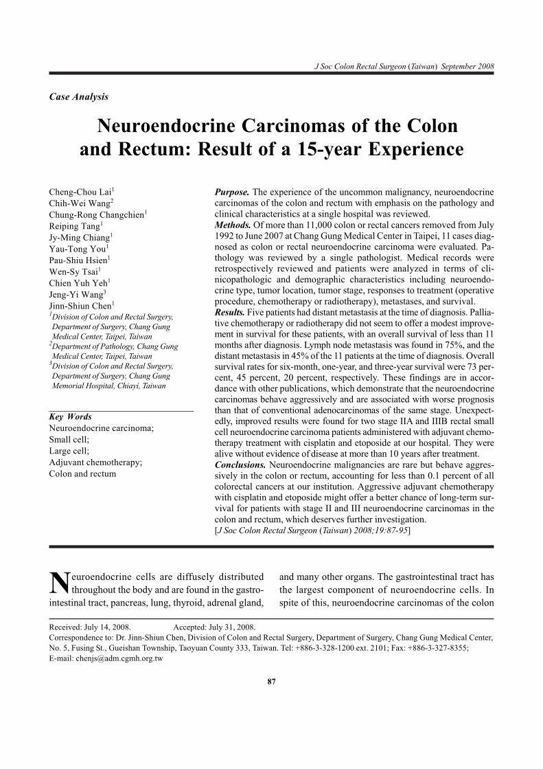
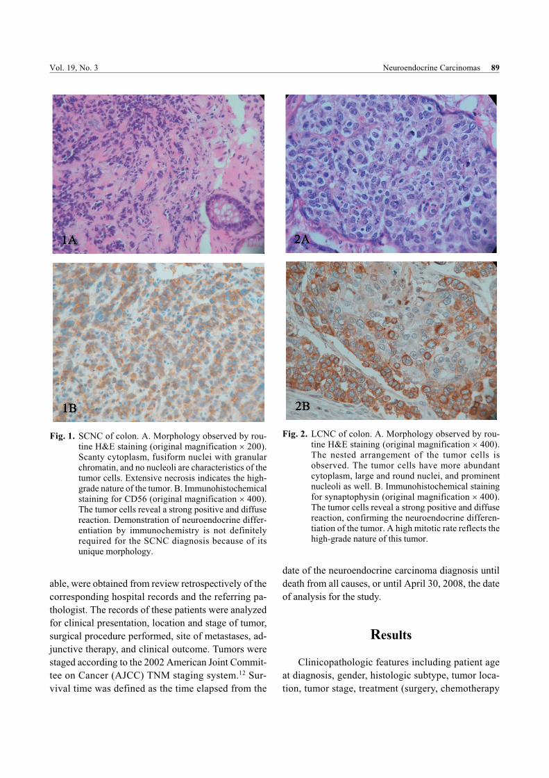
Fig. 1. SCNC of colon. A. Morphology observed by rou-tine H&E staining (original magnification � 200).Scanty cytoplasm, fusiform nuclei with granularchromatin, and no nucleoli are characteristics of thetumor cells. Extensive necrosis indicates the high-grade nature of the tumor. B. Immunohistochemicalstaining for CD56 (original magnification � 400).The tumor cells reveal a strong positive and diffusereaction. Demonstration of neuroendocrine differ-entiation by immunochemistry is not definitelyrequired for the SCNC diagnosis because of itsunique morphology.
Fig. 2. LCNC of colon. A. Morphology observed by rou-tine H&E staining (original magnification � 400).The nested arrangement of the tumor cells isobserved. The tumor cells have more abundantcytoplasm, large and round nuclei, and prominentnucleoli as well. B. Immunohistochemical stainingfor synaptophysin (original magnification � 400).The tumor cells reveal a strong positive and diffusereaction, confirming the neuroendocrine differen-tiation of the tumor. A high mitotic rate reflects thehigh-grade nature of this tumor.

and radiotherapy), the distant metastatic site and sur-
vival are outlined in Table 1. Patient demographics are
shown in Table 2. Two tumors were located in the as-
cending colon, two in the sigmoid colon, one in the
descending colon, five in the rectum, and one case of
synchronous lesion in the cecum and sigmoid colon.
The SCNC were diagnosed on the basis of routine
hematoxylin and eosin (H&E) staining due to its
unique morphology. Immunohistochemical tech-
niques for tumor markers synaptophysin, chromo-
granin, neuron-specific enolase (NSE), or CD56 were
performed to confirm the diagnosis of all LCNC. All
cases exhibited the typical histological characteristics
of high-grade neuroendocrine carcinoma within at
least 50 percent of the tumor, including a densely cel-
lular, solid growth pattern, with cells arranged in nests
having minimal intercellular stroma. The nuclear fea-
tures also suggested neuroendocrine differentiation,
with a “stippled or salt-and-pepper” chromatin pattern.
Stages of the tumors at the time of diagnosis were
as follows: two AJCC stage II, four stage III, and five
stage IV. Distant metastases were noted at the time of
diagnosis in nearly half patients (5/11, or 45 percent),
90 Cheng-Chou Lai, et al. J Soc Colon Rectal Surgeon (Taiwan) September 2008
Table 1. Clinicopathologic features of colorectal neuroendocrine carcinoma
CaseAge (yr)/Gender
NEType
Location Stagea
Surgery Chemotherapy Radiotherapy MetastasesSurvival
(mo)
1 60/F SCNC Rectum ��A APR CDDP,VP-16b
60 Gy, pelvis None 186, A/NED2 73/M SCNC Rectum ���C�IV LAR 5-FU, LV
b30 Gy, brain None�Brain 14, DOD
3 41/F SCNC Rectum ���B LAR, BSO CDDP,VP-16b
None None 139, A/NED4 38/F LCNC Rectum IV S-loop
colostomy,Biopsy
None None Carcinomatosis 2, DOD
5 44/F LCNC Ascend IV RH,hepatectomy
Folfoxc
None Liver 7, DOD
6 41/M LCNC Sigmoid ���C�IV HAR 5-FU, LVb
None None�Liver 7, DOD7 63/M SCNC Ascend IV Biopsy CDDP,VP-16
c25 Gy, bone Lung, Bone 5, DOD
8 65/M LCNC Rectum IV HAR Folfiric
None Liver 3, DOD9 55/M LCNC Descend ��A�IV LH CDDP,VP-16
cNone None�Lung 26, DOD
10 56/F SCNC Cecum,Sigmoid
IV Biopsy CDDP,VP-16c
30 Gy, brain Brain 11, DOD
11 64/M LCNC Sigmoid ���B HAR Noned
None None 14, A/NED
SCNC: small cell neuroendocrine carcinoma; LCNC: Large cell neuroendocrine carcinoma; APR: combine abdomino-perineal resection; LAR: low anterior resection; HAR: high anterior resection; RH: right hemicolectomy; LH: lefthemicolectomy; BSO: bilateral salpingo-oophrectomy; A/NED: alive with no evidence of disease; DOD: dead ofdisease.a. Stage groupings according to the AJCC and UICC system for cancer of the colon and rectum., 6
thedition.
b. Adjuvant chemotherapy.c. Palliative chemotherapy.d. Post operative persist perianal infection, hold adjuvant chemotherapy.
Table 2. Distribution of demographic and
clinicopathologic characteristics
Factor Mean Range
Age (yr) 54.5 38-73Male 60.2 41-73Female 47.8 38-60
N %
GenderMale 6 55Female 5 45
LocationRight colon 2 18Left colon 3 27Right and left colon 1 09Rectum 5 46
PathologySCNC 5 45LCNC 6 55
Tumor stage at diagnosisStage � 0 00Stage �� 2 18Stage ��� 4 36Stage IV 5 46
Survival6 months 8 731 year 5 453 years 2 20
such as liver (cases 5 and 8), brain (case 10), bone and
lung metastasis (case 7), and peritoneal carcino-
matosis as well (case 4).
The chemotherapeutic regimens included the fol-
lowing: two postoperative adjuvant chemotherapy with
the combination of cisplatin (CDDP) and etoposide
(VP-16) (cases 1 and 3), and two of 5-FU (5-fluo-
rouracil) and LV (leucovorin) (cases 2 and 6). In addi-
tion, three patients underwent palliative chemotherapy
with cisplatin and etoposide (cases 7, 9, and 10), one
with Folfox (case 5), and one with Folfiri (case 8). Two
patients did not receive chemotherapy. One with stage
IV rectal cancer with peritoneal carcinomatosis received
sigmoid-loop colostomy alone because of the colonic
obstruction, and refused further combined chemother-
apy/radiotherapy. She was dead of disease two months
after operation (case 4). And the other with stage IIIB
sigmoid colon cancer with regional lymph node metas-
tasis received no chemotherapy because of persistently
postoperative perianal infection and the history of
Fournier’s gangrene three years ago (case 11).
In addition, four patients received radiation ther-
apy. The dose of adjuvant radiotherapy for case 1 pa-
tient submitted to postoperative pelvic irradiation was
60 Gy. However, the side effects from radiotherapy
led her to incomplete treatment. Another two patients
underwent palliative radiation therapy for brain meta-
stases (30 Gy, cases 2 and 10), and the last received 25
Gy for painful bone metastasis (case 7). All conducted
treatment of 11 patients was listed in Table 1.
Most patients with neuroendocrine carcinoma
have poor prognosis, thus in this study only 6-month,
1-year, and 3-year survival were taken into consider-
ation. Overall survival rates for six-month, one-year,
and three-year survival were 73 percent, 45 percent,
20 percent, respectively (Table 2). Improved results
was found for two stage IIA and IIIB rectal small cell
neuroendocrine carcinoma patients administered with
adjuvant chemotherapy treatment with cisplatin and
etoposide at our hospital. They were alive without evi-
dence of disease at more than 10 years after treatment.
Discussion
During the past 15-year period between July 1992
and June 2007, only 11 patients with neuroendocrine
carcinomas of the colon and rectum were identified at
our institution. In this study, neuroendocrine car-
cinomas comprise approximately 0.1% of colorectal
malignancies that have been treated at CGMC. The
possible reasons for the lower incidence than pre-
viously reported 0.1-3.9% might be the selection
bias of a single hospital as well as the term “large cell
neuroendocrine carcinoma (LCNC)” of the colon and
rectum appeared to be well-recognized at our institu-
tion since 2000 when the WHO Classification of Tu-
mours7-9 was introduced. This classification is based
upon size, proliferative rate, and differentiation, and
comprises three subtypes of tumors: well-differen-
tiated endocrine neoplasm (both classified as car-
cinoid), and two poorly differentiated neuroendocrine
neoplasm, SCNC and LCNC. Before this tumor clas-
sification system was in place, only carcinoid tumors
and SCNC were extensively discussed in the literature.
In 2006, Kobayashi et al.15 conducted a literature
review on neuroendocrine carcinoma of the colon and
rectum. The incidence of neuroendocrine carcinoma,
rate of lymph node metastasis, rate of distant meta-
stasis at diagnosis, and survival documented in our
study and in the literature are summarized in Table 3.
The aggressive behavior of SCNC and LCNC has
been described in the literature. At the time of diag-
nosis, clinically, surgically or pathologically detect-
able metastases were present in most patients. Based
on the available literature, the rate of distant meta-
stasis appears to be between 38% and 73%, and the
rate of lymph node metastasis ranges from 60% to
87%.1,2,6,13,14 Moreover, the reported one-year survival
rate was between 15% and 46%.1,2,6,14,16 In this study,
we found the distant metastasis rate of 45% and lymph
node metastasis rate of 75%. Overall survival rates for
six-month, one-year, and three-year survival were 73
percent, 45 percent, 20 percent, respectively. Our
findings are in accordance with other publications,
which demonstrate that the neuroendocrine carci-
nomas behave aggressively and are associated with
worse prognosis than that of conventional adeno-
carcinomas of the same stage.1,2,6,14 Bernick et al.1
described a highest level of one-year survival rate
(46%) relatively similar to our result (45%). A plau-
sible explanation for this favorable prognosis is that
Vol. 19, No. 3 Neuroendocrine Carcinomas 91

chemotherapy treatment contributes to the improve-
ment in survival of patients with colorectal neuro-
endocrine carcinoma to some extent.
Clinicopathologic features of the stage II and III
patients were outlined in Table 4. In our study, several
chemotherapeutic regimens such as 5-FU/LV, Folfox,
Folfiri, and cisplatin/etoposide have been used as
adjuvant or palliative treatment. Postoperative ad-
juvant chemotherapy was given to 4 of 6 stage II or III
patients. One stage IIA patient receiving adjuvant che-
motherapy with cisplatin and etoposide remains alive
for more than 10 years with no evidence of disease re-
currence and distant metastases (case 1). Another
stage IIA patient who did not undergo postoperative
adjuvant chemotherapy was found to have brain me-
tastasis in around 24 months after the initial diagnosis.
Although treated with palliative radiotherapy and
cisplatin/etoposide chemotherapy, he died 2 months
after beginning therapy (case 9). These two cases em-
phasize the importance of adjuvant chemotherapy
92 Cheng-Chou Lai, et al. J Soc Colon Rectal Surgeon (Taiwan) September 2008
Table 3. Neuroendocrine carcinoma of the colon and rectum in the literature and this study
Author Year
Frequency incolorectalcancer (%)
No. ofcases
Distribution(right colon,left colon,
rectum, other)
Rate oflymph node
metastasis (%)
Rate ofdistant metastasisat diagnosis (%) Survival
Wick et al.13
1987 N/A 10 N/A 60 N/A MS: 5MStaren et al.
141988 1.9 13 6, 3, 4, 0 62 38 6M SR: 58%,
1Y SR: 33%,3Y SR: 25%
Gaffey et al.6
1990 N/A 24 10, 6, 8, 0 87 73 6M SR: 33%,1Y SR: 21%,3Y SR: 8%
Saclarides et al.2
1994 3.9 39 19, 11, 9, 0 79 39 6M SR: 58%,1Y SR: N/A,3Y SR: 15%
Bernick et al.1
2004 0.6 38 N/A N/A 66 6M SR: N/A,1Y SR: 46%,3Y SR: 13%
This study 2008 0.1 11 2, 3, 5, 1 75 45 6M SR: 73%,1Y SR: 45%,3Y SR: 20%
N/A: not available; MS: median survival; M: month; Y: year; SR: survival rate.Modification of Table 1 from Jpn J Clin Oncol 2006;36(5):325-328.
Table 4. Postoperative adjuvant chemotherapy for stage II and stage III patients
CaseAge (yr)/Gender
NE Type Location Stagea
Surgery Chemotherapy MetastasesSurvival (mo),
Status
1 60/F SCNC Rectum ��A APR CDDP, VP-16 None 186, A/NED2 73/M SCNC Rectum ���C�D LAR 5-FU, LV None�Brain 14, DOD3 41/F SCNC Rectum ���B LAR, BSO CDDP, VP-16 None 139, A/NED6 41/M LCNC Sigmoid ���C�D HAR 5-FU, LV None�Liver 7, DOD9 55/M LCNC Descend ��A�D LH None
bNone�Lung 26, DOD
11 64/M LCNC Sigmoid ���B HAR Nonec
None 14, A/NED
SCNC: small cell neuroendocrine carcinoma; LCNC: Large cell neuroendocrine carcinoma; APR: combine abdomino-perineal resection; LAR: low anterior resection; HAR: high anterior resection; RH: right hemicolectomy; LH: lefthemicolectomy; BSO: bilateral salpingo-oophrectomy; A/NED: alive with no evidence of disease; DOD: dead ofdisease.a. Stage groupings according to the AJCC and UICC system for cancer of the colon and rectum., 6
thedition.
b. Without receiving postoperative adjuvant chemotherapy.c. Without receiving postoperative adjuvant chemotherapy due to his Fournier’s gangrene history and persistently
postoperative perianal infection.
treatment on patients with stage II neuroendocrine
carcinoma of the colon and rectum. The American
Society of Clinical Oncology (ASCO) panel did not
recommend the routine administration of adjuvant
chemotherapy for stage II colon cancer patients but
suggested that it should be considered, particularly for
certain high-risk patients.17 Several considering fac-
tors such as tumor differentiation, tumor perforation,
number of lymph nodes examined, and T stage are
advocated when assessing the likely benefit:risk ra-
tio. There is growing evidence that the prognosis of
certain stage II patients with unfavorable prognostic
factors can be improved by adjuvant chemotherapy,
and increasingly refined tools are now available to
define those most likely to benefit.18 Thus, routine
postoperative adjuvant chemotherapy similar to that
used in high-risk stage II colon cancer is recom-
mended for patients with stage II neuroendocirne car-
cinoma of the colon and rectum. Referral of stage II
patients for individual assessment is strongly re-
commended.
Of four stage III patients, three received adjuvant
chemotherapy. The two treated with adjuvant 5-FU/
LV had distant metastasis (cases 2 and 6), and the one
with adjuvant cisplatin/etoposide remains alive for
more than 10 years with no evidence of disease re-
currence and distant metastases (case 3). This find-
ing suggests, in accord with published results19 that
systemic chemotherapy regimens for colorectal ne-
uroendocrine carcinoma should be similar to those
for small cell lung cancer. On the basis of objective
responses demonstrated in our patients, the combina-
tion of two chemotherapeutic regimens, cisplatin and
etoposide, are recommended for stage II and III tu-
mors. However, the role of adjuvant chemotherapy
requires further evaluation so that we might better
understand the overall treatment effect for patients
with neuroendocrine carcinomas of the colon and
rectum.
The combination of cisplatin and etoposide has
been considered as the reference treatment for poorly-
differentiated neuroendocrine tumors.20 Moertel et
al.21 reported that of 18 patients received palliative
chemotherapy with cisplatin and etoposide for meta-
static neuroendocrine carcinoma, 9 exhibited partial
response and 3 exhibited complete regression. An
overall regression rate of 67% was obtained with a
median duration of regression of 8 months. In our
study, there were two patients underwent palliative
chemotherapy with cisplatin and etoposide. One pa-
tient achieved a partial response and died within 11
months while the remaining one showed no response
and died within 5 months after diagnosis. Thus, pallia-
tive chemotherapy with cisplatin and etoposide can be
considered as treatment preference for colorectal
neuroendocrine carcinoma, particularly if the patient
has no other available options.
Previous reports suggest that there are geo-
graphic differences in the incidence rates of co-
lorectal neuroendocrine carcinoma, with the pre-
dominance of right-sided colon (41.6-48.7%) and
rectum (23.7-36.8%).2,6,14 Moreover, Yasui et al.22
reviewed the predominant distribution of small cell
carcinoma as well, with the disease affecting espe-
cially the right-sided colon (47.2%) and rectum
(44%). As a result of the small sample size and the
selection bias at single institution, our study showed a
distinctively predominant distribution (18% in right-
sided colon and 46% in rectum) from the publi-
cations.
Liver is the commonest site of distant metastasis.
In this report, we demonstrated a lower incidence of
liver metastasis (37.5%) than that reported in previous
studies (53.3-87.5%),2,6,14,22 possibly due to our li-
mited sample size.
Conclusion
Neuroendocrine malignancies are rare but behave
aggressively in the colon or rectum, accounting for
less than 0.1 percent of all colorectal cancers at our in-
stitution. Limited sample size and inconsistency of
treatment in our study make it difficult to generate an
exact recommendation regarding the optimal choice
of chemotherapeutic regimen. However, aggressive
adjuvant chemotherapy with cisplatin and etoposide is
still recommended on the basis of objective responses
demonstrated in our patients, which might offer a
better chance of long-term survival for patients with
stage II and III neuroendocrine carcinomas in the
colon and rectum.
Vol. 19, No. 3 Neuroendocrine Carcinomas 93
References
1. Bernick PE, Klimstra DS, Shia J, Minsky B, Saltz L, Shi W,
Thaler H, Guillem J, Paty P, Cohen AM, Wong WD. Ne-
uroendocrine carcinomas of the colon and rectum. Dis Colon
Rectum. 2004;47:163-9.
2. Saclarides TJ, Szeluga D, Staren ED. Neuroendocrine
cancers of the colon and rectum. Results of a ten-year
experience. Dis Colon Rectum 1994;37:635-42.
3. Nippon Geka Gakkai Zasshi. 2008;109:152-6.
4. Simon SR, Fox K. Neuroendocrine carcinoma of the colon.
Correct diagnosis is important. J Clin Gastroenterol. 1993;
17:304-7.
5. Staren ED, Gould VE, Jansson DS, Hyser M, Gooch GT,
Economou SG. Neuroendocrine differentiation in “poorly
differentiated” colon carcinomas. Am Surg 1990;56:412-9.
6. Gaffey MJ, Mills SE, Lack EE. Neuroendocrine carcinoma of
the colon and rectum. A clinicopathologic, ultrastructural,
and immunohistochemical study of 24 cases. Am J Surg
Pathol 1990;14:1010-23.
7. Solcia E, Capella C, Kloppel G, et al. Endocrine tumours of
the gastrointestinal tract. In: Solcia E, Kloppel G, Sobin J,
eds. Histological typing of endocrine tumours. Berlin:
Springer-Verlag, 2000:61-8.
8. Kloppel G, Heitz PU, Capella C, et al. Endocrine tumours of
the pancreas. In: Solcia E, Kloppel G, Sobin L, eds. His-
tological typing of endocrine tumours. Berlin: Springer-
Verlag, 2000:56-60.
9. Capella C, Heitz PU, Hofler H, Solcia E, Kloppel G. Re-
vised classification of neuroendocrine tumours of the lung,
pancreas and gut. Virchows Arch 1995;425:547-60.
10. Rindi G, Azzoni C, La Rosa S, Klersy C, Paolotti D, Rappel
S, Stolte M, Capella C, Bordi C, Solcia E. ECL cell tumor and
poorly differentiated endocrine carcinoma of the stomach:
prognostic evaluation by pathological analysis. Gastro-
enterology 1999;116:532-42.
11. Matsuki Y, Tanimoto A, Hamada T, Sasaguri Y. Histidine
decarboxylase expression as a new sensitive and specific
marker for small cell lung carcinoma. Mod Pathol. 2003;
16:72-8.
12. American Joint Committee on Cancer. AJCC Cancer Staging
Manual. 6th ed. New York: Springer-Verlag, 2002.
13. Wick MR, Weatherby RP, Weiland LH. Small cell neuro-
endocrine carcinoma of the colon and rectum: clinical,
histologic, and ultrastructural study and immunohistoche-
mical comparison with cloacogenic carcinoma. Hum Pathol
1987;18:9-21.
14. Staren ED, Gould VE, Warren WH, Wool NL, Bines S, Baker
J, Bonomi P, Roseman DL, Economou SG. Neuroendocrine
carcinomas of the colon and rectum: a clinicopathologic eval-
uation. Surgery 1988;104:1080-9.
15. Kobayashi H, Ueno H, Hashiguchi Y, Ishiguro M, Omata J,
Kajiwara Y, Shimazaki H, Mochizuki H. T1 neuroendocrine
carcinoma of anal canal after transanal resection for in-
tramucosal adenocarcinoma. Jpn J Clin Oncol. 2006;36:325-8.
16. Hung SS. Small cell carcinoma of the colon. A case report
and literature review. J Clin Gastroenterol. 1989;11:335-9.
17. Benson AB 3rd, Schrag D, Somerfield MR, Cohen AM,
Figueredo AT, Flynn PJ, Krzyzanowska MK, Maroun J,
McAllister P, Van Cutsem E, Brouwers M, Charette M, Haller
DG. American Society of Clinical Oncology recommenda-
tions on adjuvant chemotherapy for stage II colon cancer. J
Clin Oncol. 2004;22:3408-19.
18. André T, Sargent D, Tabernero J, O�Connell M, Buyse M,
Sobrero A, Misset JL, Boni C, de Gramont A. Current issues
in adjuvant treatment of stage II colon cancer. Ann Surg
Oncol. 2006;13:887-98.
19. Redman BG, Pazdur R. Colonic small cell undifferentiated
carcinoma: a distinct pathological diagnosis with therapeutic
implications. Am J Gastroenterol. 1987;82:382-5.
20. Mitry E, Rougier P. The treatment of undifferentiated ne-
uroendocrine tumors. Crit Rev Oncol Hematol. 2001;37:
47-51.
21. Moertel CG, Kvols LK, O�Connell MJ, Rubin J. Treatment of
neuroendocrine carcinomas with combined etoposide and
cisplatin. Evidence of major therapeutic activity in the
anaplastic variants of these neoplasms. Cancer. 1991;68:
227-32.
22. Yasui O, Tsukamoto F, Kudo K. Small cell undifferentiated
carcinoma of the ascending colon with rapid enlargement
after resection: report of a case and review of the literature.
Tohoku J Exp Med. 2006;209:361-7.
94 Cheng-Chou Lai, et al. J Soc Colon Rectal Surgeon (Taiwan) September 2008
賴正洲等 J Soc Colon Rectal Surgeon (Taiwan) 2008;19:87-95 95
病例分析
結直腸神經內分泌癌 單一醫學中心之 15年經驗
賴正洲 1 王志偉 2 張簡俊榮 1 唐瑞平 1 江支銘 1 游耀東 1
謝寶秀 1 蔡文司 1 葉建裕 1 王正儀 3 陳進勛 1
1林口長庚紀念醫院 外科部 大腸直腸肛門外科
2林口長庚紀念醫院 解剖病理科
3嘉義長庚紀念醫院 外科部 大腸直腸肛門外科
目的 結直腸神經內分泌癌為少見之惡性腫瘤。本研究回顧林口長庚紀念醫院過去治療結直腸神經內分泌癌之經驗,並就其臨床與病理特徵進行綜述。
方法 本研究回顧並收集林口長庚紀念醫院之結直腸癌及病理學資料庫於 1992 至 2007年間,經診斷罹患結直腸癌之 11,000 餘名病患中,病理診斷結果為結直腸神經內分泌癌者共 11 例。11 例病患之病理檢查結果皆由同一位病理科醫師加以重新檢閱。另回顧分析上述病患之個別醫療紀錄,並就其臨床病理及人口統計學特徵,如神經內分泌之病
理分類、腫瘤位置、腫瘤期別、治療 (手術方式、化學治療或放射線治療) 成效、轉移情形,以及存活率等進行分析。
結果 5 例病患於初次診斷時即發現有遠處轉移的情形,緩和性放射線治療或化學治療對於上述病患之存活率似無法提供適當之改善,整體存活率自診斷日起算不到 11個月。11名病患之初次診斷結果顯示其中 75% 有淋巴結轉移,45% 有遠處轉移,整體之六個月、一年與三年之存活率依序約為 73%、45% 以及 20%。上述結果與前人文獻相符,即相較於常見之同期腺癌,結直腸神經內分泌癌之侵襲性更強,預後亦較差。然而,兩
名 IIA 與 IIIB 期直腸小細胞神經內分泌癌病患在接受 cisplatin 合併 etoposide 之輔助性化學治療後,其存活率有正面且具積極意義的初步結果,治療後存活期達 10 年以上,且期間無疾病再發或轉移的情形。
結論 結直腸神經內分泌癌為一罕見但具侵襲性之疾病,占本院收治所有結直腸癌病例的 0.1% 以下。積極給予 II期與 III期結直腸神經內分泌癌患者進行 cisplatin合併 etoposide之輔助性化學治療,可能有助提高病患之存活率,此值得未來進一步研究。
關鍵詞 神經內分泌癌、小細胞、大細胞、輔助性化學治療、結腸與直腸。


![Journal of Tumour Research & Reports€¦ · neuroendocrine components [2]. The tumor may appear in various levels of the digestive tract including the oesophagus, stomach, colon](https://static.fdocuments.net/doc/165x107/60724a80318cfe68f50265c5/journal-of-tumour-research-reports-neuroendocrine-components-2-the-tumor.jpg)