Case 14.1 Pernicious anaemia - immunologyclinic.com studies_book/Case_studies_book... · afebrile...
7
Essentials of Clinical Immunology, Sixth Edition. Helen Chapel, Mansel Haeney, Siraj Misbah, and Neil Snowden. © 2014 John Wiley & Sons, Ltd. Published 2014 by John Wiley & Sons, Ltd. Case 14.1 Pernicious anaemia A 67-year-old widow presented with gradually increasing tiredness, exertional dyspnoea and ankle swelling. Two years earlier she had been found to be anaemic and had been treated with oral iron without symptomatic improvement. She had lost 6 kg in weight, but denied any history of anorexia, dyspepsia or blood loss. On examination, she was very pale and had signs of congestive cardiac failure. Laboratory investigations showed a very low haemoglobin of 54 g/l with a reduced white cell count of 3.7 × 10 9 /l (and a platelet count of only 31 × 10 9 /l). A blood film showed marked macrocytosis with a mean cell volume of 112 fl. Bone marrow examination revealed marked megaloblastic erythropoiesis with abundant iron stores. Serum vitamin B 12 was 40 ng/l (NR 170–900) but serum folate, serum iron and total iron-binding capacity were normal. Her serum contained strongly positive gastric parietal cell antibodies of IgG class (Fig. 14.8) and blocking antibodies to intrinsic factor. Antibodies to thyroid microsomal antigen were also found, although the patient was clinically and biochemically euthyroid. The patient had pernicious anaemia and was therefore started on intramuscular injections of hydroxycobalamin at 3-monthly intervals. Four days after the first injection, her reticulocyte count rose to 16%. Fig. 14.8 Gastric parietal cell antibodies detected by indirect immunofluorescence (see Chapter 19). Fluorescent cytoplasm in parietal cell Smooth muscle Case Figure 14.1 Autoantibodies to cytoplasm of gastric parietal cells (smooth muscle at lower right is unstained).
Transcript of Case 14.1 Pernicious anaemia - immunologyclinic.com studies_book/Case_studies_book... · afebrile...
Essentials of Clinical Immunology, Sixth Edition. Helen Chapel, Mansel Haeney, Siraj Misbah, and Neil Snowden.© 2014 John Wiley & Sons, Ltd. Published 2014 by John Wiley & Sons, Ltd.
Case 14.1 Pernicious anaemia
A 67-year-old widow presented with gradually increasing tiredness, exertional dyspnoea and ankle swelling. Two years earlier she had been found to be anaemic and had been treated with oral iron without symptomatic improvement. She had lost 6 kg in weight, but denied any history of anorexia, dyspepsia or blood loss. On examination, she was very pale and had signs of congestive cardiac failure.
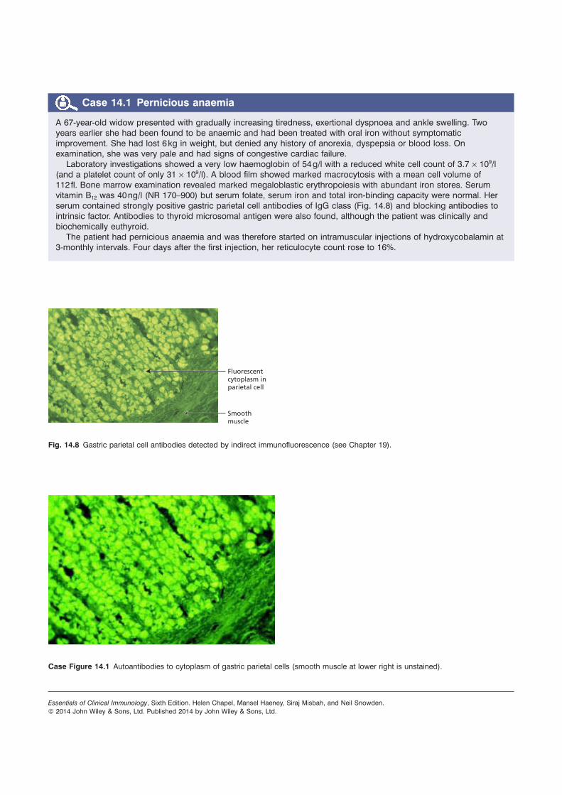
Laboratory investigations showed a very low haemoglobin of 54 g/l with a reduced white cell count of 3.7 × 10⁹/l (and a platelet count of only 31 × 109/l). A blood film showed marked macrocytosis with a mean cell volume of 112 fl. Bone marrow examination revealed marked megaloblastic erythropoiesis with abundant iron stores. Serum vitamin B12 was 40 ng/l (NR 170–900) but serum folate, serum iron and total iron-binding capacity were normal. Her serum contained strongly positive gastric parietal cell antibodies of IgG class (Fig. 14.8) and blocking antibodies to intrinsic factor. Antibodies to thyroid microsomal antigen were also found, although the patient was clinically and biochemically euthyroid.
The patient had pernicious anaemia and was therefore started on intramuscular injections of hydroxycobalamin at 3-monthly intervals. Four days after the first injection, her reticulocyte count rose to 16%.
Fig. 14.8 Gastric parietal cell antibodies detected by indirect immunofluorescence (see Chapter 19).
Fluorescent cytoplasm in parietal cell
Smoothmuscle
Case Figure 14.1 Autoantibodies to cytoplasm of gastric parietal cells (smooth muscle at lower right is unstained).

Case 14.2 Coeliac disease
A 35-year-old school cook presented to her dentist with a 30-month history of a sore mouth and tongue; she was treated with triamcinolone oral paste without improvement. Three months later she developed loose stools and generalized but vague abdominal pain. On questioning, she had felt tired for 2 years and had lost 8 kg in weight during the preceding 6 months despite a good appetite. During her second and third pregnancies she had developed moderate folic acid-deficiency anaemia. There was no family history of gastrointestinal disease and no abnormalities were found on examination.
Laboratory investigations showed a macrocytic anaemia but normal white cell, platelet and reticulocyte counts. The blood film showed many Howell–Jolly bodies (fragments of nuclear material within red blood cells) (Fig. 14.11), suggestive of hyposplenism. Bone marrow examination revealed active erythropoiesis with early megaloblastic features but no stainable iron. The appearances were those of a combined deficiency of iron and folate/vitamin B12; laboratory tests confirmed this (Table 14.5). In view of the blood film and the malabsorption of fat, coeliac disease was the most likely diagnosis. Her serum was positive for IgA antibodies to endomysium and to tissue transglutaminase, strongly supporting the clinical diagnosis. A jejunal biopsy was performed: this showed a convoluted pattern of stunted villi under the dissecting microscope, and subtotal villous atrophy with marked increase in intraepithelial lymphocytes and chronic inflammation in the lamina propria (Fig. 14.12).
The patient was started on a strict gluten-free diet with folic acid, iron and calcium supplements. One year later, she had put on 4.8 kg. A repeat jejunal biopsy showed improvement in villous architecture. This improvement following gluten withdrawal confirmed the diagnosis of coeliac disease and the patient will continue her gluten-free diet for life.
Fig. 14.11 Howell–Jowell bodies in a blood film.

Table 14.5 Laboratory investigation in Case 14.2
Investigations of anaemia
Investigations of malabsorption
Analyte Value NR Analyte Value NR
Hb 107 LOW 120–160 g/l Serum albumin 27 35–50 g/l
Mean cell volume 102 HIGH 80–90 fl Serum calcium 2.22 2.33–2.60 mmol/l
Serum iron 5.9 LOW 14–29 μmol/l Serum phosphate 0.74 0.80–1.45 mmol/l
Iron-binding capacity
85 HIGH 45–72 μmol/l Serum alk. phosphatase 70 20–85 IU/l
Serum folate 1.0 LOW 2–13 μg/l Serum IgG 8.2 7.2–19.0 g/l
Red cell folate 52 VERY LOW 165–600 μg/l Serum IgA 3.9 0.8–5.0 g/l
Serum vitamin B12 197 NORMAL 160–900 ng/l Faecal fat excretion 25 <17 mmol/day
Serum endomysial antibodies (IgA class)
Positive
Anti-tissue transglutaminase (IgA class)
Positive
Fig. 14.12 Typical histological features of coeliac disease shown in (a) low and (b) high power. Intraepithelial lymphocytes are arrowed.
Low power
High power
(a)
(b)
Case 14.3 Crohn’s disease
A 30-year-old woman was admitted with a 4-week history of increasing bloody diarrhoea and abdominal pain; she had lost 3 kg in weight. She smoked 25 cigarettes a day. On examination, she was not clinically anaemic but had a temperature of 37.8 °C. She was tender over the right iliac fossa, and had a number of hypertrophic tender anal skin tags which she thought were haemorrhoids. Sigmoidoscopy to 15 cm showed a red, granular mucosa with mucopus and contact bleeding. Laboratory investigations showed a low haemoglobin (108 g/l) with a raised C-reactive protein (CRP) (67 mg/l) but a normal white cell count. Urea and electrolytes, serum vitamin B12, folate, iron, ferritin and iron-binding capacity were normal. Her total serum proteins were 54 g/l (NR 62–82) with a serum albumin of 29 g/l (NR 35–50). Antibodies to neutrophil cytoplasmic antigens (ANCA) were not detected. Faecal examination and culture revealed no ova or Campylobacter. Clostridium difficile toxin was absent from the stools.
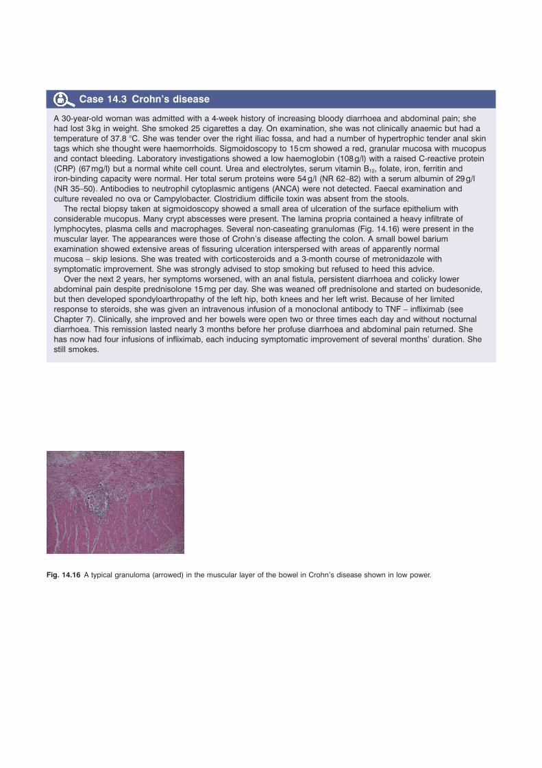
The rectal biopsy taken at sigmoidoscopy showed a small area of ulceration of the surface epithelium with considerable mucopus. Many crypt abscesses were present. The lamina propria contained a heavy infiltrate of lymphocytes, plasma cells and macrophages. Several non-caseating granulomas (Fig. 14.16) were present in the muscular layer. The appearances were those of Crohn’s disease affecting the colon. A small bowel barium examination showed extensive areas of fissuring ulceration interspersed with areas of apparently normal mucosa – skip lesions. She was treated with corticosteroids and a 3-month course of metronidazole with symptomatic improvement. She was strongly advised to stop smoking but refused to heed this advice.
Over the next 2 years, her symptoms worsened, with an anal fistula, persistent diarrhoea and colicky lower abdominal pain despite prednisolone 15 mg per day. She was weaned off prednisolone and started on budesonide, but then developed spondyloarthropathy of the left hip, both knees and her left wrist. Because of her limited response to steroids, she was given an intravenous infusion of a monoclonal antibody to TNF – infliximab (see Chapter 7). Clinically, she improved and her bowels were open two or three times each day and without nocturnal diarrhoea. This remission lasted nearly 3 months before her profuse diarrhoea and abdominal pain returned. She has now had four infusions of infliximab, each inducing symptomatic improvement of several months’ duration. She still smokes.
Fig. 14.16 A typical granuloma (arrowed) in the muscular layer of the bowel in Crohn’s disease shown in low power.

Case 14.4 Hepatitis A
An 18-year-old man presented with a 10-day history of anorexia, nausea and upper abdominal discomfort. Two weeks earlier, he had experienced some mild arthralgia in his fingers which lasted for 2 days. He normally smoked 20 cigarettes and drank two to three pints of beer each day, but had done neither for several days. He had noticed that his urine was much darker than normal. There was no significant medical history. On examination, he was afebrile but jaundiced. There were no needle tracks on his arms. His liver was just palpable and tender.
Hepatitis was diagnosed and confirmed by routine investigations. His serum bilirubin was 48 μmol/l (NR 1–20) with raised liver enzyme levels [aspartate transaminase 895 IU/l (NR 5–45); alanine transaminase 760 IU/l (NR 5–30)], and an alkaline phosphatase of 128 IU/l (NR 20–85). A monospot test for infectious mononucleosis was negative. Hepatitis B surface antigen (HBsAg) was also negative but he had detectable IgM antibodies to hepatitis A virus. He was managed conservatively at home. There is no active treatment for hepatitis A infection, although rest may be beneficial. The clinical and biochemical evidence of hepatocellular damage subsided over the next 4 weeks but he continued to feel vaguely unwell for several months. A further blood sample after 6 months showed IgG antibody to hepatitis A.
Case 14.5 Hepatitis C-induced liver disease
A 29-year-old man was noted to have abnormal liver function tests on routine ‘well-man’ medical screening in the private health sector. Apart from an episode of cellulitis and septic arthritis aged 24 years, he had been fit and well. He had no symptoms of liver disease and his GP notes showed no record of jaundice. Liver function testing, subsequently repeated twice, showed a mild chronic hepatitis with serum alanine transaminase levels of 140–210 IU/l (NR 0–50), and normal serum levels of alkaline phosphatase, bilirubin and albumin. He had no detectable antibodies to smooth muscle or mitochondria. Virus serology showed no evidence of exposure to hepatitis A or B. However, he was positive for hepatitis C antibody but negative for hepatitis C virus RNA by polymerase chain reaction (PCR). He underwent a liver biopsy, which showed normal liver architecture. There was mild non-specific portal tract inflammation but no piecemeal necrosis or fibrosis.
The source of his chronic hepatitis C liver disease was uncertain. He denied any use of intravenous drugs and had received no transfusions of blood or blood products. However, he was adorned with tattoos on his limbs and trunk. Tattooing carries a risk of transmitting hepatitis C of up to 3% in countries in which HCV is not endemic. It has been suggested that up to 40% of current carriers were infected by tattooing. The risk is increased by multiple tattooing, work by an amateur tattooist, and if the tattooist is hepatitis C positive.
He was told that his chronic hepatitis was likely to progress only slowly and over decades provided he abstained from alcohol, but that his liver biopsy would need to be repeated every 2 years or so, as an indicator of the potential need for antiviral therapy.

Case 14.6 Chronic active hepatitis
A 43-year-old woman presented with a 5-month history of weight loss (6 kg), anorexia, irritability and generalized pruritus. On examination, she was icteric with numerous spider naevi, scratch marks, palmar erythema and hepatosplenomegaly. Investigations showed a low haemoglobin (95 g/l) with a normal white cell count but an erythrocyte sedimentation rate of 140 mm/h. The prothrombin time was prolonged but urea and electrolytes, calcium and phosphate concentrations were normal. Although the serum albumin was normal (41 g/l), the total serum proteins were raised at 93 g/l (NR 62–82) with a raised serum bilirubin of 34 μmol/l (NR 1–20), alanine transaminase of 152 IU/l (NR 5–30), and aspartate transaminase of 164 IU/l (NR 5–45). The alkaline phosphatase level was normal (83 IU/l). Her serum immunoglobulins showed an increased IgG level of 44 g/l (NR 7.2–19.0) with normal IgA and IgM levels. No paraprotein was present on serum electrophoresis.
Antinuclear antibodies (IgG class) were strongly positive to a titre of 1/10 000 and antibodies to dsDNA were positive (60% binding; normal < 30%). Her serum was positive for antibodies to smooth muscle (Fig. 14.24) to a titre of over 1/1000 (see Chapter 19). HBsAg and hepatitis C antibody were absent and alpha-fetoprotein (AFP) was not detected. The immunological picture strongly favoured a diagnosis of autoimmune hepatitis. She was therefore started on prednisolone (30 mg/day) and vitamin K, with dramatic improvement. Her serum bilirubin, transaminases and prothrombin time returned to normal over the next fortnight. A diagnostic liver biopsy was performed: this showed chronic active hepatitis with cirrhosis. She was continued on prednisolone (15 mg/day) and is fully reassessed every 6 months, including a repeat liver biopsy as appropriate.
Fig. 14.24 Smooth muscle antibodies staining actin in a blood vessel (Indirect immunofluoresence using patient serum and rat liver sections – see Chapter 19).
Case 14.7 Primary biliary cirrhosis
A 62-year-old woman presented with a 6-week history of generalized itching and progressive shortness of breath. She also had a dragging feeling in the right upper quadrant of her abdomen. There was no history of weight loss, anorexia or jaundice. She smoked 25 cigarettes a day. On examination, she had many scratch marks but no xanthomas, xanthelasmas or jaundice. A large right-sided pleural effusion was present, with smooth, firm moderate enlargement of the liver. She was thought to have a bronchial carcinoma with hepatic secondaries.
Investigations showed a haemoglobin of 131 g/l, a normal white cell count and an erythrocyte sedimentation rate of 93 mm/h. Prothrombin time, urea and electrolytes, calcium, phosphate, total proteins, serum albumin and serum bilirubin were normal. However, the alkaline phosphatase was 1050 IU/l (NR 20–85), aspartate transaminase 166 IU/l (NR 5–45), and alanine transaminase 121 IU/l (NR 5–30). HBsAg and hepatitis C antibody were not detected. A chest X-ray confirmed the right pleural effusion but showed no evidence of malignancy or tuberculosis. The pleural effusion was aspirated three times; on each occasion, malignant cells were absent, culture was non-contributory, the fluid had the characteristics of a transudate and pleural biopsies were normal.
During her stay in hospital, however, the patient became obviously jaundiced, with a rise in serum bilirubin from 8 to 32 μmol/l. She also developed ascites and a palpable spleen. In view of her progressive obstructive jaundice she underwent a laparotomy; no surgically correctable cause could be found but a liver biopsy was taken. This showed the typical changes of primary biliary cirrhosis. Immunological tests were first performed at this stage – rather late! Antimitochondrial antibodies were present (Fig. 14.25) to a titre of 1/10 000. Serum immunoglobulins showed a polyclonal rise in IgM to 6.20 g/l (NR 0.5–2.0) with normal IgG and IgA levels. She was given cholestyramine to control her itching and ursodeoxycholic acid therapy. In the 4 years since diagnosis she has been reasonably well.
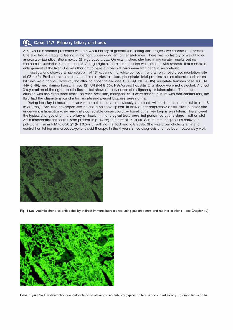
Fig. 14.25 Antimitochondrial antibodies by indirect immunofluorescence using patient serum and rat liver sections – see Chapter 19).
Case Figure 14.7 Antimitochondrial autoantibodies staining renal tubules (typical pattern is seen in rat kidney – glomerulus is dark).