Case 107: 43-Year-Old Lady with Severe Upper Abdominal Pain.
13
Case 107: 43-Year-Old Lady with Severe Upper Abdominal Pain
-
Upload
osborne-harrell -
Category
Documents
-
view
214 -
download
0
Transcript of Case 107: 43-Year-Old Lady with Severe Upper Abdominal Pain.

Case 107: 43-Year-Old Lady with Severe Upper Abdominal
Pain

Ultrasound of Gall Bladder Region

Laproscopic view of the gallbladder underneath the liver(air has been pumped in)
The surgeon aspirates the bile from the gall bladder

The vessels and cystic duct are identified and exposed
the cystic duct is cut after stapling both sides

The artery & vein are isolated and cut (make sure you don’t cut the hepatic artery or tear the portal vein!)
the gall bladder is dissected from the liver bed

Gallbladder with marked dilatation and dull serosal surface. Note the injected blood vessels

Gallbladder opened showing numerous multifaceted calculi
and markedly thickened wall.

Low power photomicrograph showing ulcerated mucosal surface

Medium power view showing the numerous inflammatory cells, the identity of which cannot be discerned at this power.

Medium power microscopic section of the muscularis of the gallbladder showing infiltration by numerous
inflammatory cells.

Another area showing ulcer with fibrinous exudate and debris at the base of the ulcer.
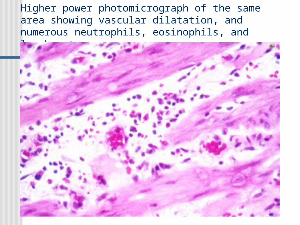
Higher power photomicrograph of the same area showing vascular dilatation, and numerous neutrophils, eosinophils, and lymphocytes
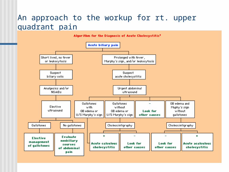
An approach to the workup for rt. upper quadrant pain