Cartilage - Weeblyjumed16.weebly.com/.../88514776/cartilage_lec_2017.pdf · Cartilage Dr. Heba...
38
1 Cartilage Dr. Heba Kalbouneh Assistant Professor of Anatomy and Histology
Transcript of Cartilage - Weeblyjumed16.weebly.com/.../88514776/cartilage_lec_2017.pdf · Cartilage Dr. Heba...

1
Cartilage
Dr. Heba KalbounehAssistant Professor of Anatomy and Histology

Cartilage is a specialized type of connective •
tissue designed to give support, bear weight and withstand tension, torsion and bending
Avascular•
Low metabolic rate•

FUNCTION OF CARTILAGE
1. Firm consistency of the extracellular matrix allows the tissue to bear mechanical stresses without permanent distortion.
2. Support soft tissues.
3. Cartilage is a shock-absorbing and sliding area for joints and facilitates bone movements.
4. Cartilage is also essential for the development and growth of long bones both before and after birth.

Components of Cartilage
Perichondrium
Outer fibrous
Inner cellular
Cells
Chondroblasts
Chondrocytes
Fibers
Collagen
Elastic
Ground Substance
Proteoglycans
Glycosaminoglycans
glycoproteins

Cells of cartilage:
Chondroblasts: typical protein synthesizing cells.
Chondrocytes: situated in lacuna. Usually seen in isogenous groups.
Lacuna= space occupied by chondrocyte.
Isogenous group= cells originating from the
mitotic activity of one chondrocyte.

Chondrogenesis

A. Mesenchyme is
the precursor for all
types of cartilage .
B. Mitosis and early
differentiation
produces a tissue
with condensations
of rounded cells
called chondroblasts.

C. Chondroblasts are then separated from one another again by their production of various matrix components, which collectively swell with water and form the very extensive ECM.

D. Multiplication of
chondroblasts within the
matrix gives rise to
isogenous cell aggregates
surrounded by a
condensation of territorial
matrix .
In mature cartilage, this
interstitial mitotic activity
ceases and all chondrocytes
typically become more
widely separated by their
production of matrix.

As the amount of matrix increases the chondroblasts become separated
from each other and are, from this time on, located isolated in small cavities
within the matrix, the lacunae. Concurrently the cells differentiate into mature
cartilage cells, chondrocytes
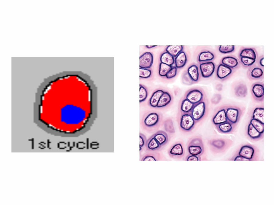

The matrix near the isogenousgroups of chondrocytes contains larger amounts of GAGs than the matrix further away from the isogenous groups. This part of the matrix is termed territorial matrix . In H&E stained sections the territorial matrix is more basophilic, i.e. it stains darker. The remainder of the matrix is called the interterritorial matrix
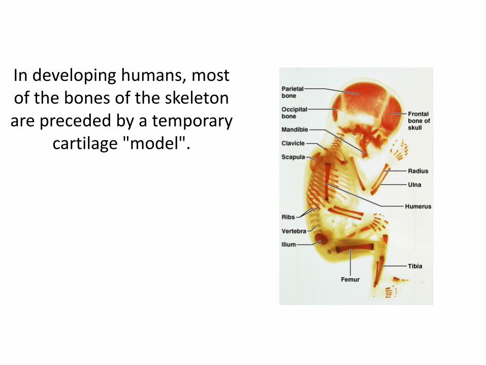
In developing humans, most of the bones of the skeleton are preceded by a temporary
cartilage "model".

Perichondrium: present in all types of cartilage except fibrous and articularcartilages
Outer fibrous: dense irrregular connective tissue, fibroblasts and type I collagen fibers
Inner cellular: contains undifferentiated cells (chondrogenic), essential for growth
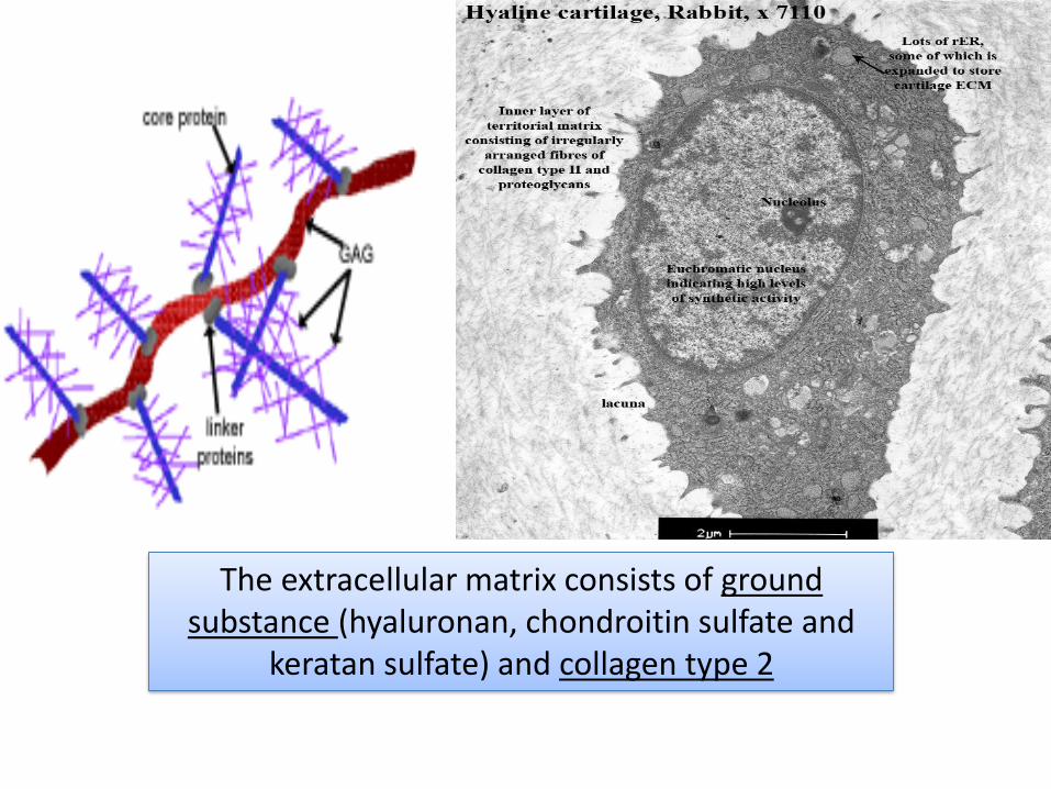
The extracellular matrix consists of ground substance (hyaluronan, chondroitin sulfate and
keratan sulfate) and collagen type 2

Types of cartilage
Hyaline Elastic Fibrous
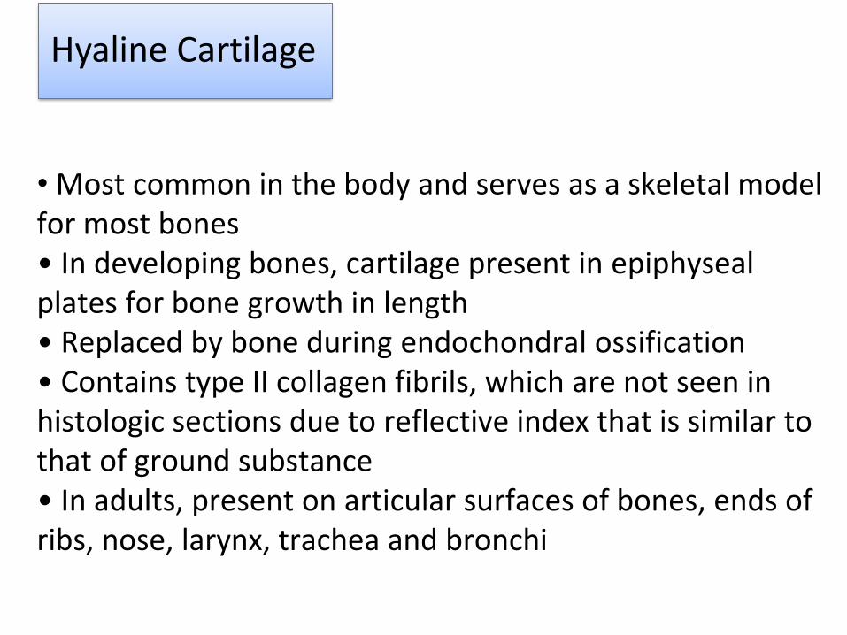
Hyaline Cartilage
• Most common in the body and serves as a skeletal model for most bones• In developing bones, cartilage present in epiphysealplates for bone growth in length• Replaced by bone during endochondral ossification• Contains type II collagen fibrils, which are not seen inhistologic sections due to reflective index that is similar tothat of ground substance• In adults, present on articular surfaces of bones, ends of ribs, nose, larynx, trachea and bronchi

Distribution of hyaline cartilage
Epiphysealgrowth plate
Costal cartilage
Thyroid cartilage
Fetal skeleton
Nose
Trachea and bronchi


Hyaline Cartilage

Hyaline Cartilage
22

ELASTIC CARTILAGE
Similar to hyaline cartilage but has elastic fibers running in all directions in addition to collagen.
Found in auricle of ear, walls of external auditory canals, eustachian tubes, epiglottis
Maintains shape, deforms but returns to shape; flexibility of organ; strengths and supports structures.
In contrast to hyaline cartilage, which can calcify with aging, the matrix of elastic cartilage does not calcify, and the cartilage maintains its high flexibility
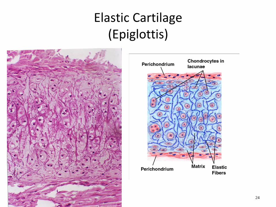
Elastic Cartilage(Epiglottis)
24

Distribution of elastic cartilage
Ear pinna
External auditory tube
Eustachian tube
Epiglottis


Fibrocartilage
Is a form of connective tissue transitional between dense connective tissue and hyaline cartilage.
Chondrocytes may lie singly or in pairs, but most often they form short rows between dense bundles of collagen fibers.
Collagen type I is dominant in fibrous cartilage.
Is typically found in knee joint (menisci), intervertebral disks, symphysis pubis. Is found at insertion of tendons into bones
It is difficult to define the perichondrium because of the fibrous appearance of the cartilage and the gradual transition to surrounding tissue types.

Fibrocartilage
29


Growth of Cartilage
Interstitial growth
Appositional growth
Growth from outsideGrowth from within
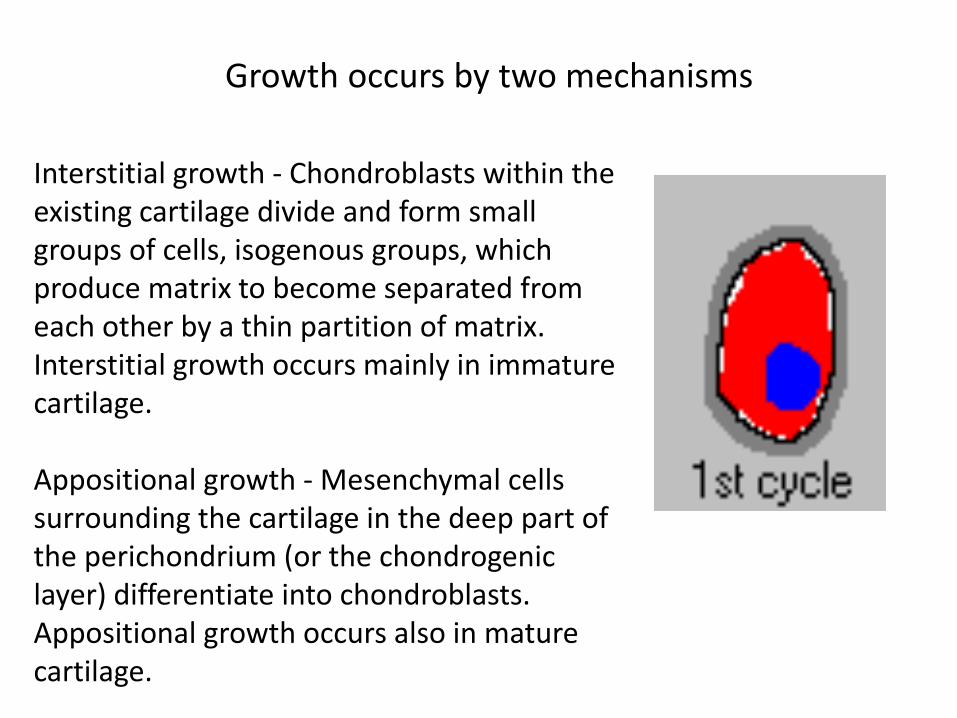
Interstitial growth - Chondroblasts within the existing cartilage divide and form small groups of cells, isogenous groups, which produce matrix to become separated from each other by a thin partition of matrix. Interstitial growth occurs mainly in immature cartilage.
Appositional growth - Mesenchymal cells surrounding the cartilage in the deep part of the perichondrium (or the chondrogeniclayer) differentiate into chondroblasts. Appositional growth occurs also in mature cartilage.
Growth occurs by two mechanisms

Growth in the Epiphyseal Plate
34

Clinical Problems
• Degenerative changes
• Herniation of the intervertebral disc


Herniated Disc/ ruptured disc/ slipped disc

Elastic cartilage and gravity are the reason why it seems our ears keep growing


![Cartilage - facultymembers.sbu.ac.irfacultymembers.sbu.ac.ir/rajabi/ppt toPDF/Cartilage [Compatibility Mode].pdfFibrocartilage • Fibrous Cartilage • is a form of connective tissue](https://static.fdocuments.net/doc/165x107/6012989a4318862a0e5813ae/cartilage-topdfcartilage-compatibility-modepdf-fibrocartilage-a-fibrous.jpg)