Carrara S et al. Endoscopic ultrasound-guided application ...€¦ · a gastric fold. VIDEO...
43
Jurgensen C et a. EUS-guided alcohol ablation of an insulinoma. Gastrointest Endosc. 2006;63:1059-62. Carrara S et al. Endoscopic ultrasound-guided application of a new hybrid cryotherm probe in porcine pancreas: a preliminary study. Endoscopy 2008;40:321-6.
Transcript of Carrara S et al. Endoscopic ultrasound-guided application ...€¦ · a gastric fold. VIDEO...

Jurgensen C et a. EUS-guided alcohol ablation of aninsulinoma. Gastrointest Endosc. 2006;63:1059-62.
Carrara S et al. Endoscopic ultrasound-guided applicationof a new hybrid cryotherm probe in porcine pancreas: apreliminary study. Endoscopy 2008;40:321-6.

Diagnostic imaging of pancreatic NETs:”take home message” 1
• To date the predominants imaging modalities forpancreatic endocrine tumors are CE spiral CT, EUS and SRS(Octreoscan). They provide the most cost-effective andaccurate means for detecting/diagnosing and staging mostcases of pancreatic NETs•in case of a suspected pancreatic lesion CE MDR-CT remains thefirst staging method of choice. It has replaced DSA and hasachieved similar or only slightly lower accuracy than EUS indetecting pancreatic tumors
• EUS had the highest accuracy in assessing tumor size and lymph nodeinvolvement and remains the first choice in diagnosis of small tumors
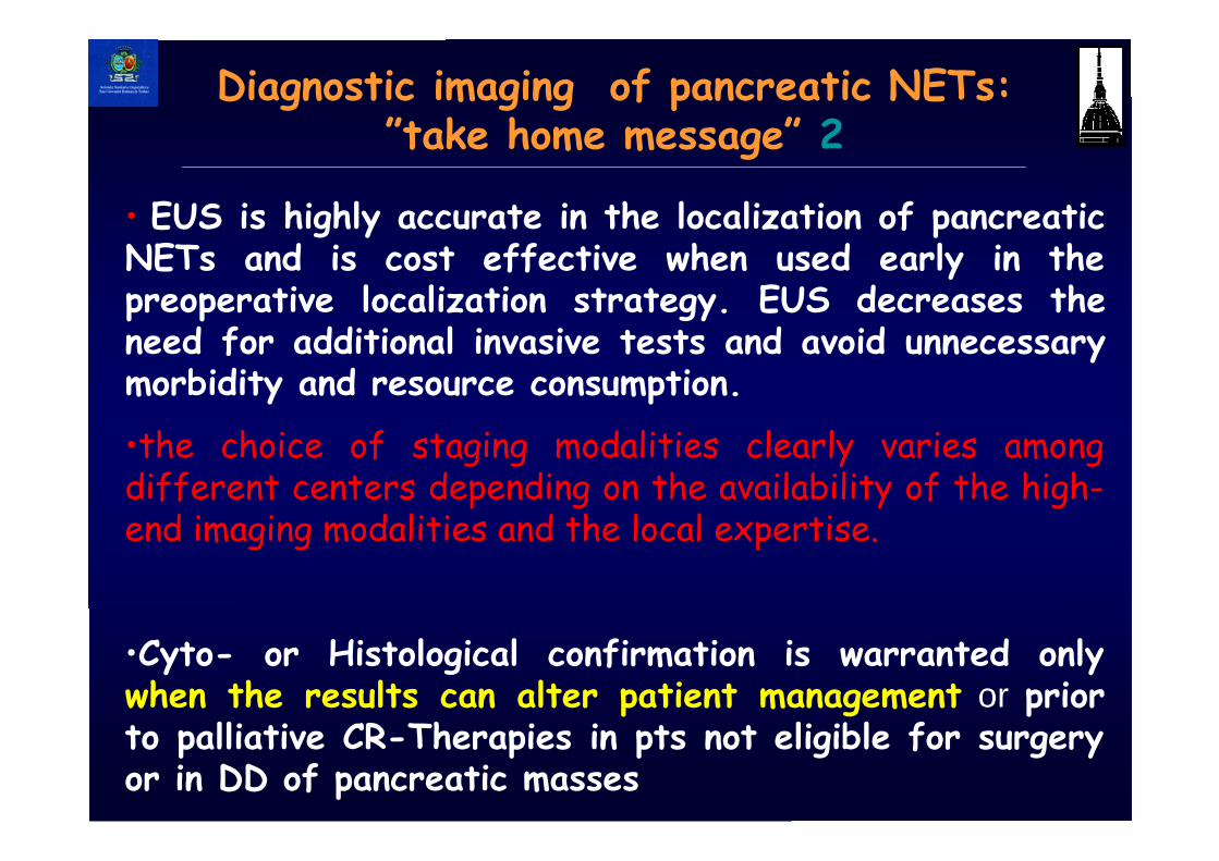
Diagnostic imaging of pancreatic NETs:”take home message” 2
• EUS is highly accurate in the localization of pancreaticNETs and is cost effective when used early in thepreoperative localization strategy. EUS decreases theneed for additional invasive tests and avoid unnecessarymorbidity and resource consumption.
•the choice of staging modalities clearly varies amongdifferent centers depending on the availability of the high-end imaging modalities and the local expertise.
•Cyto- or Histological confirmation is warranted onlywhen the results can alter patient management or priorto palliative CR-Therapies in pts not eligible for surgeryor in DD of pancreatic masses

ENDOSCOPY AND EUSFOR THE DIAGNOSTIC MANAGEMENT OF GI
WALL NETs (so-called CARCINOIDs)
• Accurate diagnosis, localization and pre-operativestaging are MANDATORY in order to offer thepatient the best treatment (the best cost-benefitratio in the single case)

ENDOSCOPY
Allow us to detect and diagnose,by means of targeted endoscopicbiopsies, neuro-endocrineneoplasias localized in themucosa and submucosa of all theGI sites that an endoscope canreach
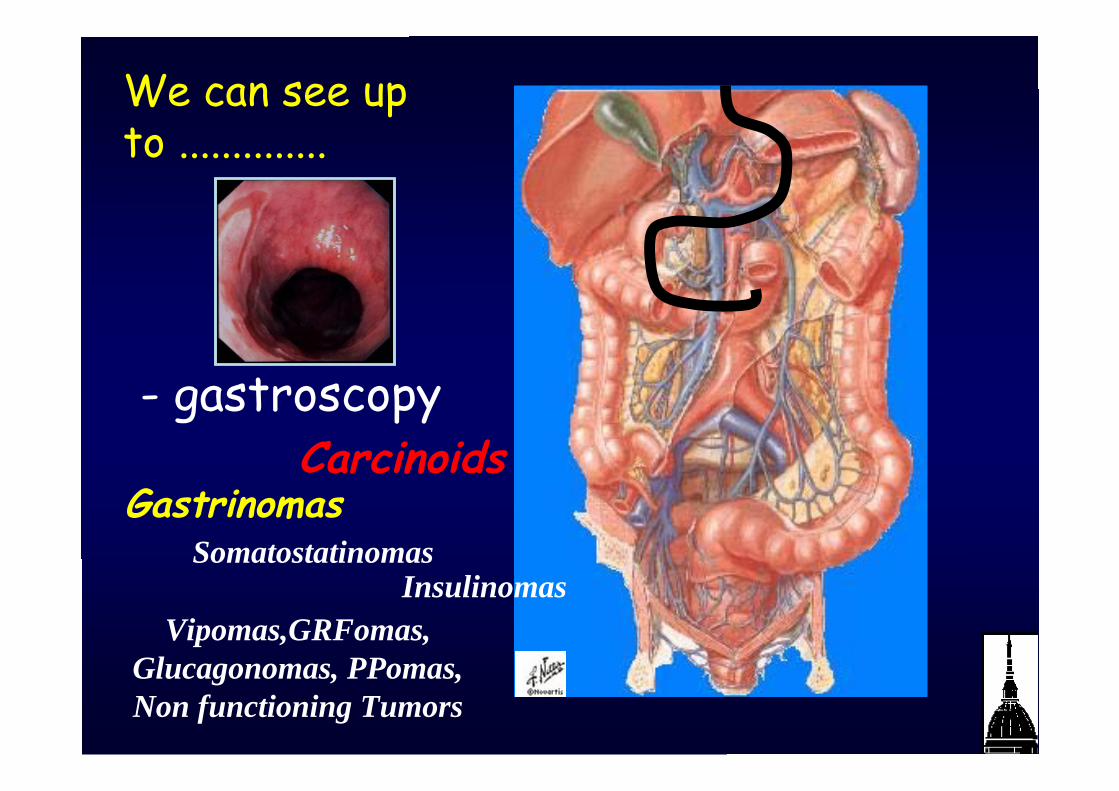
We can see upto ..............
- gastroscopyCarcinoids
Gastrinomas
InsulinomasSomatostatinomas
Vipomas,GRFomas,Glucagonomas, PPomas,Non functioning Tumors

We can seeup to............
- colonscopyCarcinoids
Somatostatinomas
Enteroglucagonomas
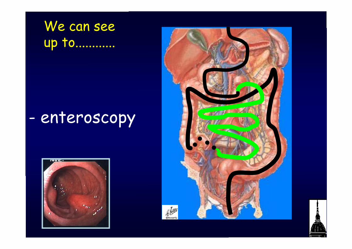
- enteroscopy
We can seeup to............

“PUSH-TYPE”ENTEROSCOPY
INTRA-OPERATIVE OR LAPAROSCOPICALLY-ASSISTED ENTEROSCOPY

VIDEO CAPSULE

Mouth to Cecum (or..to anum)
Teeth Epiglottis
Small IntestineIleocecal valve
Wall of right colon
Multiple telangiectasia ona gastric fold

VIDEO CAPSULE: LIMITs
1. Recorded images: impossible to wash the field, comeback on a doubtful point, change vision angle etc.
2. Movements sometimes too fast: you can miss also biglesions
3. No operative capabilities both diagnostic andtherapeutic
4. Contraindicated in case of stenosis

VIDEO CAPSULE: LIMITs
Unsuitable in case of stenoses, extensive adhesions, history ofsmall bowel resection
or, as in the case of carcinoid tumors with retractedmesentery, should be sed with caution or preceded
by the “patency capsule”
(an ingestible and dissolvable, a disintegration time-controlled capsule with an external scanner)

DOUBLE-BALLOON ENTEROSCOPY Today the double-balloon enteroscopy and more recently the single
balloon, allow us, with time-consuming and invasive examinations, mostlywith double approach (oral and anal), to endoscopically visualize, biopsy
and, in selected cases, to treat lesions all over the small intestine
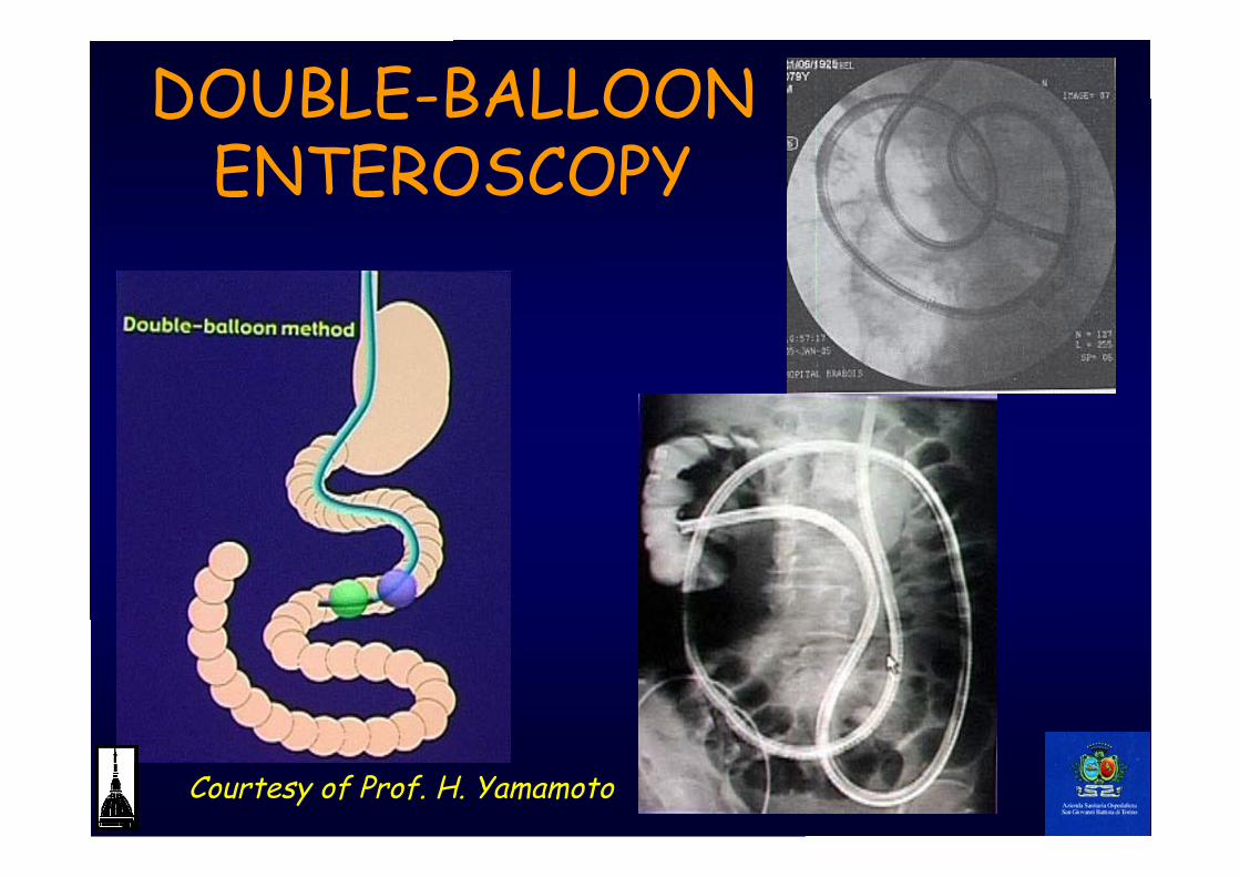
DOUBLE-BALLOONENTEROSCOPY
Courtesy of Prof. H. Yamamoto

DOUBLE-BALLOONENTEROSCOPY
Courtesy of Prof. H. Yamamoto

DOUBLE-BALLOON ENTEROSCOPY
Neuroendocrine tumors of the small bowelappear as subepithelial lesions, mostly
yellowish, that can be ulcerated when theyexceed 2 cm
Courtesy of Prof. H. Yamamoto

ENTEROSCOPY (wireless andballoon endoscopy)
Preliminary studies seem to demonstrate some potentialin detection and diagnosis of small bowel carcinoids.
2 papers compared video capsule endoscopy vsCT/enteroclysis:
1. One fails to demonstrate better results with video capsule.Johanssen S et al. The yield of wireless capsule endoscopy in the detection of
neuroendocrine tumors in comparison with CT enteroclysis. Gastrointest Endosc. 2006; 63(4):660-5.
2. Video capsule detected 9 small bowel NETs that were notvisualized after CT and enteroclysis.
van Tuyl SA et al. Detection of small-bowel neuroendocrine tumors by video capsuleendoscopy. Gastrointest Endosc. 2006; 64(1):66-72

ENTEROSCOPY (wireless and balloon endoscopy)
Preliminary studies seem to demonstrate some potential indetection and diagnosis of small bowel carcinoids.
1. Double or single balloon enteroscopy should be useful indetecting and biopsy tiny NEts of the small intestine, morefrequently in the ileum
2. but so far only few literature data **Yamaguchi Tet al. Multiple carcinoid tumors of the ileum preoperatively diagnosed
by enteroscopy with the double-balloon technique.Gastrointest Endosc. 2005;62(2):315
*Scherübl H et al. Double-balloon enteroscopy for the detection of midgutcarcinoids. Gastrointest Endosc. 2005;62(6):994.
Hystological confirmation of a diagnosis suspected with a video capsuleand/or curative endoscopic resection when facing small superficial
lesions, limited to mucosa and submucosa, as in other GI sites
Yamagishi H et al. Endoscopy. 2007;39 Suppl 1:E243-4.

ENDOSONOGRAPHY (EUS)
• Can visualize the layersof the GI wall (5-9),allowing to identify alsotiny lesion (2-3 mm) andto accurately stage thedepth of wall invasionand/or the locoregionalnodal involvement
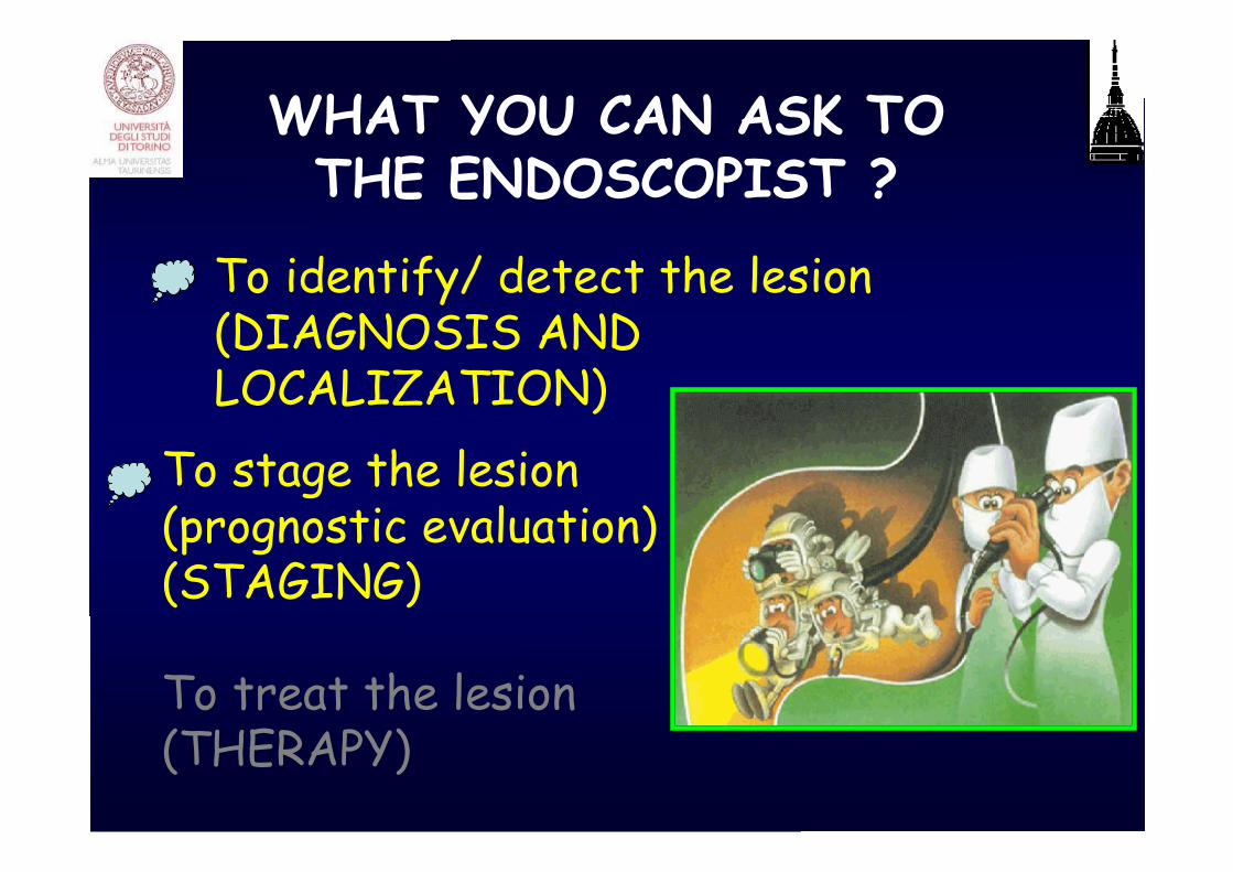
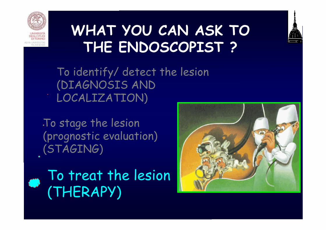
WHAT YOU CAN ASK TOTHE ENDOSCOPIST ?
To identify/ detect the lesion(DIAGNOSIS ANDLOCALIZATION)
To stage the lesion(prognostic evaluation)(STAGING)
To treat the lesion(THERAPY)

• Locoregional staging of lesions localized inthe wall of the esophagus, stomach,duodenum and colon-rectum, alreadyidentified and diagnosed by means ofendoscopic biopsies
EUS IN THE ASSESSMENT OF GITRACT NETs
•Localization of submucosallesions endoscopicallyinvisible (i.e. duodenalgastrinomas), even of 2 mmin diameter
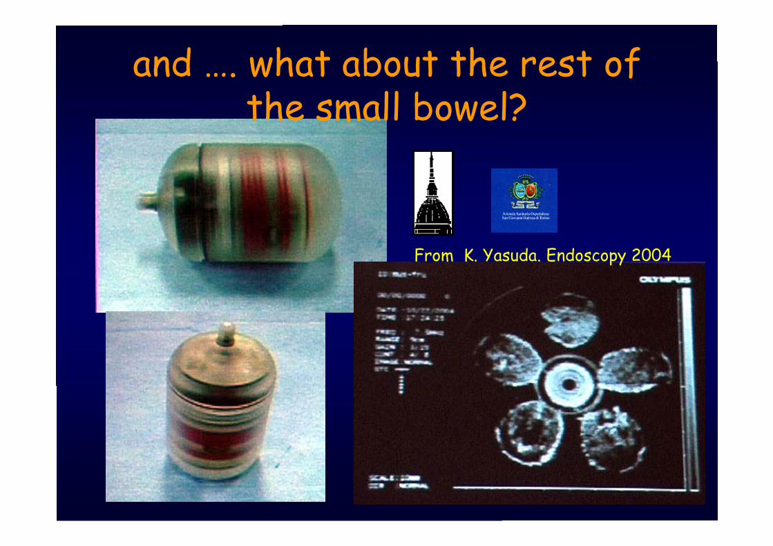
and …. what about the rest ofthe small bowel?
From K. Yasuda. Endoscopy 2004

and …. what about the rest ofthe small bowel?
From K. Yasuda. Endoscopy 2004
Today is also possible, with the new balloonendoscopes, to evaluate and stage, by meansof miniprobes, NETs of the small bowel, so
far beyond the grasp of EUS
Fukumoto A et al. Usefulness of EUS with double balloonenteroscopy for diagnosis of small bowel diseases.Gastrointest Endosc. 2007; 65: 412-420.
and …. what about the rest ofthe small bowel?

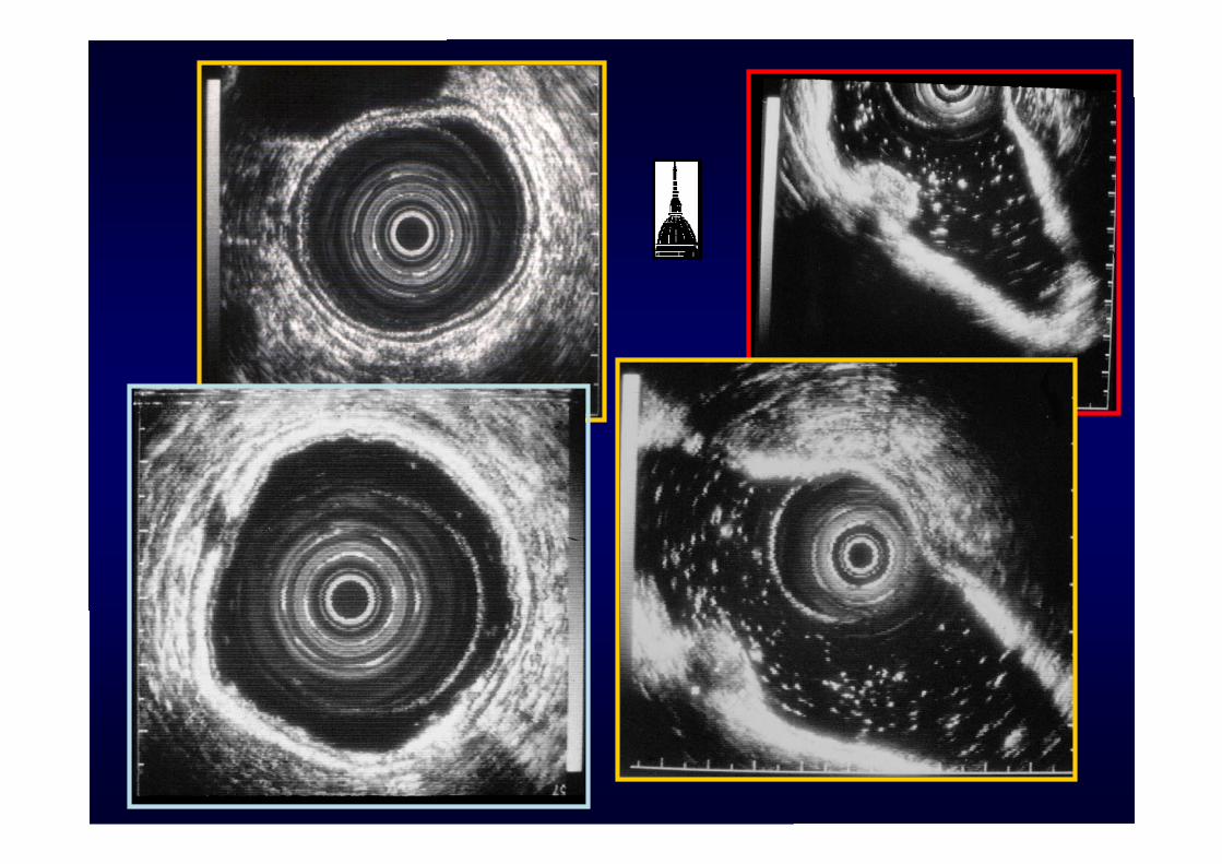
• Hypo-echoic, round or oval, well demarcatedlesions, mainly in the 2nd and 3rd wall layer,sometimes with transmural invasion of the GIwall
EUS IN THE ASSESSMENT OF GITRACT NETs

• Preoperative EUS is mandatory for evaluating tumorsize and depth of invasion; these features areconsidered to be important metastatic risk factorsand the main determinants of appropriate therapy(endoscopic excision, local excision or radicalresection)
CLINICAL IMPACT PROGNOSTIC VALUE
EUS IN THE ASSESSMENT OF GITRACT NETs
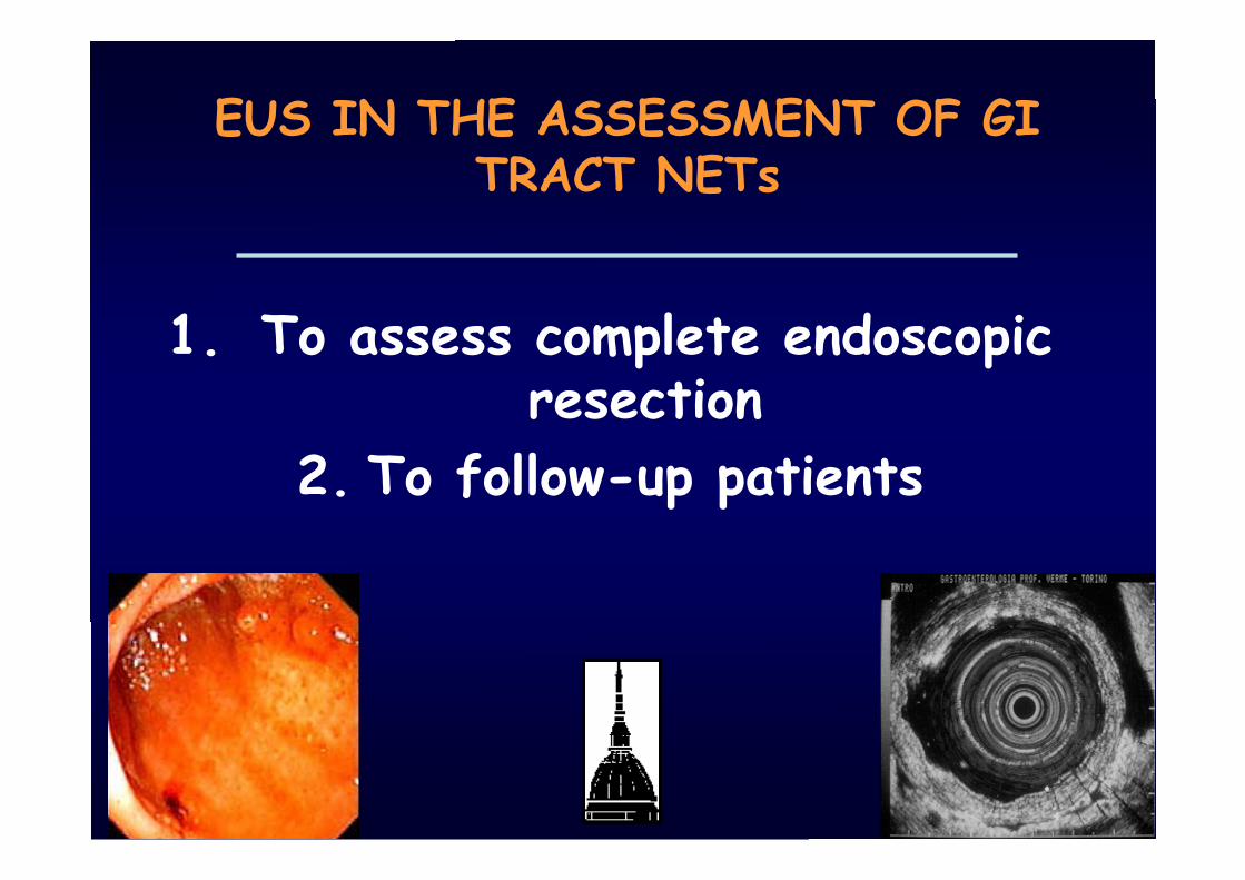
1. To assess complete endoscopicresection
2. To follow-up patients
EUS IN THE ASSESSMENT OF GITRACT NETs
WHAT YOU CAN ASK TOTHE ENDOSCOPIST ?
To identify/ detect the lesion(DIAGNOSIS ANDLOCALIZATION)
To stage the lesion(prognostic evaluation)(STAGING)
To treat the lesion(THERAPY)

ENDO THERAPY = CURATIVE THERAPY
Mucosal and/or submucosal Tumors
< 1 cmEsophagusStomachDuodenumColon
< 1.5 cm Rectum
Without NODAL involvement
GI TRACT NETs (carcinoids): ROLEOF ENDOSCOPIC TECHNIQUES
DIAGNOSTICEUS
THERAPEUTICEUS
GEP NETs: ROLE OF ENDOSCOPICTECHNIQUES

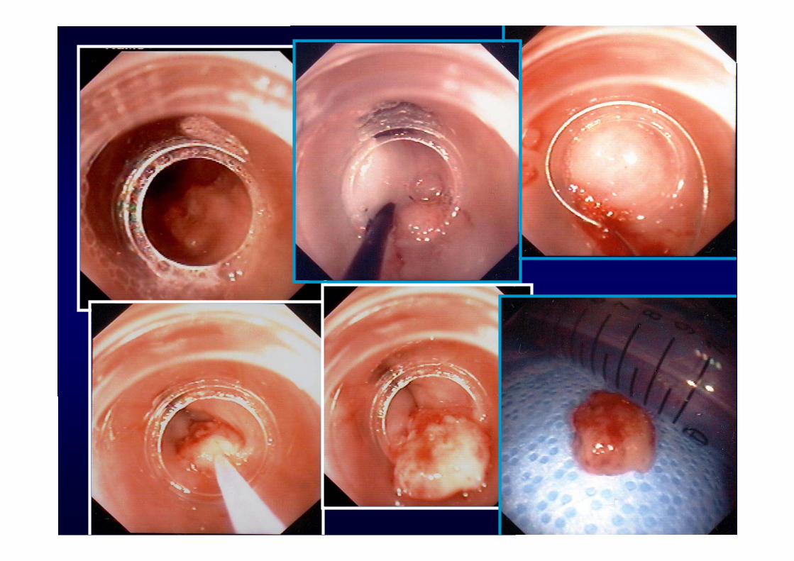
High-frequency probe EUS-assisted endoscopicmucosal resection: A therapeutic strategy forsubmucosal tumors of the GI tract (Waxman I et al. Gastrointest Endosc2002)
“Carcinoid of the GI tract can bemanaged safely, quickly, and easily
with HFPE-assisted EMR”.

“Use of EUS-guided Injectionin endoscopic resection ofsolid submucosal tumors”
Sun S et al. Endoscopy 2002
16 lesions; 9 in the muscolaris propria: noperforations; two bleedings endoscopically treated
stomach
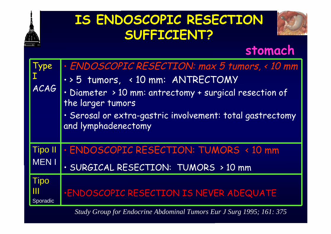
•ENDOSCOPIC RESECTION IS NEVER ADEQUATETipoIIISporadic
• ENDOSCOPIC RESECTION: TUMORS < 10 mm
• SURGICAL RESECTION: TUMORS > 10 mm
Tipo IIMEN I
• ENDOSCOPIC RESECTION: max 5 tumors, < 10 mm• > 5 tumors, < 10 mm: ANTRECTOMY• Diameter > 10 mm: antrectomy + surgical resection ofthe larger tumors• Serosal or extra-gastric involvement: total gastrectomyand lymphadenectomy
TypeIACAG
Study Group for Endocrine Abdominal Tumors Eur J Surg 1995; 161: 375
IS ENDOSCOPIC RESECTIONSUFFICIENT?
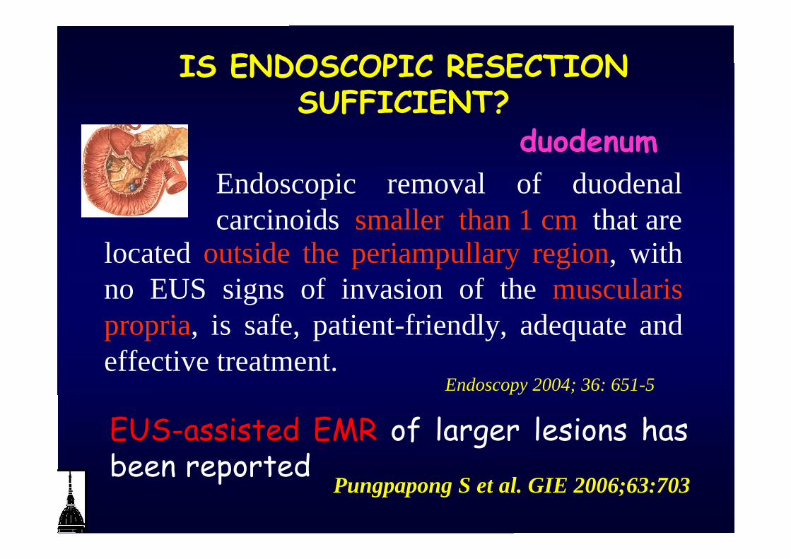
duodenumEndoscopic removal of duodenalcarcinoids smaller than 1 cm that are
located outside the periampullary region, withno EUS signs of invasion of the muscularispropria, is safe, patient-friendly, adequate andeffective treatment.
Endoscopy 2004; 36: 651-5
IS ENDOSCOPIC RESECTIONSUFFICIENT?
EUS-assisted EMR of larger lesions hasbeen reported
Pungpapong S et al. GIE 2006;63:703
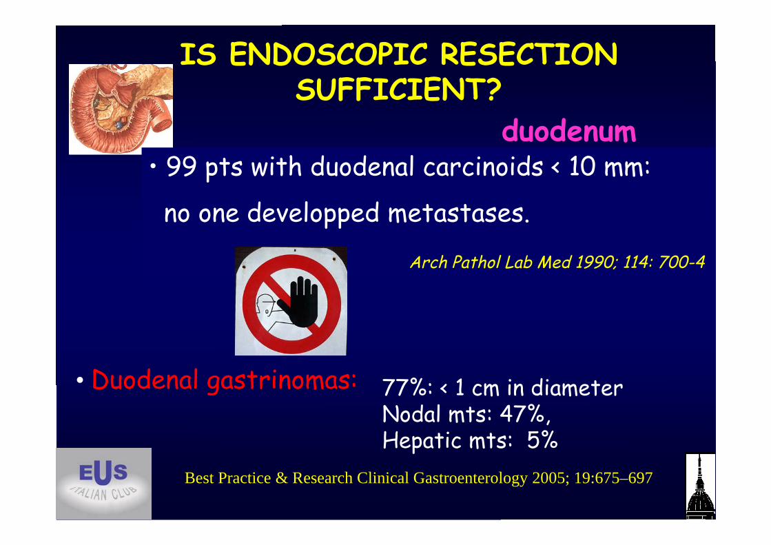
duodenum• 99 pts with duodenal carcinoids < 10 mm:
no one developped metastases.
Arch Pathol Lab Med 1990; 114: 700-4
• Duodenal gastrinomas: 77%: < 1 cm in diameterNodal mts: 47%,Hepatic mts: 5%
Best Practice & Research Clinical Gastroenterology 2005; 19:675–697
IS ENDOSCOPIC RESECTIONSUFFICIENT?
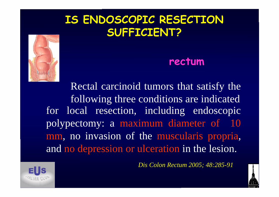
rectum
Rectal carcinoid tumors that satisfy thefollowing three conditions are indicated
for local resection, including endoscopicpolypectomy: a maximum diameter of 10mm, no invasion of the muscularis propria,and no depression or ulceration in the lesion.
Dis Colon Rectum 2005; 48:285-91
IS ENDOSCOPIC RESECTIONSUFFICIENT?

Endoscopyand EUS inGEP tumors LOC/STAGING
DIAGNOSI TISSUTALE(BIOPSY/EUS-FNA)
TERAPIAEMR/HF-MPs
EUS-FNIID-HIFU
Diagnosis
“The future treatment of patients with NEtumors will be tumor-biology based andbiotherapies will be tumor-targeted. With theadvent of the new analogs and drugs everypatients will get a “tailor-made” therapy”
adapted from: Oberg K. The Oncologist 1998;3:339
Requirements for a correcttherapeutic approach:

ASSESSMENT AND MANAGEMENT OFPATIENTS WITH SUSPECTED GEP NETs :
The ideal team (The dream team)The ideal team (The dream team)
ExpertPathologist
EUS/endoscopy
Expert
RadionuclearimagingExpert
ExpertRadiologist
Expert Clinician
Dedicatedsurgeon