Carp mucus and its role in mucosal defense - WUR
183
Carp mucus and its role in mucosal defense Maria C. van der Marel 2012 Carp mucus and its role in mucosal defense Maria C. van der Marel
Transcript of Carp mucus and its role in mucosal defense - WUR

Carp mucus and its role in mucosal defense
Maria C. van der Marel
20
12
C
arp m
ucu
s and
its role
in m
uco
sal defen
se
M
aria C. van
der M
arel

Carp mucus and its role in mucosal defense
Maria C. van der Marel

Thesis committee Thesis supervisors Prof. dr. ir. H.F.J. Savelkoul Professor of Cell Biology and Immunology Wageningen University Prof. dr. D. Steinhagen Professor of Fish Diseases University of Veterinary Medicine ,Hannover, Germany Thesis coどsupervisor Prof. dr. J.H.W.M. Rombout Associate professor, Cell Biology and Immunology Group Wageningen University Other members Prof. dr. ir. M.W.A. Verstegen, Wageningen University Prof. dr. V. Kiron, University of Nordland, Bodø, Norway Prof. dr. B. Schröder, University of Veterinary Medicine, Hannover, Germany Dr. ir. J.W. Schrama, Wageningen University This research was conducted under the auspices of the Graduate School of Wageningen Instiどtute of Animal Sciences (WIAS).
Carp mucus and its role in mucosal defense
Maria C. van der Marel
Thesis submitted in fulfilment of the requirements for the degree of doctor
at Wageningen University by the authority of the Rector Magnificus
Prof.dr. M.J. Kropff, in the presence of the
Thesis Committee appointed by the Academic Board to be defended in public
on Monday November 26, 2012 at 1.30 p.m. in the Aula.

Maria C. van der Marel Carp mucus and its role in mucosal defense PhD Thesis, Wageningen University, The Netherlands (2012) With references, with summaries in English, German and Dutch ISBN: 978ど94ど6173ど427ど3

Contents
Chapter 1 General introduction 7
Chapter 2 Intestinal mucus of common carp, Cyprinus carpio L.; biochemical and histochemical analysis 17
Chapter 3 Characterization of skin mucus layer in common carp, Cyprinus carpio L. 29
Chapter 4 Changes in skin mucus of common carp, Cyprinus carpio L., after exposure to water with a high bacterial load 41
Chapter 5 Molecular cloning and expression of two éどdefensin and two mucin genes in common carp (Cyprinus carpio L.) and their upどregulation after éどglucan feeding 53
Chapter 6 Differential soybean meal induced mucus composition in intestinal segments of common carp, Cyprinus carpio L. 65
Chapter 7 Chemotaxis towards, adhesion to, and growth in carp gut mucus of two Aeromonas hydrophila strains with different pathogenicity for common carp, Cyprinus carpio L. 81
Chapter 8 Intestinal mucus of challenged carp 93
Chapter 9 General discussion 105
References 117
Summaries and aknowledgements 135 Summary Samenvatting
Zusammenfassung Acknowledgements
About the author 145 Curriculum vitae
List of publications
Appendix I
Biochemical and histochemical study on the intestinal mucosa of the common carp Cyprinus carpio L., with special consideration of mucin glycoproteins
Appendix II Biochemical and histochemical effects of perorally applied endotoxin on intestinal mucin glycoproteins of the common carp Cyprinus carpio
Appendix III Changes of intestinal mucus glycoproteins after peroral application of Aeromonas hydrophila to common carp (Cyprinus carpio)

The slimy is docile. Only at the very moment when I believe that I possess it, behold by a curious reversal, it possesses me. Here appears its essential charどacter: its softness is leechどlike…
(Sartre)

Chapter 1
General introduction

General introduction
8
1 Body surfaces of multiどcellular organisms are defended by epithelia, which provide a physical barrier between the internal milieu and the pathogensどcontaining external world [1]. In fish, this barrier is formed by the integument and alimentary tract. Fish integument is a large organ that is continuous with the lining of all the body openings, and also covers the fins. In addition to its protective functions, fish skin may serve important roles in communication, sensory perception, locomotion, respiration, ion regulation, excretion, and thermal regulation [2]. The intestine of fish also performs many tasks [3]. The main function of the alimentary tract of any animal is the acquisition of food with subsequent assimilation of vital nutrients [4]. However, it also plays a role in osmoregulation and defense against pathogens [3]. The integumentary and intestinal barrier can be divided into the extrinsic, intrinsic, and immunological barriers. The extrinsic barrier is the first barrier pathogens and toxins encounter and is mainly formed by mucus which counters pathogens and toxins. The intrinsic barrier consists of epithelial cells and cell junctions. Immunological barriers are formed by the innate immune system and the adaptive immune system that are present in the intestinal wall. Active immune responses occurring in the skin are suggested by prevention of colonization at the skin by vaccination against bacteria (e.g. Vibrio sp.) and parasites (e.g. Ichthyiophthyrius multifilliis), but the mechanisms are still largely unknown. Immune cells, cytokines and antibodies were all described at the epithelial surface and were shown to respond to infections and vaccinations. An infectious pathogen can enter the fish through the skin, gills, or the gut [5]. Infections occur only when pathogens can colonize or cross the above mentioned barriers [1]. Most aspects of barrier function are actively controlled. Increase in mucus production and infiltration of immune cells occur when the epithelium senses pathogens or tissue damage. Disturbances to the general homeostasis of the fish, as with other physiological systems, can affect the intestinal barrier. The barrier function is disturbed by factors, such as suboptimal water temperature, hypoxia, low water quality, suboptimal diet, acute and chronic stress, exposure to pathogens, and starvation [5].
Mucus In mucus, the watery gel on the surface of the epithelium, immune functions usually prevent the entry of pathogens and toxins [1]. Mucus is a complex viscous adherent secretion that covers the surface of most epithelia and represents an interface between the environment and the interior milieu [6]. In vertebrates, mucus is a secreted viscous colloid produced by, and covering, mucous membranes, and is typically produced from mucous cells found in muどcous glands. The range of roles for fish mucus is very large and includes respiration, ionic and osmotic regulation, reproduction, excretion, disease resistance, communication, feeding, nest building and protection [6]. Mucus is secreted in higher vertebrates, fish and amphibians, and all vertebrates, including fish, have more or less the same composition. Even the slime trail of snails and earthworms has a chemical composition similar to that of mammalian mucus [7]. Mucus lines all organs that are exposed to the external environment including the respiratory tract, the gastrointestinal tract, the reproductive tract, the oculoどrhinoどotolaryngeal tracts and in fish and amphibians also the skin [8, 9]. In fish, mucus is considered to be predominantly found on the surface of skin and gills, but it also forms the gut lining being

General introduction
9
1merely produced by goblet cells [6]. Underneath the mucus layer, a dense forest of highly diverse glycoproteins and glycolipids are resent, the soどcalled glycocalyx [10]. Mucus is kept close to the surface of the intestine by the glycolayx, which consists of glycoprotein filaments extending from the interior of absorptive cells to the exterior forming a buffering network above the cell surface [11]. Mucus is primarily composed of water (approx. 95%) [8] and mucus glycoproteins (mucins and nonどmucins) but also comprises many other substances. Substances that have been identified in mucus and include such chemical compounds as cytokines [12], peptides [13, 14], lysozyme [15], lipoprotein [16], complement [17], lectins [18ど20], proteases [21] and antibodies [22ど24]. Some components have a defensive purpose [9] whereas others may act to modulate the organization
and properties of the gel [25]. In fish, the mucins contained in the intestinal goblet cells appear to be speciesどspecific. Some species secrete predominantly negatively charged mucins [e.g. Shi drum Umbrina cirrosa; 26] and some secrete predominantly neutral mucins [e.g. common dentex Dentex dentex; 27]. There also appears to be heterogeneity in the glycoproteins within the goblet cells [26].
Mucus in health and disease Mucus helps to protect epithelial surfaces in many different ways. It is a lubricant that protects against mechanical damage while enabling rapid removal of various types of aggression (such as pathogens or irritants) from the mucosal surfaces. It is a regulated nutrient medium in which whole ecosystems thrive, usually symbiotically [1]. Mucus allows exchange between the luminal content and the epithelial lining [9]. The major components of the mucosal barrier are expressed constitutively, as continuous mucus production and secretion are required to replace the mucus that is degraded by bacteria and, in the intestinal tract, which is lost with the movement of luminal contents [28]. Unlike the systemic immune system, the mucosal immune system continuously encounters, monitors and regulates a thriving array of microbes and toxins that are always present and continuously changing. The mucosal barrier itself is not a static barrier as its constituents and the released products are modulated by the microenvironment and by neural, endocrine and immune factors [28] (Fig. 1). In the mucosal barrier, the recognition of pathogens leads to the production of host inflammatory factors that, in turn, induce the differentiation of secretory cells and alterations in mucus composition through altered transcription of mucins and glycosyltransferases that interact with them. The recognition of pathogenどassociated molecular patterns (PAMPs) activates Tollどlike receptor (TLR) and NODどlike receptor (NLR) signaling pathways, which leads to alterations in the expression of mucin and antimicrobial genes (Fig.1) [28]. The continuous regulation of mucous secretions in general helps to keep microbes and toxins out of the system. The majority of bacteria, yeasts, protozoa and worms that live in the muどcous ecosystem of the human gut generally cause the host no trouble. However, many of them can switch from being commensals to pathogens if they become overgrown within this niche, especially if they penetrate the mucus blanket and adhere to the surfaces and/or peneどtrate the epithelial cells [1].

General introduction
10
1
Fig. 1 Regulation of the mucus barrier adopted from McGuckin et al. [28]. The recognition of pathogens leads to the production of host inflammatory factors that, in turn, induce the differentiation of secretory cells and alterations in mucus composition through altered transcription of mucins and the glycosyltransferases that act on them. The recognition of pathogenどassociated molecular patterns (PAMPs) activates Tollどlike receptor (TLR) and NODどlike receptor (NLR) signaling pathways, which leads to alterations in the expression of mucin and antimicrobial genes. IL, interleukin; TH, T helper cell. In addition to its protective functions, mucus can also be involved in disease processes. Many commensal bacteria possess specific adhesins that bind specifically to mucus [29] and some parasitic organisms produce their own layers of mucus to evade the immune system [30]. Furthermore, a hyperど or hyposecretion of mucus is related to some diseases. In humans, hypersecretion of mucus is seen in cystic fibrosis [31], bronchitis, asthma [32] and in middle ear infections, with mucus gels serving as the matrix in which pathogens, allergens, and in the case of cholersterol gallstones trap nuclei and promote growth [33]. Hypersecretion can be induced as a response to proinflammatory cytokines, ATP, bacterial exoproducts (e.g. from Pseudomonas aeruginosa), and host proteases [34ど38]. It is present in dry eye syndrome [39] and in some forms of ulcer disease. Additionally, mucus expression and composition is altered in cancers of epithelial origin [40].

General introduction
11
1Mucins General Mucus gels typically have mucin concentrations of 2 to 5%. In the gel, each mucin monomer is linked to other mucin monomers by nonどcovalent interactions and disulphide bonds [1]. Mucins are heavily glycosylated filamentous proteins which impart viscoelastic and rheological properties to mucosal layers. Mucins are built from a central core protein, the apoどmucin, to which carbohydrate side chains are attached (Fig. 2). Mucins have a high relative molecular mass (from several hundred to several thousand kDa) [9] and differ structurally and chemically from serum glycoproteins and proteoglycans of connective tissues [7]. Differences in glycosylation can be induced by altered assembly of carbohydrate sideどchains or altered expression of core proteins [41].
Fig. 2 Schematic view of a mucin molecule according to LaFitte et al. [42] The primary functions of mucins are protection and lubrication of epithelial surfaces [9, 43]. Mucins have a negatively charged protruding structure and act as a selective barrier protecting the cell. Besides protection and lubrication, mucins appear to be also involved in more complex biological processes such as epithelial cell renewal and differentiation, cell signaling, and cell adhesion [44]. The secretion of mucins, which are stored in the vesicles of the goblet cells, is continuous (basal secretion) or can occur as a reaction on a specific stimulus (stimulated secretion) [45]. Mucins are known to be qualitatively and/or quantitatively abnormal in a number of diseases such as inflammatory bowel disease, respiratory disease and cancer [cf. 46].

General introduction
12
1 Many mucins are specific to a particular mucosal surface forming an environmental niche that can support its own microどecosystem [1]. On the basis of their structural characteristics and physiological purposes, mucins have been categorized into two major types. One type is thought to be monomeric and primarily, but not exclusively, synthesized and located at the apial epithelial cell surface and is referred to as “membraneどbound” mucins. The secretory type consists of oligomeric mucins based on the cysteineどrich sequences located in the Nど and Cどterminal regions facilitating the formation of disulphide bridges and forming filamentous or networks of multimers that are apically secreted by goblet cells. These can be gelどforming or nonどgelどforming, and are thought to be responsible for the rheological properties of mucus [25]. The main difference between members of these two types is the presence of a hydrophobic Cどterminal transmembrane domain that serves to anchor membraneどbound mucins to the plasmalemma [44]. Cellどattached mucins range from 100 to 500 nm in length and, although much shorter than secreted mucins, are long enough to reach well beyond the glycocalyx where they help to make initial specific interactions between cells [1]. Membraneどanchored cellどsurface mucin glycoproteins are a major constituent of the glycocalyx in all mucosal tissues [10]. Secreted mucins are extremely long molecules reaching up to several microns in length, longer than the diameter of many types of cells [1] and have a molecular mass between 0.5 and 40 MDa [47, 48]. These huge molecules are formed by end to end linkage of numerous mucin monomers, each about 1 micrometer in length [49, 50]. Cell surface mucins are cleaved into two subunits in the endoplasmic reticulum and inserted into the membrane and Nどglycosylated. In the endoplasmic reticulum, secreted mucins are Nどglycosylated and dimerised via their Cどterminal domains. Both cell surface and secreted mucins are Oどglycosylated in the Golgi apparatus. Following completion of Oどglycosylation, the dimers of the secreted mucins undergo Nどterminal oligomerization and are packed into granules for secretion (Fig. 3) [28]. Protein core The apomucin is a linear polypeptide [51], which contains at least one protein domain [44]. Some mucin monomers are amongst the longest peptides known with a molecular mass exceeding 5 gigaどDaltons and some are even polymorphic. Mucin monomers are encoded by different mucin genes (MUC for human and Muc for other species) [9]. Some are expressed in all or almost all epithelia studied indicating their involvement in general epithelial functioning. Expression of other mucins is confined to specific tissues. In a one single epithelium, two mucins can be highly expressed [46]. Centrally positioned within the apomucins, specific tandem repeated motifs (“tandem repeats”) are present [44]. Tandem repeats are particularly rich in proline, threonine and serine and are also designated as “PTS domains” [9]. Proline is presumed to be necessary to achieve a conformation that will permit the very close packing of the carbohydrate chains [7]. The oligosaccharide chains are bound to the hydroxyl side chains of serine and threonine and arranged in a “bottle brush” configuration around the protein core (Fig. 2). Many of the smaller membrane bound mucins are not considered true mucins since they only share the PTS repeats and glycosylation with other mucins [8]. In addition to the “bottle brush” regions of Oどlinked oligosaccharides (one to several hundred carbohydrate chains), there are Nどlinked sulphateどbearing oligosaccharides located near both ends of mucin monomers comprising 2

General introduction
13
1to 3 by weight % [1] (Fig. 2). Additional postどtranslational modifications, including sialylation or sulphation, are commonly observed on mature mucin glycoproteins [44]. The “bottle brush” regions are separated by “naked” protein regions which are cysteineどrich and are the most hydrophobic regions of mucins (Fig 2.). These naked proteins are sufficient hydrophobic that they may fold into globular beads which are stabilized by disulfide bonds [1] leading to enormous complexes [9]. About 90% of sulfhydryl groups in secreted mucins are involved in SどS bonds [1]. The “bottle brush” sections are resistant to proteolysis because they are protected by the carbohydrate sheath however, the naked regions can be hydrolyzed by a variety of proteolytic enzymes [7].
Fig 3 Mucin biosynthesis according to McGuckin et al. [28]. Cell surface mucins ( ) are cleaved into two subunits in the endoplasmic reticulum, inserted into the membrane and Nどglycosylated ( ). In the endoplasmic reticulum, secreted mucins ( ) are Nどglycosylated and dimerize via their Cどterminal domains. Both cell surface and secreted mucins are Oどglycosylated in the Golgi. Following completion of Oどglycosylation, the dimers of the secreted mucins undergo Nどterminal oligomerization ( ) and are packed into granules ( ) for secretion. Carbohydrate side chains The carbohydrate side chains constitute up to 80% of a mucins total mass [9, 25] and give an elongated and rigid structure to the molecule, while they also contribute to the rheological/viscoelastic properties of mucus secretions [44]. Due to their high glycosylation, many functions of the mucins depend on their carbohydrate chains, which offer wide possibilities of interactions with their environment in addition to participating to the mechanical properties of the mucus [9]. Furthermore, certain bacteria bind specific

General introduction
14
1 oligosaccharide ligands. It is likely that the primary function of the oligosaccharide diversity is the enhancement of bacterial adherence and the facilitation of their removal by mucociliary transport. By providing competing receptors for cellどsurface glycoconjugates, mucins may also trap bacteria and make them less successful in their attempts to colonize the epithelium. Thus, the array of oligosaccharides expressed on the mucins of an individual may play a key role in governing the susceptibility to infection [25]. Mucins contain negatively charged sugars [52] which interact to form the protective gel [53]. However, this also creates a negatively charged barrier that ions and water must cross to be absorbed [54]. The oligosaccharide chains consist of 5–15 monomers that exhibit moderate branching [8]. The chains are built up in a stepwise fashion through the transfer of one sugar at a time from sugar nucleotides by the action of specific glycosyltransferases. In this way the sequence of the sugars in the chains is exactly and genetically determined [7]. In the gastrointestinal tract, shorter, and perhaps sparser, carbohydrate sideどchains are found in the goblet cell mucins of the lower crypt [41]. Typical mammalian mucins contain galactose, fucose, Nどacetylglucosamine, Nどacetylgalactosamine, sialic (neuraminic) acids, sulphate and mannose [8, 9, 25] but not uronic acid [7]. The Oどglycosidic linkages are found between a hydroxylated amino acid (serine or threonine) and Nどacetylgalactosamine [7ど9, 25, 41].
ßどdefensins In addition to forming a relatively impervious gel, which acts as a lubricant, a physical barrier, and a trap for microbes, mucus also provides a matrix for a rich array of antimicrobial peptides (AMPs) [10]. AMPs are a part of the barrier function as they represent the firstどline or host defense against pathogens [55]. As component of the innate immune system their role is to directly neutralize invading microbes [56] and act as part of the nonどspecific response of multiどcellular organisms but cannot recognize specific antigens [55]. Human neutrophils contain large amounts of three üどdefensins (HNPど1ど3), and smaller amounts of a fourth, HNPど4. AMPs are a group of small endogenous antibiotic peptides with a broad range of activity and a large diversity in structure and function. AMP consists of 18ど45 amino acids including six to eight conserved cysteine residues. The molecules have a cationic or amphipathic charge which allow them to interact with bacterial cell membranes and develop antimicrobial activity by a pore forming mechanism [57]. Among the AMPs, the defensins are an important peptide family [55]. Defensins have been identified in many multicellular organisms including plants [58, 59], insects [60], invertebrates [61] and vertebrates [62]. Mammalian defensins can be classified into three subfamilies based on the arrangement of the canonical six cysteine motif and the disulfide bridges that stabilize the ßどsheet structure. The different subfamilies of defensins are thought to share a common ancestry, of which the ßどdefensins (BDs) are probably the ancestral gene [63]. Defensins protect the skin and the mucous membranes of the respiratory, genitourinary and gastrointestinal tracts by acting as effectors of the innate immunity and as enhancers of antigenどspecific humoral and cellular immune responses. Human BDs are endogenous antibiotics with broad bactericidal activity towards Gramどpositive and Gramどnegative bacteria and can affect a few pathogenic fungi, capsulate viruses and protozoa [55]. Neutrophils and almost all epithelial cells contain large

General introduction
15
1amounts of üどdefensins and ßどdefensins. Monocytes and macrophages generally lack defensins, but they release messengers that induce the synthesis of éどdefensins in epithelial cells. In addition to their antimicrobial and immunomodulatory effects, defensins possess antiviral and toxinどneutralizing properties. Induction of éどdefensins in epithelial cells is mediated by cellどsurface Tollどlike receptors or cytoplasmic peptidoglycan receptors that can recognize pathogenどassociated molecules. Mutations in Nod2, a cytoplasmic peptidoglycan receptor, are associated with reduced levels of intestinal defensins and ileal Crohn's disease. Human defensin genes show marked copyどnumber polymorphism. High level constitutive expression of defensins may afford protection against HIVど1 and other defensinどsensitive pathogens. Antimicrobial activity of defensins in fish has been documented as well [64]. Furthermore, human BDs are thought to be potent immunomodulaters indicating their important role in regulating both innate and adaptive immunity [65]. The expression of human BDs has been shown to vary between different tissues and have different induction patterns (constitutive expression vs. upregulation). This differential expression and induction suggest involvement of multiple signaling pathways in the regulation of these processes [65].
Aim and outline of this thesis In fish, like higher vertebrates, the cells that form the intestinal epithelium are protected from pathogen and toxin offences by a layer of mucus [6]. Mucins are considered to be the major component of this protecting layer. Although all surfaces of fish are covered with mucus, knowledge about piscine mucins is scarce. Based on histochemical data, the number and content of intestinal goblet cells in fish and the composition of mucins is considered to be similar to that of mammals. However, nothing is known about the biochemical composition of piscine glycoproteins and their role in the first line of defense. This thesis investigates high molecular glycoproteins (HMG) in naive carp mucus of the intestine and skin. In chapter two, intestinal mucus HMGs and their glycosylation from adult carp at two ages was analyzed. As in mammals, mucus composition has been found to be ageどrelated [66, 67]. As mentioned previously, mucus lines all of the organs that are exposed to the external environment. In contrast to most vertebrates, in fish this also includes the skin [8, 9]. As mucus HMG composition depends on the mucus origin, naive carp mucus was also monitored in skin and reported in chapter three. Numerous interactions between microorganisms and the carbohydrate side chains of mammalian mucins as well as an influence on mammalian mucin gene expression have been demonstrated [references in 10, 68ど70]. The mucus has a dual role in relation to microbiota as, on the one hand, it protects the underlying mucosa from undesired interactions with microbes such as pathogens, while on the other hand it provides an initial adhesion site, nutrient source, and matrix on or in which bacteria can proliferate and thrive. Interactions of bacteria with fish skin mucus have also been described [71]. In chapter four, carp were exposed to water with a high bacterial load. The influence of this challenge on carp skin HMGs is discussed. The intestinal mucus HMGs that are described in chapter two, originated from the intestinal tract between intestinal bulb. In chapter six mucus HMGs from different intestinal segments

General introduction
16
1 was isolated. Differences in the composition of mucus HMGs between these segments were examined. For mammals, a differential mucin expression was be found between various mucosal tissues [46]. In the human gastrointestinal tract site specific patterns of MUC gene expression have been observed [72]. Therefore, in chapter five, the gene expression of two mucin genes with homology to secreted mucin genes in other vertebrates is examined in different tissues, like skin, gills and intestine. The protective role of the mucus barrier does not solely rely on the mucins, but AMPs also play an important role. Human BDs are known to be expressed to a variable degree between different tissues [65]. In chapter five the gene expression of two carp éどdefensins in different tissues are reported. Continuous regulation of mucus secretions helps preventing inflx of microbes and toxins [1]. Dietary components are known to influence mucus quantity and quality as well as composition in mammals [73]. The influence of feeding the immunomodulatory éどglucan on mucin and ßどdefensin gene expression in mucosal tissues of carp has also been monitored in chapter five. Alteration of mucin structure and/or function by éどglucans is possible as they interact with innate signaling pathways (including TLR signaling) in mucus producing cells [74]. Besides having a positive effect on health as found for éどglucan, food components may also have a negative effect on gut health. Soybean meal (SBM) containing diets are known to induce an inflammatory response in the hindgut of certain fish species, like carp [75]. For inflammatory bowel diseases in humans genetic mutations in mucin genes, changes in sulphation, degree of glycosylation, mucin mRNA, protein levels and degradation of mucins have been described. In Chapter six, changes in mucus HMGs and staining for AMPs in different intestinal segments upon a SBM induced enteritis are monitored and discussed. Although continuous regulation of mucus secretions helps to keep microbes and toxins away most of the time, pathogens are s able to sometimeovercome the mucus barrier layer [1]. Some of the characteristics that promote bacterial colonization of the mucosal surface include chemotactic activity towards mucus [76] followed by bacterial adherence to mucins coating mucosal cells [77] and subsequent growth in mucus. These characteristics were examined for two bacteria strains using intestinal mucus in chapter seven. In chapter eight changes of intestinal mucus HMG samples upon different bacterial challenges are evaluated. Finally, in chapter nine, the overall results obtained from this study, will be summarized and discussed and implications for future research are indicated.

Chapter 2
Intestinal mucus of common carp, Cyprinus carpio L.; biochemical and histochemical analysis van der Marel, M.C.

Intestinal mucus of carp
18
2
This chapter is an updated version of the article “Biochemical and histochemical study on the intestinal mucosa of the common carp Cyprinus carpio L., with special consideration of mucin glycoproteins” [78, see appendix I] and contains additional information.
Abstract Mucins are high molecular weight glycoproteins produced by specialized cells (goblet cells) and secreted on mucosal surfaces. Released glycoproteins form a biofilm protecting the underlying epithelium. Aim of the present study was to find and characterize biochemical and histochemical properties of intestinal mucin glycoproteins of virus and parasiteどfree common carps (Cyprinus carpio L.) and compare the results of secreted mucus from carp of two different ages. The presence of carbohydrates in mucin glycoproteins could be demonstrated with histochemical methods, but generally, no obvious differences in specific staining for mucin glycoproteins were observed in contrast to biochemical techniques. Biochemical staining methods displayed differences in structure and composition of intestinal glycoproteins: Released intestinal glycoproteins contained two types of mucin glycoproteins: large mucins which were highly glycosylated and type 2 smaller mucins which were weakly glycosylated. Thus, structure and composition of intestinal glycoproteins of carps were similar to those found in mammals. Differences in secreted mucus glycoproteins could be found for younger and older carp. The larger glycoproteins which are thought to form the mucus layer changed only slightly, where as the smaller glycoproteins showed more differences.
Introduction Intestinal tissue is exposed to a permanent challenge of bacteria, parasites, viruses and toxins from the luminal content. The epithelial cells are protected against pathogen offences by a mucus layer, which covers the intestinal epithelium of all vertebrates including fish [6]. Mucin glycoproteins (“mucins”) are considered to be the major component of this protecting biofilm. Mucins are produced by goblet cells, which are scattered in many epithelial systems of aquatic vertebrates and particularly in the gut. The high molecular weight glycoproteins (HMG) form a waterどinsoluble layer of adherent mucus on epithelial cells, and have a high content of oligosaccharides. Oligosaccharides are believed to mediate the adhesion of microbial pathogens [79, 80] and to prevent glycoprotein degradation by proteases of microbial origin. Although all surfaces of fish are covered with mucus, knowledge about piscine mucins is scarce. Based on histochemical data, the number and content of intestinal goblet cells in fish and the composition of secreted mucins in secretory tissues is considered to be similar to that of mammals; the production of secretory vesicles with neutral, acidic or sulphated glycoproteins has been described for fish [6, 81, 82]. Histochemical studies also indicate that fish with parasitic infection display goblet cell hyperplasia and hypertrophy, whereby their intracellular mucins shifted towards acidic and sulphated glycoprotein types [81, 83, 84].

Intestinal mucus of carp
19
2
However, nothing is known about the biochemical composition of piscine glycoproteins contributing to the intestinal first line of defence. Therefore, in the present study intestinal goblet cells of the common carp were characterized primarily by biochemical and histochemical methods to estimate their carbohydrate contents. The results provide a basis for further studies on intestinal glycoproteins of the carp.
Materials and Methods Animals and sampling Parasite and virus free sibling carp from a single crossing (E20xR8, Wageningen Agricultural University, the Netherlands) were used. Carp raised and kept in filtered recirculated tap water were used to examine the physiological mucus layer of carp. Hereto 12 to 18 month old carp (n=10) with a body weight of 88.3 ± 9.7 g and four year old carp (n=4) with a body weight of 92.8 ± 10.7 g were used. Carp were starved three days before sampling to reduce faeces on intestinal mucus. For sampling, carp were killed by bath immersion with 500 mg lど1 tricaine (Sigma, Germany), weighed and subsequently dissected. The intestinal tract from directly behind the intestinal bulb until the anus was removed, weighed (younger carp: 1.33 ± 0.23 g, 4 year old carp 1.39 ± 0.38 g) and cooled on ice for further processing on ice. The intestines of all carp were used to isolate secreted mucus and of younger carp to isolate nonどsecreted mucus from the intestinal goblet cells. Mucus from all intestines was used to isolate high molecular weight glycoproteins (HMG). HMG from the younger carp was used for a lectin binding assay to determine the terminal glycosylation pattern. Furthermore 4 mm long gut sections were taken 4 cm behind the intestinal bulb, and fixed with Bouin´s solution and used for histochemistry. Histochemistry After fixation, samples were carefully dehydrated and embedded in paraffin wax. Sections of 5 µm thickness were stained for the presence of complex carbohydrates. Mucin carbohydrates were visualized with periodicどacid Schiff (PAS) Alcian Blue 8GX pH 1.0 (AB1.0) and pH 2.5 (AB2.5) as well as AB2.5/periodicどAcidどSchiff (ABどPAS) [85]. The AB1.0 method stains sulphated glycoconjugates; the AB2.5 method stains acidic glycoconjugates; the PAS reaction visualises neutral glycoconjugates and the ABどPAS visualises neutral and acidic glycoproteins. All sections were studied with a light microscope (Zeiss light microscope Axiphot, Zeiss, Germany). In addition, terminal sugar residues were specifically stained with a variety of lectins (Table 1). Hereto, sections were incubated with several biotin labelled lectins in a concentration of 10 µg ml
ど1 in 0.1 M phosphate buffered salt solution (PBS), pH 7.2 for 30 min at room temperature. Lectin binding was visualized with peroxidase (PO) conjugated streptavidin and a diaminoどbenzidineどhydrogen peroxidase system (DAB, Biogenex, Germany). The sugar binding specificity was tested by the addition of 0.1 M inhibitory sugar solution [86]. To detect endogenous peroxidase activity, sections were also incubated with DAB alone.
Intestinal mucus of carp
20
2
The goblet cell staining, for pHどstains and for terminal sugars, was estimated on an arbitrary scale (no; very weak; weak; moderate; strong; very strong response). Table 1 Lectins used in histochemistry and ELISA and the terminal sugars they bind to. Acronym Lectin Binding specificity Source ConA* Canavalia ensiformis ü–man and ü–Dどglc Sigma, Germany DBA* Dolchios biflorus üどgalNAc Sigma, Germany MALど1‡ Maackia amurensis I üNeuNAc(2т3)gal Vector, USA PNA Arachis hypogaea é–gal(1т3)galNAc Sigma, Germany RCAど1* Ricinus communis I galNAc and éどgal Vector, USA SNA* Sambucus nigra üNeuNAc(2т6)gal Vector, USA UEAどI* Ulex europaeus I üどLどfuc Sigma, Germany WGA Tricium vulgare (glcNAc)2 and NeuNAc Sigma, Germany
* used in lectin ELISA. ‡ former known as MAA
Isolation of intestinal mucus HMG Intestines of carp were opened longitudinally and cut into small pieces of 3ど4 mm. Subsequently, glycoproteins were isolated as described previously [87]. In brief, to isolate secreted glycoproteins, tissue pieces were incubated for 20 minutes in 100 mL isolation buffer. The isolation buffer consisted of a phosphate buffer (PBS without calcium and magnesium (PAA, Germany), 1% sodiumpyruvate and 0.6% Hepes) with antibiotics (0.03% amphotericin B, 0.001% gentamycin, 100 IE/ml penicillin and 0.01% streptomycin) and a protease inhibitor (1% dithiothreitol). Intestinal tissue was removed from the isolation buffer and the isolation buffer containing the secreted mucus HMG was centrifuged for 30 min at 12 000 x g. The supernatant was collected and frozen at ど20 °C until further processing. Nonどsecreted mucus HMG were released from goblet cells by subsequent incubation of the tissue pieces for 30 min in a second buffer [87]. This buffer contained another protease inhibitor (0.02 % EDTA) and 0.005% dimercaptoethanol, but was otherwise equal to the first isolation buffer. Goblet cells were disrupted by means of an ultrasonic unit (Ultra Turrax T8, IKAどWerke, Germany). The suspension was centrifuged at 10,000 x g for 30 min, and the supernatant was collected and homogenised by gentle stirring. All samples were concentrated by ultrafiltration (Amicon, USA, exclusion limit 30,000 Da) to a final volume of 2 ml. Concentrated mucus samples were subjected to downward gel filtration on a 34 x 0.9 cm Sepharose CLど4B column (Sigma, Germany, flow rate 5.2 ml hど1
, fraction size 1.3 ml, 40 fractions). Aliquots of each fraction were used for carbohydrate content determination by periodicどacidどSchiff (PAS) reaction (absorbance at 550 nm) [88, 89] and for protein content determination by Bradford reaction (absorbance at 580 nm) [90]. From the optical density (OD) of the samples a biphasic profile (Fig. 1) was obtained after downward gel filtration and subsequent staining for carbohydrates and proteins. For further analysis of protein and for carbohydrate content, a distribution in three areas was made (peak 1: fraction 7ど10, transition area: 11ど15 and peak 2: 16ど22, elution volume respectively 7.8ど13, 13ど19.5 and 19.5ど28.6 ml). Mucin ratio between size areas (peak 1: TA: peak 2) was expressed as a percentage and was calculated with corrected carbohydrate ODs: (summated OD per size area) x (summated OD from fraction 7ど22)ど1. Furthermore, the protein : carbohydrate ratio

Intestinal mucus of carp
21
2
(P:C) was calculated to determine the degree of glycosylation. A high glycosylation is reflected by a low P:C ratio as the amount of carbohydrates increases relatively to the protein core. Mucin amount was determined with the summated OD values of peak 1, TA and peak 2 by PAS reaction of the samples and of a standard of pig gastric mucin. Determination of the terminal glycosylation pattern The terminal glycosylation pattern was determined by lectin ELISA and the fractions were pooled per size area (peak 1, TA, peak 2). Fraction pools were incubated overnight in microtiter plates (Nunc Maxisorb, Germany) at room temperature. After blocking with 1% bovine serum albumin (BSA) solution in PBS, samples were incubated with biotin labelled lectins (10 µg ml
ど1 in 0.1 M PBS) for 30 min at room temperature. The following lectins were used: ConA, DBA, RCAどI, SNA, UEAどI (table 1). Lectin binding was visualized by streptavidinど horseradishど peroxidase (HRP) incubation for 30 min at room temperature and orthophenylenediamine (OPD)どperoxide (Dako Chemicals, Denmark). After 15 min, the reaction was stopped by the addition of 0.5 M sulphuric acid and the optical density was measured in a plate reader (BMG, Germany) at a wavelength of 485 nm. To be able to compare the importance of the individual terminal sugars, the total OD of each lectin was divided by the total OD of all lectins and expressed as percentage. Furthermore, per lectin the distribution of the lectin binding over the size pools was calculated (OD size pool/ total OD).
Fig. 1 Elution profiles of nonどsecreted (a) and secreted (b) intestinal mucus high molecular weight glycoproteins (HMG; per g gut weight) after staining for carbohydrates (�) and proteins (�). Values are median (25 ど75% quartiles). Statistics If not indicated else, results are expressed as median value and 25% ど75% quartiles. Data were statistically analysed with a tどtest (for normally distributed data) or with a Rank Sum test (nonどnormally distributed data). Differences were considered significant at p < 0.05.

Intestinal mucus of carp
22
2
Results Histochemistry Goblet cells were regularly distributed in the intestinal epithelium between the enterocytes. Most goblet cells had a rounded, thick appearance and could be stained for carbohydrates. With the glycoprotein staining the presence of neutral, acidic and sulphated glycoconjugates could be monitored (Fig. 2).
Fig. 2 General carbohydrate histochemical staining (a) PAS, (b) AB 2.5, (c) AB 1.0 and (d) ABどPAS of the intestinal mucosa of the common carp. Goblet cells (*) and mucous layer (т) covering the intestinal mucosa of common carp show a positive, intense staining.
Fig. 3 Lectin histochemical staining (a) UEAI, (b) RCA and (c) DBA (see Table 1) on goblet cells (*), positive for the intestinal mucous layer (т).

Intestinal mucus of carp
23
2
Goblet cells responded in two different ways to the staining for sulphated glycoconjugates (AB 1.0). One population of cells was weakly stained, while a second population showed a stronger reaction. Overall the cells were stained very weak to weak. Acidic glycoconjugates (AB2.5) were weakly stained. Neutral glycoconjugates (PAS reaction) as well as neutral and acidic glycoproteins (ABどPAS) were stained weak to moderate. Lectin staining of goblet cell content for specific carbohydrates (Fig. 3.) displayed a weak or negative reaction with UEAどI, WGA, as well as MALど1. A faint to moderate reaction was observed with ConA, PNA, and SNA. A moderate reaction was observed with RCA and a moderate to strong reaction with DBA. Reactivity of mucus layer to all lectins used was generally stronger than reactivity of mucous cell content. Isolation of intestinal mucus HMG Nonどsecreted (epithelial) and secreted (luminal) glycoproteins could be isolated separately with the methods applied. From samples a biphasic profile (Fig. 1) was obtained after downward gel filtration and subsequent staining for carbohydrates and proteins. In between the first peak (peak 1), which contains the largest molecules and the second peak (peak 2) a transition area (TA) could be found. The secreted mucin amount which could be isolated per mg of gut weight of the younger fish was approximately four times higher for secreted as for nonどsecreted mucus (Table 2). Total glycosylation was similar between nonどsecreted and secreted mucus. However, when observing the glycosylation of the three HMG size pools, a lower glycosylation (higher P:C) could be observed for the nonどsecreted glycoproteins than for the secreted glycoproteins. The difference was significant for molecules in peak 1 and peak 2. Highest glycosylation could be observed in peak 1 and the lowest in peak 2 for both nonどsecreted as for secreted mucus. In peak 2, the highest percentage of protein could be found and for secreted mucus also the highest percentage of carbohydrates. When comparing nonどsecreted with secreted mucus in younger carp, significantly more protein and carbohydrates could be found in peak 1 and significantly less in peak 2. When comparing older carp to younger carp, amount of secreted mucin which could be isolated per g per mg of gut weight was significantly higher in older carp. The secreted mucus of older carp had the lowest Protein:Carbohydrate ratio (P:C), indicating a higher glycosylation. Total glycosylation was significantly higher than that of younger carp. Glycosylation of molecules in peak 1 however, was hardly higher, but glycoproteins in TA and peak 2 had an approximately three times higher glycosylation. Secreted mucus of older carp had significantly more protein in peak 1 than in TA and also significantly more as the younger carp in peak 1 of their secreted mucus. Terminal glycosylation pattern The terminal glycosylation pattern was determined by lectin ELISA for the secreted and nonどsecreted mucus of younger carp. OD values of UEAどI (αどDどfucose) were around detection level and lectin binding of UEAどI was therefore not used for further analysis. OD values of all other lectins had a high variation. Total lectin binding was higher for nonどsecreted mucus than for secreted mucus (Table 3). For both secretedど and nonどsecreted mucus the lectins which predominantly bound were DBA and ConA (Table 3). SNA bound relatively stronger to secreted mucus than to nonどsecreted mucus (Table 3).
Intestinal mucus of carp
24
2
Table 2 Total mucin weight of carp intestinal mucus, glycosylation expressed as Protein:Carbohydrate ration for the total mucus as well as for the individual size fractions. Also shown are protein and carbohydrate distribution over the three size areas (peak 1, TA, peak 2). Shown are median and 25% ど 75% quartiles.
Younger carp secreted mucus (ys)
Younger carp nonどsecreted mucus (yn)
Older carp secreted mucus (os)
Significant differences (P<0.05)
Mucin amount (g) 1.7 (1.3 ど 1.9)
0.4 (0.4 ど 0.6) 2.9 (2.4 ど 3.3) ys >yn; os > ys
Glycosylation (P:C) peak 1 TA peak 2 Total
0.5 (0.4 ど 0.8) 2.4 (1.3 ど 2.9) 5.1 (3.9 ど 5.8) 2.9 (2.5 ど 3.5)
1.1 (0.9 ど 1.3)3.5 (2.7 ど 4.5)6.6 (6.1 ど 8.9)2.8 (2.3 ど 3.1)
0.3 (0.3 ど 0.5) 0.5 (0.4 ど 0.7) 1.3 (1.1 ど 1.5) 1.0 (0.9 ど 1.0)
ys < yn os < ys ys < yn; os < ys os < ys
Protein distribution (%) peak 1 TA peak 2
8 (6 ど 9) 11 (10 ど 12) 80 (80 ど 85)
27 (24 ど 28) 19 (17 ど 21) 56 (54 ど 57)
13 (10 ど 16) 8 (7 ど 9) 80 (76 ど 82)
ys < yn; os > ys ys < yn ys > yn
Carbohydrate distribution (%) peak 1 TA peak 2
38 (32 ど 40) 14 (9 ど 17) 50 (46 ど 54)
60 (54 ど 70) 17 (14 ど 18) 24 (15 ど 28)
32 (30 ど 33) 14 (14 ど 15) 54 (53 ど 57)
ys < yn ys > yn
Mucus glycoproteins from different size fractions for lectin binding were also analysed for their distribution over the size fractions. Generally, the distribution over the size fractions was different for nonどsecreted and secreted mucus. Nonどsecreted glycoproteins from peak 1, generally had lower amounts of oligosaccharides than those from TA, whereas secreted glycoproteins from TA had higher amounts of oligosaccharides than those from peak 2 (Table 3). For secreted mucus no or little lectin binding was found in peak 2, except for ConA.
Discussion General mucus HMG characteristics In mammals, the most abundant macromolecules in mucus are mucins, large polypeptides with tandemly repeated sequences and rich in threonine and/or serine with hydroxyl groups. These amino acids are Oどlinked to oligosaccharides [52]. In large intestinal mucins, carbohydrate content may account for up to 80% of its weight, but in gastric mucins approximately only 50% of its weight [91, 92]. For fishes, the presence of mucins was deducted from genomic sequences in the pufferfish, Fugu rubripes [93].

Intestinal mucus of carp
25
2
Table 3 Total OD, importance of the individual lectin (total OD lectin /total OD all lectins) and distribution over the size pools per lectin (OD size pool/ total OD)
secreted mucus nonどsecreted mucus significant differencesTotal OD DBA ConA RCAどI SNA
3 (1 – 8) 5 (4 – 5) 2 (1 – 2) 3 (1 – 3)
25 (17 – 48) 19 (15 – 27) 5 (4 – 10) 4 (3 – 5)
nonどsecreted < secreted nonどsecreted < secreted nonどsecreted < secreted
(total OD lectin /total OD all lectins) in percent DBA ConA RCAどI SNA
22 (15 – 49) 36 (27 – 51) 12 (10 – 17) 15 (11 – 22)
55 (28 – 71) 30 (21 – 44) 10 (7 – 13) 5 (4 – 9)
nonどsecreted < secreted
OD size pool/total OD) in percent DBA peak 1 TA peak ConA peak 1 TA peak 2 RCAどI peak 1 TA peak 2 SNA peak 1 TA peak 2
55 (36 ど 67) 44 (32 ど 63) 0 (0 ど 1) 17 (14 – 22) 68 (57 – 77) 14 (8 – 22) 33 (29 – 34) 67 (61 – 71) 0 (0 – 0) 26 (11 – 30) 73 (68 – 83) 2 (2 – 5)
17 (14 ど 18) \ 66 (56 ど 72) 15 (11 ど 21 9 (5 – 11) 24 (23 – 32) 65 (55 – 73) 15 (14 – 20) 64 (50 – 72) 18 (8 – 24) 17 (15 – 20) 49 (45 – 64) 32 (18 – 34)
nonどsecreted < secreted secreted < nonどsecreted nonどsecreted < secreted nonどsecreted < secreted secreted < nonどsecreted nonどsecreted < secreted nonどsecreted < secreted secreted < nonどsecreted nonどsecreted < secreted secreted < nonどsecreted
In contrast to a variety of protective proteins and their functions described in skin and gut mucus of fish, knowledge about fish mucins is scarce. The present paper presents some specific features of these molecules, which どat least for mammalsど are closely attributed to functional characteristics. In accordance to Aristoteli and Willcox [94], histochemical methods were applied to identify structural components of intracellular glycoproteins, and lectin analyses to display the presence of their specific carbohydrates. As many fixatives change glycoprotein reactions by interactions between the fixatives themselves and glycoproteins [6]. In a second step, further biochemical investigations were done. Hereto, intestinal nonどsecreted and secreted mucin glycoproteins were isolated and analysed. The biochemical approach overcomes the difficulties of histochemical methods to fixate and quantify extracellular secreted glycoproteins.

Intestinal mucus of carp
26
2
With the histochemical methods, numerous scattered goblet cells containing neutral, acidic and sulphated glycoproteins could be detected in the intestinal epithelium. Acidic and sulphated glycoproteins are reported to inhibit bacterial adhesion and glycoprotein degradation by proteases, and goblet cells with such mucins usually are detected in parasiteどinfected fishes [81]. With biochemical methods, intestinal mucin glycoproteins were isolated separately from the luminal content and from the epithelium. By isolation with gelfiltration, a biphasic elution profile was received both for protein and carbohydrate contents. In rats, similar elution profiles were obtained, and the intestinal mucins displayed similar molecular sizes [87]. The first peak of the biphasic profile is thought to represent adherent mucins (AM) and the second peak contains the soどcalled luminal mucins (LM) [95]. Functionally, large molecules (AMs) are regarded to form the mucus layer, which adheres to the epithelium. Smaller molecules (LMs) are thought to represent the soluble, luminal mucus. Generally it can not be excluded that isolated small mucin molecules also contain different “non mucin” glycoproteins. Regarding to Enss et al.[95] isolated small glycoproteins are considered to represent mainly small mucin glycoproteins. Molecules of peak 1 had biochemical characteristics of mucins with high molecular weight and high carbohydrate contents. This means that mucus HMG of high molecular weight were highly glycosylated, resembling mammalian HMG and this suggests that mucin glycoproteins in vertebrates are highly conserved. Peak 2 HMG were less glycosylated than HMG from peak 1 or TA. In secreted mucus separation, these glycoprotein types are thought to represent soluble mucins, which originate from degradation of glycoproteins by proteases of microbial origin [94], or by digestive enzymes from the host. In nonどsecreted mucin glycoproteins, peak 2 molecules most likely represent small mucin glycoproteins, which are not completely synthesised yet [95]. Small molecules are generally less glycosylated and bind in lower intensities to carbohydrate specific lectins than glycoproteins with higher molecular weight. These aspects indicate that peak 2 HMG of epithelial separations contain glycoproteins in an early state of synthesis. In mammals, the protein core of the glycoproteins is synthesised first and serves as a structural basis for subsequent glycosylation [45]. The dense molecular packing of the protein core with carbohydrate side chains in vesicles is fundamental for the general properties of mucosal glycoproteins, which includes protease resistance, swelling and gel formation due to the uptake of water and binding of ions [51]. In addition, carbohydrate side chains allow adhesion of bacteria [96], which also was recorded from the skin and the intestinal tract of fishes [97, 98]. In the present study, intestinal isolates of mucus HMG were investigated according to known mammalian oligosaccharides. Carbohydrate side chains in mucins from mammalian mucosal surfaces are composed of a limited array of oligosaccharides: Nどacetylどαどgalactosamine (galNAc), Nどacetylどβどglucosamine ((glcNAc)2), galactose, mannose, fucose and neuraminicどacid (NeuNAc) [45]. The presence of galNAc, (glcNAc)2, galactose, mannose and NeuNAc in the content of piscine intestinal goblet cell was shown by lectin histochemistry and lectin ELISA for both nonどsecreted and secreted mucus HMG. Especially the terminal presence of mannose and galNAc was demonstrated. GalNAc is typically the first monosaccharide that connects serine or
Intestinal mucus of carp
27
2
threonine in particular forms of protein Oどglycosylation. Mannose is a typical component in Nどglycosylation [53], and is bound to the protein core early during synthesis [45]. Fucose (via UEAどI) however, in compliance with Fiertak & Kilarski [99], could not be detected by means of the lectin approach. In a gas chromatographic analysis of mucus isolated from the intestinal tract of the rainbow trout Oncorhynchus mykiss, however, fucose was demonstrated [100]. This lack of fucose, indicated in the present study, may have two reasons: In intestinal mucins of carp, fucose could be located in a position, which どby steric reasonsど did not allow a lectin linkage. On the other hand, fucose is totally missing and does not play an important, physiological role in untreated carp. In conclusion, goblet cells of the intestinal mucosa of carp secrete large glycoproteins, which in structure and composition show a high similarity to those found in mammals. Secreted molecules have a high molecular weight and are highly glycosylated with the similar order of monosaccharides at terminal positions as recorded for their mammalian counterparts. Such glycoproteins form the basis for the mucus layer, which in fishes covers the epithelia of the skin, the gills and the intestine, and shields piscine cells from environmental challenges. Differences between intestinal mucus HMG The significantly higher mucin amount per mg of gut weight in older carp (compared to younger carp) might be caused by an increased relief of the mucosal folds present in older fish. However, in older carp the submucosal tissues is also more pronounced. To obtain more information about differences in mucus amount between younger and older carp, the thickness of the mucosal layer had to be measured as well as the viscosity. As glycosylation of peak 1 hardly differed between older and younger carp, the larger glycoproteins in the mucus, which are thought to play a major role in the formation of the structure of the mucus gel [87, 95], appeared not to be affected over time. Total glycosylation of older carp was significantly higher (lower (P:C) than that of younger carp. Glyosylation may differ with age [101] and in the present study an increased glycosylation (lower P:C ratio) in TA and peak 2 in older carp was found. This increased glycosylation might reflect the exposure to a larger array of microどorganisms. The smaller molecules are thought to be able to trap invading microどorganisms as microどorganisms can bind to certain carbohydrates [71, 102]. Viscosity may differ with age [101]. The older carp in this study had significantly more protein in peak 1 as in TA. The increased amount of the structure protein might indicate that the mucus of older fish is more firm. However, to be certain viscosity measurements must be made. Mucus is produced in the goblet cells and their content is a mixture of mature and immature mucus glycoproteins, which are not yet fully glycosylated [95]. This is reflected in the lower glycosylation of the nonどsecreted glycoproteins compared to the secreted glycoproteins. The mucus molecules that are important in the formation of the gel layer (peak 1) [87], are likely to have a lower turnover rate as the molecules that are thought to be able to trap invading microどorganisms (peak 2). Unchallenged mammals have a mucus layer which is continuously replaced [103]. A higher release rate of smaller mucus HMG would explain the significantly higher protein and carbohydrates percentages found in peak 1 and TA of nonどsecreted mucus compared to secreted mucus. In conclusion, intestinal mucus HMG of younger and older carp differ, especially the HMG found in TA and peak 2 and less those of peak 1 which are thought to form the mucus layer.


Chapter 3
Characterization of skin mucus layer in common carp, Cyprinus carpio L. van der Marel, M.C.
1
Caspari, N.1 Neuhaus, H.
Meyer, W.2
Enss, M.どL.3
Steinhagen, D.1 Manuscript in preparation 1Fish Disease Research Unit, Centre of Infectious Diseases, University of Veterinary Medicine Hannover, Germany 2Anatomical Institute, University of Veterinary Medicine Hannover, Germany
3Institute for Laboratory Animal Schience and Animal Facility, Medical School of Hannover, Germany

Skin mucus of carp
30
3
Abstract Mucosal surfaces form the primary interface between the animal and its environment. The outermost constituent of this barrier is a mucus gel that mostly contains water and high molecular weight glycoproteins (HMGs). In fish, skin, gills and intestinal tract are the main mucosal surfaces. Goblet cells of the skin mucosa of fish secrete HMGs. HMGs of common carp skin mucus were analyzed using histological, histochemical and biochemical techniques. HMGs were highly glycosylated and the presence of Nどacetylgalactosamine, galactose, fucose, mannose and neuraminic acid could be demonstrated. A high glycosylation with fucose and neuraminic acid indicated a negative net charge of mucins, which reduces the ability of pathogens to attach. This study shows that HMGs from carp skin have a high similarity to those found in the intestinal mucus of carp and even in mammals. This confirms the idea of an evolutionary conserved mucosal barrier system.
Introduction The fish mucosal surface of skin, gills and intestinal tract are the primary interface between the animal and its environment. The outermost constituent of this barrier is a mucus gel which forms a layer of adherent mucus covering the cells of the epithelium. This mucus blanket is secreted from goblet cells in the epithelium. Proposed roles of mucus in the fish system are numerous, and consist of respiration, ionic and osmotic regulation, reproduction, locomotion and defence against microbial infections (review Shephard et al. 1994). Mucus includes many components, two of which are predominantly important in gel formation. These two gelどforming components are water and high molecular weight glycoproteins (HMGs), called “mucins” [1, 51ど53, 104]. Most studies on fish mucus have focused on the qualitative immunohistochemical staining of goblet cell contents [105ど107], or on goblet cell numbers and distribution in response to a variety of stressors [107ど116]. The few studies in which mucus was isolated focussed on viscosity [108, 117] or the characterisation of particular substances within the mucus [6, 112, 117, 118]. Studies directly dedicated to fish mucins are relatively rare. The available histological data indicates that fish skin mucins are quite similar in their composition to the mucins of mammalian epithelial tissues [115, 119, 120]. This study presents histological analysis and biochemical data on mucus HMGs in carp skin.
Materials and Methods Fish Parasite free, virus free and clinically healthy sibling common carp Cyprinus carpio L. from a single crossing (E20 x R8, Wageningen Agricultural University, Netherlands) were used throughout the study. Carp were raised and kept in filtered recirculated tap water at 20ど23° C. For this study, 12ど18 month old carp were removed from their aquarium in a small plastic container with water (to avoid damage to the mucosal layer) and euthanised by adding 500

Skin mucus of carp
31
3
mg tricaine methane sulfonate and 600 mg sodium bicarbonate (Sigma, Munich, Germany)
per litre. All fish were weighed and measured. Histology and histochemistry Four carp with an average body weight of 92.5 g ± 14.2 were euthanised for histological and histochemical examination and then immersed in Bouin's fluid for four days at room temperature. From each fish, skin samples were taken from three different locations of the dorsoどlateral body region: 5mm cranial, medial and caudal from the lateral line. Samples were deどcalcified with ethylene diamino tetra acid (EDTA), washed, dehydrated in a series of graded ethanol and embedded in paraffin wax. From the embedded material, 4 µm sections were cut. Paraffin embedded material was stained with haematoxylinどeosin (HE) and for the presence of complex carbohydrates. Carbohydrate staining included AB 1.0 [121], AB 2.5, PAS and the combined staining AB2.5どPAS (Table 1). Table 1 Carbohydrate staining of carp skin goblet cell HMGs
Acronym Staining method Binding specificity Staining intensity‡
PAS PeriodicどAcid Schiff periodate reactive vicinal diols (neutral glycoproteins)
moderate to strong
AB1.0 AcianBlueどpH 1.0 sulphated glyconjugates moderate to strong AB2.5 AcianBlueどpH 2.5 acidic glycoproteins moderate to strong
AB2.5/PAS AcianBlueどpH 2.5/ PeriodicどAcid Schiff
acidic glycoproteins, periodate reactive vicinal diols and mixtures of the former two
moderate
‡ intensity was estimated on an arbitrary scale (none; faint; weak; moderate; strong; very strong response).
Since the stratum superficiale was only stained very weakly and the goblet cells stained intensively, the pHどstains were evaluated on the basis of the goblet cell reaction. Furthermore, the number of responding goblet cells was counted for the AB1.0 and AB 2.5 and HE stained sections in an area comprising all the cells above 100 epithelial stratum basal cells. The number of stained goblet cells was counted twice per fish and per location. As values differed largely between the three different locations and no correlation between location and number of stained goblet cells could be observed (data not presented), an average and standard deviation (SD) of all measurements per fish was calculated. In addition, terminal sugar residues were specifically stained with different lectins (see Table 2). Paraffin embedded sections were incubated with several biotin labelled lectins in a concentration of 10 µg ml
ど1 in 0.1 M phosphate buffered salt solution (PBS), pH 7.2 for 30 min at room temperature. The following lectins were used: PNA, ConA, DBA, WGA, UEAどI (Sigma, Munich, Germany), MALど1, RCAど1, and SNA (Vector, Burlingame, USA) (Table 2). Lectin binding was visualized with peroxidase (PO) conjugated streptavidin and a diaminoどbenzidineどhydrogen peroxidase system (DAB, Biogenex, Hamburg, Germany). The sugar binding specificity was tested by the addition of 0.1 M inhibitory sugar solution (Brooks et al. 1997). To detect possible endogenous peroxidase activity, sections were incubated with DAB alone. Goblet cell staining was estimated on an arbitrary scale (no; faint; weak; moderate; strong; very strong response).

Skin mucus of carp
32
3
Isolation of high molecular weight glycoproteins Skin mucus was isolated as described previously [122]. Carp were transferred individually to a container filled with 100 ml of 100 mM ammonia bicarbonate, pH 7.8, agitated by hand for 10 min on ice and then removed from the container. The mucus containing solution was mixed with 100 ml of ice cold protease inhibitor phenyl methyl sulphonyl fluoride (PMSF, 1 mM), and ultracentrifuged at 13,000 x g for 30 min at 4°C (Beckman Coulter, Palo Alto CA). The supernatant was collected and stored at ど80°C. Samples were concentrated by ultrafiltration (Amicon, Beverly, MA, exclusion limit 30 kDa) to a final volume of 2 ml. Table 2 Lectin binding to carp skin goblet cell HMGs. Acronym Lectin Target sugars Lectin binding‡ PNA Arachis hypogaea é–gal(1т3)galNAc none to faint ConA Canavalia ensiformis ü–man and ü–Dどglc faint DBA Dolchios biflorus üどgalNAc weak MALど1 Maackia amurensis I üNeuNAc(2т3)gal moderate RCAど1 Ricinus communis I galNAc and éどgal none to faint SNA Sambucus nigra üNeuNAc(2т6)gal moderate WGA Tricium vulgare (glcNAc)2 and NeuNAc faint to weak UEAどI Ulex europaeus I üどLどfuc none ‡ intensity was estimated on an arbitrary scale (no; faint; weak; moderate; strong; very strong response).
Size exclusion chromatography From six euthanized carp (139.5 g ± 9.2) skin mucus was isolated and concentrated. Concentrated samples containing high molecular weight glycoproteins (HMGs) were subjected to downward gel chromatography on a 34 x 0.9 cm Sepharose CLど4B column (Sigma, Munich, Germany, flow rate 5.2 ml hど1
, fraction size 1.3 ml, separation range: >70 kDa). Fractions were analyzed for carbohydrate by the PAS reaction at 550 nm and for protein with the Bradford assay at 580 nm in a multiwell plate reader (BMG, Offenburg, Germany) [89, 90, 123]. After gel filtration, a biphasic elution profile was obtained for protein and for carbohydrate content which allowed a distribution in three areas (Peak I (fractions 7ど10), transition area (TA; fractions 11ど15) and Peak II (fractions: 16ど22) with elution volumes of respectively 9.1ど14.3, 14.3ど20.8 and 20.8ど29.9 ml). Protein ratio between size areas (Peak I: TA: Peak II) was expressed as a percentage and was calculated with corrected optical density (OD): (summated OD size area) x (summated OD fraction 7ど22)ど1. Carbohydrate ratio between size areas was calculated in the same way. The protein (P) : carbohydrate ratio (C) was calculated to determine the degree of glycosylation. Mucin amount was determined with the summated OD values of the samples by PAS reaction and the summated OD values of the pig gastric mucin standard. As each fish has a different body surface, a body surface correction factor was used for the ODs of each fish. This was done to improve the comparison of the protein and carbohydrate data as well as mucin amount between different days. The body surface factor (1.82 (1.68 ± 1.91)) is a modified Fulton’s condition factor [124] and includes the total fish length to account for the mucus on the fish tail.

Skin mucus of carp
33
3
This body surface factor was calculated as follows: (body weight x 100) x (total length^3)ど1 All HMGs results were expressed as median and 25ど75% percentiles. Determination of the terminal glycosylation pattern For mucus from the size exclusion chromatography, terminal glycosylation pattern was determined by lectin ELISA as described previously [78, 125]. Therefore fractions were pooled per size area (Peak I, TA, Peak II). Fraction pools were incubated in microtiter plates (Nunc Maxisorb, Wiesbaden, Germany) overnight at room temperature. After blocking with 1% bovine serum albumin (BSA) solution in PBS, samples were incubated with biotin labelled lectins (10 µg ml
ど1 in 0.1 M PBS) for 30 min at room temperature. The following lectins were used: ConA, DBA, RCAどI, SNA, UEAどI (Table 2). Lectin binding was visualized by streptavidinど horseradishど peroxidase (HRP) incubation for 30 min at room temperature and orthophenylenediamine (OPD)どperoxide (Dako Chemicals, Glostrup, Denmark). After 15 min, the reaction was stopped by the addition of 0.5 M sulphuric acid and the OD was measured in a plate reader (BMG, Offenburg, Germany) at a wavelength of 485 nm. Results were expressed as median and 25ど75% percentiles. Furthermore, the binding of each lectin was expressed as the percentage of the total binding of all lectins tested and compared to the percentage binding to intestinal mucus HMGs. Also the binding of the lectins ConA, DBA, RCA and SNA to each fraction was expressed as the percentage of the total binding to all fractions and compared to the percentage binding to intestinal mucus HMGs. Highどperformance liquid chromatography of mucus Skin mucus samples were also analysed for the presences of monosaccharides by reverseどphase HPLC with the method described by Anumula [126]. In brief, concentrated mucus samples were hydrolysed with 20% trifluoroacetic acids and then derivatized with excess anthranilic acid in the presence of sodium cyanoborohydride. Antranilic acid derivatives of monosaccharides were separated on a HPLC column using a 1ど butylamineどphosporic acidどtetrahydrofuran mobil phase. All the separations were carried out at room temperature using a flow rate of 1ml/min. Solvent A consisted of 0.25% 1どbutylamine, 0.5 % phosphoric acid, 1% tetrahydrofuran (inhibited) in water. Solvent B consisted of equal parts of solvent A and acetonitrile. The elution program was 5% B isocratic for 25 min followed by a linear increase to 15% B at 50 min. The column was washed for 15 min with 100% B and equilibrated for 15 min with 5% B. Elution was monitored by measuring the fluorescence (excitation wavelength, 230 nm; emission wavelength, 425 nm). Sugars were identified by comparing retention times (RT) of peaks in the samples with RT of peaks of standard sugar solutions. Standard sugar solutions were galacotose (gal) RT 12.1, Nどacetylneuraminic acid (NeuNAc) RT 12.6 min, glucose (glc) RT 12.8 min, mannose (man) RT 13.2, fucose (fuc) RT 18.2 min and NどAcetylgalactosamine (galNAc) RT 23.4. Of each identified peak in the samples, surface areas of eluted peaks were compared with the surface area of peaks of standard sugar solutions with a known amount of monosaccharide.

Skin mucus of carp
34
3
Results Histological evaluation The presence of neutral, acidic and sulphated glycoconjugates could be visualised in goblet cells of carp skin. In the area comprising all the cells above 100 epithelial stratum basal cells 47.1 (± 7.4) goblet cells could be observed (HE stain). In this area, 31.5 (± 6.7) cells containing carboxylated acid glycoconjugates (AB 2.5 positive; Fig. 1A) and 20.3 (± 5.2) cells containing sulphated glycoconjugates (AB 1.0 positive) could be found. In all reactions skin goblet cells were stained with a similar moderate intensity, only acidic glycoproteins stained somewhat stronger (Table 1). Lectin histochemistry displayed the contents of skin goblet cells and showed no or a faint reaction with ConA, PNA, RCAどI and UEAどI (Table 2). With WGA, a faint to weak reaction was observed, while DBA, MALど1 and SNA (Fig. 1B) produced a weak to moderate staining with goblet cells.
Fig. 1 Staining of acidic glycoproteins with AB2.5 (Fig. 1A) and lectin binding of SNA (Fig. 1B) in goblet cells and the mucus layer of carp skin. Size exclusion chromatography Released HMGs could be isolated from the skin of carp. With lyophilized pig gastric mucin as standard and corrected with body surface correction factor, the total glycoprotein content was calculated to be approximately 15.8 g (15.3 ど 16.0) per fish. Glycosylation index (P : C ratio) of the HMGs was 1.2 (0.8ど 1.7). Table 3 Carbohydrate and protein ratios of carp skin HMGs. Values are median and 25 ど 75 percentiles.
Peak I: TA: Peak II Protein ratio between size fractions (%) 13.9 (9.1 ど 17.2) 30.1 (24.9 ど 39.1) 52.1 (47.9 ど 56.8)Carbohydrate ratio between size fractions (%)
38.0 (37.0 ど 45.8) 32.9 (24.8 – 38.7) 30.1 (29.4 – 36.6)
Protein/carbohydrate ratio per size fraction
0.6 (0.5 ど 0.7) 1.3 (1.1 ど1.4) 2.1 (0.9 ど 3.0)
A B

Skin mucus of carp
35
3
After downward gel filtration, a biphasic elution profile was obtained for both the protein and carbohydrate contents (Fig. 2). Between these peaks, a transition area with a lower glycoprotein content was found. In the peak I, the highest carbohydrate and the lowest protein amounts could be detected (Table 3). In peak II on the other hand, the highest protein and the lowest carbohydrate amounts could be detected. This low P : C ratio in the first peak and the somewhat higher PC ratio in the second peak (Table 3) indicates that HMGs from peak I have higher levels of glycosylation than HMGs of peak II. However, all three pooled skin mucus size fractions demonstrated a high level of glycosylation (low P:C ratio). The molecules in TA showed the highest glycosylation level.
Fig. 2 Elution profile of protein ( ) and carbohydrate ( ) content of carp skin HMGs on a CL4B column. Values are median and 25 ど 75 percentiles. Terminal glycosylation pattern of skin HMG The following lectins were bound to isolated glycoproteins: RCAど1, ConA, DBA and SNA (Table 4). The presence of fucose via a ligation of UEAどI could hardly be detected in the carp skin mucins. Skin mucins showed the highest lectin binding for DBA and ConA (Fig. 3), indicating the presence of high amounts of üどgalNAc, ü–man and ü–Dどglc. For ConA and SNA, lectin binding was almost equally distributed over the three size fractions (Fig. 4). DBA and RCA however showed a lower binding to peak II than to peak I and TA. Table 4 Lectin binding to pooled size fractions of carp skin HMGs from the skin of the common carp. Values are median and 25 ど 75 percentiles. Lectin* Peak I TA Peak II UEA1 0.03 (0.01 ± 0.07) 0.04 (0.00 ± 0.08) 0.01 (0.00 ± 0.03) SNA 0.30 (0.24 ± 0.35) 0.36 (0.31 ± 0.43) 0.25 (0.21 ± 0.30) RCA 0.41 (0.25 ± 0.57) 0.32 (0.21 ± 0.45) 0.16 (0.10 ± 0.24) DBA 1.70 (1.45 ± 1.81) 1.46 (1.14 ± 1.78) 0.40 (0.35± 0.75) ConA 0.98 (0.78 ± 1.05) 1.18 (0.94 ± 1.33) 1.04 (0.85 ± 1.18) * see Table 2I for specific binding to terminal sugars

Skin mucus of carp
36
3
Fig. 3 Distribution of lectin binding (%) to skin mucus per HMG size fraction (Peak I, Transition Area (TA) and Peak II). Used lectins: UEAどI ( ), SNA ( ), RCAどI ( ), DBA ( ), ConA ( )
Fig. 4 Lectin binding: distribution over HMG size fractions (%) per lectin. Peak I ( ), TA ( ), Peak II ( ). Monosaccharide composition Skin mucus samples were analysed for the composition of monosaccharides by reverseどphase HPLC. For all samples, a differentiation on basis of retention time could not be made between glc/man and NeuNAc/gal and hence they were analysed as a mix. Of the tested sugars fucose was the most predominant one and galNAc could not be detected (Table 5). Of the identified sugars (fuc, glc/man, NeuNAc/gal), a total of 3.0 ± 1.0 µg could be collected per fish. One peak at RT 16 min. could not be identified with the standard sugar solutions used. The peak had approximately the same area size as NeuNAc/gal. Table 5 Monosaccharides that were quantified in hydrolysed carp skin mucus.
Tested monosaccharide % of totalfuc galNAc glc/man NeuNAc/gal
68. ± 16.5 0.0 ± 0.0 19.6 ± 8.8 12. ± 8.3

Skin mucus of carp
37
3
Discussion In contrast to a variety of protective proteins and their functions described in the skin and gut mucus layer in fish, knowledge about its constituting molecules (soどcalled “mucins”) is scarce. However, fish gill and intestinal mucins appear to be similar to mammalian mucins [127]. The present paper presents some characteristics of mucin molecules which, at least for mammals, are closely attributed to functional characteristics. In accordance to Aristoteli and Willcox (2003), histochemical methods and lectin analysis were applied to identify structural components of intracellular glycoproteins and to display the terminal glycosylation. With histochemical methods, skin goblet cells were stained with a similar intensity (moderately), only acidic glycoproteins were stained somewhat more intensively. This indicates that skin goblet cells contain similar amounts of neutral, acidic and sulphated glycoproteins. Acidic and sulphated glycoproteins are reported to inhibit both the adhesion of some bacteria and the degradation by proteases of glycoproteins. Goblet cells with such mucins are more frequently found in parasiteどinfected fish [128]. When comparing skin and intestinal goblet cells comparable individuals [78], the skin goblet cells stained stronger for carbohydrates, indicating a higher or different amount of glycoproteins in skin. Furthermore, also lectin histochemistry suggests that glycoproteins in skin goblet cells differ from intestinal goblet cells as the lectins ConA, PNA, DBA, RCAどI, and UEAどI bound less and MALど1 better to skin sections than to gut sections [78]. Parallel to the histochemical approach, mucin glycoproteins were isolated from the epithelium of carp skin for biochemical characterization to allow for a better quantification of extracellular secreted glycoproteins. After isolation and gel filtration, a biphasic elution profile was obtained for both protein and carbohydrate contents. Similar elution profiles were reported for intestinal mucus of rat and carp [78, 87]. The HMGs of peak I had similar biochemical characteristics to mammalian mucins with a high molecular weight, and as indicated by a low P:C ratio were highly glycosylated. These HMGs are considered to form a gel layer covering the underlying skin epithelium [51] and provide protection for epithelial cells from external challenges or pathogen penetration [80]. Furthermore, a second peak of smaller glycoproteins could be identified. These smaller HMGs coどelute with various “non mucin” glycoproteins, but smaller isolated HMGs are considered to represent mainly small mucin glycoproteins; they may be a result of bacterial mucin degradation [129], or may have been prematurely secreted as shown by a lower glycosylation (a higher P:C ratio). In the present study, peak II HMGs were less glycosylated than larger HMGs. However, the clearly higher glycosylation of skin HMGs of this size compared to similar sized intestinal HMGs [78], might indicate that other mucinどlike glycoproteins are released next to the large mucinどlike glycoproteins. In general, the protein core of the glycoproteins is synthesized first and serves as a scaffold for subsequent glycosylation [45]. The dense molecular packing of the protein core with carbohydrate side chains is fundamental for the general properties of mucosal glycoproteins, which include protease resistance, swelling and gel formation due to the uptake of water and binding of ions [51]. In addition, glycans also interact with lectins of microorganisms or parasites and make the mucin domain resistant to proteolytic enzymes from pathogens [94]. As a consequence the dense glycosylation of smaller mucus molecules might play a role in the

Skin mucus of carp
38
3
defence against bacteria. Carbohydrate side chains in mucins from mammalian mucosal surfaces are composed of a limited array of oligosaccharides: Nどacetylどαどgalactosamin (galNAc), Nどacetylどβどglucosamin ((glcNAc)2), galactose (gal), mannose (man), fucose and neuramic acid (NeuNAc) [45]. Oligosaccharides which are bound directly to the protein core of mucin HGMs are composed primarily of the Oどlinked galNAc, gal, the Nどlinked (glcNAc)2, man, fucose and NeuNAc. The carbohydrate side chains Oどlinked to the protein core through galNAc are often terminated by fucose, NeuNAc or sulphated groups, which give the mucin a negative net charge at neutral pH [130]. For crude skin mucus samples of rainbow trout, Nどacetylgalactosamine,
Nどacetylglucosamine,
2どdeoxyribose, glucose, mannose, ribose, xylose, and inositol appeared as free carbohydrates. Hydrolysis of the mucus material liberated the
monosaccharides fucose and galactose and increased amounts of Nどacetylgalactosamine and Nどacetylglucosamine [100] . The unidentified sugar in this study is probably ribose [131]. Ribose and 2どdeoxyribose were the predominant sugars in rainbow trout skin mucus [100]. Ribose is probably not a predominant sugar in carp skin mucus, as the peak area was not large. Fucose is also a predominant sugar in hydrolysed rainbow trout skin mucus [100]. In the present study fucose was by far the most predominant sugar in carp skin mucus. In mammals, fucose content of intestinal mucus or anal gland mucus, may vary to a great extent but may be especially important for the viscoelasticity of the mucus [132]. Although fucose was the predominant monosaccharide detected by HPLC, no terminal bound fucose could be detected by lectin histochemistry and only in low levels in the lectinどELISA. This result corresponds with previous findings on cyprinid intestinal mucins [78, 99]. The lack of fucose, is probably caused by steric hindrance, in which fucose could be located in a position that does not allow a lectin linkage. Galactose could be detected in relatively high amounts in hydrolysed skin mucus samples of rainbow trout. Unfortunately in the present study, no differentiation could be made in the HPLC between galactose and NeuNAc. Terminal galactose could be detected by lectin histochemistry and lectinどELISA, but only in small amounts. Slightly higher amounts of galactose are reported for carp intestinal mucins [78]. Especially immature mucins are thought to have a high amount of terminal galactose [133]. This may indicate that the smaller sized mucus molecules found in peak II are not immature as they are supposed to be for intestinal mucus. The sugar galNAc could not be detected with the method used in this study for carp skin mucus. In skin mucus of rainbow trout galNAc was only present in a low concentration [100]. Although galNAc was not detectable by HPLC, galNAc does seem to be present as terminal galNAc was detected in low amounts by histochemistry with DBA. DBA stained stronger compared to PNA staining, indicating that a restricted amount of galNAc is present in the form of Nどacetylどβど1ど3どgalactosamine. In mammals, (glcNAc)2 forms a considerable part of the mucin carbohydrate side chains. The weak staining of the goblet cells, observed in this study, might be due to the demonstrated NeuNAc, since NeuNAc often binds as terminal sugars to glcNAc and hereby hides the glcNAc [134].

Skin mucus of carp
39
3
Also the binding of ConA was lower for skin mucins than for intestinal mucus of carp [78]. ConA, which detects primarily mannose in the core of Nどglycosylated glycoproteins, could not be detected in skin goblet cells of rainbow trout [135]. NeuNAc is terminally bound to oligosaccharides under complete assembly [125] and gives a negative charge to glycoproteins [52]. NeuNAc is thought to protect core sugars against glycosidases, degradation and prevents adhesion of some pathogenic micro organisms, such as bacteria, fungi and virus particles on the skin surface [136]. The presence of NeuNAc in skin mucus of rainbow trout was not described [100]. In lectin histochemistry SNA and MALど1 stained well, but in lectin ELISA SNA responded only weakly and MALど1 was not tested. However, SNA was found in a relatively higher percentage in skin mucus compared to gut mucus [78]. In conclusion, goblet cells of the skin mucosa of carp secrete large glycoproteins which show a high similarity in structure and composition to those found in mammals and the intestinal mucus of carp. These findings confirm the idea of an evolutionary conserved barrier system. A biphasic profile was found for both carbohydrates and proteins, with peaks of similar molecule size ranges, which are comparable to intestinal mucus. As for intestinal mucus, secreted molecules from peak I were highly glycosylated. Such glycoproteins form the basis for the mucus layer, which in fishes covers the epithelia of skin, gills and intestinal tract, and shields piscine cells from environmental challenges. The presence of GalNAc, galactose, fucose, mannose and NeuNac acid could be demonstrated in skin mucus. GalNAc appears to be abundantly present and NeuNac might play a more important role in skin mucus than in gut mucus. HMGs from the second peak were also highly glycosylated indicating a different glycoprotein constitution or glycosylation between the skin and gut, which may be correlated to the different functions, although both organs can be considered as important mucosal barriers in fish. Acknowledgements We are thankful for the generous technical support provided by HansどJürgen Hedrich, Institute for Laboratory Animal Science, Medical School Hannover, and Ulrich Neumann, Clinic for Poultry, University of Veterinary Medicine Hannover. The authors acknowledge the financial support by the European Community’s Improving Human Potential Programme under contract [HPRNどCTど2001ど00241], [PARITY] and the DFG (Deutsche Forschungsgemeinschaft).


Chapter 4
Changes in skin mucus of common carp, Cyprinus carpio L., after exposure to water with a high bacterial load van der Marel, M.C.
1
Caspari, N.1 Neuhaus, H.
Meyer, W.2
Enss, M.どL.3
Steinhagen, D.1 Journal of Fish Diseases, 2010, 33, 431ど439 1 Fish Disease Research Unit, Centre of Infectious Diseases, University of Veterinary Medicine Hannover,
Germany 2 Anatomical Institute, University of Veterinary Medicine Hannover, Germany
3 Institute for Laboratory Animal Schience and Animal Facility, Medical School of Hannover, Germany

Skin mucus of carp after bacterial exposure
42
4
Abstract Water in aquaculture systems may contain a high load of microorganisms. Reduction in overall bacterial tank water load improves fish health and growth parameters. In this study, the effect of an increase of overall bacterial load in tank water on carp skin mucus was assessed. Intracellular and released high molecular weight glycoproteins (HMGs) of carp skin mucus were analysed for changes using histological, histochemical and biochemical techniques. Increase of bacterial load did not induce obvious clinical responses in carp, but the skin of exposed carp responded quickly. The amount of skin mucus HMGs isolated increased as well as their total glycosylation. An increased goblet cell number was observed for all carbohydrate stainings, but most clearly for acidic glycoconjugates. A change in the terminal presence of some sugars was also seen. After the initial response of carp, an adaptation to the higher bacterial load in the water appeared to occur as mucins had a higher glycosylation. The changes observed suggest that these skin mucus adaptations are part of a primary defence mechanism of mucosal epithelia, even at a low pathogenic pressure.
Introduction The mucosal surface of the skin, gills and intestinal tract is the primary interface between a fish and its environment. The outer constituent of this barrier is a mucous gel which forms a layer of adherent mucus covering the epithelial cells and is secreted from goblet cells in the epithelium. There are many proposed roles of mucus in fish, including respiration, ionic and osmotic regulation, reproduction, locomotion and defence against microbial infections ewed [reviewed by 6]. Mucus consists of many components, two of which are predominantly important in gel formation. These are water and glycoprotein conjugates with a large content of high molecular weight oligosaccharides, called ‘mucins’ [1, 51ど53, 104]. In mammals, alterations in mucus composition are often associated with severe disease signs like diarrhoea. In fish, excess mucous production on skin and gills was reported after exposure to various noxes, such as adverse pH conditions, toxins or pathogenic micro organisms. Altered mucus secretion is considered to modulate exchange processes during respiration, ammonia excretion or ionic and osmotic regulation and thus may have a serious impact on fish physiology. Altered mucus secretion is also considered an important factor in the protection against pathogen invasion. For infection, pathogens have to attach to and penetrate the mucous layer. The pathogenic effect of pathogens such as Entamoeba histolytica, [137] or Helicobacter pylori [138] to the epithelium of the mammalian intestinal tract was found to depend on the ability of these pathogens to colonize, degrade or alter the secretory profile of host mucins on infection. Peroral application of endotoxin [139] or motile aeromonads[140] has been found to alter the profile of secreted intestinal high molecular weight glycoproteins (HMGs) in carp. Lipopolysaccharides and one aeromonad induced mild alterations, whereas another strain of Aeromonas sp. induced bigger changes [140]. In recirculating aquaculture systems, the water may contain a high load of microorganisms. Empirical data during farm operations showed that a reduction of overall bacterial load of

Skin mucus of carp after bacterial exposure
43
4
tank water improved fish health and growth parameters. This observation lead to the study reported here in which the effect of water with a high bacterial load on the mucus cover of carp skin was investigated. Intracellular and released HMGs of carp skin mucus were analysed for changes using histological, histochemical and biochemical techniques.
Material and methods Fish Parasite free, virus free and clinically healthy sibling common carp (Cyprinus carpio L.) from a single crossing (E20 x R8, Wageningen Agricultural University, Netherlands) were used throughout the study. Carp were raised and kept in filtered recirculated tap water at 20ど23° C. For this study, 12ど18 months old carp were used. For mucus collection, the carp were killed by adding 500 mg tricaine methane sulphonate LЪ1 (Sigma). All fish were weighed and measured. Water with a high bacterial load To create tank water with a high bacterial load Aeromonas hydrophila, isolated from organically polluted water from a commercial duck farm, was added to the tank water. The A. hydrophila was biochemically characterized using the API 20 NE system (Bio Mérieux). The strain was tested negative for the type three secretion system (TTSS), which is required for pathogenesis of A. hydrophila [141], using the method of Yu et al. The bacteria were propagated on standard blood agar) and then suspended in 0.9 % saline. In exposure experiments, A. hydrophila was added to tank water with a bacteria load of approximately 9 x 10⁹ CFU ml
ど1. Carp (n = 60) were exposed for 20 days. Before exposure and
on day 1, 3, 6, 13 and 20 postどexposure, 10 carp were collected. On each sampling day, tank water was sampled for monitoring bacterial numbers which were counted in a cytometer under phase contrast optics. Before addition of A. hydrophila, the tank water had a bacterial load of 1 x 10³ CFU. Concentrations of 9 x 10⁴ CFU (day 1), 5 x 10⁴ (day 3), 6 x 10⁴
(day 6), 7 x 10
4 (day 13), and 6 x10⁴ (day 20) CFU ml
ど1 were found after exposure. For sampling, carp were removed from their tank in a small plastic container with water (to avoid damage to the mucosal layer). ). Of the ten fish sampled on each occasion, four carp (88.1 g ± 10.5) were processed for skin histology and six carp (132.9 g ± 19.3) for skin mucus collection. Histology and histochemistry Killed carp were immersed in Bouin’s fluid for 4 days at room temperature. From each fish, skin samples were taken from three different locations of the dorsolateral body region: cranial, medial and caudal from the lateral line. Samples were deどcalcified with ethylene diamino tetra acid (EDTA), washed, dehydrated in a series of graded ethanols, embedded in paraffin wax or glycol methacrylate (Technovit) and 4 ´m sections cut. Glycol methacrylate embedded material was used to measure epidermis thickness, which was determined with the CUE 3 program (Olympus, Version 4.5; Galai Productions Ltd). Per fish and per location thickness was measured three times each from five different fields.

Skin mucus of carp after bacterial exposure
44
4
Paraffinどembedded material was stained with haematoxylinどeosin (H&E) and for the presence of complex carbohydrates. Mucin carbohydrates were visualized histochemically [85] with periodicどacid Schiff (PAS) alcian blue 8GX pH 1.0 (AB1.0) and pH 2.5 (AB2.5) as well as AB2.5/periodicどAcidどSchiff (ABどPAS). The AB1.0 method stains sulphated glycoconjugates; the AB2.5 method stains acidic glycoconjugates; the PAS reaction visualises neutral glycoconjugates. All sections were studied by light microscopy (Zeiss). The pHどstains were evaluated on the basis of the goblet cell reaction. The number of responding goblet cells was counted for the AB1.0ど, AB 2.5ど, and H&E stained sections in an area comprising all the cells above 100 epithelial stratum basal cells. The number of stained goblet cells was counted twice per fish and per location and an average plus standard deviation of all measurements per fish was calculated. Besides the goblet cells, also eosiniphilic granular cells, rodlet cells and neutrophils were counted. Results are not shown as no differences between days postどexposure to water with a high bacterial load were observed. Terminal sugar residues were specifically stained with different lectins (see Table 1). Paraffinどembedded sections were incubated with several biotinどlabelled lectins at a concentration of 10 ´g mLЪ1 in 0.1 m phosphateどbuffered saline (PBS), pH 7.2 for 30 min at room temperature. The following lectins were used: PNA, ConA, DBA, WGA, UEAどI (Sigma), MALど1 (formerly known as MAA), RCAど1 and SNA (Vector) (Table 1). Lectin binding was visualized with peroxidase (PO) conjugated streptavidin and a diaminoどbenzidineどhydrogen peroxidase system (DAB; Biogenex). The sugarどbinding specificity was tested by the addition of 0.1 m inhibitory sugar solution [86]. To detect endogenous peroxidase activity, sections were incubated with DAB alone. Goblet cell staining was estimated on an arbitrary scale (no; faint; weak; moderate; strong; very strong response). Table 1 Lectins used in histology and ELISA in this study and their target terminal sugars. Acronym Lectin Target sugarsPNA ConA DBA MALど1 RCAど1 SNA WGA
Arachis hypogaea Canavalia ensiformis Dolchios biflorus Maackia amurensis I Ricinus communis I Sambucus nigra Tricium vulgare
é–gal(1т3)galNAc ü–man and ü–Dどglc üどgalNAc üNeuNAc(2т3)gal galNAc and éどgal üNeuNAc(2т6)gal (glcNAc)2 and NeuNAc
Isolation of HMGs from carp skin Six euthanized carp were individually transferred to a container filled with 100 ml of 100 mM ammonia bicarbonate, pH 7.8 [122], agitated by hand for 10 min on ice and then removed from the container. The mucusどcontaining solution was mixed with 100 ml of ice cold protease inhibitor phenyl methyl sulphonyl fluoride (PMSF, 1 mM) and ultracentrifuged at 13 000 x g for 30 min at 4°C (Beckman CoulterA). The supernatant was collected and stored at ど80°C. Samples were concentrated by ultrafiltration (exclusion limit 30 kDa; Amicon) to a final volume of 2 mL.

Skin mucus of carp after bacterial exposure
45
4
Concentrated samples containing HMGs were subjected to downward gel chromatography on a 34 x 0.9 cm Sepharose CLど4B column (flow rate 5.2 ml hど1
, fraction size 1.3 ml; Sigma). Fractions were analysed for carbohydrate by the PAS reaction at 550 nm and for protein by the Bradford at 580 nm in a multiwell plate reader (BMG) [89, 90, 123]. A biphasic profile (Fig. 1) was obtained from all samples after downward gelfiltration and subsequent staining for carbohydrates and proteins. The first peak of the biphasic profile is considered to represent mucins which adhere to the epithelium (EM), and the second peak mucin constituents mixed with luminal proteins (LM) [95]. For further analysis of protein and for carbohydrate content three size areas were used [Peak I (EM): fractions 7–10, transition area (TA): fractions 11–15 and Peak II (LM): fractions 16–22, elution volumes 9.1–14.3, 14.3–20.8 and 20.8–29.9 mL, respectively]. The mucin ratio between size areas (Peak I: TA: Peak II) was expressed as a percentage and was calculated with corrected carbohydrate ODs: (summated OD per size area) × (summated OD from fraction 7ど22)ど1. The protein:carbohydrate ratio (PC) was calculated to determine the degree of glycosylation. A high glycosylation is reflected by a low PC ratio as the amount of carbohydrates increases relative to the protein core. Mucin amount was determined using the summed optical density (OD) values of the samples by PAS reaction and the summed OD values of a pig gastric mucin standard. As each fish has a different body surface, a size correction factor was used for the ODs of each fish to improve comparison of the skin mucin amount between sampling days. A size correction factor was used for protein and carbohydrate data from skin mucus from individual carp. This size correction factor [(weight × 100) × (total length3)Ъ1; 1.74 (1.69 ± 1.95)] is a modified Fulton’s condition factor [124] and includes the total fish length to account for the tail skin mucus. Results were expressed as median and 25ど75% quartiles and analysed using an ANOVA on ranks.
Fig. 1 Protein and carbohydrate content of glycoproteins (corrected with a modified Fulton’s factor) in skin mucus eluted on a sepharose CLど4どB column on day 0 and day 3 postどexposure to water with a high bacterial load. Median and 25ど75 quartiles of six carp.

Skin mucus of carp after bacterial exposure
46
4
Determination of the terminal glycosylation pattern The terminal glycosylation pattern was determined by lectin ELISA as described before [78]. For this fractions were pooled per size area (Peak I, TA, Peak II). Fraction pools were incubated in microtiter plates (Nunc Maxisorb) overnight at room temperature. After blocking with 1 % bovine serum albumin solution in PBS, samples were incubated with biotin labelled lectins (10 µg ml
ど1 in 0.1 M PBS) for 30 min at room temperature. The following lectins were used: ConA, DBA, RCAどI, SNA, UEAどI (Table 1). Lectin binding was visualized by incubation with streptavidinどhorseradishどperoxidase (HRP) and orthophenylenediamine (OPD)どperoxide (Dako Chemicals) for 30 min at room temperature. After 15 min, the reaction was stopped by the addition of 0.5 m sulphuric acid, and the optical density was measured in a plate reader (BMG) at a wavelength of 485 nm. Results were expressed as median and 25–75% quartiles.
Results No clinical alterations were observed in fish during exposure to the water with a high bacterial load. Histology and histochemistry After exposure to water with a high bacterial load, epidermal thickness significantly decreased on days 6, 13 and 20 compared to the control. Differences between the number of goblet cells were not apparent with the naked eye because of a high variation in numbers. However, after counting, an increase in goblet cell number (with H&E stain) was observed (Table 2) after exposure. These changes were not significant. With increasing duration of the exposure, the number of goblet cells containing acid glycoconjugates (AB 2.5 positive) was increased (significantly higher on days 6 and 20 compared to the control). The numbers of goblet cells with sulphated glycoconjugates (AB1.0 positive) remained unchanged. Staining intensity of the goblet cells was moderate and relatively uniform for all carbohydrate stains (Fig. 2). Staining intensity remained relatively stable after exposure to water with a high bacterial load (Fig. 2, Table 3). Table 2 Epidermal thickness and number of stained goblet cells of carp (H&E, AB1.0, AB2.5) after Aeromonas hydrophila exposure. Days post A. hydrophila exposure Control 1 3 6 13 20
Epidermis thickness (µm)
106.4 ± 18.8 97.6 ± 18.8 84.1 ± 14.4 81.1 ± 6.0a 79.5 ± 8.1a 78.2 ± 10.7a
Goblet cell number HE stain AB1.0 stain AB2.5 stain
42.0 ± 2.5 20.3 ± 5.5 32.8 ± 6.5
47.7 ± 4.920.5 ± 5.837.3 ± 6.0
44.2 ± 12.820.7 ± 6.832.6 ± 12.1
47.5 ± 3.124.4 ± 5.544.9 ± 7.0b
45.6 ± 4.7 25.0 ± 6.8 42.1 ± 8.0
50.5 ± 9.4 21.6 ± 3.6 48.1 ± 10.5b
aEpidermis thickness was significantly lower on day 6, 13 and 20 compared to control. bThe number of goblet cells which stained for acidic high molecular weight glycoproteins was significantly higher on days 6 and 20 compared to control.

Skin mucus of carp after bacterial exposure
47
4
Fig. 2 Skin of carp stained for (a) neutral glycoconjugates (PAS), (b) sulphated glycoconjugates (AB1.0) and (c) üNeuNAc(2т6)gal (SNA). Sections are shown of untreated individuals. After bacterial exposure, changes in glycoprotein content were not obvious, but could be identified by statistical analysis When lectin binding patterns were considered, the goblet cell content was not clearly changed in carp skin after exposure to water with a high bacterial load. However, an alteration in the terminal presence of üNeuNAc(2т3)gal was seen at day 1 postどexposure by a significantly reduced binding of MALど1 (Table 3). The terminal presence of üどgalNAc could be recognized at day 6 postどexposure by a significantly reduced binding of DBA (Table 3). Release of HMGs from carp skin After exposure to water with a high bacterial load, the amount of HMGs isolated increased and had doubled by day 3 (Table 4). Mucin quantity then decreased and by day 20 the quantity was significantly lower compared to day 3. By day 13, the relative amount of adherent mucins had increased while the relative amount of luminal mucins (LM) decreased (Table 4). However, this difference in the distribution of the mucin molecules over the different size fractions was not significant. Directly after exposure, total glycosylation of mucus HMGs increased and then decreased as indicated by a higher PC ratio. On day 13, molecules of all size fractions had the highest PC ratio, which indicated reduced glycosylation throughout the range of HMG molecules. However, the main reduction in glycosylation was found for molecules from TA and LM (Table 5). The lowest PC ratio was found for molecules of all size fractions on day 20, which indicated an increased glycosylation throughout the range of HMG molecules.

Skin mucus of carp after bacterial exposure
48
4
Table 3 Intensity of carbohydrate staining and lectin binding to carp skin goblet cell high molecular weight glycoproteins after Aeromonas hydrophila exposure Days post A. hydrophila exposure
Control 1 3 6 13 20
Histology Periodic acidどSchiff (PAS)
Moderate to strong
Moderate Moderate to strong
Moderate to strong
Moderate Moderate to strong
AB/PAS Moderate to strong
Moderate Moderate Moderate to strong
Moderate to strong
Moderate to strong
AB1.0 Moderate to strong
Moderate to strong
Moderate Moderate to strong
Moderate to strong
Moderate to strong
AB2.5 Moderate to strong
Moderate Moderate Moderate to strong
Moderate to strong
Moderate to strong
Lectin binding ConA No to faint No to
faint No to faint No to faint No to faint No to faint
DBA Weak Weak Weak No to fainta Weak to moderate
Weak
MALど1 Weak to moderate
Fainta Weak to
moderate Weak to moderate
Weak Weak to moderate
PNA No to faint No to faint
No to faint No to faint Faint No to faint
RCAど1 No to faint No to faint
No to faint No to faint No to faint No to faint
SNA Weak to moderate
Weak to moderate
Weak to moderate
Weak Moderate Weak to moderate
UEAどI No to faint No to faint
No to faint No No to faint No
WGA faint to weak faint to weak
faint to weak
faint to weak
faint to weak
faint to weak
aBinding of MALど1 was significantly lower on day 1 than on all other day, except for day 13. Binding of DBA was significantly lower on day 6 than on all other days.
Table 4 Percentage of mucin weight compared to control and distribution of carp skin mucin high molecular weight glycoproteins (%) over the three size fractions after Aeromonas hydrophila exposure (mean ± SD) Days post A. hydrophila exposure Control 1 3 6 13 20
Mucin weight (%) compared to control
100 ± 14 143 ± 64 206 ± 34a 131 ± 60 133 ± 58 73 ± 39a
Distribution Adherent mucins Transition area Luminal mucins
36 ± 17 35 ± 14 29 ± 11
42 ± 11 30 ± 3 28 ± 8
46 ± 14 31 ± 5 23 ± 9
50 ± 19 28 ± 6 23 ± 13
41 ± 16 32 ± 8 27 ± 22
45 ± 8 28 ± 11 27 ± 13
aMucin quantity on day 3 was significantly higher compared to control. On day 20, mucin quantity was significantly lower compared to day 3.

Skin mucus of carp after bacterial exposure
49
4
Table 5 Protein/carbohydrate ratio of skin high molecular weight glycoproteins (HMGs) from Aeromonas hydrophilaどexposed carp of different size fractions (AM: Adherent mucins, TA: , LM: Luminal mucins). Protein/carbohydrate ratio is an index for glycosylation. Carbohydrate content was monitored by periodic acidどSchiff reaction, and the protein by Bradford assay Days post A. hydrophila exposure
Size fractions Control 1 3 6 13 20
AM TA LM Total
0.6 (0.5 ど 0.7) 1.3 (1.1 ど 1.4)a
2.1 (0.9 ど 3.0) 1.2 (0.8 ど 1.7)
0.3 (0.2 – 0.3) 0.9 (0.7 – 1.2)a
2.2 (1.9 – 2.7) 1.0 (0.9 ど 1.2)
0.3 (0.3 ど 0.6) 2.4 (1.9 – 3.3)a
3.2 (2.5 – 5.2) 1.6 (1.6 ど 1.8)c
0.5 (0.4 – 0.6)1.4 (1.1 – 2.7)2.8 (2.8 – 4.7)1.6 (1.0 ど 2.6)
0.6 (0.4 – 0.6) 2.8 (1.6 – 3.3)a
5.3 (3.9 – 7.9)b
2.3 (1.4 ど 2.8)c
0.2 (0.2 – 0.4) 0.8 (0.7 – 0.9)a
0.4 (0.4 – 0.6)b
0.4 (0.4 ど 0.5)a,caOn day 3 and 13 the glycosylation of HMGs from TA was significantly lower compared to control and day 1. On day 20 the glycosylation of HMGs from TA was significantly higher compared to control and day 3 and day 13. bOn day 13 the glycosylation of luminal mucins was significantly lower compared to day 1. On day 20 the glycosylation of luminal mucins was significantly higher compared to control and days 1, 3 and 13. cOn day 3 and day 13 the total glycosylation of the HMGs was significantly lower compared to day 1. On day 20 the total glycosylation of the HMGs was significantly lower compared to control and days 3, 6 and 13.
Terminal glycosylation pattern of skin HMGs Lectin binding to molecules from carp mucin size pools changed after exposure to water with a high bacterial load (Table 6). UEAどI binding was weak in all size fractions on all sampling days. Lectin binding to HMGs was lower on days 1, 3, 6 and 13 compared to the control. Binding of ConA, DBA, RCA and SNA was generally lowest on day 3. Lectin binding to HMGs was generally higher on day 20 compared to the control. On day 20, binding to adherent mucin molecules was significantly higher than at day 3 for ConA, DBA and RCAど1. For DBA, binding to TA was also significantly higher on day 20 compared to day 3 and day 6; binding to LM was significantly higher on day 20 compared to day 1 and day 3. For RCAど1, binding to TA was also significantly higher on day 20 compared to day 3.
DISCUSSION During the present study the bacterial load of the water was increased from 1 × 10³ CFU ml
ど1 before exposure to approximately 5 till 9 × 10⁴ CFU ml
ど1 after addition of A. hydrophila. Exposure to water with a high bacterial load did not induce obvious clinical responses in carp, but the skin of exposed carp responded quickly with increased mucous secretion. At 3 days postどexposure, significantly more (1.0 mg) HMGs were isolated from the skin of challenged carp, versus approximately 0.5 mg from control carp. This coincides with the reduced glycosylation of the HMG which suggests a hypersecretion of ‘immature mucins’ which would allow adherent bacteria to be washed off.

Skin
mucus of carp
after bacterial exposure
50
4
aOn day 20, binding to adherent mucin molecules was significantly higher than at day 3 for ConA, DBA and RCAど1. For DBA binding to TA was also significantly higher on day 20 compared to day 3 and day 6, binding to LM was significantly higher on day 20 compared to day 1 and day 3. For RCAど1 binding to TA was also significantly higher on day 20 compared to day 3.
UEAどI
SNA
RCAどI
DBA
ConA
Table 6 Lectin binding to separated high molecular weight glycoproteins from the skin of the common carp exposed to Aeromonas hydrophila. Lectins were applied to glycoproteins from the pooled fractions of adherent mucins (AM), transition area (TA) and luminal mucins (LM). Shown are summated median OD values and 25 ど 75 percentiles
AM TA LM
AM TA LM
AM TA LM
AM TA LM
AM TA LM
0.22 (0.05 ど 0.48) 0.19 (0.04 ど 0;31) 0.31 (0.08 ど 0.49)
2.12 (1.09 ど 2.18) 3.20 (1.60 ど 3.69) 3.52 (2.12 ど 3.98
2.35 (1.65 ど 2.85) 2.77 (0.60 ど 3.66) 2.61 (2.33 ど 2.80)
9.24 (7.91 ど 10.41) 5.72 (4.34 ど 8.82) 11.94 (11.22 ど 14.15)
5.76 (4.75 ど 7.90) 12.98 (9.58 ど 22.77) 11.95 (7.70 ど 15.23)
Control
Days post A. hydrophila exposure
0.28 (0.13 ど 0.59) 0.14 (0.06 ど 0.37 0.24 (0.13 ど 0.59)
1.83 (1.70 ど 1.98) 2.32 (1.50 ど 3.42) 2.68 (2.43 ど 2.88)
2.09 (1.71 ど 2.97) 1.09 (0.39 ど 1.90) 2.09 (1.51 ど 2.90)
6.31 (5.09 ど 8.86) 1.36 (0.75 ど 3.63) 5.30 (2.92 ど 9.11)a
3.73 (3.35 ど 5.27) 9.87 (4.98 ど 16.21)7.59 (4.96 ど 9.91)
1
0.10 (0.07 ど 0.11) 0.01 (0.00 ど 0.62) 0.02 (0.00 ど 0.20)
0.63 (0.32 ど 0.92) 0.94 (0.48 ど 1.28) 1.17 (0.56 ど 1.29)
1.03 (0.36 ど 1.49)* 0.48 (0.14 ど 1.55)* 0.25 (0.15 ど 0.91)
4.21 (3.60 ど 4.82)a 2.00 (1.10 ど 7.09)a 2.36 (1.30 ど 4.72)a
3.22 (1.61 ど 4.30)a 5.78 (2.81 ど 7.08) 3.69 (1.24 ど 5.38)
3
0.21 (0.14 ど 0.44) 0.79 (0.19 ど 1.24) 0.43 (0.11 ど 0.79)
1.38 (0.64 ど 1.63) 1.82 (1.27 ど 2.58) 1.71 (1.21 ど 1.98)
2.72 (0.67 ど 3.22) 3.28 (0.74 ど 3.83) 2.47 (0.68 ど 3.28)
6.36 (5.30 ど 9.10) 8.16 (3.79 ど 9.69)a 6.63 (6.08 ど 9.94)
3.90 (2.28 ど 7.18) 6.81 (3.65 ど 11.17) 5.21 (4.17 ど 8.74)
6
0.26 (0.14 ど 0.62) 0.17 (0.10 ど 0.46) 0.37 (0.01 ど 0.51)
1.23 (0.79 ど 1.32) 1.86 (1.45 ど 3.85) 2.23 (0.39 ど 2.47)
2.40 (1.43 ど 2.70) 1.56 (0.89 ど 2.09) 2.70 (1.75 ど 4.44)
6.67 (4.07 ど 15.07)4.56 (4.25 ど 4.61) 7.59 (3.09 ど 19.26)
2.43 (1.98 ど 6.45) 10.55 (9.90 ど 16.25)3.41 (1.34 ど 9.72)
13
0.11 (0.01 ど 0.82) 0.97 (0.39 ど 1.88) 0.06 (0.02 ど 0.27)
3.50 (2.50 ど 5.30) 6.02 (4.04 ど 7.44) 5.05 (3.48 ど 7.55)
7.49 (3.58 ど 12.15)* 8.23 (6.11 ど 12.05)* 8.84 (4.25 ど 14.60)
18.08 (9.88 ど 25.93)a 30.01 (25.03 ど 42.21)a 31.74 (13.89 ど 51.58)a
11.18 (9.46 ど 11.82)a 15.95 (7.38 ど 24.56) 18.64 (13.14 ど 23.64)
20

Skin mucus of carp after bacterial exposure
51
4
The mucous blanket is secreted from goblet cells in the epithelium, and mucous secretion in many studies is estimated from altered goblet cell numbers or staining properties in histological or histochemical observations [83, 112]. A rapid increase of skin mucus secretion after exposure of rainbow trout to acidified [142] or organically fertilised water [110] was suggested based on such histological investigations, as trout skin responded with an initial increase in the number of goblet cells, which was followed by a sharp decline because of mucus release from mature cells. In this study, an increase in goblet cell number was also observed for all carbohydrate stainings. The increase in the number of goblet cells was most clearly observed for the acidic glycoconjugates. A clear decrease in epidermal thickness was also observed. On days 6, 13 and 20 postどexposure, the thickness of the epidermis was significantly thinner compared to the control. A decrease in the epidermal thickness has been correlated with stress [143]. Only minor differences in the intensity of carbohydrate staining and lectin binding could be observed in the skin goblet cells after exposure to water with a high bacterial load. However, a significantly lower presence of terminal bound üNeuNAc(2т3)gal was found on day 1 compared to the other days. Neuraminic acid provides negative charge to the mucin molecules and reduces bacterial binding [144]. A decreased presence of terminal bound sugars on days 1, 3, 6 and 13 after exposure was also observed for released mucins, although the decrease was not significant. However, the decreased presence of the sugars was reflected on day 13 by a significantly increased P:C ratio (low glycosylation) for the TA and the LM. On day 20 postどexposure, glycosylation had clearly increased compared to day 13 in the TA and the LM. This is in agreement with the higher binding of the lectins used, as found in the binding assay. Mannose (ConA) and galNAc (DBA, RCAど1) especially showed a higher binding. In conclusion, this study indicates that carp skin mucosa responds rapidly to water with a high bacterial load, even if the bacteria involved are considered to be nonどpathogenic, with an increased secretion of mucin molecules especially of low glycosylated ones. This corresponds to wellどcharacterized reactions of mucosal tissue in mammals to bacterial challenges, which were associated with a goblet cell hyperplasia and an increased release of mucins, mainly of Muc2 and Muc5AC/Muc5B molecules [145 and references therein]. The release of these mucins from mucosal epithelia was found to be triggered by bacterial products or live bacteria ([146 and references therein]). Our data, together with previous observations on the application of lipopolysaccharide to the intestinal mucosa of carp [139] suggest that this is a primary defence mechanism of mucosal epithelia over a wide range of organisms. After the initial response of carp to bacteria an adaptation to the higher bacterial load in the water seems to take place as mucins have a higher glycosylation. Similar responses were observed from the mucosa of germどfree rats [123] and zebrafish [147] after colonisation with the resident microbial flora. Acknowledgement The authors acknowledge the financial support of the European Community’s Improving Human Potential Programme under contract [HPRNどCTど2001ど00241] [PARITY].


Chapter 5
Molecular cloning and expression of two éどdefensin and two mucin genes in common carp (Cyprinus carpio L.) and their upどregulation after éどglucan feeding van der Marel, M.C.
1 Adamek, M.
1
Gonzalez, S.F.3
Frost, P.2
Rombout, J.H.W.M.3
Wiegertjes, G.F.3 Savelkoul, H.J.F.3
Steinhagen, D.1 Fish and Shellfish Immunology, 2012, 31: 494ど501 1Fish Disease Research Unit, Centre of Infectious Diseases, University of Veterinary Medicine Hannover, Bünteweg 17, Dど30559 Hannover, Germany 2Polish Academy of Sciences, Institute of Ichthyobiology & Aquaculture in Goヘysz, Kalinowa 2, 43ど520 Chybie, Poland 3Cell Biology and Immunology Group, Wageningen Institute of Animal Sciences, Wageningen University, Marijkeweg 40, 6709 PG, Wageningen, The Netherlands

Cloning of éどdefensin and mucin genes
54
5
Supplemental files can be found on: http://www.sciencedirect.com/science/article/pii/S1050464811004712
Abstract In this study, we described the partial structure, mRNA tissue distribution and regulation of two carp mucin and two éどdefensin genes. This is the first description of these genes in fish. The genes might provide relevant tools to monitor feedどrelated improvements of fish health under aquaculture conditions. Carp mucin 2 and mucin 5B genes show a high similarity to their mammalian and avian counterparts. The carp éどdefensin 1 and éどdefensin 2 genes cluster together well with their piscine family members. The influence of a éどglucan immunomodulant on the expression of these genes in mucosal tissues could be confirmed for the first time. Muc5B expression was significantly increased in the skin. For Muc2 no significant upど or downどregulation could be observed. Significantly higher expression levels of éどdefensin 2 in gills and both éどdefensin genes in skin were found. Thus, the mucosal system can be influenced by the addition of éどglucans to the food.
Introduction Body surfaces of multicellular organisms are defended by epithelia, which provide a physical barrier between the internal milieu and the external world. In fish, skin, gills and intestine are mucosal barriers, in which the epithelium is covered by a mucus overlay [6]. In this mucus layer, particles, bacteria or viruses are entrapped and removed from the mucosa by the water current or, in the intestinal tract, by peristaltic movements [148]. Infections occur only when a pathogenic organism can colonise and/or invade mucosal barriers [1]. Mucus is a complex fluid and its composition varies throughout the epithelial surface. The main components of the mucus layer are large filamentous, highly glycosylated glycoproteins called mucins. Mucins are strongly adherent and play a major role in the defence of the mucosae [9, 43]. Mucins give mucus its viscous properties and form a matrix in which a diverse range of antimどicrobial molecules can be found [28]. Based on biochemical characterisation, 19 genes are currently assigned to the mucin family [see 149, 150] and are named "MUCどnumber" for humans or "Mucどnumber" for other species [9]. While in mammals the structure of mucin type genes and their critical role in the infection process in the gastrointestinal tract [145] or airways [151] are confirmed, to our knowledge mucin genes are yet to be identified in fish. Besides mucins, antimicrobial peptides (AMPs) are a part of the barrier function as they are the first challenge for pathogens [28]. Fish mucus contains antimicrobial peptides such as piscidins [57] and defensins [9]. Defensins are most effective in killing microbes by comproどmising cell membrane integrity [152, 153]. Antimicrobial activity of defensins has been documented in mammals [review by 154] as well as in fish [64]. Homologues to éどdefensin 1 (BD1) and éどdefensin 2 (BD2) were recently identified from in silico studies in several fish species [155, 156].

Cloning of two carp éどdefensin and two mucin genes
55
5
Despite the physical barrier function of mucus and their bioどactive substances, protection of fish against infectious diseases is a major challenge in aquaculture worldwide, and losses due to infectious diseases limit profitability. The use of antibiotics and vaccination has partially alleviated this problem. Probiotics and prebiotics, such as éどglucans, are gaining more and more interest for use in the therapy and prevention of human diseases as the antibiotic resisどtance development and antibiotic residues in fish have raised concern [157, 158]. For many fish species, the immuneどmodulatory activity of éどglucan has been reported [159ど164]. Reどcent preliminary research data indicates that éどglucan promotes an antimicrobial response [165]. Furthermore, éどglucans can potentially affect mucin structure and/or function as they interact with innate signaling pathways in mucus producing cells. In this study, we described the partial structure and mRNA tissue distribution of two carp mucin and two éどdefensin genes. Furthermore, the influence of feeding the immunomodulant éどglucan on gene expression in mucosal tissues has been monitored in this study.
Materials and methods Animals and sampling For identification of genes and gene expression in naïve four year old, parasite and specifiedどvirus free sibling carp (92.1 ± 4.6 g) from a single crossing (E20xR8, Wageningen University, the Netherlands) were used. Carp were fed with commercial carp feed (Pro Aqua, Skretting, Germany). Brain, liver, kidney, head kidney, spleen, skin, gills, first intestinal segment and second intestinal segment were used for mRNA tissue. For the éどglucan experiment, ten month old (78.4 ± 9.0 g) UR (PASどIIA, Poland) which were raised under pond conditions, were transferred to a recirculation system with 90 l aquaria. Carp were then fed with pellets (1% body weight per day) containing 0% éどglucans (supplied by TETRA, Germany). After two weeks the carp were divided into two treatment groups (n = 5 for each group). The first group continued on the 0% éどglucans diet, while the other group was fed with the same feed that was supplemented with éどglucans. Fish were sampled after 14 days of feeding, based on results of other groups [166, 167] and manufacturer’s suggestions. Tissue samples for qPCR analysis were taken from skin, gills, first intestinal segment and second intestinal segment. See Supplementary File 1 for diet composition and information. cDNA production Total RNA was extracted using the Trizol reagent (Invitrogen, Germany) from 20 mg of collected tissue. Any remaining genomic DNA was digested with 2 U of DNase I (Fermentas, Germany). cDNA was synthesised from 900 ng total RNA. To find (partial) sequences of two mucin and two éどdefensin genes, a mix of skin and intestine cDNA was synthesised with the SMART RACE cDNA amplification kit (Clonetech, USA). For gene expression, cDNA was synthesised using the 200 U Maxima RT and a mix of 25 pM random hexamer primers, 25 pM oligo dT(18) and 0.5 mM dNTP mix (Fermentas, Germany). cDNA samples were further diluted 1:20 prior to realどtime quantitative PCR analysis.

Cloning of éどdefensin and mucin genes
56
5
Amplification and sequencing of carp mucin and éどdefensin genes For the secreted mucin genes Muc2 and Muc5 B as well as the éどdefensin genes BD1 and BD2, primers were designed on the basis of known vertebrate sequences (Table 1). For carp Muc5B several primers were used (see Supplemental File 2 for primers and cloning strategy). The primers were used in an endpoint PCR, performed with the Advantage 2 PCR kit (Clonetech, USA) with a Mastercycler gradient (Eppendorf, Germany). Products amplified by PCR were ligated and cloned with the StrataClone PCR cloning kit (Stratagene, Germany). DNA was isolated from colonies with the NucleoSpin Plasmid kit (MachereyどNagel, Germany) and sequenced (Eurofins MWG Operon, Germany). Sequences were checked for homologues in the GenBank using the program BLAST (http://www.ncbi.nlm.nih.gov/blast). Structural analysis of the genes was conducted at the protein level (nucleotide translation using http://expasy.org/tools/dna.html). Sequences were aligned with ClustalW2 (http://www.ebi.ac.uk/Tools/msa/clustalw2). Phylogenetic trees (Supplemental Files 3, 4 and 5) were depicted on the overall amino acid sequences by the neighbourどjoining method implemented in the Mega5.05 [168]. Newly identified carp sequences were used to design qPCR primers (Table 1). Table 1 Endpoint primers for partial sequences of Mucin 2 (Muc2, 805 bp), Mucin 2 (Muc5B, 3212 bp), éどdefensin 2 (BD2, 64 bp) and the full sequence of éどdefensin 1 (BD1, 204 bp) as well as qPCR primers for these genes and reference genes 40S ribosomal protein S11 (40S), 40S ribosomal protein S18b
(18S). Primer Primer sequence (5’ど3’) Gene Usage
Muc2FW1 Muc2RV1
CAGCAYSTGGGGARACTTCCAC CATCGATGTTGTGTTCCTCAC
Muc2 endpoint PCR
FL1どfw 5_rv3a
AATTATAAAAGGACAAGTGCTGACAGGTAG TCCGCAGGKYTKRTAGTGCC
Muc5B endpoint PCR
pDefB1_F1 pDefB1_R1
tcatccgaagataccaac agggaacataattttcagtt
BD1 endpoint PCR
Def2ど128a TGGACRTGTGGGTAYRGAGGACTCT BD2 endpoint PCR Muc2c_F Muc2c_R
TGACTGCCAAAGCCTCATTC CCATTGACTACGACCTGTTTCTC
Muc2 qPCR
Muc5b_F Muc5b_R
CAGCCCTCTTCCTCTTTCATC CCACTCATCTTTCCTTTCTCTTC
Muc5B qPCR
Def1_rt1F Def1_rt1R
CTTGCTTGTCCTTGTCGT CCCTTGCCACAGCCTAA
BD1 qPCR
Def2_rt2_F Def2_rt2_R
GGGATTCGATTTGGACGTGTGG GTGGACAACCCTGGTGACTAACA
BD2 qPCR
q40S.FW1 q40S.RV1
CCGTGGGTGACATCGTTACA TCAGGACATTGAACCTCACTGTCT
40S qPCR
Cyca_18S_qF1 Cyca_18S_qR1
AAACGGCTACCACATCCAA TTACAGGGCCTCGAAAGAGA 18S qPCR
a Raceprimer. b qPCR primers for the reference genes were designed by Gonzalez et al. [169].

Cloning of two carp éどdefensin and two mucin genes
57
5
Expression analysis To perform plasmid based quantification recombinant plasmids were constructed. The PCR was performed using Advantage 2 PCR kit (Clontech, USA). The products were ligated into the pGEM–T Easy vector (Promega, USA) and propagated in JM109 competent E. coli bacteria (Promega, USA). The plasmids were isolated with the GeneJET Plasmid Miniprep Kit (Fermentas, Germany). Quantitative realどtime PCR (qPCR) was used for expression analysis. For each gene and each sample qPCR was performed in duplicate. The reactions were performed using the Maxima SYBR Green 2× mastermix (Fermentas, Germany), in Stratagene Mx3005P cycler (Agilent, USA). Briefly, mastermix was prepared as follows: 1× Maxima SYBR Green mastermix (with 10 nM of ROX), 200 nM of each primer, 5.0 ´l of 20× diluted cDNA and nuclease free water to a final volume of 25 ´l. The amplification program included an initial denaturation at 95 °C for 10 min, followed by 40 cycles with denaturation at 95 °C for 30 s, annealing at 55 °C for 30 s and elongation at 72 °C for 30 s. At the end of the run dissociation was performed. For gene quantification of the samples, a standard curve from 102 to 107 of gene copies (Supplementary File 3) was prepared using the recombinant plasmids. Two reference genes were used (Table 1). For normalisation 40S ribosomal protein S11 was used as this one was the most stable reference gene between tissues [cf. 170]. The level of common carp Muc2, Muc5B, BD1 and BD2 gene expression was shown as copy number of the gene normalised against the reference gene (1 × 10⁵ copies of 40S ribosomal protein S11). Normalised copy number = mRNA copies per PCR for target gene/(mRNA copies per PCR for reference gene/105). Differences in expression of the target genes in tissues from carp fed different éどglucan diets are shown as fold increase between the two feed groups: éどglucan enriched or control diet, according to the following formula: Fold increase = normalised copy number of the target gene found in the tissue of éどglucan fed carp/average of normalised copy numbers of the target gene in tissue from nonどéどglucan fed carp Differences in gene expression upon éどglucan feeding were tested for significance (p г 0.05) by means of a Students tどtest using SigmaPlot12 (Systat Software, USA).
RESULTS Sequence analysis of mucin genes The sequence found with the primers Muc2FW1 and Muc2RV1 showed a high similarity to Muc2 genes of zebrafish and other vertebrates and will be called Muc2 onwards. With the primers FL1どfw and 5_rv3a a 3212 bp sequence (excluding primers) was found. This sequence showed in a BLAST analysis a high similarity to the sequence of a mucinどlike gene of zebrafish as well as to the sequence of Muc5B genes of zebrafish and of other vertebrates and will from this point on be referred to as Muc5B. The cDNA sequences for the mucin carp genes were deposited in GenBank (GenBank ID JF343440 and JF343438). As mucin genes have long sequences, only fragments that aligned with the carp genes are shown (Fig. 1 and Fig. 2).

Cloning of éどdefensin and mucin genes
58
5
A BLAST analysis revealed conserved domains in both mucin sequences: von Willebrand factor D domains, and C8 domain (Fig. 1 and Fig. 2). In addition, the Muc5B sequence contained Trypsin InhibitorどLike (TIL) cysteine rich domains (Fig. 2). Phylogenetic analysis revealed that the translated amino acid sequences of carp Muc2 and Muc5B were closely clustered with sequences from zebrafish and other vertebrates, as seen in Supplementary File 4.
Fig. 1 Multiple alignment of the amino acid (aa) sequences of carp Muc2 with Muc2ど(like) genes of zebrafish (predicted, partial), human, mouse, zebra finch (predicted) and Western clawed frog (predicted). Symbols indicate identities (*) and similarities (: and .). Conserved domains (von Willebrand factor D domain aa 2–121) and (C8 domain aa 179–248) are marked in the carp sequence.

Cloning of two carp éどdefensin and two mucin genes
59
5
Fig. 2 Multiple alignment of the amino acid (aa) sequence of carp Muc5B with Muc5Bど(like) genes of zebrafish (predicted), human, mouse, zebra finch (predicted) and Western clawed frog (predicted), as well as a Mucどlike gene of zebrafish (predicted). Conserved domains (von Willebrand factor D domain aa 72–20, 409–571, 869–1027, C8 domain aa 254–321, 606–680 and TIL domain aa 324–380, 686–743) are marked in the carp sequence. Sequence analysis of éどdefensin genes With the pDefB1_F1 and pDefB1_R1 the full coding sequence of a gene with high similarity to piscine BD1どlike genes was found. With the race primer Def2ど128 a partial sequence with high similarity to BD2どlike genes of fish was found. The cDNA sequences for these éどdefensin carp genes were deposited in GenBank (GenBank ID JF343439 and JF343441).

Cloning of éどdefensin and mucin genes
60
5
The translated amino acid sequence of the carp BD1 and BD2 genes were compared to known piscine BD1 and BD2 sequences by multiple alignment analysis (Fig. 3 and Fig. 4). The protein sequence in the regions containing the potential secondary structure of the éどdefensins [as described by 155] were identical to the zebrafish sequence for DB1. Furthermore, the carp BD1 protein sequence had a sixどcysteine motif identical to piscine and higher vertebrate BD1 (Fig. 3). As for carp BD2, alignment of one of the three possible secondary structure containing regions was made, as only partial protein sequences were obtained. In this region 7 out of 9 amino acids are identical for carp and D. rerio. The partial sequence is encoding two of six conserved cysteins (Fig. 4). Furthermore, the translated amino acid sequence of the carp genes was used in a phylogenetic analysis (Supplementary Files 5 and 6). The carp éどdefensins (BD1 and BD2) have highest similarity to those in D. rerio, but less to other fish species and cluster poorly with those from mammalian and avian species.
Fig. 3 Multiple alignment of the amino acid (aa) sequence of carp éどdefensin 1 with full éどdefensin 1ど(like) genes known from fish species. Symbols indicate identities (*) and similarities (: and .). Probable regions for éどstrands based on human éどdefensin 2 [155] are marked as well as conserved cysteins.
Fig. 4 Multiple alignment of the amino acid sequence of carp éどdefensin 2 with full éどdefensin 2ど(like) genes known from fish species. Symbols indicate identities (*) and similarities (: and .). Probable regions for carp éどstrands based on human éどdefensin 2 [155] are marked as well as conserved cysteins.

Cloning of two carp éどdefensin and two mucin genes
61
5
Tissue specific expression of carp mucins and éどdefensins Expression of carp Muc and éどdefensin genes could not be detected in kidney, head kidney and spleen when cDNA of naïve carp was analysed by RTどqPCR. Low expression (30–100 normalised copy number) of BD2 was detected in liver (Fig. 5). An intermediate expression, with 100–1000 normalised copy number, could be detected for BD1 in skin. High expression with > 1000 normalised copy number was not found for BD1 or BD2 in any of the examined tissues. Low expression of Muc5B was detected in brain and liver (Fig. 5). High gene expression of Muc2 in both the first and second intestinal segments and Muc5B in the skin and gills was seen.
Fig. 5 Expression of carp BD1, BD2, Muc5B and Muc2どlike in liver, brain, first intestinal segment, second intestinal segment, gills and skin from naïve fish. Data are presented as copy numbers of mRNA normalised against 40S mRNA from the same sample. *copy numbers below 10 could be observed. Influence of éどglucan feeding on expression of carp mucins and éどdefensins The expression of carp Muc and éどdefensin genes was analysed in skin, gills and intestine from carp with a éどglucan feeding regimen (0.1% MacroGard) relative to the control fish (0% éどglucan). Differences in the expression could be observed in all tested tissues (Fig. 6) upon éどglucan feeding A significant increase of BD1 and BD2 mRNA could be detected in the skin (4.0 and 2.8 fold respectively) and of BD2 mRNA in the gills (1.5 fold). Furthermore, a significant increase of Muc5B mRNA could be detected in the skin (2.7 fold). A nonどsignificant decrease in the amount of Muc2 and Muc5B mRNA could be observed in the gills (0.5 and 0.9 of the expression in control). Muc2 mRNA was also nonどsignificantly decreased in the first and second intestinal segment (0.7 and 0.9 of the expression in control) (Fig. 6).
Discussion Homology of carp mucins with vertebrate mucins In the present study two partial mucinどlike sequences from common carp were cloned and sequenced. Accurate assembly of the mucin genes is difficult due to the large size of the central tandem repeats [39], which is probably why attempts to fully sequence the mucin genes in this study were unsuccessful. Carp mucin sequences had high homology to two mammalian and avian gel forming mucins:Muc2 and Muc5B. For mammals it is known that gel forming mucins present strong structural similarities [9].

Cloning of éどdefensin and mucin genes
62
5
Fig. 6 Difference in the expression of carp BD1, BD2, Muc5B and Muc2 in tissues from carp after MacroGard éどglucan feeding regimen relative to expression in these tissues from no éどglucan feeding control fish. Copy numbers of mRNA were normalised against 40S mRNA from the same sample. The data are presented as a fold increase of normalised mRNA copies in tissues of carp fed with éどglucan feed to normalised mRNA copies in tissues of carp fed with nonどéどglucan feed. * indicates significant (p < 0.05) increases as tested with a Students tどtest. However, for MUCどtype mucins, unifying sequence homology is not seen, implying that they may have evolved through convergency [149]. Dekker et al. [149] therefore suggested an adaptation of mucin nomenclature to distinguish at least two separate families, one being the MUCどtype mucins located within the human 11p15 locus. The most identifiable relationships are found for the mucins within this locus: MUC2, MUC5AC, MUC5B and MUC6. These mucins have von Willebrand factor domains containing several type D domains: D1 and D2 are present within the Nどterminal propeptide, whereas the remaining D domains are required for multimerisation. Von Willebrand factor can also be found in secreted mucins of humans humans [52]. In both carp mucins, von Willebrand factor D domains were found, indicating a relationship to this MUC family, however these domains are also found in other nonどmucin proteins [149]. However, the carp sequences found in the present study also share C8 domains with other vertebrate mucins. C8 domains contain 7 or 8 conserved cysteine residues that overlap with other domains. C8 domains can be found in diseaseどrelated proteins including von Willebrand factor and mucin humans [52]. The cysteinどrich domains of MUC2, MUC5AC and MUC5B are so far unique to these mucins [149]. In the Muc5B sequence also TIL domains were found. TIL domains typically contain ten cysteine residues, that form five disulphide bonds. Gel forming mucins of the type observed in mammals have arisen by the combination of the VWDどVWEどTIL module with PTS, CysD, and cysteineどknot domains [171]. The presence of von Willebrand factor D, C8 and TIL domains strongly supports the conclusion that the carp mucins are real Muc2 and Muc5B genes, instead of genes which only resemble those (Mucどlike genes). Analysis of tissues and organs for mucin expression in carp revealed a clear separation in the site of expression between the two mucins. Muc5B was expressed with low copy numbers in brain and liver and with high copy numbers in skin and gills. Muc5B appears to be homologous to mammalian Muc5B. MUC5B has been found to be mainly expressed in the mucous glands of the respiratory mucosa and salivatory glands, as well as in the gall bladder, pancreas and cervix [9]. Carp Muc2 was only, but highly, expressed in the first and second intestinal segment. In human tissues this mucin is also expressed in the intestine, but can also

Cloning of two carp éどdefensin and two mucin genes
63
5
be found in bronchi [9]. In contrast to this, carp Muc2 expression was not detected in the gills. Contrary to the respiratory tract of mammals, in the gills of carp Muc5B seems to be more important than Muc2. Homology of carp éどdefensins with piscine éどdefensins In addition to the mucin genes, two éどdefensin encoding genes from carp were also partially sequenced in the present study. Defensins have a broad antimicrobial spectrum ranging from Gramどnegative to Gramどpositive bacteria, mycobacteria, fungi and enveloped viruses. Based on the organisation of three intra molecular disulphide bonds between cysteine residues, defensins are termed üど and éどdefensins [172]. To our knowledge piscine éどdefensins have so far only been identified through in silico studies [155, 156]. These defensinどhomologues share the common features of vertebrate defensins, including small size, net cationic charge and six conserved cysteine amino acids in the mature region. Based on their cysteine arrangement, the identified fish defensinどlike peptides resemble the éどdefensin family members in birds and mammals [155]. In carp BD1 and BD2 these conserved cysteines were also present. The two sequences obtained from carp cluster well with, and are closely related to piscine BD1 and BD2 and cluster poorly with those from mammalian and avian species. In human, BD1 is constitutively expressed. Analysis of éどdefensin expression in tissues from naïve carp revealed limited constitutive expression, as BD1 was only expressed at intermediate levels (100–1000 normalised copy numbers) in skin and BD2 only at low levels (10–100 normalised copy numbers) in liver. In contradiction to this, Oncorhynchus mykiss show a widespread constitutive expression at both mucosal and systemic sites, especially with high expression of éどdefensin 3 (BD3) and éどdefensin 4 (BD4). However, BD1 and BD2 were expressed at low levels [156]. For D. rerio, BD2 was only expressed at low levels in the gut, while BD1 and BD3 were more highly expressed in all tissues examined [155]. Therefore, the expression profile of the éどdefensin genes appears speciesどdependent. Influence of éどgluan feeding on mucin and éどdefensin expression éどGlucans derived from plants, bacteria or fungi are recognised by receptors from the innate immune system, like Cどtype lectins (Dectinど1 [173] and TLR2/6 [174]), and therefore have immuneどmodulatory properties when administered to mammals [174] and fish [175]. éどGlucans have been shown to be effective immuneどmodulators in a number of bacterial, viral and parasitic infections [176]. When prebiotics, such as éどglucans, promote health responses in fish, less chemotherapeutics may be required, holding the potential to increase efficiency and sustainability of aquaculture production [177]. The present data show an effect of éどglucan feeding on the expression of mucusどrelated genes in carp. For the mucin encoding genes carp Muc5B and Muc2, differences in expression were observed in carp with different éどglucan feeding regimens. Consistent, but not significant, downどregulation of Muc2 in the intestine and gills was seen in the glucan fed fish, while Muc5B was significantly increased in skin, with slight upどregulation in gills. An upどregulation of Muc2 expression after éどglucan feeding was described in the intestine of chicken [178] and pigs [179], and an increased expression of Muc2 and Mub5B has been observed in mammals after bacterial [174, 180ど182] and nematode infections [183]. In addition to the mucin encoding genes, an effect of éどglucan feeding could also be observed on the expression of carp éどdefensin genes. The expression levels for both carp éどdefensins were significantly

Cloning of éどdefensin and mucin genes
64
5
higher in the skin of éどglucan fed carp, with BD2 significantly higher in gills. A regulation of BD1 and BD2 was not observed in the mucosal tissues of O. mykiss challenged with Yersinia ruckeri, but in these fish BD3 was increased in gills [183]. The present study shows that carp éどdefensins can be upどregulated, although their precise role in infections and immune defence remains to be elucidated. Even though different mucin and defensin genes are expressed in skin and intestine, the regulation of both in the skin of carp after feeding éどglucans suggests that not only the mucosal system of the intestine, can be influenced. This underscores the interconnection of mucosal tissues in the body, potentially permitting the application of functional feed additives to improve fish skin health. In summary, with the two éどdefensins and two mucin genes (partially) sequenced in the present study, important molecules for monitoring the mucosal defence in carp, are now available for futher analysis. The mucin genes were highly conserved and showed a high similarity to Muc2 and Muc5B. The éどdefensins showed high similarity to piscine BD1 and BD2. The éどdefensin expression in naïve carp was low (BD1 intermediate expression in skin and BD2 low expression in liver). Mucin expression on the other hand was high in certain mucosal tissues (Muc5B: skin and gills, Muc2: intestine). Expression levels of BD1 (skin), BD2 (skin, gills) and Muc5B (skin) could be significantly increased by the addition of éどglucans to the food. This indicates the relevance of these genes to monitor feedどrelated improvement of fish health under aquaculture conditions. Acknowledgements The authors acknowledge the financial support by the DFG (Deutsche Forschungsgemeinschaft), the European Community’s Improving Human Potential Programme under contract [HPRNどCTど2001ど00241], [PARITY] and the European Community’s Seventh Framework Programme (FP7/2007ど2013) under Grant agreement number PITNどGAど2008ど214505.

Chapter 6
Differential soybean meal induced mucus composition in intestinal segments of common carp, Cyprinus carpio L. van der Marel, M.C.
1 Pröpsting, M.J.
1
Battermann, F.2 Hübner, A.1 JungどSchroers, V.1 Rombout, J.H.W.M.
3
Steinhagen, D.1 Submitted 1Fish Disease Research Unit, Centre of Infectious Diseases, University of Veterinary Medicine Hannover, Bünteweg 17, Dど30559 Hannover, Germany, 2Department of Physiological Chemistry, University of Veterinary Medicine Hannover, Bischofsholer Damm 15, Dど30173 Hannover, Germany 3Cell Biology and Immunology Group, Wageningen Institute of Animal Sciences, Wageningen University, Marijkeweg 40, 6709 PG, Wageningen, The Netherlands

Carp intestinal mucus after soybean meal
66
6
Abstract The alimentary tract is a possible site where pathogens and toxins can enter. The alimentary tract is protected, amongst others, by mucus. In this study, tissue samples and crude mucus preparations from different parts of the intestinal tract of Cyprinus. carpio (from intestinal bulb onto the hindgut) were examined using histological, histochemical and biochemical techniques. Furthermore, the response of the intestinal mucosal layer and intestinal mucus of C. carpio to acute soybean meal (SBM)どinduced enteritis was investigated. In the present study, an indication for a different protein core of mucus high weight molecular glycoproteins (HMGs) for first and second segment could not be found. However, differences in mucus glycosylation could be found. Along the gut axis, the size of the major protein peaks were not similar, which can be caused by a different glycosylation. Also differences in staining for the antimicrobial peptide betaどdefensin 2 were found. Furthermore, changes in HMGs upon SBMどdiet were found similar those found in inflammatory bowel disease IBD in humans. Initial changes include: changes in mucin composition, the presence of BD3 and of bacteria in internal organs. After the initial changes, all values measured returned back to the initial preどSBM diet values.
Introduction Barrier function of the alimentary tract The main function of the alimentary tract of fish as well as any other vertebrates is the acquisition of food with subsequent assimilation of vital nutrients and defense against pathogens [4]. In fish the alimentary tract also plays a role in osmoregulation [3]. The intestinal barrier is formed by extrinsic, intrinsic, and immunological barriers. the first barrier pathogens and toxins encounter is the extrinsic barrier. This barrier is mainly formed by mucus that counters pathogens and toxins. The intrinsic barrier consists of epithelial cells and cell junctions. Molecules and cells of the innate and the adaptive immune system that are present in the intestinal wall form the immunological barriers. The extrinsic barrier mucus consists mostly of two components which are both important in gel formation [1, 51ど53, 104]. These components are water and high molecular weight glycoproteins (HMGs), called “mucins”. Mucins exhibit a high content of oligosaccharides and form a waterどinsoluble layer. Preventing pathogens from penetrating the mucus layer relies not only on mucus flow and mucin composition, but also on other mucus components such as antimicrobial peptides or lysozyme. Knowledge about fish mucus and especially about its HMGs is still scarce. In addition, the composition of the commensal microbiota and therefore also of potential pathogens in fish is still limited. The gastrointestinal tract of fish can be subdivided into four topographical regions: the headgut, foregut, midgut and hindgut [184]. The headgut is composed of the mouth and pharynx and the foregut is formed by esophagus and stomach [185]. In carp as an agastric fish the foregut only comprises the esophagus. The midgut or intestine accounts for the greatest proportion of the gut length and here the chemical digestion of ingested food is continued and absorption of nutrients mainly occurs. In carp the anterior intestine bulges to form an
Carp intestinal mucus after soybean meal
67
6
intestinal bulb, which functions in temporary food storage: however, gastric glands and a pylorus are lacking. The hindgut is the final section of the gut, which includes the rectum [186] In the absorbing parts of the gut, there are differences in barrier function between regions. In teleosts, the anterior intestine including the pyloric ceca, which are present in some species, are the main nutrientどabsorbing regions. The lumen in this region contains high nutrient concentrations, typically relative low numbers of bacteria, and is lined by an epithelium of high paracellular permeability. In the posterior intestine however, bacterial numbers are higher and free nutrient concentrations drop. As the nutrient content drops along the intestinal tract and bacteria and bacterial toxins rise in concentration, the need for a tighter barrier increases [5]. Therefore, in the absorbing parts of the gut, differences in the barrier function between regions are found. Soybean meal and enteritis Optimal feeding of fish has been subject of extensive study. As, in aquaculture, feed costs often comprise more than 50% of total production costs [187, 188] a cheap and reliable source of protein is needed for both economic and sustainability reasons. Soybean Glycine max L. has a great potential as protein and/or oil source for fish feed [189]. However, soybean meal (SBM) containing diets are known to have the potential to induce an inflammatory response in the hindgut of certain fish species. So far most studies have been conducted on S. salar and have mainly focused on the effect of SBMどcontaining diets on intestinal morphology, growth, enzyme activity and metabolism. A negative influence was observed for these parameters [190ど194] and on disease resistance [195] as well as immune relevant genes [196]. In common carp Cyprinus carpio L. which were fed a diet where 20% of the protein was replaced by SBM, enteritis was induced in the hindgut [75]. Contrary to previous observations made with S. salar, C. carpio start to recover or adapt to the SBM feed from the fourth week after the SBM feeding. In humans, increased gut permeability has been described following enteritis. If changes after enteritis persist a chronic inflammation can develop [197]. Chronic inflammatory bowel diseases (IBD) are well described for humans. In IBD, genetic mutations in mucin genes, changes in sulphation, degree of glycosylation, mucin mRNA, protein levels and degradation of mucins have been described [198]. In the research on SBM induced enteritis in fish no attention has been paid so far to the role of mucins. The present study In C. carpio, the presence of intestinal glycoproteins that are similar in structure and composition to those found in mammals has been documented [78], but until now data did not include a differentiation of functional intestinal segments based on mucus composition. In this study, tissue samples and crude mucus preparations from different parts of the intestinal tract of C. carpio (from intestinal bulb onto the hindgut) were examined using histological, histochemical and biochemical techniques. A distinction was made between nonどsecreted mucus HMGs present in the goblet cells and secreted mucus HMGs present in the lumen. Furthermore, in this study, the response of the intestinal mucosal layer and of intestinal mucus of C. carpio to acute SBMどinduced enteritis was examined.
Carp intestinal mucus after soybean meal
68
6
Materials and methods Fish and rearing conditions Clinically healthy, parasite and virus free C. carpio from a single crossing (Wageningen Agricultural University, Netherlands) were used throughout the study. From day 4 post fertilization until four weeks of age, larvae were fed Artemia salina nauplii. To examine possible differences in mucin composition between intestinal segments, C. carpio (120 ± 21 g, strain E20 x R8) which had been fed with commercial trout pellets (Trouvit) were used. To examine a possible effect of SBM on mucin composition C. carpio were transferred to another system and kept in recirculating, filtered, UVどsterilized water at 23 °C. C. carpio were then fed a fishmeal based diet without soybean protein (0SBM). For experimentation, fish were switched to an experimental diet (20SBM), in which fish meal, fish oil and wheat were exchanged for 20% SBM. Feed was produced as extruded sinking pellets (Skretting, Aquaculture Research Centre, Stavanger, Norway) and were formulated to be isoどnitrogenous and isoどenergetic on a crude protein and a crude lipid basis. The fish (221.7 ± 47.2 g, stain R8 x R3) were manually fed 4% of their body weight per day, which was divided into two equal servings and given by hand. Both diets (0SBM and 20SBM) have been described earlier by Uran et al. [75]. Sampling C. carpio were starved for two days before sampling to reduce fecal contamination of the intestinal mucus. C. carpio were euthanized with 0.03 % tricaine methane sulphonate (TMS; Crescent Research Chemicals, USA), buffered with 0.06% sodium bicarbonate to a pH of 7.2. The entire intestinal tract was removed, cooled on ice and divided into three parts: intestinal bulb, rest of the first segment and second segment. Small tissue pieces (3 ど 4 mm) were taken from the intestinal segments for histology and the remaining tissue was used for mucus isolation. Fish were sampled at the start of the experiment at week 0 (n=8) and weeks 1, 2 and 3. (at each of these time points n=5). Furthermore, one bacterial swab was taken from internal organs (liver, kidney and spleen) from these fish for microbiological examination. Swabs were cultivated on blood agar plates for two days at 25 °C. Bacteria were identified with the API system (BioMerieux, France). Differences to control were tested to One Way ANOVA on Ranks. Histology and immunohistochemistry By means of histology and immunohistochemistry, the composition and amount of complex carbohydrates within the mucus in the goblet cells as well as the presence of antimicrobial substances was examined. Hereto fresh intestinal tissue was immersed in Carnoy’s fluid (staining for complex carbohydrates, segment analysis samples) [199] or Bouin's fluid. Samples were immersed in Bouin's fluid for at least four days at room temperature. Samples were immersed in Carnoy’s for two hours at room temperature and then transferred to, and washed in isopropanol. All samples were then washed, dehydrated in a series of graded ethanol and embedded in paraffin wax. Subsequently, 5 ´m paraffin sections were cut.

Carp intestinal mucus after soybean meal
69
6
Mucin carbohydrates were visualized with periodicどacid Schiff (PAS), Alcian Blue 8GX pH 1.0 (AB1.0) as well as a AB2.5 (segment analysis samples) or AB2.5/Crossmann (OSBM and 20 SBM) [85]. The PAS reaction visualizes neutral glycoconjugates; the AB1.0 method stains sulphated glycoconjugates; the AB2.5 method stains acidic glycoconjugates. All sections were studied or photographed with a light microscope (Zeiss light microscope Axiphot, Zeiss, Germany). The presence of antimicrobial substances (ßどdefensin 2 (BD2), ßどdefensin 3 (BD3) and lysozymeどmuramidase (Lys)) was examined by immunoどhistochemical indirect visualization. Hereto, sections were deどparaffinised in xylene and carefully hydrated through descending concentrations of ethanol. Sections for lysozyme staining were incubated for 30 min with 0.1% trypsin (pH 7.8, 37°C) to demask the proteins. Subsequently, all sections were preどtreated for 20 minutes with NGS (normal goat serum). Then sections were incubated with the first antibody which was diluted with 1% BSA (bovine serum albumin) in PBS (phosphate buffered saline). The polyclonal primary antibody BD2 was diluted 1:500, BD3 1:1000 and Lys 1:100, and all three were obtained from rabbit immunized with human peptides (Biologo, Germany). Following overnight incubation at 4°C, the reaction was detected by the EnVision system (Dako, Germany). Control sections were incubated without the first antibody to test for unspecific binding of the visualization substrate. Furthermore, sections were incubated without the primary, and without the secondary antibody, but with the visualization substrate to test for endogenous enzyme activity. Antibody specificity has been previously described [200]. The presence of antimicrobial substances in the goblet cells was estimated on an arbitrary scale (none, few, several, many) for both the number of folds with stained goblet cells and the number of stained goblet cells per fold. The carbohydrateどstains were evaluated on the basis of the goblet cell reaction. The number of responding goblet cells was counted in four intestinal folds each over a fixed length (390 µm) of epithelial lining per fold. The mean and standard deviation of all measurements per fish were calculated. Furthermore, staining intensity of the goblet cells (faint, weak, moderate, strong, very strong) was evaluated on an arbitrary scale. The number of goblet cells was statistically analyzed with a one way ANOVA. Goblet cell staining intensity was statistically analyzed with a KruskallどWallis one way ANOVA on ranks. Differences were considered as significant at a probability of error of p г 0.05. Isolation of HMGs from intestinal mucus For mucus isolation, the intestinal segments were opened longitudinally and cut into small pieces of 3ど4 mm. Subsequently, secreted HMGs were isolated with isolation medium as described previously [140]. In brief, tissue pieces were incubated for 20 minutes in 100 ml isolation buffer containing antibiotics and protease inhibitors (Amphotericin B, dithiothreitol, sodium pyruvate, HEPES). The isolation buffer was collected, centrifuged for 30 min at 12,000 x g, and the supernatant was collected and frozen at ど20 °C until further processing. Nonどsecreted mucus HMGs were released from goblet cells by subsequent incubation of the tissue pieces for 30 min in a buffer containing EDTA, with the addition of the same antibiotics and protease inhibitors as in the isolation medium for secreted mucus. Goblet cells were disrupted by means of an ultrasonic unit (Ultra Turrax T8, IKAどWerke, Staufen, Germany).
Carp intestinal mucus after soybean meal
70
6
The suspension was centrifuged at 10,000 g for 30 min, and the supernatant was collected and homogenized by gentle stirring. All samples were concentrated by ultrafiltration (Amicon, Beverly, MA/USA, exclusion limit 30 kDa) to a final volume of 2 ml and used for size exclusion chromatography. Highどperformance liquid chromatography (HPLC) Mucus samples for segment analysis were analyzed for the presences of monosaccharides by reverseどphase HPLC with the method described by Anumula [126]. In brief, the mucus samples were hydrolyzed with 20% trifluoroacetic acids and then derivatised with an excess of anthranilic acid in the presence of sodium cyanoborohydride. Antranilic acid derivatives of monosaccharides were separated on a HPLC column using a 1どbutylamineどphosphoric acidどtetrahydrofuran mobile phase. All separations were carried out at ambient temperature using a flow rate of 1 ml min
ど1. Solvent A consisted of 0.25% 1どbutylamine, 0.5 % phosphoric acid,
1% tetrahydrofuran (inhibited) in water. Solvent B consisted of equal parts of solvent A and acetonitrile. The elution program was 5% of solvent B for 25 min followed by a linear increase to 15% of solvent B at 50 min. The column was washed for 15 min with 100% of solvent B and equilibrated for 15 min with 5% of solvent B. Elution was monitored by measuring the fluorescence (excitation wavelength, 230 nm; emission wavelength, 425 nm). Sugars were identified by comparing retention times (RT) of peaks in the samples with RT of peaks of standard sugar solutions. Standard sugar solutions were galactose (gal) RT 12.1 min, Nどacetylneuraminic acid (NeuNAc) RT 12.6 min, glucose (glc) RT 12.8 min, mannose (man) RT 13.2 min, fucose (fuc) RT 18.2 min and Nどacetylgalactosamine (galNAc) RT 23.4 min. For each identified peak in the samples, surface areas of eluted peaks were compared with surface area of peaks of standard sugar solutions with a known amount of monosaccharide. Total sugar quantities and the monosaccharide percentage of the total sugar quantities were calculated. Data was analyzed with a tどtest. Differences between intestinal segments and differences between secreted and nonどsecreted mucus of the same segment were considered as significant at a p value of г 0.05. Size exclusion chromatography of mucus samples Size exclusion chromatography was performed on an ÄktaFPLCどSystem (GE Healthcare). Sepharose CLど4B (SigmaどAldrich) was used in an XK 16/40 column (GE Healthcare). PBS at pH 7.2 was used as running buffer. Up to 2 ml of concentrated mucus samples were loaded on the column. In a steady buffer stream of 1 ml min
ど1 the proteins in the mucus concentrate were separated by size. Elution of the proteins was monitored online via UVどactivity (280 nm) and conductivity. To determine the molecular weight of the eluted proteins, solutions with known molecular weights were also monitored. Hereto apoprotein (8 kDa), FK506 binding protein (12 kDa), MBP2* protein (40 kDa) and MBPどéどgalactosidase (158 kDa) were subjected to size exclusion chromatography (Fig. 1). Differences were statistically evaluated with an ANOVA on Ranks (p < 0.05).

Carp intestinal mucus after soybean meal
71
6
Results Histology Goblet cells with sulphated, acidic and neutral glycoproteins were found in all gut regions. In all stainings and all intestinal segments the goblet cells had an oval to rounded form. For carp that were kept on normal trout feed, a significant difference in goblet cell number and staining intensity could be found for some stainings between the intestinal segments (Table 1). Goblet cells in the second segment showed a significantly weaker staining than in the first segment for the AB2.5 and PAS stain (acidic and neutral glycoconjugates, respectively). The number of goblet cells which were stained in the intestinal bulb was significantly lower than the number in the first and second segment for AB1.0 and PAS. The number of AB2.5 stained goblet cells was also lower in the intestinal bulb, but the differences were not significant. Table 1 Number of stained goblet cells (mean ± SD) per 390 µm of epithelial lining per fold and median goblet cell staining intensity of three intestinal segments after staining for complex carbohydrates.
intestinal bulb 1st segment 2nd segment
Goblet cell number AB1.0 AB2.5 PAS
14.5 ± 3.5a
15.0 ± 3.8 14.5 ± 3.5a
20.4 ± 3.4b 19.2± 2.1 20.4 ± 3.4b
18.6 ±3.0b 18.0 ± 3.5 18.6 ± 3.0b
Staining intensity AB1.0 AB2.5 PAS
strong strong
ab strong
ab
strong strong
a very stronga
strong moderate
b strong
b ab Different superscripts indicate significant differences between intestinal segments (One Way ANOVA, p г 0.05)
After switching from the 0SBM to the 20SBM diet, significant differences between the 0SBM and 20SBM fish could be found for some carbohydrate stainings in the goblet cell number (Table 2). Table 2 Percent change in number of goblet cells compared to control after changing the diet from 0SBM to 20SBM. Values are averages ± standard deviation. Significant differences (One Way ANOVA, p г 0.05) to control are indicated with *. Staining Intestinal segment Time point after diet change
Week 1 Week 2 Week 3
AB1.0 1st 106 % ± 12 % 68 %± 27%*
131% ± 19* 2nd 88 % ± 3 % 93 % ± 26 % 85 % ± 9 %
AB2.5
1st
95 % ± 6 %
92 % ± 25 %
104 % ± 4 %
2nd 59 % ± 20 %*96 % ± 18 % 117 % ± 19 %*
PAS
1st
122 % ± 15 %*
97 % ± 66 %*
123 % ± 5 %*
2nd 42 % ± 13 %*64 % ± 4 %*
74 % ± 18 %*

Carp intestinal mucus after soybean meal
72
6
For the sulphated glycoconjugates (AB1.0), the number of stained goblet cells was significantly lower on week 2 post diet change and significantly higher the week after. For the acidic glycoconjugates (AB2.5) significant differences were observed in the second intestinal segment. Here the goblet cell number was significantly lower at week 1 after switching to 20SBM and significantly higher at week 3 after switching to 20SBM. For the neutral glycoconjugates (PAS), significant differences could be found for both intestinal segments for all sampling points compared to the 0SBM. Here, a significantly higher goblet cell number was found for the first intestinal segment and a significantly lower number for the second segment. No clear effect of switching to 20SBM could be observed on the presences of supranuclear vacuoles. Immunohistochemistry For carp that were kept on a normal feeding regime, goblet cells could be stained with the antibodies against BD2, BD3 and lysozyme humoral substances (Table 3). However, goblet cells were mostly stained at the base of the intestinal fold. In the first segment only a few goblet cells were stained on the flank base. Although goblet cells were visible in flank and tips of the folds they showed low levels of antibody binding. Table 3 Staining for the presence of antimicrobial peptides in the base and the flank base of intestinal folds of first and second segments of naïve carp. Number of intestinal folds in which goblet cells could be stained (folds stained) and the number of stained goblet cells per stained fold (goblet cells stained) are indicated. Shown are median (min – max). humoral substances presence of staining 1
stsegment 2
nd segment BD2 folds stained of:
base flank base goblet cells stained in: base flank base
many (few – many) few (none –few) several (few – several) few (none – few)
few (none – several)* none (none – none)* few (none – few)* none (none – none)
BD3 folds stained of: base flank base goblet cells stained in: base flank base
few (few – several) none (few – none) few (few – several) none (none – few)
few (none ど several) none (none – none few (none – few) none (none – none)
lys folds stained of: base flank base goblet cells stained in: base flank base
none (none – few) none (none – none) none (none – few) none (none – none)
none (none どfew) none (none – none none (none – few) none (none – none)
* indicates significant differences between first and second segment

Carp intestinal mucus after soybean meal
73
6
For lysozyme and BD3, a few goblet cells per fold, and only a few folds with stained goblet cells could be observed at the base of the intestinal folds for both first and second segments. Only for BD2 could significant differences be observed between the first and second segment (Table 3, Fig. 1). The number of goblet cells per fold which could be stained was significantly less in the second segment when compared to the first segment, and they were present in both base and flank base. The number of folds (base) with stained goblet cells was significantly lower in the second segment compared to the first segment.
Fig. 1 Goblet cell staining at the fold base for BD2. Shown are sections of the first (left) and second (right) intestinal segment of carp fed with standard trout feed.
Fig. 2 Goblet cell staining for BD3. Shown are sections of the first (left) and second (right) intestinal segment of 0SBM (top) and day 7 after switching to 20SBM samples (bottom).

Carp intestinal mucus after soybean meal
74
6
After switching from the 0SBM to the 20 SBM diet, significant differences to 0SBM could only be observed for BD3. These differences were observed only at the fold base between the control and week 1 fish post diet change (Fig 2.). The number of folds with stained goblet cells was significantly lower for the second intestinal segment at week 1 (none (noneどfew)) compared to the control (few (few ど several)). The number of goblet cells per 390 µm of epithelial lining per fold was also significantly lower at week 1 compared to control. This was found for both first intestinal segment (none (none ど none) compared to several (few ど several)) and second intestinal segment (none (none ど few) compared to few (few ど several)). HPLC Mucus samples of naïve fish were analyzed for the presence of monosaccharides by reverseどphase HPLC (Table 4). In samples from the first segment NeuNAc/gal was abundantly present. For nonどsecreted mucus the percentage of NeuNAc/gal was significantly higher in the second segment when compared to first segment. For the second segment, glc/man and fuc could be frequently detected. GalNac could not be detected in the second segment. Therefore, the percentage of glc/man was significantly lower in the first than in the second segment for secreted mucus and for fuc in nonどsecreted mucus. One peak at RT 16 min could not be identified with the standard sugar solutions used. The peak area was large for both nonどsecreted and secreted mucus in the first segment (1.7 and 2.5 times the area of NeuNAc/gal). The area of the unidentified peak was smaller for the second segment. The size area was approximately that of the glc/man peak for the nonどsecreted mucus and for secreted mucus it was 1.2 times the size area of the glc/man peak. Table 4 The presence of Nどacetylneuraminic acid/galactose (NeuNAc/gal), glucose/mannose (glc/man), fucose (fuc) and N どacetylgalactosamine (galNAc) in nonどsecreted and secreted mucus from first segment (1どns and 1どs respectively) and second segment (2どns and 2どs respectively) of naïve carp.
1どns 1どs 2どns 2どs significant differences (p<0.05)
Total amount (µg) 16.0 ± 6.3 16.8 ± 6.1 2.4 ± 0.1 9.5 ± 0.2 2どns < 2どs% of total NeuNAc/gal glc/man fuc galNAc
42.7 ± 5.7 22.6 ± 6.8 15.4 ± 5.0 19.3 ± 6.6
39.0 ± 5.029.3 ± 5.315.1 ± 4.716.7 ± 4.7
13.9 ± 2.632.0 ± 4.654.2 ± 7.20.0 ± 0.0
27.7 ± 3.447.0 ± 2.425.4 ± 1.00.0 ±0.0
2どns < 1どns 1どs < 2どs 1どns < 2どns 2どs < 1どs, 2どns < 1どns
Size exclusion chromatography In all examined samples a biphasic profile could be observed. All samples showed a major protein residue between 120 and 180 kDa (Fig. 3) peaking at approximately 150 kDa. In all samples, a second major protein residue with molecules ranging between 12 and 70 kDa could be observed. The second major protein residue showed a high variation. In all residues a major peak at approximately 40 kDa with a second flanking peak at approximately 25 kDa was observed.
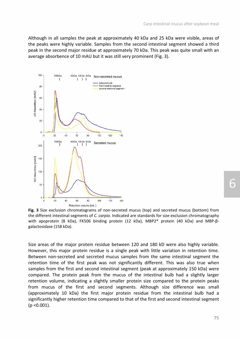
Carp intestinal mucus after soybean meal
75
6
Although in all samples the peak at approximately 40 kDa and 25 kDa were visible, areas of the peaks were highly variable. Samples from the second intestinal segment showed a third peak in the second major residue at approximately 70 kDa. This peak was quite small with an average absorbence of 10 mAU but it was still very prominent (Fig. 3).
Fig. 3 Size exclusion chromatograms of nonどsecreted mucus (top) and secreted mucus (bottom) from the different intestinal segments of C. carpio. Indicated are standards for size exclusion chromatography with apoprotein (8 kDa), FK506 binding protein (12 kDa), MBP2* protein (40 kDa) and MBPどéどgalactosidase (158 kDa). Size areas of the major protein residue between 120 and 180 kD were also highly variable. However, this major protein residue is a single peak with little variation in retention time. Between nonどsecreted and secreted mucus samples from the same intestinal segment the retention time of the first peak was not significantly different. This was also true when samples from the first and second intestinal segment (peak at approximately 150 kDa) were compared. The protein peak from the mucus of the intestinal bulb had a slightly larger retention volume, indicating a slightly smaller protein size compared to the protein peaks from mucus of the first and second segments. Although size difference was small (approximately 10 kDa) the first major protein residue from the intestinal bulb had a significantly higher retention time compared to that of the first and second intestinal segment (p <0.001).

Carp intestinal mucus after soybean meal
76
6
All samples from the SBM experiment also showed a biphasic protein profile with a major protein peak at approximately 150 kDa. For nonどsecreted mucus in the first intestinal segment, switching to the 20SBM diet lead to a significant increase in peak height (8.0 fold increase on day 7 compared to day 0). The increase became less over time (7.2 fold (significant) on day 14 and 5.3 fold on day 20). No significant differences could be found for the peak height of the secreted mucus from the first intestinal segment and of both nonどsecreted and secreted mucus from the second intestinal segment. Microbiological examination from the soybean experiment None of the fish died during the soy bean feeding experiment. From the 23 fish that were sampled from only seven no bacteria could be cultured on blood agar plates. From all other carp facultative pathogenic bacteria (fluorescent and nonどfluorescent Pseudomonas spp., Aeromonas sobria, Aeromonas veronii und Cytophagaどlike bacteria) could be isolated Bacterial content in the organs was for most fish (twelve out of sixteen) where bacteria could be isolated, low to moderate (Table 5). From none of the control fish or of the fish sampled three weeks after feed alteration, bacteria could be isolated in high numbers. On week 1 after feed alteration, significant higher bacteria intensity could be observed. Table 5 Number of carp with a no, low, moderate and high bacterial content isolated from carp organs after switching to a 20SBM diet.
Bacterial content Time point after diet change
Control (n=8) Week 1*(n=5) Week 2 (n=5) Week 3 (n=5) No 2 0 2 3
Low 3 0 1 1 Moderate 3 2 1 1 High 0 3 1 0 * Significant difference to control (p< 0 .05). Prevalence was compared to control and tested with One Way ANOVA.
Discussion HMGs and antimirobial peptides in intestinal mucus On fish mucin molecules relatively few studies have been conducted, however the data available indicate that fish mucus is similar in composition to the mucins secreted by mammalian goblet cells from epithelial tissues [78, 119, 120, 127, 201]. For C. carpio, mucus glycoproteins have so far been described under physiological circumstances and after bacterial challenge for C. carpio [78, 139, 140, 202]. For mammals, it is well known that different parts of the gastrointestinal tract have different mucus thicknesses [203, 204] and express different mucus genes [46, 205]. For carp so far two mucin genes have been found of which only one, Muc2, was expressed in the intestinal tract [206]. Since only one Muc gene has been identified in the intestinal tact it is not known wether different Muc gene profiles exist along the gut axis. However, morphological and functional differences have been previously found along the gut axis. Whethere these differens between different parts of

Carp intestinal mucus after soybean meal
77
6
intestinal tract also include differences in mucus HMGs has to our knowledge not been extensively examined so far. In this study, mucus glycoproteins within the goblet cells appeared to have a similar charge due to their glycosylation, as differences between the applied stainings could not be found. However, HPLC differences in the monosaccharide content of the mucus HMGs could be observed for both the first and second segments. Based on its molecular weight, the unidentified sugar in this study is probably ribose [131]. Ribose and 2どdeoxyribose were the predominant sugars in rainbow trout skin mucus [100], but probably not in C. carpio skin mucus (unpublished data). For the first segment, the size area of the “riboseどpeak” was the highest, indicating a different role for ribose in C. carpio skin and intestinal mucus. Fucose which is thought to be especially important for the viscoelasticity of the mucus, may vary strongly in intestinal mucus or anal gland mucus of mammals [132]. The fucose content of C. carpio intestinal mucus was dependent on the origin of the mucus (first or second segment). For nonどsecreted mucus in the second segment, fucose was the most predominant sugar, as was also observed for C. carpio skin mucus (unpublished data). Nonどsecreted mucus is thought to be present in a native form, whereas secreted mucus might be altered by environmental influences such as bacterial degradation. In the first segment NeuNAc/gal was predominant. The sugar galNAc could not be detected with the method used in the second segment, which was also the case for C. carpio skin mucus (unpublished data). In general, on the monosaccharide level, C. carpio skin mucus resembled the mucus of the second segment more than the mucus of the first segment. Besides sugar analysis, protein content was examined by size exclusion chromatography. In all examined samples a biphasic profile could be observed. Nonどsecreted and secreted mucus samples from the different intestinal segments contained almost similar protein profiles: their residues are between 120 to 180 kDa and 12 to 70 kDa. The proteins found between 120 to 180 kDa most likely represent mucins which adhere to the epithelium, as they are similar to the adherent mucins (AMs) from previous reports [95]. The proteins found between 12 to 70 kDa probably consist of mucin constituents mixed with luminal proteins, the soどcalled luminal mucins (LM) [95]. Smaller molecules (LMs) probably represent the soluble luminal mucus. The AMs form a framework in which smaller molecules might be entrapped. This could explain why molecules smaller than the retention size of the ultrafiltration filter could be found. LM might be essential for protection of the intestinal surface as higher bacterial adhesion to smaller mucus molecules has been observed [207]. Mucin degradation by pathogenic bacteria has been suggested earlier for C. carpio [140, 208] and might largely take place at the carbohydrate side chains of the mucins. In humans, it is known that bacteria with extracellular glycosidases may contribute to the damage of intestinal mucins [209]. In clinically healthy C. carpio, bacterial degradation of the protein core of mucus HMGs appears to be limited, since secreted and nonどsecreted mucus have comparable sized mucins indicated by their similar protein profiles. However, between different intestinal segments differences in carbohydrate staining could be observed indicating differences in pH of the mucus. Differences between the first and second segment in the staining intensity of the glycoproteins in the goblet cells support the role of mucus in the defence against pathogens. This fits well in the immunological role as previously suggested for the second segment [210]. The mucus from the intestinal bulb might have also

Carp intestinal mucus after soybean meal
78
6
a different function in defense against bacteria as differences in carbohydrate staining to that of first and second intestinal segment could be seen. A possible different role for mucus in the defense against bacteria is underlined by the different staining for BD2. Staining for BD2 was more pronounced for tissue samples of the first than for the second segment. It is probable that under physiological circumstances, the release of BD2 in the first segment is high enough so that BD2 molecules can also be functional in the second segment. Staining for BD3 and lysozyme was low and similar between the first and second segment. Therefore under unchallenged conditions the humoral substances BD3 and Lys appear to be less important in C. carpio. Besides the LMs also AMs were found. Functionally, large molecules (AMs) are regarded to form the mucus layer, which adheres to the epithelium. The main function of these proteins appears to be some kind of stabiliser or carrier for the mucus. In the present study, size areas of the major protein residue between 120 and 180 kD were variable in height, but were a single peak with little variation in retention time. Retention volume of the first peak were similar for samples from the first and second intestinal segment (peak at approximately 150 kDa), but was slightly but significantly larger (approximately 140 kDa) for mucus of the intestinal bulb. Size shifts of around 10 kDa are often caused by a difference in the glycosylation pattern of a protein. The smaller size of the molecules might play a role in food transportation, as smaller molecules are considered to be washed away easier. The intestinal bulb has a food storage function, with the mucus enveloping the food for easier transport. Furthermore a large part of the digestion has already taken place in the intestinal bulb. Mucus alterations upon SBM SBM is used in fish feed as it is a cheap source of protein. The use of soy bean in fish feed however, is sometimes causing problems as SBM containing diets are known to induce an inflammatory response in the hindgut of certain fish species. Contrary to previous observations made with S. salar, C. carpio start to recover or adapt to the SBM feed from the fourth week after the SBM feeding [75]. After SBM feeding, the second gut segment of C. carpio did not show external disease symptoms as found previously for carp by Uran et al. [75]. A clear loss of supranuclear vacualistion could not be found. A reduction in goblet cell number in the AB2.5 staining as found by Uran et al. [75] however could also be observed. This indicates that the carp in this study suffered from an acute but mild enteritis process. In carp, the enteritis process is accompanied by damages to the intestinal epithelium [75]. In the present study, a higher number of bacteria could be isolated from internal organs after SBM feeding. This might be explained by damage to the intestinal epithelium. The increased bacteria number in the internal organs indicates that the mucosal barrier as first line of defence was compromised, supporting the theory that the intestinal barrier was affected. For humans, increased gut permeability has been described following enteritis. If changes after enteritis persist a chronic inflammation can develop [197]. Between the control and week 3 after 20SBM feeding, no difference could be found in bacterial numbers in intestinal organs, indicating that the intestinal barrier function recovers over time. All samples from the SBM experiment showed a biphasic protein profile with a major protein peak at approximately 150 kDa. The size of the large mucins did not change markedly. However, switching to the 20SBM diet led to a significant increase in peak height of 150 kDa

Carp intestinal mucus after soybean meal
79
6
proteins in nonどsecreted mucus of the first segment, whereas the amount of 150 kDa in secreted mucus remained relatively stable. The obseved increase in peak heightbecame less over time. Mucus has a high turnover rate as mucus traps pathogens that can be removed from the body through a constant flow of mucus. The increase in the nonどsecreted mucus indicates that the SBM diet induces an increased mucus synthesis. The amount of newly synthesised mucus might increase the mucus flow, which would explain why the amount of secreted mucus remains stable. An increased mucus flow stimulates the removal of pathogens from the entire subsequent intestine, which might hereby prevent pathogens from entering the soy bean damaged intestinal epithelium. Changes in mucin levels during enteritis have been described for chronic inflammatory bowel diseases (IBD) in humans. In IBD, genetic mutations in mucin genes, changes in sulphation, degree of glycosylation, mucin mRNA, protein levels and degradation of mucins have been described. Changes of immunological or bacterial factors during an initial or ongoing inflammation can influence mucin production, which could have further adverse effects on mucosalどbacterial interactions, hereby sustaining the chronic character of the inflammation [198]. In ulcerative colitis (a form of IBD) mucin protein levels [198], as well as the expression of MUC2 were reduced [211]. MUC2 is the structural component of the colonic mucus layer in humans [211]. Reduced Muc2 levels were also observed in interleukin (IL)ど10 knockout mice that develop colitis [211]. The expression of mucin 2 proteins and the antiどinflammatory cztokine ILど10 by type 2 T helper cells and activated macrophages which induces its expression seems to be essential in the control of enteritis in mice. The increase in mucus glycoproteins as seen in the first intestinal segment in carp under SBM diet decreased over time, which indicates that mucus composition returns to the state before switching to the SBMどdiet. An initial upどregulation of ILど10 at week 1, followed by a downどregulation to slightly below or to the level before the enteritis induction was observed previously for C. carpio [75]. In the present study, a 20SBM diet induced changes in the mucus, indicating that the mucosal layer showed altered mucus secretion upon 20SBM feeding. In the first segment the amount of sulphated glycoconjugates decreased, suggesting that similar to IBD reports [198] SBM feeding also leads to changes in mucus sulphation in C. carpio. In addition, mucus from SBMどfed C. carpio showed a different glycosylation pattern as was also reported for IBD [198]. The different glycosylation pattern is indicated by the significantly increased amount of neutral glycoconjugates in the first intestinal segment and the significantly decreased amount of acid and neutral glycoconjugates in the second intestinal segment. Besides the changes in mucus HMs also a difference in staining for BD3 could be observed. One week after switching to the SBMどdiet, the amount of goblet cells which were stained for the antimicrobial peptide BD3 was significantly reduced. This might indicate that BD3 was released to the intestinal lumen to fight pathogens, as BD3 in humans has a strong bactericidal activity [212]. No goblet cells could be stained at week 1 after the diet switch, this might indicate that BD3 was released more rapidly than it could be produced. After the initial depletion of the goblet cells, synthesis and secretion of BD3 seemed to return to their initial equilibrium, as on week 2 or 3, no differences compared to the control could be observed anymore.

Carp intestinal mucus after soybean meal
80
6
Conclusion In conclusion, no indication for a different protein core of mucus HMGS for first and second segment could be found. This is in contrast to what is found for humans and other mammalian species analyzed. In the present study, only differences in mucus glycosylation could be observed. Along the gut axis, between the intestinal segments and the intestinal bulb a difference in the size of the major protein peak could be observed. This difference however can also be caused by a different glycosylation. A difference between first and second segment could be observed for the antimicrobial peptide BD2. Furthermore it was found that SBMどdiet induces a change in mucins in C. carpio similar to that found in IBD in humans. Initial changes included; changes in mucin composition, the presence of BD3 and of bacteria in internal organs. After the initial changes, all values measured returned back to the initial preどSBM diet values. This might indicate a recovery of the mucosal layer and thus a recovery of the primary barrier between the C. carpio and its surrounding. Changes in the mucosal layer form only a small part in the complex inflammation process of soybean mediated enteritis with its many symptoms such as the normal supranuclear vacuolisation of the absorptive cells in the intestinal epithelium [213]. The recovery of the extrinsic mucus layer may help C. carpio in recovering from SBMどenteritis and not developing a chronic inflammation, as through the restoration of the barrier the C. carpio is no longer constantly exposed to new pathogens. Acknowledgments The authors acknowledge the financial support by the DFG (Deutsche Forschungsgemeinschaft).

Chapter 7
Chemotaxis towards, adhesion to, and growth in carp gut mucus of two Aeromonas hydrophila strains with different pathogenicity for common carp, Cyprinus carpio L. van der Marel, M.C. Schroers, V. Neuhaus, H. Steinhagen, D. Journal of Fish Diseases, 2008, 31, 321ど330 Fish Disease Reseach Unit, Centr of Infectious Diseases, University of Veterinary Medicine Hannover, Germany

Carp gut mucus and bacterial behaviour
82
7
Abstract Characteristics that promote bacterial colonization of the intestinal mucosal surface were examined in two strains of the common fish pathogen Aeromonas hydrophila, with different pathogenicity. The characteristics examined were chemotactic activity towards mucus, bacterial adherence to mucus and growth in mucus. Intestinal gut mucus of healthy common carp was used. The results indicate that chemotaxis is not necessary for a bacterium to become pathogenic, but it may be a necessary parameter for a bacterium to be an obligate pathogen. Adhesion also seems to be a factor influencing pathogenicity. The results suggest that higher adhesion to mucus and subsequent growth is associated with differences in pathogenicity.
Introduction Many infectious diseases are initiated by bacterial colonization of mucosal surfaces [77]. Aeromonas spp. are bacteria that are widespread throughout aquatic environments such as fresh surface water or marine water and are probably one of the leading causes of bacterial enteritis in humans [214]. Aeromonas hydrophila is a common pathogen and has been involved in heavy mortalities in farmed and feral fishes [215]. It is considered to be an opportunistic agent that only provokes clinical signs in stressed fish or fish affected by concurrent infections. In experimental infections, clinical signs could only be invoked when bacteria were injected intraどperitoneally [216]. Oral, anal or bath application is seldom used experimentally, as few or no pathological changes are observed when common disease indicators, such as blood parameters, are monitored [217, 218]. It appears that an intact mucosal surface is capable of forming a protective barrier against A. hydrophila [219]. The mucosal epithelial surface is protected against pathogens by the mucus layer and the mucins it contains. However, the ability of bacteria to bind to mucus components may actually facilitate the colonization of mucosal epithelial surfaces by immobilizing the bacteria in the mucus gel. For successful colonization, it is necessary that bacteria establish themselves in the mucus gel once they are bound to mucus. Normal erosion of the mucus gel is likely to wash away adherent bacteria. Therefore, to achieve successful colonization the replication rate of bacteria attached to mucus must be equal to or higher than the rate of erosion of the mucus layer itself. Thus, it would be advantageous for bacteria if they can use mucus as a nutrient source for their growth [71]. Some of the characteristics that promote bacterial colonization of the mucosal surface are chemotactic activity towards mucus (cell contact) [76] followed by bacterial adherence to mucins coating the mucosal cells[77] and subsequent growth in mucus. Several bacteria species are known to show chemotactic responses towards mucus [96, 97, 100, 220ど222]. However different strains of the same species may show a different chemotactic behaviour. For instance, Hazen et al. [220] showed that different isolates of A. hydrophila have a different chemotactic index for largemouth bass skin mucus and that some isolates are nonどchemotactic. Based on the chemotactic behaviour of the isolates examined, Hazen et al. [220] suggested that there could be a difference in pathogenicity between A. hydrophila strains.

Carp gut mucus and bacterial behaviour
83
7
This view is supported by the observation that different strains of bacteria may also show a different adhesion to mucus [223]. In a study of the intestinal bacterial microbiota of carp, Namba et al. [224] demonstrated that most isolates with high adhesive ability to intestinal mucus from carp belong to Aeromonas sp. In addition A. hydrophila can grow in mucus supplemented media [71, 225]. Whether different strains have a different growth in mucus is not known. Although Ascencio et al. [71] state that mucus can serve as a carbon and nitrogen source for A. hydrophila, it is not known whether carbon, nitrogen or phosphate are limiting factors for growth. In this study we examined chemotactic behaviour, adhesion and growth of two A. hydrophila strains isolated from fish.
Material and methods Animals Parasite and virusどfree sibling common carp, Cyprinus carpio L., from a single crossing (R8S8 × R3S8, Wageningen University, The Netherlands) were used for mucus isolation. Carp were raised and kept in filtered reどcirculated tap water. For all experiments, approximately 4どyearどold carp with a mean body weight of 99.1 g (± 20.6 g) and a standard length of 14.1 cm (± 1.1 cm) were used. The carp were placed in a 400 L tank with filtered tap water 3 days before sampling, and were starved to reduce faecal contamination of intestinal mucus. Fish were killed by bath immersion with 500 mg LЪ1 tricaine (Sigma, Munich, Germany) and subsequently dissected. The entire intestinal tract from pseudogaster to anus was removed, weighed and cooled on ice. Bacteria Aeromonas hydrophila strain 38 (A38) was a gift from the FriedrichどLoeffler Institute, Federal Research Institute for Animal Health, Germany. Aeromonas hydrophila strain 60 (A60) was a gift from the Department of Veterinary Pathobiology, Royal Veterinary and Agricultural University Copenhagen, Denmark. Both A. hydrophila strains were isolated from carp with skin ulcers. Edwardsiella tarda was a gift from the LowerどSaxony State Office for Consumer Protection and Food Safety and was isolated from diseased catfish. Bacteria were routinely grown in veal infusion medium and stored at 109 CFU mL
Ъ1 at Ъ80 °C. To remove all possible nutrients, the bacterial suspension was centrifuged at 10 500 g for 10 min and the supernatant removed. For the infection and adhesion experiments, bacteria were resuspended in physiological salt solution. For the chemotaxis and growth experiments, bacteria were resuspended in a nine salt solution (NSS) (Marden, Tunlid, Malmcronafriberg, Odham & Kjelleberg 1985). NSS is a carbon, nitrogen and phosphorus free solution and was adjusted with 0.5% NaCl to 0.9% salinity. Infection experiment To determine whether the A. hydrophila strains used (A38 and A60) had different virulence, 100 carp were orally intubated either with physiological salt solution (control fish) or with 107
CFU of one of the A. hydrophila strains. All carp were intubated with a volume of 300 ´L. Thirtyどtwo carp were used per treatment. Carp were kept at 20 °C and eight fish per bacterial

Carp gut mucus and bacterial behaviour
84
7
strain were examined 1, 3, 6 and 10 days after oral intubation. Carp were killed, dissected for pathological–anatomical examination and swabs taken from liver, kidney and spleen for microbiological examination. Swabs were cultivated on blood agar plates for 2 days at 25 °C. Bacteria were identified with the API system (BioMerieux, France). Isolation of mucus Mucus was isolated from carp that were starved for 3 days to empty the gut. Intestines of untreated carp (mean gut weight 1.37 g ± 0.34 g) were opened longitudinally and cut into 3ど4 mm small pieces. Subsequently, secreted luminal mucus was isolated with isolation medium as described previously [87, 139]. In brief, tissue pieces were incubated for 20 min in 100 mL isolation buffer containing protease inhibitors. The isolation buffer was collected and centrifuged for 30 min at 12 000 g. Subsequently, the supernatant was collected and frozen at ど20 °C until further processing. All samples were concentrated by ultrafiltration (Amicon, Beverly, MA/USA, exclusion limit 30 000 Da) to a final volume of 2 mL. Mucus was sterilized by freezing at ど80 °C for at least a week. Sterility was checked by plating out 100 µL of mucus and incubation for 2 days at 25 °C. For chemotaxis and growth experiments, the mucus was pooled (3ど6 fish) and subsequently UV radiated for at least 1 h. For the adhesion experiments, mucus of eight fish was used. Protein content was determined by Bradford reaction (580 nm; BMG, Germany) [90]. Carbohydrate content was determined by the periodic acidどSchiff [226]reaction (540 nm) [88, 89]. Labelling of live bacteria For adhesion experiments bacteria were stained with a green fluorescent nucleid acid stain (Syto 9, Invitrogen, Germany; 2 µL per 109 bacteria). Stained bacteria were spectrophotometrically analysed (470 nm excitation, 520 nm emission, BMG, Germany). Chemotaxis Chemotaxis assays were performed using a 10どwell transmigration chamber (Neuroprobe, Gaithersburg, MD, USA). NSS with mucus (5 mg protein mL
Ъ1) or without mucus (control) was
placed in the lower wells. Bacterial suspension (109 CFU mLЪ1) was placed into the upper wells
of the transmigration chamber. Besides the two A. hydrophila strains, a strain of the obligate fish pathogenic bacterium E. tarda was used as a positive control. In all assays, NSS was used as a negative control in the upper and lower wells, to check for random migration. All treatments were done in duplicate. Upper and lower compartments of the transmigration chamber were separated by a polyvinylpyrrolidone (PVP)どtreated polycarbonate filter with 2 ´m pores (Neuroprobe). Filled transmigration chambers were incubated for approximately 16 h at 25 °C. Cells were harvested from upper and lower wells and spectrophotometrically analysed (470 nm excitation, 520 emission). UVmigrated bacteria = UVlower well /( UVupper well + UVlower well) Chemotactic index (CI) was calculated with fluorescence values (UV) as: CI = UVmigrated bacteria in assayed conditions/ UVmigrated bacteria in negative control

Carp gut mucus and bacterial behaviour
85
7
Adhesion For adhesion experiments, mucus of carp was separated by means of downward gel filtration on a 34 × 0.9 cm Sepharose CLど4B column (Sigma, flow rate 5.2 mL hど1
, fraction size 1.3 mL, 40 fractions). The fractions pooled into three fraction pools of glycoproteins with different molecular size (30ど670 kDa, 670ど2000 kDA, >2000 kDa). Twentyどfive microlitres of each sample was added in triplicate to a 96どwell black polystyrene plate (Nunc, Denmark). As positive controls, bovine serum albumin (BSA) (1 mg mL
ど1) and pig gastric mucin (5 mg mL
ど1,
Invitrogen) were used. Then 75 ´L of coating buffer (16.8 g sodium hydrogen carbonate, 21.2 g sodium carbonate per litre, pH 9.6) was added to each well and left overnight at room temperature. After washing with physilogical salt solution, 25 µL of 109 CFU mL
ど1 fluorescent labelled bacteriaどsolution (A38 or A60) was added. A strain of E. tarda served as positive control. Bacteria were forced to the plate bottom by centrifugation (163 g, 12 s) and then incubated for 30 min in the dark at room temperature. Plates were washed and liquid was removed by centrifugation. For spectrophotometrical measurements 50 µL of physiological salt solution was added and the fluorescence generated by adherent bacteria was recorded with a microplate reader (BMG, Germany). Data were corrected on the basis of carbohydrate content of the mucus fractions applied and gut weight was used as an indication of mucus quantity [78]. Binding of mucus to the plate was confirmed by a lectin binding assay using ConA as described by Neuhaus et al.[139]. Growth Both A. hydrophila strains were incubated with and without mucus and compared for growth. 1 × 10⁸ CFU were incubated in the following media: (i) Complete energy and nutrient depletion (NSS) (ii) NSS supplemented with mucus (0.8 mg protein mL
ど1)
(iii) veal infusion medium A38 (5 × 10⁷ CFU mL
ど1) was also incubated in several experimental media. NSS was
supplemented with 1.1 g Lど1 ammonium sulphate, 0.27 g Lど1 potassium hydrogen phosphate and 2.0 g Lど1 glucose or mucus. The following media were used: (i) Complete energy and nutrient depletion (NSS) (ii) Carbon limitation: NSS supplemented with ammonium sulphate and potassium hydrogen phosphate (iii) Nitrogen limitation: NSS supplemented with glucose and potassium hydrogen phosphate (iv) Phosphorus limitation: NSS supplemented with glucose and with ammonium sulphate (v) Enriched medium: NSS supplemented with ammonium sulphate, potassium hydrogen phosphate and glucose (vi) NSS supplemented with mucus (1.7 mg/mL) Growth was monitored after 24 h by measurement of the optical density (595 nm) and by serial dilution and plate counts. Fold increase after 24 hours was calculated as follows: CFUafter 24 h in total medium /CFUinocculated.

Carp gut mucus and bacterial behaviour
86
7
Statistics The chiどsquare test was used to compare the impact of the two Aeromonas strains on fish mortality and bacteria counts in internal tissues in the infection study. For the adhesion study, results were analysed using a Rank sum test. For growth studies Student’s tどtest was used. P < 0.05 was considered to be significant.
Results Infection experiment
Clinical changes None of the fish given 107 CFU died during the infection experiment. However, after intubation some fish did show clinical signs, such as redness of one or more fins, fin erosions or congestion of blood vessels. Some control fish (4 of 32) showed external changes, but signs of infection were more often observed in the bacteriaどtreated fish, where external changes were seen more frequently in the A38どtreated fish (14 of 32) than in the A60どtreated fish (7 of 32). However, results were not significantly different (P = 0.062). Redness of the skin was only observed in one fish in the control and A60 groups. However, five fish from the A38 group had a reddened skin. In the A38 fish, congestion of blood vessels was also observed. Pathological changes of internal organs were not observed in any fish. Microbiological examination Bacteria were isolated from some fish treated with 1 × 10⁷ CFU A38 and A60 and also from control fish. Bacteria were isolated from one or more of the examined organs on all sampling days and were identified as Pseudomonas putida, Staphylococcus spp., Gaffkya tetragena, Acinetobacter lwoffii and Shewanella putrefaciens. Aeromonas hydrophila was reどisolated from only one fish (A38, day 6). In general, bacteria were isolated more frequently from A38どtreated fish (8 of 32) than from control or A60どtreated fish (2 and 3 of 32, respectively). Chemotaxis Syto9 stained A38, A60 and E. tarda equally. Bacterial vitality (motility and percentage of mobile bacteria) was not negatively influenced by staining or the presence of mucus, as determined visually under a fluorescence microscope 0, 1, 2, 3 and 20 h after incubation at 25°C. In the first migration experiment, A. hydrophila were incubated overnight with 5 mg mucus protein mL
Ъ1 as chemoattractant in the lower well of the chemotaxis chamber. In this assay, a CI less than one was recorded for the A. hydrophila strains examined. The A38 and A60 strains had a CI of 0.75 and 0.63, respectively. E. tarda on the other hand showed a high chemoattractive movement towards the mucus (CI = 3.52). In a second migration experiment, different mucus concentrations (0.3, 1 and 3 mg mucus protein mL
Ъ1) were used in a 3 h
chemotactic assay with the A38 strain. At low mucus concentrations (0.3 and 1 mg mLЪ1), no
differences in CI (CI = 0.95) were found. Only at a higher mucus concentration (3 mg mLЪ1
mucus protein), a slight difference in CI was found (CI = 1.28). The viscosity of mucus might slow bacterial movement. Therefore, the A38 strain was also incubated in a solution

Carp gut mucus and bacterial behaviour
87
7
supplemented with 3 mg mucus protein mLЪ1 and the movement towards NSS observed.
More bacteria moved into NSS when incubated in mucus than when incubated with NSS (CI = 1.42). It appears that the viscosity of mucus at this specific concentration is not able to slow bacteria. The increased bacterial movement might indicate a chemokinetic effect of gut mucus on A. hydrophila. Adhesion Aeromonas hydrophila strain A38 adhered more strongly to mucus glycoproteins of all size fractions tested than the A60 strain (see Fig. 1). Bacteria from both strains adhered most strongly to molecules with a size of 670–2000 kDa and weakest to molecules with a size of 30–670 kDa. The fluorescence intensity of bacteria which adhered to mucus molecules of 30 to >2000 kDa was 13.3 times higher for the A38 than for the A60 strain. Edwardsiella tarda showed an even higher fluorescence intensity (59.5 times higher than the A60 bacteria adhering to mucus, see Fig. 1).
Fig. 1 Adhesion of the facultative pathogen Aeromonas hydrophila strains A38 and A60, and the obligate pathogen Edwardsiella tarda to intestinal carp mucus (n = 8) of different molecular size ranges. All values represent mean and standard deviation of absorbancy measurements of fluorescent labelled bacteria. Mean values with different superscripts are significantly different (Rank sum test, P < 0.05). Growth Bacterial growth was determined by measuring OD and by serial dilutions with plate counts for all cultures in the growth experiments. However, OD values were only used to estimate which dilution was necessary for the serial dilution assays. OD values were not used for data processing, since for mucusどsupplemented NSS cultures no correlation was found between OD values and the CFU mL
Ъ1 cell culture. The total yield of CFU after 24 h starvation in NSS was approximately the same as the amount of CFU with which the culture was inoculated (Figs 2 & 3). Both strains grew rapidly in the

Carp gut mucus and bacterial behaviour
88
7
normal culture medium (veal infusion medium, Fig. 2). In this medium, the A60 strain produced a 2.8 times higher yield than the A38 strain. Addition of mucus to NSS cultures clearly increased bacterial growth after 24 h. In this medium, the A38 strain produced a 2.3 times higher yield than the A60 strain. Veal infusion medium was a better medium for growth for the A60 strain than NSS supplemented with mucus (total yield four times higher). A38, on the other hand, grew better in mucusどsupplemented NSS than in veal infusion medium (Fig. 2).
Fig. 2 Fold increase of cellどforming units of Aeromonas hydrophila strains A38 and A60 after 24 h cultivation in a nutrient depleted medium (NSS), a nutrient depleted medium supplemented with 0.8 mg mL
Ъ1 mucus protein and a normal culture medium (veal infusion medium). All values represent mean and standard deviation. Mean values with different superscripts are significantly different (Student’s tどtest, P < 0.05).
Fig. 3 Fold increase of cellどforming units of Aeromonas hydrophila strain A38 after 24 h cultivation in nutrient depleted media, complete nutrient depleted medium (NSS), carbon limited medium (C lim.), nitrogen limited medium (N lim.), phosphorus limited medium (P lim.), medium supplemented with carbon, nitrogen, phosphorus and glucose (Enriched) and medium supplemented with 2.0 mg mL
Ъ1 mucus protein (Mucus). All values represent mean and standard deviation. Mean values with different superscripts are significantly different (Student’s tどtest, P < 0.05).

Carp gut mucus and bacterial behaviour
89
7
A38 grew better in mucusどsupplemented NSS than A60 and was therefore used to determine which nutrients are limiting for A. hydrophila and which may be obtained from mucus. Cultures with NSS supplemented with carbon, nitrogen and phosphorus from inorganic sources promoted bacterial growth differently: bacterial growth was only slightly increased in media with carbon or nitrogen depletion, in comparison with completely depleted medium (NSS). Medium with phosphorus depletion allowed up to 100どfold increase in bacteria compared with NSS. None of the inorganic supplements enhanced bacterial growth as much as supplementation with mucus (Fig. 3).
Discussion Aeromonas hydrophila is a facultative pathogen of fish and can cause secondary infections [227]. In general, A. hydrophila can not cause disease in healthy, wellどconditioned fish [228]. Carp used in the present infection experiments were healthy, parasite and virus free and kept in wellどfiltered tap water. It is therefore not surprising that no severe clinical signs were observed in this experiment after bacterial intubation [229]. Mild redness of fins and skin was rarely observed in control fish and fish treated with Aeromonas hydrophila strain A60. Facultative pathogens, commonly resident in fish, can cause these signs in stressed fish. The carp in this experiment were stressed due to transfer to their new environment, starvation and oral application. Previous experiments with platys, Xyphophorus spp. [229] showed that A. hydrophila can induce mortality. Fish that died showed clinical symptoms, such as redness in gills, head and vent [229]. All survivors, however, did not show any pathological changes. These findings are in accordance with the results of the present infection experiment, where fish treated with 10
7 CFU A38 did not show severe clinical signs. A. hydrophila could be reどisolated more often from A38 h than from A60どtreated fish. This is in agreement with Kawula et al. [229], who only reどisolated A. hydrophila from organs of fish which died from the infection. Fish treated with A38 showed clinical signs more often than the A60どtreated fish. In addition, bacteria were isolated more often from internal organs after A38 treatment. This suggests that A. hydrophila A38 is more capable of destabilizing the intestinal barrier. The higher occurrence of clinical signs and higher number of fish where bacteria could be isolated from internal organs indicate that A38 is more virulent than A60. In order to act as a pathogen, bacteria need to have contact with the host. This can be coincidental or the result of directed chemotactic movement. In a chemotaxis, capillary assay movement towards mucus of fish was observed for some A. hydrophila strains [220]. The chemotaxis response for A. hydrophila strains towards mucus found in the present experiment and as reported by Hazen et al. (1982) is a low response (approximately 1). The present study showed that gut mucus of carp did not induce a chemotactic migration of the two A. hydrophila strains used, but it might induce an increased chemokinesis, which results in enhanced migration of the bacteria and thus allows it to more readily reach the fish epithelium. Greater chemoattractive movement towards mucus from a different location is not to be expected as intestinal mucus is equally chemoattractant as skin mucus and more

Carp gut mucus and bacterial behaviour
90
7
chemoattractant than gill mucus [230]. O´Toole et al.(1999) found that a wild type Vibrio anguillarum had a much higher chemotactic response towards intestinal mucus (relative response approximately 140) than A. hydrophila. This large difference in chemotactic response might be a reason why A. hydrophila is an opportunistic pathogen and V. anguillarum is a more frequent causative agent of disease. The mucus used in the chemotaxis experiment came from healthy virus and parasiteどfree fish. Hazen et al. (1982) found differences in chemotactic activity of Aeromonas bacteria towards surface mucus from healthy largemouth bass and surface mucus from red soreどinfected largemouth bass. It is, however, debatable whether this difference is caused by excretion of antibodies as suggested by Hazen et al., (1982), or by an alteration of the major constituent of mucus, glycoproteins. During infection, mucus glycoproteins (mucins) are known to have altered molecular size and glycosylation pattern [76, 139, 219, 231]. An upどregulation of the expression of an MUCどencoding gene that codes for the mucin protein core, is also recorded in the context of bacterial infection [232]. Altered diet also influences mucus composition [233]. Since diseased fish usually change or stop their food intake such an influence should be excluded. Therefore, when comparing mucus from diseased and nonどdiseased fish the mucus should always originate from artificially infected fish kept under controlled conditions. Mucus should also be corrected for protein content as it is known that total mucus production can be modulated in diseased fish [125, 139, 234, 235]. Bacteria must attach to host mucus in order to become pathogenic. However, adhesion to mucus is not necessarily a sign of pathogenicity or virulence. For instance, probiotics are chosen for their ability to bind to mucus [236]. Nevertheless, adhesion of pathogens to the mucosal surface is the first step in the pathogenesis of most infectious diseases [96, 237, 238]. In infection experiments, it is observed that more virulent strains of a bacterium show a higher adhesion than less virulent strains. This was reported for example in Yersinia enterocolitica [102, 239] and Pseudomonas cepacia [240]. The higher adherence of A38 therefore suggests that this strain has a higher virulence than A60. When bacteria bind to mucus, they bind to the major constituent of mucus, i.e. glycoproteins [71]. Glycoproteins are highly glycosylated proteins [1, 51ど53, 104], where the protein core is usually not accessible to pathogens due to the carbohydrate sideどchains [241]. Therefore, bacteria usually bind predominantly to the carbohydrate sideどchains of the glycoproteins [71, 77, 102, 239, 240, 242]. Mantle and Husar [102] found that some bacteria can bind better to mucus glycoproteins when some of the carbohydrate sideどchains are cleaved off from the protein core. A possible explanation is that the bacteria can now bind to carbohydrates which lie closer to the protein core or even to the core itself [102, 240]. Glycoproteins of high molecular weight are considered to represent matured, fully glycosylated proteins. Smaller glycoproteins may either be newly synthesized glycoproteins, where not all the glycan side chains are already formed or they may be matured glycoproteins where glycan side chains have already been cleaved by bacteria or intestinal enzymes [96, 139, 243]. The higher adherence of A. hydrophila in this study to mucus molecules sized 670–2000 kDa compared with molecules of 30–670 kDa or >2000 kDa might therefore be explained by a difference in the carbohydrate side chains of the glycoproteins. In order to become pathogenic, bacteria must be able to use mucus for growth once they have adhered to it. They need to multiply fast to prevent their being flushed out of the gut. Both A. hydrophila strains grew in mucusどsupplemented media. A38 however had a higher

Carp gut mucus and bacterial behaviour
91
7
total yield than A60 when cultured under the same conditions. Both strains did not grow in nutrient depleted medium. This agrees with previous findings for A. hydrophila [71]. Culture of A38 in inorganic nutrientどrich (phosphorus limited or enriched) medium enhanced bacterial growth, but not as strongly as did mucus supplementation. This might indicate that the bacteria can use mucus as an energy source. Optical density values were not used in the present experiments to process growth data, since for mucusどsupplemented NSS cultures no correlation could be found between OD values and the bacterial content measured as CFU mLЪ1 cell culture. Aeromonas hydrophila utilizes mucus and therefore most likely degrades it. With increasing growth of bacteria, mucus might have a changed OD. Different bacteria might process mucus in a different way or to a different degree. Furthermore, different mucus composition (e.g. due to diet or disease) might also influence how bacteria process mucus. Therefore, OD measurements on mucus should be considered unreliable when monitoring bacterial growth. When only OD values are used to fit data to growth models [225], bacterial suspensions should be centrifuged and the bacteria diluted again in a known volume of a medium devoid of mucus. It is doubtful if growth models are fitted correctly when no reliable blank value is available. The present study indicates that chemotaxis is not necessary for a bacterium to become pathogenic, as both of the A. hydrophila strains used did not move towards mucus. However, it might be a necessary parameter for an obligate pathogenic bacterium, as the pathogenic bacterium E. tarda cultured under the same conditions as A. hydrophila did show a strong chemotactic movement towards gut mucus. Adhesion also seems to be a factor influencing pathogenicity as E. tarda showed a very high adhesion to mucus. A38 showed a 13 times higher adhesion to mucus than the A60 strain. This agrees with the findings of the infection experiment where A38 was apparently more pathogenic than the A60 strain. The A38 strain also showed a higher growth in mucus than the A60 strain. A faster multiplication of bacteria in mucus increases its potential pathogenicity as the flushing out of bacteria due to mucus erosion is reduced. A higher adhesion and subsequent growth may lead to differences in pathogenicity. Acknowledgements The authors acknowledge the financial support of the DFG.


Chapter 8
Intestinal mucus of challenged carp van der Marel, M.C.

Intestinal mucus of challenged carp
94
8
This chapter is an updated version of the articles “Biochemical and histochemical effects of perorally applied endotoxin on intestinal mucin glycoproteins of the common carp Cyprinus carpio., with special consideration of mucin glycoproteins” [139, see appendix II] and “Changes of intestinal mucus glycoproteins after peroral application of Aeromonas hydrophila to common carp (Cyprinus carpio)” [140, see appendix III] and contains additional information.
Abstract Mucins are high molecular weight glycoproteins produced by specialized cells (goblet cells) and secreted on mucosal surfaces. Released glycoproteins form a biofilm protecting the underlying epithelium. Aim of the present study was study the effect of different challenges on the intestinal mucus from common carp, Cyprinus carpio. Here to carp were exposed to a mechanical challenge, a challenge with a bacterial endotoxin or to a challenge with one of two strains of A. hydrophila strains (A38 and A60). Intestinal mucus was isolated and used to determine mucin weight, glycosylation, protein and carbohydrate distribution over different molecule size. Subsequently, terminal glycosylation was characterized by a lectinどbinding assay. With the lectin ELISA the five monosaccharides which primarily occur in the glycosylation of piscine mucins were tested and differences in terminal glycosylation were found. Changes occurred quickly after challenge. With the isolation of the HMG and the determination of protein and carbohydrate content, differences between the control and treated fish could be observed for mucin content, glycosylation and distribution of carbohydrate and protein over the size fractions. After the bacterial challenges three trends could be observed. For each bacterial challenge each trend was observed at different times, indicating different mechanisms leading to the observed changes. The first trend indicates that a bacterial challenge leads to release of mucus from the goblet cells, but this release is not sufficient to keep the amount of mucus stable over the next days. The second trends indicates that a bacterial challenge leads to a degradation of secreted molecules followed by a release of newly synthesized high glycosylated molecules. The third trend indicates a shift in the equilibrium between release and degradation of mucus molecules.
Introduction One of the major routes of infection in fish is through the gastrointestinal tract [244]. An overwhelming number of infectious diseases are initiated by bacterial colonisation of mucosal surfaces [77]. The epithelium is protected from chemical, enzymatic, and mechanical damage by overlaying mucus. This waterどinsoluble mucus layer on the epithelial cells also plays an important role in host defence mechanisms. Mucus consists mostly of water and high molecular weight glycoproteins (HMG), called “mucins”. Both are important in gel formation [1, 51, 53, 104]. Mucins are produced by goblet cells, which are scattered in many epithelial

Intestinal mucus of challenged carp
95
8
systems of aquatic vertebrates and particularly in the gut. Mucus HMG exhibit a high content of oligosaccharides, which are believed to mediate adhesion of microbiata [79, 80, 92, 97] and to prevent the glycoproteins from degradation by proteases of microbial origin. Based on histochemical data, the number and content of intestinal goblet cells in fish are considered to be similar to those of mammals. As in mammals, a production of secretory vesicles with neutral, acidic or sulphated glycoproteins has been described [6, 81]. Histological and histochemical studies also indicate that fishes with parasitic infections, like mammals, display goblet cell hyperplasia and hypertrophy, whereby as a chronic reaction their intracellular mucins shift towards acidic and sulphated glycoprotein types [81, 83, 84]. Although all fish surfaces are covered with a mucus layer, knowledge concerning piscine mucins and their responses to noxes is scarce. In the present study, the biochemical composition of carp glycoproteins and the changes upon a challenge is studied, using a mechanical, an endotoxin or a live bacteria challenge. Therefore intestinal goblet cells of the common carp were characterized with biochemical and histochemical methods.
Materials and Methods Challenges Parasite and virus free sibling carp from a single crossing (R3 x R8, Wageningen Agricultural University, The Netherlands) were used. Carp were raised and kept in filtered recirculated tap water. Carp were used to examine the effect of a mechanical challenge (induced by application of physiological salt solution) (body weight 88.3 ± 18.7, gut weight 1.10 ± 0.25 g), of an endotoxin challenge (LPSどexperiment; body weight 76.4 ± 12.9, gut weight, 1.06 ± 0.2 g)), and of two live bacteria strains (A. hydrophilaどexperiment; body weight 81.6 ± 19.2, gut weight, 1.00 ± 0.28 g) on intestinal mucus (see Table 1). Fish were treated by peroral application. Carp were starved three days before treatment to reduce faeces on intestinal mucus. Before intubation, the animals were anaesthetized by bath immersion with 150 mg lど1 tricaine (Sigma, Germany). For LPSどexperiment, 150 µg LPS of Escherichia coli O55:B5 (Sigma, Germany) per gram body weight dissolved in PBS was used. PBS was taken for the control fish. In the A. hydrophilaどexperiment, carp were perorally intubated with a physiological salt solution (control fish) or one of the two A. hydrophila strains (A38 or A60). These control fish were used in the mechanical challenge as treated fish. All fish were intubated with a total volume of 300 µl containing 107 CFU bacteria. Bacteria Two strains of motile Aeromonads were used, that were isolated from diseased commercially farmed carp. The two strains were examined by standard biochemical diagnostic methods and classified as Aeromonas hydrophila. A. hydrophila strain 38 (A38) was a gift from the FriedrichどLoefflerどInstitute, Federal Research Institute for Animal Health, Germany. A. hydrophila strain 60 (A60) was a gift from the Department of Veterinary Pathobiology, Royal Veterinary and Agricultural University Copenhagen, Denmark [245]. Bacteria were routinely grown on sheep blood agar at 25°C and stored in veal infusion medium at 109 bacteria mL
ど1 at

Intestinal mucus of challenged carp
96
8
–80°C. To remove all possible nutrients, bacterial cells were harvested by centrifugation at 10,500 x g for 10 min and the supernatant was removed. Bacteria were reどsuspended in physiological salt solution. The used strains differed in adhesion ability to mucus, growth rate in mucus, in vitro cytotoxicity and presence of a type III secretion system (TTSS). A38 adhered more strongly to mucus and had a higher growth rate [208]. When monolayers of EPCどcells were incubated with A60 or with physiological salt solution, no changes in cell morphology or monolayer coherence were observed over a period of 24 h. However, when EPCどmonolayers were incubated with A38 over this time period, the EPCどmonolayer had detached completely from the bottom of the culture vessel, and only a few viable cells could be recognised [140]. On a genomic level the TTSS was found for A38, but not for A60 (unpublished data). For both A. hydrophilaどstrains, both live and dead bacteria were not detectable by periodicどacidどSchiff reaction. Table 1 Experimental setup of the mechanical challenge, LPSどexperiment and A. hydrophilaどexperiment Age Experiment (treatment) Sampling time points
after intubation (days) Fish per time point (n=)
12ど18 months LPSどexperiment (PBS: ctrl) 1, 2, 3, 5, 8 2 12ど18 months LPSどexperiment (LPS) 1, 2, 3, 5, 8 8 ca 4 year mechanical challenge (none: ctrl) ど 4 ca 4 year mechanical challenge (NaCl),
A. hydrophilaどexperiment (NaCl: ctrl)1, 3, 6, 10 8
ca 4 year A. hydrophilaどexperiment (A38) 1, 3, 6, 10 8 ca 4 year A. hydrophilaどexperiment (A60) 1, 3, 6, 10 8
Sampling and analysis For sampling, carp were killed by bath immersion with 500 mg lど1 tricaine (Sigma, Deisenhofen, Germany). They were weighed and subsequently dissected. The complete intestinal tract from behind the intestinal bulb to anus was removed, weighed and cooled on ice. From the intestine of all fish secreted intestinal mucus was isolated. Furthermore, from the LPSどtreated and control carp (Table 1) also nonどsecreted mucus (present in the intestinal goblet cells) was isolated. Isolated intestinal mucus was processed and the high molecular weight glycoproteins (HMG) were analysed by gelchromotography and subsequent protein and carbohydrate determination. Isolation, processing, analysis and calculations were done as described in Chapter 2. From all untreated and control samples a biphasic profile was obtained after downward gel filtration and subsequent staining for carbohydrates and proteins. In between the first peak (peak 1), which contains the largest molecules and the second peak (peak 2) a transition area (TA) could be found. Gel filtration, carbohydrate and protein staining as well as subsequent calculations were done as described in Chapter 2. Fractions from the gel chromatography were pooled into peak 1, peak 2 and a transition area (TA) between these peaks. These pools (except the mechanical challenge pool) were used for a lectinどELISA (cf. Chapter 2) to determine terminal glycosylation.

Intestinal mucus of challenged carp
97
8
Statistics If not indicated else, results are expressed as median value and 25% ど75% quartiles. Data were statistically analysed with an analysis of variance (ANOVA for normally distributed data, and ANOVAどonどRanks for nonどnormally distributed data). Differences were considered significant at p < 0.05. If not indicated else, treatments were compared to the control group.
Results Animals All fish appeared healthy without clinical alterations throughout the study. Isolation of intestinal mucus HMG
Mechanical challenged carp Mucus remained relatively unchanged after a peroral application of PBS. No significant effect othe amount of mucus which could be isolated, was found (Table 2). However, total glycosylation was significantly higher on all days from carp exposed to a mechanical challenge compared to untreated carp and for TA on some days (Table 3). Table 2 Total mucin weight (in g per mg gut weight) of carp intestinal mucus HMG of mechanically challenged, bacterial challenged as well as LPS challenged carp. Shown are median and 25% ど 75% quartiles and significant differences to control (p<0.05). Challeng
e ctrl day 1 day 2 day 3 day 6/5 day 10/8
Secreted mucus
mechanic
al A38 A60 LPS
2.9 (2.4 ど 3.3) 2.3 (1.6 ど 3.5) 2.3 (1.6 ど 3.5) 1.8 (1.6 ど 1.9)
2.2 (1.7 ど 3.6) 1.2 (0.8 ど 1.6) 2.5 (1.6 ど 3.3) 2.0 (1.5 ど 2.1)
No data No data No data 1.0 (0.7 ど 1.0)
2.5 (2.1 ど 3.0) 1.2 (1.0 ど 1.5) 3.6 (3.1 ど 4.6) 1.3 (1.1ど 1.6
3.9 (3.7 ど 4.1) 1.5 (1.4 ど 1.7) 2.9 (2.2 ど 3.8) 1.6 (1.5ど 2.0)
1.4 (1.4 ど 1.5) 1.0 (0.9 ど 1.3) 1.8 (1.7 ど 3.2) 1.8 (1.5 ど 2.0)
Nonどsecreted mucus LPS 0.6 (0.5 ど 0.6) 0.4 (0.4 ど 0.6) 0.6 (0.6 ど 0.7) 0.8 (0.7 ど 1.1) 0.9 (0.8 ど 1.2) 0.7 (0.6 ど 0.8)
7 A38/A60 challenged carp The amount of mucus that could be isolated decreased significantly after oral intubation with A. hydrophila 38 whereas it did not significantly change after intubation with A. hydrophila 60 (Table 2). On day 3 and day 10 the amount of mucus which could be isolated was significantly lower for A38 compared to A60. On day 1 and 3 after A38 challenge, molecules of TA showed a significantly higher protein to carbohydrate ratio (P:C) (Table 3), indicating a lower glycosylation as well as a higher protein percentage. The significantly higher P:C ratio was also observed after the challenge with A60 on day 1 for TA, peak 2 as well as the total mucus. In contrast to the challenge with A38, the A60 challenge resulted in a significantly higher protein percentage for TA on day 6 and 10 (Table 4).

Intestinal mucus of challenged carp
98
8
LPS challenged carp Mucus amount which was secreted decreased significantly on day 2 after LPS challenge (Table 2). On day 2, molecules from TA and total mucus were less glycosylated. Also changes in protein and carbohydrate distribution were observed (Table 4, Table 5). The percentage of molecules found in TA increased significantly. On day 3 a significantly higher glycosylation of molecules in peak 2 could be observed. On day 5 a significantly higher amount of nonどsecreted mucus could be isolated compared to control. Terminal glycosylation As UEAどI results were below or around level of detection the results were not statistically analysed. After LPS challenge only few significant differences in terminal glycosylation could be observed (Table 6). A stronger binding of SNA (binding to üNeuNAc(2т6)gal) could be found on day2 for the secreted mucus after LPSどchallenge. After this challenge ConA (binding to ü–man and ü–Dどglc) showed a stronger binding compared to control in peak 1 on day2 for the nonどsecreted mucus and on all days for the secreted mucus. No significant changes could be found in peak 2 after LPS or after A38/A60 challenge. However, after A38 and A60 significant changes in terminal glycosylation could be found in the total OD per tested sugar as well as for the percentage of the total OD in peak 1 and in TA. On day 10 the percentage of the OD found in TA was significantly lower for all lectins compared to control. Total OD on this day was higher for both A38 and A60 for RCAど1 (binding to galNAc and éどgal) as well as for SNA for A60. On day 1 only for A60 for conA a significantly lower total binding was observed and on day 3 a lower binding to RCA. On day 6 a higher binding could be observed for RCA for A38.
Discussion Mechanical challenged carp When carp were exposed to a mechanical challenge, total glycosylation and therefore the amount of carbohydrates relative to the amount of protein was significantly higher compared to untreated carp. The increased carbohydrate portion might be a microbial defence mechanism. In rats, changes in mucus composition were also observed after mechanical challenge and food uptake [123, 246, 247]. LPS In the present study, carp were perorally intubated with LPS. Lipopolysaccharide (LPS)どproteinどcomplexes are mainly synthesized by gram negative bacteria. LPS is an endotoxin, which is an integrated part of the bacterial membrane. Endotoxin has, at least in mammals, a local and systemic immuneどinducing function in inflammation reactions [248, 249]. Toxic effects in fish are mediated by different factors as in mammals or have not been identified yet. In many in vitro studies on cells of lower vertebrates such as fish, extremely high concentrations of LPS (µg mlど1) have been used before immune responses could be induced [250].

Intestin
al m
ucus o
f challenged ca
rp
99
8
Table 3 Glycosylation of intestinal mucus HMG, expressed as P:C ratio after a mechanically challenge, bacterial challenge as well as a LPS challenge. Shown are median and quartiles and significant (sign.) differences to control (p<0.05).
Sign,differences to ctrl ctrl < d1 d1, d3 < ctrl ctrl < d1, d3 ctrl < d1 ctrl < d2 ctrl < d1 ctrl < d1 d3 < ctrl d1, d3, d6, d10 < ctrl ctrl < d1 ctrl < d2 ctrl < d1
day 10/8
0.6 (0.3 ど 0.7) 0.6 (0.5 ど 0.7) 0.3 (0.1 ど 0.4) 0.7 (0.5 ど 0.8) 0.7 (0.6 ど 0.8) 0.6 (0.4 ど 0.6) 0.5 (0.5 ど 0.5) 0.4 (0.3 ど 0.4) 1.0 (1.0 ど 1.5) 1.7 (1.2 ど2.2) 0.5 (0.4 ど 1.0) 0.8 (0.6 ど 0.8) 0.4 (0.4 ど 0.8) 4.3 (3.5 ど 4.7) 3.6 (1.8 ど 11.5) 0.7 (0.4 ど 0.7) 0.6 (0.6 ど 0.6) 0.4 (0.3 ど 0.4) 2.1 (2.0 ど 2.4) 1.4 (1.2 ど 1.7)
day 6/5
0.2 (0.2 ど 0.3) 0.3 (0.3 ど 0.4) 0.2 (0.1 ど 0.2) 0.6 (0.5 ど 1.0) 1.5 (1.1 ど 2.0) 0.3 (0.3 ど 0.3) 0.4 (0.3 ど 0.5) 0.4 (0.3 ど 0.5) 1.8 (1.7 ど 2.4) 2.4 (1.7 ど 2.8) 0.6 (0.6 ど 0.6) 0.4 (0.4 ど 0.6) 0.7 (0.6 ど 0.8) 3.5 (2.9 ど 4.7) 2.5 (1.2 ど 3.2) 0.4 (0.4 ど 0.5) 0.4 (0.4 ど 0.5) 0.4 (0.4 ど 0.5) 2.5 (1.8 ど 2.6) 2.0 (1.6 ど 2.1)
day 3
0.3 (0.2 ど 0.3) 0.4 (0.3 ど 0.5) 0.3 (0.2 ど 0.4) 0.7 (0.6 ど 0.7) 1.1 (0.9 ど 1.5) 0.2 (0.2 ど 0.3) 0.8 (0.4 ど 1.0) 0.4 (0.3 ど 0.4) 1.9 (1.5 ど 2.3) 1.7 (1.3 ど 2.0) 0.7 (0.6 ど 0.8) 0.5 (0.4 ど 0.8) 0.9 (0.8 ど 0.9) 3.4 (3.1 ど 3.9) 0.9 (0.7 ど 2.5) 0.4 (0.4 ど 0.5) 0.5 (0.5 ど 0.6) 0.5 (0.5 ど 0.5) 2.0 (1.9 ど 2.4) 1.2 (0.9 ど 2.1)
day 2
No data No data No data 0.5 (0.4 ど 0.6) 1.3 (1.0 ど 1.5) No data No data No data 6.0 (5.4 ど 7.3) 1.8 (1.6 ど 2.0) No data No data No data 5.8 (5.5 ど 7.1) 2.0 (1.5 ど 2.4) No data No data No data 4.8 (4.4 ど 5.0) 1.7 (1.5 ど 1.7)
day 1
0.2 (0.1 ど 0.2) 0.5 (0.2 ど 0.8) 0.3 (0.2 ど 0.5) 0.6 (0.4 ど 0.8) 2.7 (1.6 ど 3.0) 0.2 (0.2 ど0.3) 0.7 (0.4 ど 1.1) 0.5 (0.3 ど 0.7) 2.2 (1.9 ど 2.6) 3.9 (2.8 ど 5.1) 0.8 (0.6 ど0.9) 1.1 (0.6 ど 1.5) 1.0 (0.9 ど 1.2) 4.0 (3.6 ど 5.7) 4.6 (4.0 ど 6.3) 0.5 (0.4 ど 0.6) 0.8 (0.6 ど 1.0) 0.8 (0.7 ど 0.9) 2.4 (2.2 ど 3.2) 4.1 (2.7 ど 4.2)
ctrl
0.3 (0.3 ど 0.5) 0.2 (0.1 ど 0.3) 0.2 (0.1 ど 0.3) 0.4 (0.4 ど 0.6) 1.2 (0.8 ど 1.5) 0.5 (0.4 ど 0.7) 0.2 (0.2 ど 0.3) 0.2 (0.2 ど 0.3) 2.0 (1.6 ど 2.4) 1.7 (1.5 ど 2.0) 1.3 (1.1 ど 1.5) 0.7 (0.6 ど 0.9) 0.7 (0.6 ど 0.9) 5.0 (3.7 ど 5.6) 4.5 (4.1 ど 6.4) 1.0 (0.9 ど 1.0) 0.5 (0.4 ど 0.5) 0.5 (0.4 ど 0.5) 2.9 (2.2 ど 3.2) 2.3 (1.9 ど 2.5)
Challenge
Peak 1 Secreted mucus
mechanical A38 A60 LPS
Nonどsecreted mucus LPS
TA Secreted mucus
mechanical A38 A60 LPS
Nonどsecreted mucus LPS
Peak 2 Secreted mucus
mechanical A38 A60 LPS
Nonどsecreted mucus LPS
Total Secreted mucus
mechanical A38 A60 LPS
Nonどsecreted mucus LPS

Intestinal m
ucus of challenged
carp
100
8
Peak 1 Secreted mucus mechanical A38 A60 LPS Nonどsecreted mucus LPS TA Secreted mucus mechanical A38 A60 LPS Nonどsecreted mucus LPS Peak 2 Secreted mucus mechanical A38 A60 LPS Nonどsecreted mucus LPS
Table 4 Protein distribution in percent of mucus HMG for peak 1 after a mechanically challenge, bacterial challenge as well as a LPS challenge. Shown are median and quartiles and significant differences to control (p<0.05).
13(10 ど 16) 16 (12 ど 25) 16(12 ど 25) 6(6 ど 7) 26(24 ど 27) 8(7 ど 9) 8(7 ど 12) 8(7 ど 12) 15(8 ど 17) 20(18 ど 23) 80(76 ど 82) 77(63 ど 83) 77(63 ど 83) 75(75 ど 80) 54(51 ど 55)
ctrl
13(10 ど 16) 23(13 ど 24) 12(8 ど12) 7(5 ど 8) 31(24 ど33) 7(5 ど9) 17(12 ど18) 11(7 ど15) 31(25 ど32) 31(30 ど32) 82(77 ど 85) 63(58 ど 69) 78(73 ど 84) 63(60 ど 68) 38(36 ど 44)
day 1
No data No data No data 5(4 ど 7) 29(26 ど 38) No data No data No data 35(23 ど 38) 32(27 ど 34) No data No data No data 60(58 ど 71) 36(33 ど 41)
day 2
21(15 ど 27) 25(12 ど 33) 15(12 ど 20) 7(6 ど 9) 42(38 – 47) 7(7 ど 12) 21(15 ど 23) 10(10 ど 12) 23(19 ど 25) 32(29 ど 35) 66(65 ど 77) 54(52 ど 62) 75(68 ど 80) 70(66 ど 72) 22(21 ど 27)
day 3
16(13 ど 16) 21(20 ど 24) 19(14 ど 22) 9(7 ど 11) 35(31 ど 43) 7(6 ど 8) 14(14 ど 20) 16(11 ど 17) 23(20 ど 28) 29(28 ど 30) 78(72 ど 81) 64(61 ど 65) 69(56 ど 70) 70(58 ど 71) 38(30 ど 40)
day 6/5
21(9 ど 35) 30(25 ど 35) 27(15 ど 32) 9(6 ど 10) 28(26 – 32) 8(8 ど 15) 12(10 ど 16) 19(16 ど 21) 15(12 ど 15) 30(29 ど 39) 61(53 ど 83) 54(51 ど 58) 55(49 ど 67) 76(74 ど 82) 40(28 ど 45)
day 10/8
ctrl < d3, d5 ctrl < d1, d3 ctrl < d6, d10 ctrl < d1, d2, d3, d5 ctrl < d1, d2, d3, d5, d8 d1, d2, d5 < ctrl d1, d2, d3, d5, d8 < ctrl
Significant differences to ctrl

Intestin
al m
ucus o
f challenged ca
rp
101
8
Table 5 Carbohydrate distribution in percent of mucus HMG for peak 1 after a mechanically challenge, bacterial challenge as well asa LPS challenge. Shown are median and quartiles and significant differences to control (p<0.05).
Significant differences to ctrl
d1, d2, d3, d5, d8 < ctrl ctrl < d1, d2, d3, d5, d8
day 10/8
52 (41 – 58) 31 (29 – 33) 36 (26 – 48) 33 (26 – 37) 58 (49 – 67) 9 (9 – 13) 19 (17 – 20) 21 (14 – 22) 29 (24 – 34) 28 (26 – 33) 39 (34 – 46) 51 (47 – 53) 44 (37 – 52) 37 (34 – 42) 16 (10 – 18)
day 6/5
41 (32 – 50) 29 (23 – 33) 44 (37 – 47) 33 (25 – 36) 42 (38 – 54) 15 (12 – 17) 16 (15 – 24) 13 (12 – 16) 28 (24 – 29) 22 (20 – 26) 44 (38 – 51) 53 (53 – 55) 41 (40 – 47) 41 (39 – 45) 30 (27 – 35
day 3
39 (33 – 43) 29 (27 – 30) 34 (29 – 45) 31 (29 – 33) 46 (46 – 53) 19 (17 – 20) 12 (11 – 17) 18 (18 – 19) 26 (23 – 27) 26 (19 – 31) 4537 – 49) 58 (53 – 59) 47 (38 – 53) 43 (41 – 45) 25 (23 – 29)
day 2
No data No data No data 27 (25 ど 29) 40 (34 – 46) No data No data No data 25 (24 ど 30) 29 (27 ど 30) No data No data No data 47 (42 – 51) 30 (27 – 35)
day 1
29 (25 – 33) 28 (25 – 35) 32 (19 – 36) 30 (27 ど 34) 43 (41 ど 49) 18 (16 – 20) 16 (15 – 18) 16 (15 – 19) 35 (31 – 36) 29 (22 – 31) 53 (48 – 55) 54 (48 – 56) 53 (44 – 65) 37 (32 – 41) 28 (25 – 30)
ctrl
32 (30 – 33) 32 (27 – 43) 32 (27 – 43) 40 (38 – 44) 54 (45 – 61) 14 (14 – 15) 18 (16 – 20) 18 (16 – 20) 16 (16 – 18) 23 (22 – 28) 54 (53 – 57) 49 (41 – 53) 49 (41 – 53) 45 (39 – 46) 21 (17 – 30)
Challenge
Peak 1 Secreted mucus mechanical A38 A60 LPS Nonどsecreted mucus LPS TA Secreted mucus mechanical A38 A60 LPS Nonどsecreted mucus LPS Peak 2 Secreted mucus mechanical A38 A60 LPS Nonどsecreted mucus LPS

Intestinal mucus of challenged carp
102
8
Table 6 Significant differences in terminal glycosylation of mucus HMG between control (ctrl) and treated carp. Shown are total OD per tested sugar and per lectin the percentage of the total OD which could be found in a size pool.
A. 38 secreted mucus
A. 60 secreted mucus
LPS challenge secreted mucus
LPS challenge nonどsecreted mucus
total OD per tested sugar
RCA, d6: ctrl < A38 RCA, d10: A38 < ctrl
ConA, d1: A60 < ctrl RCA, d3: A60 < ctrl RCA, d10: A60 < ctrl SNA, d1, d10: A60 < ctrl
SNA, d2: ctrl < LPS
% of OD found in peak 1
DBA, d10: ctrl < A38 RCA, d10: ctrl < A38
DBA, d6: A60 < ctrl DBA, d10: ctrl < A60 SNA, d1, d10: ctrl < A60
ConA, d1, d2, d3, d5, d8: ctrl < LPS
ConA, d2: ctrl < LPS
% of OD found in TA
DBA, d10: A38 < ctrl ConA, d10: A38 < ctrl RCA, d10: A38 < ctrl SNA, d10: A38 < ctrl
RCA, d10:A60 < ctrl SNA, d10: A60 < ctrl
% of OD found in peak 2
In this study, the cell membrane products were capable to induce a significantly lower mucus amount on day 2 after challenge probably by the formation of pathogenどassociated molecular patterns (PAMPs). It is likely that LPS has mimicked a bacterial challenge, as a large quantity of LPS was given. LPS is usually given a higher dose to fish as to mammals before inducing a reaction, because responses to LPS in fish are not as intense as they are in mammals [250]. The significant changes on day 2 in glycosylation, protein and carbohydrate distribution indicate a degradation of molecules from peak 1, as the amount of carbohydrates in peak 1 decreased although not significantly. This degradation of mucus might explain the significantly lower amount of mucus on this day caused by a washing out effect. The first peak (peak 1) of the biphasic profile is thought to represent mucins which adhere to the epithelium, while the second peak (peak 2) represents mucin constituents mixed with luminal proteins [95]. The changes on day 2 with the secreted mucus were preceded with similar nonどsignificant changes in the nonどsecreted mucus on day 1, indicating that different mucus was quickly synthesized upon LPS administration and subsequently released. After day 1, the mucus molecules in peak 2 and TA, which was synthesized in the days after the LPS application, was higher glycosylated as well as the mucus which was secreted from day 3 onwards. Small mucus molecules that are higher glycosylated are thought to be able to trap bacteria more easily. On day 5 the amount of nonどsecreted mucus was significantly higher, probably to cope with ongoing secretion and maybe caused gene upどregulation. Although changes in glycosylation were found, only a few significant differences in terminal glycosylation could be detected. This is in compliance with histological and lectinどhistochemical data from these fish [139]. In response to endotoxin treatment no significant changes in goblet cell number and general carbohydrate histochemistry were observed and only slight alterations in the lectin binding pattern of goblet cell content were found. The

Intestinal mucus of challenged carp
103
8
changes in terminal glycosylation as derived from the lectinどELISA were predominantly found in the larger HMG on day 2. This indicates that the intestine responded quickly with a slight alteration of the terminal glycosylation of the HMG, which are thought to be the framework of the mucus layer. A38/A60 challenged carp In the present study two strains of A. hydrophila were used to mimic a bacterial challenge. Aeromonas spp. are Gramどnegative bacteria, which are widespread in aquatic environments [251, 252]. They are commonly found in both fresh and salt water [252ど256] and are also part of the normal intestinal microflora of healthy fish [257]. Bacterial virulence is determined by a complex array of bacterial traits. Some of these traits have been examined for different isolates of A. hydrophila, including chemotactic behaviour [208, 220], growth, adhesion [208]. Another bacterial trait which has been linked to virulence is the presence a type III secretion system (TTSS) which injects antiどhost virulence determinants into host cells. TTSS has been found for different isolates of A. hydrophila [141, 258, 259]. The virulence factors, growth in mucus and adhesion [208] were more pronounced in A38 as in A60. Furthermore, in A38 a TTSS was found (unpublished data) and A38 has a clear in vitro cytotoxic effect [140], whereas A60 did not show a virulent effect for these factors. The significant decrease of mucus amount after A38 challenge can be explained by the strong degradation of the mucus molecules and a possible washing out effect. Apparently the fish were not able to produce and secrete enough mucus to compensate for degradation and washing out. This might be caused by a possible downどregulation of the bacterial expression of MUCどgenes. A60 appears to be less invasive as the mucus amount appears to be unaffected. Whether the mucus is hardly degraded or the mucus layer is restored through secreted mucus remains unclear. On day 1 and 3 molecules of TA showed a significantly higher P:C ratio after A38 challenge, meaning that relative less carbohydrates were present. This observation is supported by the higher protein percentage on these days. This indicates that molecules are either degraded or that more molecules which are not ready synthesized are released, which may be supported by the medium sized HMG (in TA). On day 1 after the challenge with A60 a significantly higher P:C ratio was also observed on for TA, peak 2 as well as the total mucus. However, after A60 challenge a higher protein percentage for TA could only be observed on day 6 and day 10. The two A. hydrophila strains led to a similar response after challenge, however upon the A38 challenge a faster response could be observed. This difference is probably due to the faster growth rate of A38. Differences in the P:C ratio were found in the first days after bacterial application which is accompanied by a changed terminal glycosylation (lectinどELISA). However, most differences were found on the last day of sampling. All changes in terminal glycosylation pattern were found in the larger molecules. Whether the bacteria changed terminal glycosylation or if goblet cells produced HMG with a different terminal glycosylation is still unclear.

Intestinal mucus of challenged carp
104
8
Trends after bacterial challenge
Terminal glycosylation With the lectin ELISA the five monosaccharides (fucose, mannose, Nどacetylどüどgalactosamine and Nどacetylどéどgalactosamine, neuraminic acid) [6, 260], which primarily occur in the glycosylation of piscine mucins were tested and differences in terminal glycosylation were found. Oligosaccharides are believed to mediate the adhesion of microbial pathogens and to prevent glycoprotein degradation by proteases of microbial origin [71, 77, 102, 239, 240, 242]. ODどvalues of UEAど1 (üどLどfucose), were around or below detection level in naïve, control and treated samples. Therefore üどLどfucose does not seem to play an important role in the terminal glycosylation of intestinal mucus HMG of carp. In a gas chromatographic analysis of mucus isolated from the intestinal tract of the rainbow trout Oncorhynchus mykiss, however, fucose was demonstrated [100]. Changes in the other terminally bound sugars occurred quickly after challenge and for the bacterial challenge also on the last day of sampling. However, it is not unlikely that not all changes were monitored in the analysis, as lectin ELISA results showed a high variation. General characteristics of intestinal mucus hmg With the isolation of the HMG and the determination of protein and carbohydrate content, differences between the control and treated fish could be observed for mucin content, glycosylation and distribution of carbohydrate and protein over the size fractions. After the bacterial challenges three trends could be observed. Firstly, the increase and subsequent decrease of secreted mucus amount. This combined with the data from the nonどsecreted mucus indicates that a bacterial challenge leads to release of mucus from the goblet cells, but this release is not sufficient to keep the amount of mucus stable over the next days. Secondly, upon bacterial challenge glycosylation of smaller molecules which are thought to bind to bacterial components, firstly decreased (day 1 for A38 and A60, day 2 for LPS) and then increased. This indicates a degradation of secreted molecules followed by a release of newly synthesized high glycosylated molecules able to trap bacteria. Thirdly after challenge relatively more protein could be found in TA, indicating a shift in the equilibrium between release and degradation of mucus molecules. For each bacterial challenge each trend was observed at different times, indicating different mechanisms leading to the observed changes. That the mucosal system reacts differently after a LPS challenge compared to a bacteria challenge, might have following reasons. Firstly the LPS was from E. coli and not from A. hydrophila. Secondly, LPS is only the lipopolysaccharid from the cell membrane, but the bacteria consist of many more components. Thirdly, LPS will be washed out of the intestinal system, however, bacteria have the possibility to establish themselves and grow in the intestinal tract. The differences observed between A38 and A60 can be explained by the clear differences in several pathogenic factors.

Chapter 9
General discussion

General discussion
106
9
Aim of this thesis Body surfaces are defended by epithelia, which provide a physical barrier between the internal milieu and the pathogenどcontaining external world [9]. Protection of fish against pathogen entry from the surrounding water largely relies on the integrity of external surfaces in conjunction with the capacity to induce innate and adaptive immune responses. The importance of an intact fish skin for pathogen defense was clearly shown in live experiments on ragど1 deficient zebrafish, lacking adaptive immunity. They showed normal life expectancy in water containing large numbers of fish pathogens, however within three weeks of a fin injury, they died from bacterial infection [261]. External surfaces of fish are formed by a mucosal, nonどkeratinized epidermal tissue which is covered by a mucous layer [262]. Mucus is a complex viscous adherent secretion and represents an interface between the environment and the interior milieu [9]. The mucus gel is the first barrier pathogens encounter and protects the epithelial cells against chemical, enzymatic, microbial, and mechanical challenges [10]. Mucus is produced primarily by goblet cells and composed of gel forming macromolecules, mucins and other glycoproteins that can hold water [6]. The first general aim of this study was to gather information on the mucus layer of naïve carp. The mucosal barrier is not a static barrier, as its constituents and their release are modulated by the microenvironment and by neural, endocrine and immune factors [28]. The immune functions in mucus usually prevent the entry of pathogens and toxins. For mammals, it is known that mucin glycosylation [72] and mucus secretion can be altered upon bacterial infection [77]. Infection of mucosal surfaces can result in a rapid release of stored mucin granules to bolster the barrier and exclude pathogens [1]. The second general aim of this study was to describe changes within the mucus upon different challenges.
Mucus of naïve carp Mucins This thesis investigated carp mucus high molecular glycoproteins (HMG) and antimicrobial peptides of different mucosal tissues of naïve carp. With biochemical methods, carp mucus HMGs was isolated from the intestine (Chapter 2) and skin (Chapter 3). After isolation and gel filtration, a biphasic elution profile was received both for protein and carbohydrate contents of skin mucus HMGs and of secreted and nonどsecreted HMGs of intestinal mucus. Protein profiles of nonどsecreted and secreted mucus samples from the different intestinal segments had similar protein profiles. For intestinal mucus of rats, similar elution profiles were obtained [87]. Mucus is produced in the goblet cells and their content is a mixture of mature and immature mucus glycoproteins, which are not yet fully glycosylated [95]. This is reflected in the lower glycosylation of nonどsecreted mucus HMGs of all sizes compared to the secreted glycoproteins as found in this thesis. The mucus HMGs of peak 1 from skin and intestine had similar biochemical characteristics to mammalian mucins with a high molecular weight, and as indicated by a low protein to carbohydrate (P:C) ratio were highly glycosylated. In this peak, for nonどsecreted and secreted

General discussion
107
9
mucus samples from the different intestinal segments proteins between 120 to 180 kDa were found (Chapter 6). The proteins found in peak 1 probably represent mucins which adhere to the epithelium, the soどcalled adherent mucins (AMs). These HMGs which are thought to play a major role in the formation of the structure of the mucus gel [51, 87, 95] and provide protection for epithelial cells from external challenges or pathogen penetration [80]. Retention volume of the first peak was similar for samples from the first and second intestinal segment (peak at approximately 150 kDa), but was slightly but significantly larger compared to mucus of the intestinal bulb approximately (140 kDa). Size shifts of around 10 kDa are often caused by a difference in the glycosylation pattern of a protein, which might also have occurred here. For all samples, a second peak of smaller glycoproteins could also be identified. In this peak, for nonどsecreted and secreted mucus samples from the different intestinal segments, proteins between 12 to 70 kDa were found. These smaller HMGs coどelute with various “non mucin” glycoproteins, but smaller isolated HMGs are considered to represent mainly small mucin glycoproteins. For skin mucus HMGs and intestinal HMGs of secreted and nonどsecreted mucus, peak 2 HMG were less glycosylated than HMG from peak 1 or the transition area (TA). These smaller HMGs may be a result of bacterial mucin degradation [129], or may have been prematurely secreted as shown by a lower glycosylation (a higher P:C ratio) or represent small mucin glycoproteins, which are not completely synthesized yet. Mucin can act as a carbon source for gut associated bacteria, certainly in the distal colon where the availability of carbohydrates is limited. Skin HMGs of peak 2 were clearly higher glycosylated compared to similar sized intestinal HMGs. This might indicate that other mucinどlike glycoproteins are released next to the large mucinどlike glycoproteins. This could be an indication that the glycogalyx of skin is different to that of the intestine. For the general properties of mucosal glycoproteins, the carbohydrate side chains of mucus HMGs are fundamental [51]. Carbohydrate side chains in mucins from mammalian mucosal surfaces are composed of a limited array of oligosaccharides: Nどacetylどαどgalactosamin (galNAc), Nどacetylどβどglucosamin ((glcNAc)2), galactose (gal), mannose (man), fucose and neuramic acid (NeuNAc) [45]. Oどlinked carbohydrate chains are added to the apoprotein in the Golgi apparatus during biosynthesis via Nどacetylgalactosamine (galNac) that is coupled to the hydroxyl group of either a serine or threonine residue via an Oどglycosidic linkage. Then galactose and/or Nどacetylglucosamine (glcNAc) residues are added to this initial galNAc to form the various core structures. The presence of terminally bound galNAc, galactose, mannose and neuramic NeuNAc in the content of piscine goblet cells was shown by lectin histochemistry for intestine (Chapter 3) and skin (Chapter 3). Staining for (glcNAc)2 or fucose however was low. Using HPLC, the monosaccharide content of mucus HMGs was analyzed. For both skin mucus and mucus from the second intestinal segment, the sugar galNAc could not be detected. Although galNAc was not detectable by HPLC, galNAc might to be present in low amounts as terminal galNAc, as this was detected in low amounts by histochemistry with DBA in skin. Also the fucose, which is thought to be especially important for the viscoelasticity [132], was dependent on the origin of the mucus. Fucose is found to vary strongly in intestinal mucus or anal gland mucus of mammals [132]. Fucose was the most dominant sugar for nonどsecreted mucus in the second segment as well as in skin mucus. In the first segment NeuNAc/gal was dominant. In general, on a monosaccharide level, C. carpio skin mucus (Chapter 3) resembled

General discussion
108
9
the mucus of the second intestinal segment more than the mucus of the first segment (Chapter 2). In this thesis, numerous goblet cells in the epithelium of intestine and skin could be stained with histochemical methods for neutral, acidic and sulphated glycoproteins. Acidic and sulphated glycoproteins are reported to inhibit bacterial adhesion and glycoprotein degradation by proteases, and goblet cells with such mucins usually are detected in parasiteどinfected fish [81]. Overall skin goblet cells (Chapter 3) stained stronger for carbohydrates compared to intestinal goblet cells (Chapter 2), indicating a higher or different amount of glycoproteins in skin. Goblet cells of different intestinal segments appeared to have similar charged HMGs, as differences between the applied carbohydrate stainings could not be found. Between different intestinal segments differences in carbohydrate staining could be observed indicating differences in pH of the mucus (Chapter 6). Differences between the first and second segment in the staining intensity of the glycoproteins in the goblet cells support the role of mucus in the defense against pathogens. This fits well in the immunological role suggested for the second intestinal segment [210]. The mucus from the intestinal bulb might have also a different function in defense against bacteria as differences in carbohydrate staining when comparing to carbohydrate staining of first and second intestinal segment. When bacteria bind to mucus, they bind to the major constituent of mucus, i.e. glycoproteins [71]. The protein core is usually not accessible to pathogens due to the carbohydrate sideどchains [241]. Therefore, bacteria usually bind predominantly to the carbohydrate sideどchains of the glycoproteins [71, 77, 102, 239, 240, 242]. By offering binding sites similar to those of epithelial cells, mucin can prevent pathogen adhesion to the underlying epithelial cells, and further translocation into the mucosa. The array of oligosaccharides expressed on the mucins of an individual may play a key role in governing the susceptibility to infection [25]. In this thesis, the terminal presence of galNAc, galactose, mannose and neuramic NeuNAc could also be detected by lectin ELISA for skin mucus (Chapter 3) and nonどsecreted intestinal mucus (Chapter 2). Although terminal galactose could be detected by lectin histochemistry and lectinどELISA, only small amounts were detected. Slightly higher amounts of galactose were found for carp intestinal mucins than for skin mucins. Especially immature mucins are thought to have a high amount of terminal galactose [133]. This may indicate that for skin, smaller sized mucus associated mucins are not immature, as they are thought to be for intestinal mucus. Besides histological and biochemical analysis of naïve carp mucus, also partial sequences of two genes were found that show a high homology to mammalian and avian Muc2 and Muc5B genes (Chapter 5). This is the first description of cloning and expression studies on fish mucin genes. The presence of von Willebrand factor D, C8 and TIL domains of the found genes strongly supports the conclusion that the carp mucins are real Muc2 and Muc5B genes, instead of genes which only resemble those (Mucどlike genes). Analysis of tissues and organs for mucin expression in carp revealed a clear separation in the site of expression between the two mucins. Muc5B was expressed with low copy numbers in brain and liver and with high copy numbers in skin and gills. Human MUC5B has been found to be mainly expressed in the mucous glands of the respiratory mucosa and salivary glands, as well as in the gall bladder, pancreas and cervix [9]. Carp Muc2 was only, but highly, expressed in the first and second intestinal segment. In human tissues this mucin is also expressed in the intestine, but can also be found in bronchi [9]. In contrast to this, carp Muc2 expression was not detected in the gills.

General discussion
109
9
Contrary to the respiratory tract of mammals, in the gills of carp Muc5B seems to be more important than Muc2. Antimicrobial peptides In addition to the mucin genes, two ßどdefensin encoding genes from carp were also partially sequenced in the present study (Chapter 5). Defensins have a broad antimicrobial spectrum ranging from Gramどnegative to Gramどpositive bacteria, mycobacteria, fungi and enveloped viruses [172]. To our knowledge piscine ßどdefensins have so far only been identified through in silico studies. Fish defensinどhomologs, as found in in silico studies, share the common features of vertebrate defensins, including small size, net cationic charge and six conserved cysteine amino acids in the mature region. Based on their cysteine arrangement, the identified fish defensinどlike peptides resemble the ßどdefensin family members in birds and mammals [155]. In carp BD1 and BD2 these conserved cysteins were also present. Analysis of ßどdefensin expression in tissues from naïve carp revealed limited constitutive expression, as BD1 was only expressed at intermediate levels (100ど1000 normalized copy numbers) in skin and BD2 only at a low levels (10ど100 normalized copy numbers) in liver. In contradiction to this, O. mykiss show a widespread constitutive expression at both mucosal and systemic sites, especially with high expression of éどdefensin 3 (BD3) and éどdefensin 4 (BD4). However, BD1 and BD2 were expressed at low levels [156]. For D. rerio, BD2 was only expressed at low levels in the gut, while BD1 and BD3 were more highly expressed in all tissues examined [155]. Therefore, the expression profile of the ßどdefensin genes appears speciesどdependent. When staining the first and second segment for antimicrobial peptides (Chapter 6), no difference between segments was found for BD3 and lysozyme. Staining for these humoral substances was low and therefore BD3 and Lys appear to be less important in C. carpio under unchallenged conditions. Staining for BD2 was more pronounced for the first than for the second segment. It is probable that under physiological circumstances, the release of BD2 in the first segment is high enough so that BD2 molecules can also be functional in the second segment. Factors influencing the mucus layer The mucus layer is thought to be a dynamic fast responding system as evidenced by the reaction found after a short mechanical challenge (Chapter 8). When carp were exposed to a peroral application of a physiological salt solution, total glycosylation and therefore the amount of carbohydrates relative to the amount of protein was significantly higher compared to untreated carp. The increased carbohydrate portion might be a microbial defense mechanism. In rats, changes in mucus composition were also observed after mechanical challenge and food uptake [123, 246, 247]. The range of factors which can influence mucus production is large, for instance ageどdependent glycosylation of mucins has been described for mammals [101]. In this thesis glycosylation of intestinal HGMs from peak 1 hardly differed between older and younger carp (Chapter 2). The HMGs responsible for the structure of the mucus gel therefore appear not to be affected over time. However, an increased glycosylation of HMGs from peak 2 was found for older carp compared to younger carp. This increased glycosylation might therefore reflect the exposure to a larger array of microどorganisms. The smaller molecules are thought to be

General discussion
110
9
able to trap invading microどorganisms as microどorganisms can bind to certain carbohydrates [71, 102]. Starvation is one of the internal factors that can negatively affect the barrier function [5]. Fasting is a situation experienced by fish in the wild and seems to be well tolerated by many fish species [263ど267]. Numerous species can starve for many months and then recover fully after refeeding [266]. Fasting experiments usually examine changes after several days, weeks or months. Extensive data on short term starvation, such as performed in this thesis to limit fecal contamination on mucus, is lacking for carp. In sea bass starved for 11 days, mucus lysozyme content doubled compared to the initial value [268]. Ketone concentration in mucus was significantly elevated in trout starved for 28 or 56 days [269]. The effect of starvation on fish mucus HMGs has not been examined. In the present thesis, all fish were starved for a short period to limit fecal contamination on mucus as this fecal contamination interferes with carbohydrate and protein analysis. How this starvation period influences structure and formation of mucin HMGs is unclear. However, for chicken, an effect of starvation on mucins has been found. Relative amounts of intestinal mucin mRNA and protein increased in the duodenum and jejunum of chicks starved for 72 hour, and mucus adherent layer thickness decreased throughout the small intestine. These changes may affect intestinal digestive function and defense [270]. Bacteria Bacteria are capable of influencing the mucus layer. An overwhelming number of infectious diseases are initiated by bacterial colonisation of mucosal surfaces and this colonization is preceded by bacterial attachment to epithelial cells, or to mucins coating these mucosal cells [77]. In order to act as a pathogen, bacteria need to have contact with the host. This can be coincidental or the result of directed chemotactic movement. The first characteristic that promotes bacterial colonization of the mucosal surface is therefore chemotactic activity towards mucus (cell contact) [76]. The A. hydrophila strains A38 and A60 used in this thesis to test chemotactic activity towards mucus appeared to be not very pathogenic as none of the fish given 107 CFU died. This coincides with the low chemotactic response found for these strains in the present thesis (Chapter 7). A low chemotaxis response for A. hydrophila strains towards mucus was also reported by Hazen et al. [220]. Although chemotaxis appears not necessary for a bacterium to become pathogenic, it might be a necessary parameter for an obligate pathogenic bacterium, as the pathogenic bacterium E. tarda cultured under the same conditions as A. hydrophila did show a strong chemotactic movement towards gut mucus. Once bacteria have cell contact they must adhere to mucins coating the mucosal cells in order to become pathogenic [77]. Although the mucus layer and the mucins it contains usually protect the mucosal epithelial surface against pathogens, the ability of bacteria to bind to mucus components may facilitate the colonization of mucosal epithelial surfaces by immobilizing the bacteria in the mucus gel. However, adhesion to mucus is not necessarily a sign of pathogenicity or virulence. For instance, probiotics are known for their ability to bind to mucus [236]. Nevertheless, adhesion of pathogens to the mucosal surface is the first step in the pathogenesis of most infectious diseases [96, 237, 238]. This coincides with the results from the present study. Here fish that were intubated with strain A38 showed more clinical symptoms than those intubated with A60 and A38 had a higher adherence to mucus than A60 (Chapter 7). In addition, bacteria were isolated more often from internal organs after A38

General discussion
111
9
treatment. This suggests that A. hydrophila A38 is more capable of destabilizing the intestinal barrier. In other infection experiments, it was also observed that more virulent strains of a bacterium show a higher adhesion than less virulent strains [102, 239, 240]. This was also supported by the very high adhesion to mucus from E. tarda. For successful colonization, it is necessary that after adherence bacteria establish themselves in the mucus gel. Normal erosion of the mucus gel is likely to wash away adherent bacteria. Therefore, to achieve successful colonization the replication rate of bacteria attached to mucus must be equal to or higher than the rate of erosion of the mucus layer itself. Thus, it would be advantageous for pathogenic bacteria if they can use mucus as a nutrient source for their growth [71]. Both A. hydrophila strains used in this thesis grew in mucusどsupplemented media, but did not grow in nutrient depleted medium (Chapter 7). This agrees with previous findings for A. hydrophila [71]. Culture of the more pathogenic strain in inorganic nutrientどrich (phosphorus limited or enriched) medium enhanced bacterial growth, but not as strongly as did mucus supplementation. This might indicate that the bacteria can use mucus as an energy source (e.g. mucins act as a carbon source). The more pathogenic strain A38 did not only showed a higher adhesion but also had a higher total yield when cultured in mucus compared to the A60 strain. A faster multiplication of bacteria in mucus increases its potential pathogenicity as the flushing out of bacteria due to mucus erosion is reduced. A higher adhesion and subsequent growth may lead to differences in pathogenicity as found in this study. In this thesis an effect of peroral application of a bacterial product (LPS), and two A. hydrophila strains on intestinal mucus (Chapter 8) and the effect of water with a high load of A. hydrophila on skin mucus (Chapter 4) was examined. Differences between the control and treated fish could be observed for mucin content, glycosylation of mucus HMG, differences in terminal glycosylation (intestine) and distribution of carbohydrate and protein over the size fractions. After the bacterial challenges three response trends could be observed. Firstly, secreted mucus amount increased and subsequently decreased. This indicates that a bacterial challenge leads to release of mucus from the goblet cells, but this release is not sufficient to keep the amount of mucus stable over the next days. Secondly, upon bacterial challenge glycosylation of smaller molecules which are thought to bind to bacterial components, firstly decreased (day 1 for A38 and A60, day 2 for LPS in intestinal mucus, day 13 for skin mucus) and then increased. This indicates a degradation of secreted molecules followed by a release of newly synthesized highly glycosylated molecules able to trap bacteria. Thirdly, after challenge relatively more protein could be found in TA, for both intestinal mucus as well as skin mucus. This indicates a shift in the equilibrium between release and degradation of mucus molecules. For each bacterial challenge each trend was observed at different times, indicating different mechanisms leading to the observed changes. That the mucosal system reacts differently after a LPS challenge compared to a bacteria challenge, might have following reasons. Firstly the LPS was from E. coli and not from A. hydrophila. Secondly, LPS is only the lipopolysaccharide from the cell membrane, but the bacteria consist of many more components. Thirdly, LPS will be washed out of the intestinal system, however, bacteria have the possibility to establish themselves and grow in the intestinal tract. The differences observed between A38 and A60 can be explained by the clear differences in adhesion and

General discussion
112
9
growth in mucus. For skin mucus changes could be observed at a later time point than for the intestinal mucus.Wether this late response is caused by the bacterial strain or by the origin of the mucus (skin instead of intestinal) could not be determined in this study. It remains to be clarified if different mechanisms take place upon the same bacterial challenge between different mucosal tissues. Food Besides bacteria, dietary factors (fibers, proteins, and antiどnutritional factors) have the potential to affect synthesis and secretion of mucin from the goblet cells [73]. For instance, supplementation with ovine serum immunoglobulin [271] or Lどthreonine [272] was found to alter mucin production. Probiotics modulate intestinal barrier function and modulate abnormal stimulation of MUC genes under inflammatory conditions [273, 274]. Furthermore, as mentioned before probiotics are known for their ability to bind to mucus [236]. Probiotics and prebiotics, such as éどglucans, are gaining more and more interest for use in the therapy and prevention of human diseases [157, 158]. For many fish species, the immunomodulatory activity of éどglucan has also been reported [159ど164]. Recent preliminary research data indicate that éどglucan promotes an antimicrobial response [165]. In the present study éどglucans were fed to carp. éどGlucans can potentially affect mucin structure and/or function as they interact with innate signaling pathways in mucus producing cells [74]. The influence of feeding the immunomodulatory éどglucan on mucin and ßどdefensin gene expression in mucosal tissues of carp has been monitored in this study. ßどGlucans derived from plants, bacteria or fungi are recognized by receptors from the innate immune system, like Cどtype lectins (Dectinど1 [173] and TLR2/6 [174]), and therefore have immunomodulatory properties when administered to mammals [174] and fish [175]. ßどGlucans have been shown to be effective immunomodulators in a number of bacterial, viral and parasitic infections [176]. When prebiotics, such as ßどglucans, promote health responses in fish, less chemotherapeutics may be required, holding the potential to increase efficiency and sustainability of aquaculture production [177]. The present data show an effect of ßどglucan feeding on the expression of mucusどrelated genes in carp (Chapter 5). For the mucin encoding genes carp Muc5B and Muc2, differences in expression were observed in carp with different ßどglucan feeding regimens. Consistent, but not significant, downどregulation of Muc2 in the intestine and gills was seen in the glucan fed fish, while Muc5B was significantly increased in skin, with slight upどregulation in gills. An upどregulation of Muc2 expression after ßどglucan feeding was described in the intestine of chicken [178] and pigs [179], and an increased expression of Muc2 and Mub5B has been observed in mammals after bacterial [174, 180ど182] and nematode infections [183]. In addition to the mucin encoding genes, an effect of ßどglucan feeding could also be observed on the expression of carp éどdefensin genes. The expression levels for both carp ßどdefensins were significantly higher in the skin of ßどglucan fed carp, with BD2 significantly higher in gills. A regulation of BD1 and BD2 was not observed in the mucosal tissues of rainbow trout challenged with Yersinia ruckeri, but in these fish BD3 was increased in gills [156]. The present study shows that carp ßどdefensins can be upどregulated, although their precise role in infections and immune defense remains to be elucidated. In summary, expression levels of BD1 (skin), BD2 (skin, gills) and Muc5B (skin) could be significantly increased by the addition of ßどglucans to the food. This indicates the relevance of these genes to monitor feedどrelated improvement of

General discussion
113
9
fish health under aquaculture conditions. Even though different mucin and defensin genes are expressed in skin and intestine, the regulation of both in the skin of carp after feeding ßどglucans suggests that not only the mucosal system of the intestine, can be influenced. This underscores the interconnection of mucosal tissues in the body, potentially permitting the application of functional feed additives to improve fish skin health. Inflammation Besides having a postive effect on mucus, dietary substances can also negatively influence mucus formation, structure and function. For instance, dextran sulfate sodium induces a colitis in mouse [275]. Muc gene expression was altered upon dextran sodium sulphate treatment and in mice fed soy Glycine max L. [276]. Soybean meal (SBM) containing diets are known to induce an inflammatory response in the hindgut of certain fish species. Soy bean meal is added to fish feed as it is a cheap and reliable source of protein, which is needed for both economic and sustainability reasons. Soybean has a great potential as protein and/or oil source for fish feed [189]. Between different fish species differences in the reaction upon soy bean have been observed. For instance, rainbow trout react less strongly to SBMどcontaining diets than Atlantic salmon [277]. Contrary to salmon, carp start to recover or adapt to the SBM feed from the fourth week after SBM feeding [75] So far most studies have been conducted on S. salar and have mainly focused on the effect of SBMどcontaining diets on intestinal morphology, growth, enzyme activity and metabolism. A negative influence was observed for these parameters [190ど194] and on disease resistance [195] as well as immune relevant genes [196]. The study on SBMどinduced enteritis in carp conducted by Uran et al. [75] focused on the morphology of the hindgut by using histology and expression of proど and antiどinflammatory cytokines in isolated intestinal epithelial leukocytes by real time quantitative PCR. In SBMどinduced enteritis a significant increase in goblet cells can be observed for carp and salmon [75, 213]. However, so far no studies have been conducted on the effect of SBMどinduced enteritis or a comparable intestinal inflammation on the composition of mucus. In this thesis, the response of the intestinal mucosal layer and of intestinal mucus of carp to acute SBMどinduced enteritis was examined using bacterial, histological, histochemical and biochemical techniques (Chapter 6). An intact mucus layer is essential for fish health as a primary defect in mucins could breach the epithelial barrier or lead to altered mucosalどbacterial interactions [198]. After SBM feeding, the second gut segment of carp showed mild signs of an enteritis process comparable to that described for salmon and previously found for carp. The enteritis process was accompanied by an increase in goblet cells in the second segment which could be stained for acidic glycoconjugates as found previously for SBMどenteritis in carp by Uran et al. [75]. Damages to the intestinal epithelium as previously found however could not be observed. Although no severe signs of an inflammation could be observed in the present study, a higher number of bacteria could be isolated from internal organs on week 1 and 2 after SBM feeding. This indicates that the mucosal barrier as first line of defense was compromised, supporting that the intestinal barrier was affected. In humans, increased gut permeability has been described following enteritis. If changes after enteritis persist a chronic inflammation can develop [197]. Chronic inflammatory bowel diseases (IBD) are well described for humans. In IBD, genetic mutations in mucin genes, changes in sulphation, degree of glycosylation, mucin mRNA and protein levels and degradation of mucins have been described. Changes of

General discussion
114
9
immunological or bacterial factors during an initial or ongoing inflammation can influence mucin production, which could have further adverse effects on mucosalどbacterial interactions, hereby sustaining the chronic character of the inflammation [198]. Between the control and week 3 after 20SBM no difference could be found in bacterial numbers in intestinal organs, indicating that the intestinal barrier function had recovered. In SBMどinduced enteritis in fish, changes in mucus composition have previously not been studied. In the present study, 20SBM diet induced changes in the mucus, indicating that the mucosal layer showed altered mucus secretion upon 20SBM feeding. In addition, mucus from SBMどfed carp showed a different glycosylation pattern as was also reported for IBD [198]. The different glycosylation pattern is indicated by the significantly increased amount of neutral glycoconjugates in the first intestinal segment and the significantly decreased amount of acid and neutral glycoconjugates in the second intestinal segment. The size of the mucins did not change markedly upon SBMどfeeding. However, switching to 20SBM led to an increase in 150 kDa proteins in nonどsecreted mucus of the first segment, whereas the amount of 150 kDa in secreted mucus remained relatively stable. The increase became less over time. Mucus has a high turnover rate as mucus traps pathogens that can be removed from the body through a constant flow of mucus. The increase in the nonどsecreted mucus indicates that the SBM diet induced an increased mucus synthesis. An increased mucus flow stimulates the removal of pathogens from the entire subsequent intestine, which might hereby prevent pathogens from entering the soy bean damaged intestinal epithelium. Changes in mucin levels during enteritis have been described for chronic inflammatory bowel diseases (IBD) in humans. Preventing pathogens from penetrating the mucus layer relies not only on mucus flow and mucin composition but also on other mucus components such as presence and bioactivity of antimicrobial peptides. Week 1 after switching to the SBMどdiet the amount of goblet cells which were stained for the antimicrobial peptide BD3 was significantly reduced. This might indicate that BD3 was released to the intestinal lumen to fight pathogens, as BD3 in humans has a strong bactericidal activity [212]. No goblet cells could be stained at week 1 after the diet switch, indicating that BD3 was released more rapidly than it could be produced. After the initial depletion of the goblet cells, synthesis and secretion of BD3 seemed to return to their initial equilibrium, as on week 2 or 3 no differences to control could be observed anymore. In conclusion, SBMどdiet induces a change in mucins in carp similar to that found in IBD in humans. Initial changes included changes in mucin composition, and the presence of BD3 and of bacteria in internal organs. After the initial changes, all measured values returned back to the initial preどSBM diet values. This might indicate a recovery of the mucosal layer and thus a recovery of the primary barrier between the carp and its surrounding. Changes in the mucosal layer form only a small part in the complex inflammation process of soybean mediated enteritis with its many symptoms such as the normal supranuclear vacuolization of the absorptive cells in the intestinal epithelium [213]. The recovery of the extrinsic mucus layer may help carp in recovering from SBMどenteritis and not developing a chronic inflammation, as through the restoration of the barrier the carp is no longer constantly exposed to new pathogens.

General discussion
115
9
Final conclusion In this thesis, I describe that carp mucus HMG from both skin and intestine are highly glycosylated, resembling mammalian HMG. With molecular techniques two genes, with similar domains as found in mammalian secreted mucin genes, were found and characterized. The genes have expression profiles similar to their counterparts in mammals. This suggests that mucin glycoproteins in vertebrates are highly conserved. It cannot, however, be excluded that in fish other mucin genes than in mammalians are also involved. No indication for a different protein core of mucus HMGS for first and second segment could be found. This is in contrast to what is found for humans. In the present study, only differences in glycosylation could be observed. Between these segments and intestinal bulb a difference in the size of the major protein peak could be observed. This difference, however, could also be caused by a different glycosylation. A difference between first and second segment could be observed for the antimicrobial peptide BD2. In this thesis, an alteration of mucus HMGs was found upon challenge with betaどglucan feeding, and soybeen feeding, bacterial endotoxin, and bacteria exposure. Furthermore, an effect of a mechanical challenge and age could be observed. The studies presented here form only a very small part in the research which needs to be done to be able to understand mucinどbacteria interactions and the role of mucosal antimicrobial peptides and mucins in the defense against diseases in fish and how they might be influenced. Understanding these mechanics can help improving fish health in aquaculture. For instance, intestinal mucus HMG composition from salmonids, that are fed soybean and hereby suffering from an enteritis, is probably changed as they suffer from tissue damage. In salmonids this does not improve over time as it does in carp. Artificial upregulation of the mucin production may protect the intestine of salmon, hereby aiding in pathogen defense and possibly allowing tissue repair.


References

References
118
[1] Cone R. Mucus. In: Ogra PL, Mestecky J, Lamm ME, Strober W, Bienestock J, McGhee JR, editors. Mucosal Immunology. San Diego, London: Academic Press; 1999, p. 43ど64. [2] Elliott DG. The many functions of fish integument. In: Farrell AP, editor. Encyclopedia of fish physiology: from genome to environment. London: Academic Press 2011. [3] Olsson C. Gut anatomy. In: Farrell AP, editor. Encyclopedia of fish physiology: from genome to environment. London: Academic Press 2011. [4] Bakke AM, Glover C, Krogdahl A. Feeding, digestion and absorption of nutrients. In: Grosell M, Farrell AP, Brauner CJ, editors. The multifunctional gut of fish; 2010, p. 1ど55. [5] Jutfelt F. Barrier function of the gut. In: Farrell AP, editor. Encyclopedia of fish physiology: from genome to environment. London: Academic Press 2011. [6] Shephard KL. Functions for fish mucus. Reviews in Fish Biology and Fisheries. 1994 4:401ど429. [7] Allen A. Mucus ど a protective secretion of complexity. Trends in Biochemical Sciences. 1983 8:169ど173. [8] Bansil R, Turner BS. Mucin structure, aggregation, physiological functions and biomedical applications. Current Opinion in Colloid & Interface Science. 2006 11:164ど170. [9] Roussel P, Delmotte P. The diversity of epithelial secreted mucins. Current Organic Chemistry. 2004 8:413ど437. [10] Linden SK, Sutton P, Karlsson NG, Korolik V, McGuckin MA. Mucins in the mucosal barrier to infection. Mucosal Immunology. 2008 1:183ど197. [11] Krogdahl A, Sundby A, Bakke AM. Gut secretion and digestion. In: Farrell AP, editor. Encyclopedia of fish physiology: from genome to environment. London: Academic Press 2011. [12] Lindenstrom T, Buchmann K, Secombes CJ. Gyrodactylus derjavini infection elicits ILど1 beta expression in rainbow trout skin. Fish & Shellfish Immunology. 2003 15:107ど115. [13] Cole AM, Weis P, Diamond G. Isolation and characterization of pleurocidin, an antimicrobial peptide in the skin secretions of winter flounder. Journal of Biological Chemistry. 1997 272:12008ど12013. [14] Fernandes JMO, Molle G, Kemp GD, Smith VJ. Isolation and characterisation of oncorhyncin II, a histone H1どderived antimicrobial peptide from skin secretions of rainbow trout, Oncorhynchus mykiss. Developmental and Comparative Immunology. 2004 28:127ど138. [15] Fernandes JMO, Kemp GD, Smith VJ. Two novel muramidases from skin mucosa of rainbow trout (Oncorhynchus mykiss). Comparative Biochemistry and Physiology BどBiochemistry & Molecular Biology. 2004 138:53ど64. [16] Concha MI, Molina S, Oyarzun C, Villanueva J, Amthauer R. Local expression of apolipoprotein AどI gene and a possible role for HDL in primary defence in the carp skin. Fish & Shellfish Immunology. 2003 14:259ど273. [17] Dalmo RA, Ingebrigtsen K, Bogwald J. Nonどspecific defence mechanisms in fish, with particular reference to the reticuloendothelial system (RES). Journal of Fish Diseases. 1997 20:241ど273. [18] Itami T, Ishida Y, Endo F, Kawazoe N, Takahashi Y. Hemagglutinins in the skin mucus of ayu. Fish Pathology. 1993 28:41ど47. [19] Tsutsui S, Tasumi S, Suetake H, Kikuchi K, Suzuki Y. Demonstration of the mucosal lectins in the epithelial cells of internal and external body surface tissues in pufferfish (Fugu rubripes). Developmental and Comparative Immunology. 2005 29:243ど253.

References
119
[20] Tsutsui S, Tasumi S, Suetake H, Suzuki Y. Lectins homologous to those of monocotyledonous plants in the skin mucus and intestine of pufferfish, Fugu rubripes. Journal of Biological Chemistry. 2003 278:20882ど20889. [21] Aranishi F, Mano N. Antibacterial cathepsins in different types of ambicoloured Japanese flounder skin. Fish & Shellfish Immunology. 2000 10:87ど89. [22] Cain K, Swan C. Barrier function and immunology. In: Grosell M, Farrell AP, Brauner CJ, editors. The multifunctional gut of fish; 2010, p. 1ど55. [23] Hatten F, Fredriksen A, Hordvik I, Endresen C. Presence of IgM in cutaneous mucus, but not in gut mucus of Atlantic salmon, Salmo salar. Serum IgM is rapidly degraded when added to gut mucus. Fish & Shellfish Immunology. 2001 11:257ど268. [24] LaFrentz BR, LaPatra SE, Jones GR, Congleton JL, Sun B, Cain KD. Characterization of serum and mucosal antibody responses and relative per cent survival in rainbow trout, Oncorhynchus mykiss (Walbaum), following immunization and challenge with Flavobacterium psychrophilum. Journal of Fish Diseases. 2002 25:703ど713. [25] Thornton DJ, Sheehan JK. From mucins to mucus: toward a more coherent understanding of this essential barrier. Proceedings of the American Thoracic Society. 2004 1:54ど61. [26] Leknes IL. Histochemical study on the intestine goblet cells in cichlid and poecilid species (Teleostei). Tissue & Cell. 2010 42:61ど64. [27] Carrasson M, Grau A, Dopazo LR, Crespo S. A histological, histochemical and ultrastructural study of the digestive tract of Dentex dentex (Pisces, Sparidae). Histology and Histopathology. 2006 21:579ど593. [28] McGuckin MA, Linden SK, Sutton P, Florin TH. Mucin dynamics and enteric pathogens. Nature Reviews Microbiology. 2011 9:265ど278. [29] Scharfman A, Lamblin G, Roussel P. Interactions between human respiratory mucins and pathogens Biochemical Society Transactions. 1995 23:836ど839. [30] Jain M, Karan D, Batra S, Varshney G. Mucins in protozoan parasites. Frontiers in Bioscience. 2001 6:D1276どD1283. [31] Boucher RC. New concepts of the pathogenesis of cystic fibrosis lung disease. European Respiratory Journal. 2004 23:146ど158. [32] Rose MC. Mucins: structure, function and role in pulmonary diseases. American Journal of Physiology. 1992 263:L413どL429. [33] LaMont JT, Smith BF, Moore JRL. The pathogenesis of gallstones. Hospital Practice. 1984 19:103ど104. [34] Levine SJ, Larivee P, Logun C, Angus CW, Ognibene FP, Shelhamer JH. Tumor necrosis factorどalpha induces mucin hypersecretion and MUCど2 gene expression by human airway epithelial cells. American Journal of Respiratory Cell and Molecular Biology. 1995 12:196ど204. [35] Voynow JA, Young LR, Wang Y, Horger T, Rose MC, Fischer BM. Neutrophil elastase increases MUC5AC mRNA and protein expression in respiratory epithelial cells. American Journal of Physiology. 1999 276:L835–L843. [36] Li D, Gallup M, Fan N, Szymkowski DE, Basbaum CB. Cloning of the aminoどterminal 5'どflanking region of the human MUC5AC mucin gene and transcriptional upどregulation by bacterial exoproducts. Journal of Biological Chemistry. 1998 273:6812ど6820. [37] Nadel J. Role of epidermal growth factor receptor cascade in airway hypersecretion and proposal for novel therapy. In: Salathe M, editor. Cilia and mucus. New York: Dekker, M.; 2001, p. 315ど338.

References
120
[38] Zhu Z, Homer RJ, Wang Z, Chen YP, Geba GP, Wang J, Zhang Y, Elias JA. Pulmonary expression of interleukinど13 causes inflammation, mucus hypersecretion, subepithelial fibrosis, physiologic abnormalities, and eotaxin production. Journal of Clinical Investigation. 1999 103:779ど788. [39] Argüeso P, Gipson I. Epithelial mucins of the ocular surface: structure, biosynthesis and function. Experimental Eye Research. 2001 73:281ど289. [40] Hollingsworth M, Swanson B. Mucins in cancer: protection and control of the cell surface. Nature Reviews Cancer. 2004 4:45ど60. [41] Jass JR, Walsh MD. Altered mucin expression in the gastrointestinal tract: a review. Journal of Cellular and Molecular Medicine. 2001 5:327ど351. [42] Lafitte G, Soderman O, Thuresson K, Davies J. PFGどNMR diffusometry: A tool for investigating the structure and dynamics of noncommercial purified pig gastric mucin in a wide range of concentrations. Biopolymers. 2007 86:165ど175. [43] Bates JM, Mittge E, Kuhlman J, Baden KN, Cheesman SE, Guillemin K. Distinct signals from the microbiota promote different aspects of zebrafish gut differentiation. Developmental Biology. 2006 297:374ど386. [44] Andrianifahanana M, Moniaux N, Batra SK. Regulation of mucin expression: Mechanistic aspects and implications for cancer and inflammatory diseases. Biochimica Et Biophysica ActaどReviews on Cancer. 2006 1765:189ど222. [45] Forstner G. Signal transduction, packaging and secretion of mucins. Annual Review of Physiology. 1995 57:585ど605. [46] Van Klinken BJW, Dekker J, Buller HA, Einerhand AWC. Mucin gene structure and expression: protection vs. adhesion. American Journal of Physiology. 1995 32:G613どG627. [47] Carlstedt I, Sheehan JK. Structure and macromolecular properties of mucus glycoproteins. Monographs in Allergy. 1988 24:16ど24. [48] Sheehan JK, Richardson PS, Fung DCK, Howard M, Thornton DJ. Analysis of respiratory mucus glycoproteins in asthma ど a detailed study from a patient who died in status asthmaticus. American Journal of Respiratory Cell and Molecular Biology. 1995 13:748ど756. [49] Carlstedt I, Sheehan JK. Macromolecular properties and polymeric structure of mucus glycoproteins. Ciba Foundation Symposia. 1984 109:157ど172. [50] Sheehan JK, Thornton DJ, Somerville M, Carlstedt I. The structure and heterogeneity of respiratory mucus glycoproteins. American Review of Respiratory Disease. 1991 144:S4どS9. [51] Bansil R, Stanley E, LaMont JT. Mucin biophysics. Annual Review of Physiology. 1995 57:635ど57. [52] PerezどVilar J, Hill RL. The structure and assembly of secreted mucins. Journal of Biological Chemistry. 1999 274:31751ど31754. [53] Strous GJ, Dekker J. Mucinどtype glycoproteins. Critical Reviews in Biochemistry and Molecular Biology. 1992 27:57ど92. [54] Wood CM, Bucking C. The role of feeding salt and water balance. In: Grosell M, Farrell AP, Brauner CJ, editors. The multifunctional gut of fish; 2010, p. 1ど55. [55] CorralesどGarcia LL, Possani LD, Corzo G. Expression systems of human betaどdefensins: vectors, purification and biological activities. Amino Acids. 2011 40:5ど13. [56] Hazlett L, Wu MH. Defensins in innate immunity. Cell and Tissue Research. 2011 343:175ど188.

References
121
[57] Fernandes JMO, Ruangsri J, Kiron V. Atlantic cod piscidin and its diversification through positive selection. Plos One. 2010 5:e9501. [58] Lay FT, Anderson MA. Defensins ど Components of the innate immune system in plants. Current Protein & Peptide Science. 2005 6:85ど101. [59] Thomma BPHJ, Cammue BPA, Thevissen K. Plant defensins. Planta. 2002 216:193ど202. [60] Hoffmann JA, Hetru C. Insect defensins ど inducible antibacterial peptides. Immunology Today. 1992 13:411ど415. [61] Rodríguez de la Vega RC, Possani LD. On the evolution of invertebrate defensins. Trends in Genetics. 2005 21:330ど332. [62] Lehrer RI, Ganz T. Defensins of vertebrate animals. Current Opinion in Immunology. 2002 14:96ど102. [63] Taylor K, Barran PE, Dorin JR. Structureどactivity relationships in ßどdefensin peptides. Biopolymers. 2008 90:1ど7. [64] Zhao JG, Zhou L, Jin JY, Zhao Z, Lan J, Zhang YB, Zhang QY, Gui JF. Antimicrobial activityどspecific to Gramどnegative bacteria and immune modulationどmediated NFど゛B and Sp1 of a medaka éどdefensin. Developmental and Comparative Immunology. 2009 33:624ど637. [65] Pazgier M, Hoover DM, Yang D, Lu W, Lubkowski J. Human betaどdefensins. Cellular and Molecular Life Sciences. 2006 63:1294ど1313. [66] Shub MD, Pang KY, Swann DA, Walker WA. Ageどrelated changes in chemical composition and physical properties of mucus glycoproteins from rat small intestine. Biochemical Journal. 1983 215:405ど411. [67] Turck D, Feste AS, Lifschitz CH. Age and diet affect the composition of porcine colonic mucins. Pediatric Research. 1993 33:564ど567. [68] Kubba H, Pearson JP, Birchall JP. The aetiology of otitis media with effusion: a review. Clinical Otolaryngology. 2000 25:181ど194. [69] Linden SK, Florin THJ, McGuckin MA. Mucin dynamics in intestinal bacterial iInfection. Plos One. 2008 3. [70] McNamara N, Basbaum C. Signaling networks controlling mucin production in response to Gramどpositive and Gramどnegative bacteria. Glycoconjugate Journal. 2001 18:715ど722. [71] Ascencio F, MartinezどArias W, Romero MJ, Wadstrom T. Analysis of the interaction of Aeromonas caviae, A. hydrophila and A. sobria with mucins. FEMS Medical Microbiology and Immunology. 1998 20:219ど29. [72] Corfield AP, Carroll D, Myerscough N, Probert CSJ. Mucins in the gastrointestinal tract in health and disease. Frontiers in Bioscience. 2001 6:D1321どD1357. [73] Montagne L, Piel C, Lalles JP. Effect of diet on mucin kinetics and composition: Nutrition and health implications. Nutrition Reviews. 2004 62:105ど114. [74] Volman JJ, Ramakers JD, Plat J. Dietary modulation of immune function by betaどglucans. Physiology & Behavior. 2008 94:276ど284. [75] Uran PA, Goncalves AA, TaverneどThiele JJ, Schrama JW, Verreth JAJ, Rombout J. Soybean meal induces intestinal inflammation in common carp (Cyprinus carpio L.). Fish & Shellfish Immunology. 2008 25:751ど760. [76] Ishikawa N, Horii Y, Nawa Y. Immuneどmediated alteration of the terminal sugars of goblet cell mucins in the small intestine of Nippostrongylus brasiliensisどinfected rats. Immunology. 1993 78:303ど7.

References
122
[77] Abraham SM, Sharon N, Ofek I. Adhesion of bacteria to mucosal surfaces. In: Ogra PL, Mestecky J, Lamm ME, Strober W, Bienestock J, McGhee JR, editors. Mucosal Immunology. London: Academic Press; 1999, p. 31ど42. [78] Neuhaus H, van der Marel M, Caspari N, Meyer W, Enss ML, Steinhagen D. Biochemical and histochemical study on the intestinal mucosa of the common carp Cyprinus carpio L., with special consideration of mucin glycoproteins. Journal of Fish Biology. 2007 70:1523ど1534. [79] Karlsson KA, Angström J, Teneberg S. Characteristics of the recognation of host cell carbohydrates by viruses and bacteria. In: Wadström T, editor. Molecular pathogenesis of gastrointestinal infections. New York: Plenum Press; 1991, p. 9 ど 21. [80] Moncada DM, Kammanadiminti SJ, Chadee K. Mucin and Tollどlike receptors in host defense against intestinal parasites. Trends in Parasitology. 2003 19:305ど311. [81] Bosi G, Shinn AP, Giari L, Simoni E, Pironi F, Dezfuli BS. Changes in the neuromodulators of the diffuse endocrine system of the alimentary canal of farmed rainbow trout, Oncorhynchus mykiss (Walbaum), naturally infected with Eubothrium crassum (Cestoda). Journal of Fish Diseases. 2005 28:703ど711. [82] Meyer W, Neurand K, Tanyolac A. General antiどmicrobial properties of the integument in fleece producing sheep and goats. Small Ruminant Research. 2001 41:181ど190. [83] Dezfuli BS, Pironi F, Giari L, Domeneghini C, Bosi G. Effect of Pomphorhynchus laevis (Acanthocephala) on putative neuromodulators in the intestine of naturally infected Salmo trutta. Diseases of Aquatic Organisms. 2002 51:27ど35. [84] George PV, Nadakal AM. Observations on the intestinal pathology of the marine fish, Rachycentron canadus (Gunther) infected with the Acanthocephalid worm, Serrasentis nadakali Hydrobiologia. 1981 78:59ど62. [85] Romeis B. Mikroskopische Technik. 16 ed. München; 1968. [86] Brooks SA, Leathem AJ, Schumacher U. Lectin histochemistry ど a consie practical handbook. Oxford: Bios Scientific Publishers; 1997. [87] Enss MどL, SchmidtどWittig U, Muller H, Mai UE, Coenen M, Hedrich HJ. Response of germfree rat colonic mucous cells to peroral endotoxin application. European Journal of Cell Biology. 1996 71:99ど104. [88] Mantle M, Allen A. A colorimetric assay for glycoproteins based on the periodic acid / Schiff stain Biochemical Society Transactions. 1978 6:607ど609. [89] Dubois M, Gilles KA, Hamilton JK, Rebers PA, Smith F. Colorimetric method for determination of sugars and related substances. Analytical Chemistry. 1956 28:350ど356. [90] Bradford M. A rapid and sensitive method for the quantitation of microgram quantities of protein utilizing the principle of proteinどdye binding. Analytical Biochemistry. 1976 72:248ど254. [91] Bell SL, Xu GQ, Khatri IA, Wang RQ, Rahman S, Forstner JF. Nどlinked oligosaccharides play a role in disulphideどdependent dimerization of intestinal mucin Muc2. Biochemical Journal. 2003 373:893ど900. [92] Carlstedt I, Sheehan JK, Corfield AP, Gallagher JT. Mucous glycoproteins: A gel of a problem. Essays in Biochemistry. 1985 20:40ど76. [93] Lang T, Alexandersson M, Hansson GC, Samuelsson T. Bioinformatic identification of polymerizing and transmembrane mucins in the puffer fish Fugu rubripes. Glycobiology. 2004 14:521ど527.

References
123
[94] Aristoteli LP, Willcox MDP. Mucin degradation mechanisms by distinct Pseudomonas aeruginosa isolates in vitro. Infection and Immunity. 2003 71:5565ど5575. [95] Enss ML, Muller H, SchmidtWittig U, Kownatzki R, Coenen M, Hedrich HJ. Effects of perorally applied endotoxin on colonic mucins of germfree rats. Scandinavian Journal of Gastroenterology. 1996 31:868ど874. [96] Tse SK, Chadee K. The interaction between intestinal mucus glycoproteins and enteric infections. Parasitology Today. 1991 7:163ど172. [97] Bordas MA, Balebona MC, RodriguezどMaroto JM, Borrego JJ, Morinigo MA. Chemotaxis of pathogenic Vibrio strains towards mucus surfaces of giltどhead sea bream (Sparus aurata L.). Applied and Environmental Microbiology. 1998 64:1573ど5. [98] Jöborn A, Olsson CJ, Westerdahl A, Conway PL, Kjelleberg S. Colonization in the fish intestinal tract and production of inhibitory substances in intestinal mucus and faecal extracts by Carnobacterium sp. strain K1. Journal of Fish Diseases. 1997 20:383ど392. [99] Fiertak A, Kilarski WM. Glycoconjugates of the intestinal goblet cells of four cyprinids. Cellular and Molecular Life Sciences. 2002 59:1724ど1733. [100] O'Toole R, Lundberg S, Fredriksson SA, Jansson A, Nilsson B, WolfどWatz H. The chemotactic response of Vibrio anguillarum to fish intestinal mucus is mediated by a combination of multiple mucus components. Journal of Bacteriology. 1999 181:4308ど4317. [101] Lai SK, Wang YY, Wirtz D, Hanes J. Microど and macrorheology of mucus. Advanced Drug Delivery Reviews. 2009 61:86ど100. [102] Mantle M, Husar SD. Binding of Yersinia enterocolitica to purified, native small intestinal mucins from rabbits and humans involves interactions with the mucin carbohydrate moiety. Infection and Immunity. 1994 62:1219ど27. [103] Cone RA. Barrier properties of mucus. Advanced Drug Delivery Reviews. 2009 61:75ど85. [104] Verdugo P. Goblet cells secretion and mucogenesis. Annual Review of Physiology. 1990 52:157ど176. [105] Pickering AD. The distribution of mucous cells in the epidermis of the brown trout Salmo trutta (L.) and the char Salvelinus alpinus (L.). Journal of Fish Biology. 1974 6:111ど118. [106] Pickering AD, Macey DJ. Structure, histochemistry and effect of handling on mucous cells of epidermis of char Salvelinus alpinus (L). Journal of Fish Biology. 1977 10:505ど512. [107] Yamada K, Yokote M. Morphochemical analysis of mucosubstances in some epithelial tissues of the eel (Anguilla japonica). Histochemistry. 1975 43:161ど172. [108] LopezどVidriero MT, Jones R, Reid L, Fletcher TC. Analysis of skin mucus of plaice Pleuronectes platessa L. Journal of Comparative Pathology. 1980 90:415ど420. [109] Ferguson HW, Morrison D, Ostland VE, Lumdsen J, Byrne P. Responses of mucus producing cells in gill disease of rainbow trout (Oncorhynchus mykiss). Journal of Comparative Pathology. 1992 106:225ど265. [110] Iger Y, Abraham M, Dotan A, Fattal B, Rahamim E. Cellular responses in the skin of carp maintained in organically fertilized water. Journal of Fish Biology. 1988 33:711ど720. [111] Pottinger TG, Pickering AD, Blackstock N. Ectoparasite induced changes in epidermal mucification of the brown trout, Salmo trutta L. Journal of Fish Biology. 1984 25:123ど128. [112] Buchmann K, Bresciani J, Jappe C. Effects of formalin treatment on epithelial structure and mucous cell densities in rainbow trout, Oncorhynchus mykiss (Walbaum), skin. Journal of Fish Diseases. 2004 27:99ど104.

References
124
[113] Gona O. Mucous glycoproteins of teleostean fish ど Comparative histochemical study. Histochemical Journal. 1979 11:709ど718. [114] Ledy K, Giamberini L, Pihan JC. Mucous cell responses in gill and skin of brown trout Salmo trutta fario in acidic, aluminiumどcontaining stream water. Diseases of Aquatic Organisms. 2003 56:235ど240. [115] Roberts SD, Powell MD. The viscosity and glycoprotein biochemistry of salmonid mucus varies with species, salinity and the presence of amoebic gill disease. Journal of Comparative Physiology BどBiochemical Systemic and Environmental Physiology. 2005 175:1ど11. [116] Iger Y, Balm PHM, Wendelaar Bonga SE. Cellular responses of the skin and changes in plasma cortisol levels of trout (Oncorhynchus mykiss) exposed to acidified water. Cell and Tissue Research. 1994 278:535ど542. [117] Noga EJ, Fan ZQ, Silphaduang U. Host site of activity and cytological effects of histoneどlike proteins on the parasitic dinoflagellate Amyloodinium ocellatum. Diseases of Aquatic Organisms. 2002 52:207ど215. [118] Ebran N, Julien S, Orange N, Saglio P, Lemaitre C, Molle G. Poreどforming properties and antibacterial activity of proteins extracted from epidermal mucus of fish. Comparative Biochemistry and Physiology AどMolecular and Integrative Physiology. 1999 122:181ど189. [119] Wold JK, Selset R. Glycoproteins in skin mucus of char (Salmo alpinus L). Comparative Biochemistry and Physiology BどBiochemistry & Molecular Biology. 1977 56:215ど218. [120] Asakawa M. Sialic acidどcontaining glycoprotein in the external mucus of eel, Anguilla japonica. II, Carbohydrate and amino acid composition. Bulletin of the Japanese Society of Scientific Fisheries. 1974 40. [121] Lev R, Spicer SS. Specific staining of sulphate groups with Alcian blue at low pH. Journal of Histochemistry & Cytochemistry. 1964 12:309ど&. [122] Ross NW, Firth KJ, Wang AP, Burka JF, Johnson SC. Changes in hydrolytic enzyme activities of naive Atlantic salmon Salmo salar skin mucus due to infection with the salmon louse Lepeophtheirus salmonis and cortisol implantation. Diseases of Aquatic Organisms. 2000 41:43ど51. [123] Enss ML, Grossesiestrup H, Schmidtwittig U, Gartner K. Changes in colonic mucins of germどfree rats in response to the introduction of a 'normal' rat microbial flora. Journal of Experimental Animal Science. 1992 35:110ど119. [124] Busacker GP, Adelman IR, Goolish EM. Growth. In: Schreck CB, Moyle PB, editors. Methods for fish biology. Bethesda, Maryland: American Fisheries Society; 1990, p. 363ど387. [125] Enss MどL, SchmidtどWittig U, Heim HK, Sewing KどF. Prostaglandin E2 alters terminal glycosylation of high molecular weight glycoproteins, released by pig gastric mucins:. Prostaglandins, Leukotrienes and Essential Fatty Acids. 1995 52:333ど340. [126] Anumula KR. Quantative determination of monosaccharides in glycoproteins by High Performance Liquid Chromotography with highly sensitive fluorescence detection. Analytical Biochemistry. 1994 220:275ど283. [127] Lumsden JS, Ferguson HW. Isolation and partial characterization of rainbow trout (Oncorhynchus mykiss) gill mucin. Fish Physiology and Biochemistry. 1994 12:387ど398. [128] Bosi G, Arrighi S, Di Giancamillo A, Domeneghini C. Histochemistry of glycoconjugates in mucous cells of Salmo trutta uninfected and naturally parasitized with intestinal helminths. Diseases of Aquatic Organisms. 2005 64:45ど51.

References
125
[129] Corfield AP, Myerscough N, Longman R, Sylvester P, Arul S, Pignatelli M. Mucins and mucosal protection in the gastrointestinal tract: new prospects for mucins in the pathology of gastrointestinal disease. Gut. 2000 47:589ど594. [130] Forstner JF, Oliver MG, Sylvester FA. Production, structure, and biologic relevance of gastrointestinal mucins. In: Blaser MJ, Smith PD, Ravdin JI, Greenberg HB, Guerrant RL, editors. Infections of the gastrointestinal tract. New York: Raven Press; 1995, p. 71ど88. [131] MarkoどVarga G. Highどperformance liquidどchromatographic separation of some monosaccharides and disaccharides with detection by a postcolumn enzyme reactor and a chemically modified electrode. Journal of Chromatography. 1987 408:157ど170. [132] Tsukise A, Meyer W, Nagaoka D, Kikuchi K, Kimura J, Fujimori O. Lectin histochemistry of the canine anal glands. Annals of Anatomy. 2000 182:151ど159. [133] Enss MどL, SchmidtどWittig U, Muller H, Mai UEA, Coenen M, Gartner K, Hedrich HJ. Orally applied endotoxin stimulates colonic mucin releasing cells in germfree rats. Journal of Experimental Animal Science. 1994 37:138ど148. [134] Wirth U. Vergleichende Untersuchungen zur Struktur und Funktion der Hautschichten bei Fischen unter besonderer Berücksichtigung von Hautsekreten. Hanover, Germany: University of Hannover; 1999. [135] BurkhardtどHolm P. Lectin histochemistry of rainbow trout (Oncorhynchus mykiss) gill and skin. Histochemical Journal. 1997 29:893ど899. [136] Mittal AK, Ueda T, Fujimori O, Yamada K. Histochemical analysis of glycoproteins in the unicellular glands in the epidermis of an Indian fresh water fish Mastacembelus pancalus (Hamilton). Histochemical Journal. 1994 26:666ど677. [137] EspinosaどCantellano M, MartinezどPalomo A. Pathogenesis of intestinal amebiasis: From molecules to disease. Clinical Microbiology Reviews. 2000 13:318ど331. [138] Byrd JC, Yan P, Sternberg L, Yunker CK, Scheiman JM, Bresalier RS. Aberrant expression of glandどtype gastric mucin in the surface epithelium of Helicobacter pyloriどinfected patients. Gastroenterology. 1997 113:455ど464. [139] Neuhaus H, van der Marel M, Caspari N, Meyer W, Enss ML, Steinhagen D. Biochemical and histochemical effects of perorally applied endotoxin on intestinal mucin glycoproteins of the common carp Cyprinus carpio. Diseases of Aquatic Organisms. 2007 77:17ど27. [140] Schroers V, van der Marel M, Neuhaus H, Steinhagen D. Changes of intestinal mucus glycoproteins after peroral application of Aeromonas hydrophila to common carp (Cyprinus carpio). Aquaculture. 2009 288:184ど189. [141] Yu HB, Srinivasa Roa PS, Lee HC, Vilches S, Merino S, Tomas JM, Leung KY. A type III secretion system is required for Aeromonas hydrophila AHど1 pathogenesis. Infection and Immunity. 2004 72:1248ど1256. [142] Iger Y, Wendelaar Bonga SE. Cellular responses of the skin of carp (Cyprinus carpio) exposed to acidified water. Cell and Tissue Research. 1994 275:481ど492. [143] Iger Y, Jenner HA, Wendelaar Bonga SE. Cellular responses in the skin of the trout (Oncorhynchus mykiss) exposed to temperature elevation. Journal of Fish Biology. 1994 44:921ど935. [144] Wiggins R, Hicks SJ, Soothill PW, Millar MR, Corfield AP. Mucinases and sialidases: their role in the pathogenesis of sexually transmitted infections in the female genital tract. Sexually Transmitted Infections. 2001 77:402ど408.

References
126
[145] McAuley JL, Linden SK, Png CW, King RM, Pennington HL, Gendler SJ, Florin TH, Hill GR, Korolik V, McGuckin MA. MUC1 cell surface mucin is a critical element of the mucosal barrier to infection. Journal of Clinical Investigation. 2007 117:2313ど2324. [146] Smirnova MG, Guo L, Birchall JP, Pearson JP. LPS upどregulates mucin and cytokine mRNA expression and stimulates mucin and cytokine secretion in goblet cells. Cellular Immunology. 2003 221:42ど49. [147] Cheesman SE, Guillemin K. We know you are in there: Conversing with the indigenous gut microbiota. Research in Microbiology. 2007 158:2ど9. [148] Mayer L. Mucosal immunity. Pediatrics. 2003 111:1595ど1600. [149] Dekker J, Rossen JWA, Buller HA, Einerhand AWC. The MUC family: an obituary. Trends in Biochemical Sciences. 2002 27:126ど131. [150] Patsos G, Corfield A. Management of the human mucosal defensive barrier: evidence for glycan legislation. Biological Chemistry. 2009 390:581ど590. [151] Thornton DJ, Rousseau K, McGuckin MA. Structure and function of the polymeric mucins in airways mucus. Annual Review of Physiology. 2008 70:459ど486. [152] Matsuzaki K, Sugishita K, Miyajima K. Interactions of an antimicrobial peptide, magainin 2, with lipopolysaccharideどcontaining liposomes as a model for outer membranes of Gramどnegative bacteria. FEBS Letters. 1999 449:221ど224. [153] Shai Y. Mechanism of the binding, insertion and destabilization of phospholipid bilayer membranes by alphaどhelical antimicrobial and cell nonどselective membraneどlytic peptides. Biochimica Et Biophysica ActaどBiomembranes. 1999 1462:55ど70. [154] Selsted ME, Ouellette AJ. Mammalian defensins in the antimicrobial immune response. Nature Immunology. 2005 6:551ど557. [155] Zou J, Mercier C, Koussounadis A, Secombes C. Discovery of multiple betaどdefensin like homologues in teleost fish. Molecular immunology. 2007 44:638ど647. [156] Casadei E, Wang TH, Zou J, Vecino JLG, Wadsworth S, Secombes CJ. Characterization of three novel betaどdefensin antimicrobial peptides in rainbow trout (Oncorhynchus mykiss). Molecular immunology. 2009 46:3358ど3366. [157] Gareau MG, Sherman PM, Walker WA. Probiotics and the gut microbiota in intestinal health and disease. Nature Reviews Gastroenterology & Hepatology. 2010 7:503ど514. [158] Tang MLK, Lahtinen SJ, Boyle RJ, Robert J. Probiotics and prebiotics: clinical effects in allergic disease. Current Opinion in Pediatrics. 2010 22:626ど634. [159] Figueras A, Santarem MM, Novoa B. Influence of the sequence of administration of betaどglucans and a Vibrio damsela vaccine on the immune response of turbot (Scophthalmus maximus L.). Veterinary Immunology and Immunopathology. 1998 64:59ど68. [160] Jorgensen JB, Robertsen B. Yeast ßどglucan stimulates respiratory burst activity of Atlantic salmon (Salmo salar L) macrophages. Developmental and Comparative Immunology. 1995 19:43ど57. [161] Jorgensen JB, Sharp GJE, Secombes CJ, Robertsen B. Effect of a yeast cell wall glucan on the bactericidal activity of rainbow trout macrophages. Fish & Shellfish Immunology. 1993 3:267ど277. [162] Robertsen B, Rorstad G, Engstad R, Raa J. Enhancement of nonspecific disease resistance in Atlantic salmon, Salmo salar L, by a gucan from Saccharomyces cerevisiae cell walls. Journal of Fish Diseases. 1990 13:391ど400.

References
127
[163] Sahoo PK, Mukherjee SC. Effect of dietary éど1,3 glucan on immune responses and disease resistance of healthy and aflatoxin B1どinduced immunocompromised rohu (Labeo rohita Hamilton). Fish & Shellfish Immunology. 2001 11:683ど695. [164] Samuel M, Lam TJ, Sin YM. Effect of Laminaran [é(1,3)どDどGlucan] on the protective immunity of blue gourami,Trichogaster trichopterus against Aeromonas hydrophila. Fish & Shellfish Immunology. 1996 6:443ど454. [165] Thompson I, Williamson D, Oyston P. ßどglucan dietary supplements: in vitro and ex vivo antimicrobial and immunomodulatory properties. Immunology. 2010 131:Suppl. 66. [166] Siwicki AJ, Kazun K, Glabski E, TerechどMajewska A, Baranowski P, Trapkowska S. The effect of betaど1.3/1.6ど Glucan in diets on the effectiveness of antiどYersinia ruckeri vaccine: An experimental study in rainbow trout (Oncorhynchus mykiss). Polish Journal of Food and Nutrition Sciences. 2004 13:59ど61. [167] Welker TL, Lim C, YildirimどAksoy M, Klesius PH. Effect of shortどterm feeding duration of diets containing commercial wholeどcell yeast or yeast subcomponents on immune function and disease resistance in channel catfish, Ictalurus punctatus. Journal of Animal Physiology and Animal Nutrition. 2011. [168] Kumar S, Tamura K, Nei M. MEGA3: Integrated software for molecular evolutionary genetics analysis and sequence alignment. Briefings in Bioinformatics. 2004 5:150ど163. [169] Gonzalez SF, Buchmann K, Nielsen ME. Realどtime gene expression analysis in carp (Cyprinus carpio L.) skin: Inflammatory responses caused by the ectoparasite Ichthyophthirius multifiliis. Fish & Shellfish Immunology. 2007 22:641ど650. [170] Forlenza M. Immune responses of carp: A molecular and cellular approach to infections. Wageningen: Wageningen University; 2009. [171] Lang TA, Hansson GC, Samuelsson T. Gelどforming mucins appeared early in metazoan evolution. Proceedings of the National Academy of Sciences of the United States of America. 2007 104:16209ど16214. [172] Fellermann K, Stange EF. Defensins ど innate immunity at the epithelial frontier. European Journal of Gastroenterology & Hepatology. 2001 13:771ど776. [173] Cambi A, Koopman M, Figdor CG. How Cどtype lectins detect pathogens. Cellular Microbiology. 2005 7:481ど488. [174] Goodridge HS, Wolf AJ, Underhill DM. éどglucan recognition by the innate immune system. Immunological Reviews. 2009 230:38ど50. [175] Dalmo RA, Bogwald J. ßどglucans as conductors of immune symphonies. Fish & Shellfish Immunology. 2008 25:384ど396. [176] Sakai M. Current research status of fish immunostimulants. Aquaculture. 1999 1972:63ど92. [177] Ringo E, Olsen RE, Gifstad TO, Dalmo RA, Amlund H, Hemre GI, Bakke AM. Prebiotics in aquaculture: a review. Aquaculture Nutrition. 2010 16:117ど136. [178] Cox CM, Sumners LH, Kim S, McElroy AP, Bedford MR, Dalloul RA. Immune responses to dietary éどglucan in broiler chicks during an Eimeria challenge. Poultry Science. 2010 89:2597ど2607. [179] Smith AG, Ryan M, O'Doherty JV, Reilly P, Bahar B, Sweeney T. The effects of laminarin derived from Laminaria digitata on measurements of gut health: selected bacterial populations, intestinal fermentation, mucin gene expression and cytokine gene expression in the pig. British Journal of Nutrition. 2011 105:669ど677.

References
128
[180] Radhakrishnan P, Halagowder D, Devaraj SN. Altered expression of MUC2 and MUC5AC in response to Shigella infection, an in vivo study. Biochimica Et Biophysica ActaどGeneral Subjects. 2007 1770:884ど889. [181] Shekels LL, Anway RE, Lin J, Kennedy MW, Garside P, Lawrence CE, Ho SB. Coordinated Muc2 and Muc3 mucin gene expression in Trichinella spiralis infection in wildどtype and cytokineどdeficient mice. Digestive Diseases and Sciences. 2001 46:1757ど1764. [182] Van de Bovenkamp JHB, Kortelandどvan Male AM, Buller HA, Einerhand AWC, Dekker J. Infection with Helicobacter pylori affects all major secretory cell populations in the human antrum. Digestive Diseases and Sciences. 2005 50:1078ど1086. [183] Li RW, Li CJ, Elsasser TH, Liu G, Garrett WM, Gasbarre LC. Mucin biosynthesis in the bovine goblet cell induced by Cooperia oncophora infection. Veterinary Parasitology. 2009 165:281ど289. [184] Harder W. The anatomy of fishes. 2nd ed. Stuttgart: Schweizerbart; 1975. [185] Clements KD, Raubenheimer D. Feeding and nutrition. In: Evans DL, Claiborne JB, editors. The physiology of fishes. Boca Raton: CRC Press; 2005, p. 47ど82. [186] Wilson JW, Castro LFC. Morphological diversity of the gastrointestinal tract of fishes. In: Grosell M, Farrell AP, Brauner CJ, editors. The multifunctional gut of fish; 2010, p. 1ど55. [187] ElどSayed AFM. Alternative dietary protein sources for farmed tilapia, Oreochromis spp. Aquaculture. 1999 179:149ど168. [188] Fagbenro OA. Comparative evaluation of heatどprocessed Winged bean (Psophocarpus tetragonolobus) meals as partial replacement for fish meal in diets for the African catfish (Clarias gariepinus). Aquaculture. 1999 170:297ど305. [189] Alexis MN, Nengas I. Current state of knowledge concerning the use of soy products in diets for feeding sea bass and sea bream needs for future research. National centre for marine research, AthensどGreece: American Soybean Association; 2001, p. 1ど32. [190] Baeverfjord G, Krogdahl A. Development and regression of soybean meal induced enteritis in Atlantic salmon, Salmo salar L, distal intestine: A comparison with the intestines of fasted fish. Journal of Fish Diseases. 1996 19:375ど387. [191] BakkeどMcKellep AM, Froystad MK, Lilleeng E, Dapra F, Refstie S, Krogdahl A, Landsverk T. Response to soy: Tどcellどlike reactivity in the intestine of Atlantic salmon, Salmo salar L. Journal of Fish Diseases. 2007 30:13ど25. [192] BakkeどMcKellep AM, Press CM, Baeverfjord G, Krogdahl A, Landsverk T. Changes in immune and enzyme histochemical phenotypes of cells in the intestinal mucosa of Atlantic salmon, Salmo salar L., with soybean mealどinduced enteritis. Journal of Fish Diseases. 2000 23:115ど127. [193] Corfield AP, Wagner SA, O'Donnell LJD, Durdey P, Mountford RA, Clamp JR. The roles of enteric bacterial sialidase, sialate Oどacetyl esterase and glycosulfatase in the degradation of human colonic mucin. Glycoconjugate Journal. 1993 10:72ど81. [194] Krogdahl A, BakkeどMcKellep AM, Baeverfjord G. Effects of graded levels of standard soybean meal on intestinal structure, mucosal enzyme activities, and pancreatic response in Atlantic salmon (Salmo salar L.). Aquaculture Nutrition. 2003 9:361ど371. [195] Krogdahl A, BakkeどMckellep AM, Roed KH, Baeverfjord G. Feeding Atlantic salmon Salmo salar L. soybean products: effects on disease resistance (furunculosis), and lysozyme and IgM levels in the intestinal mucosa. Aquaculture Nutrition. 2000 6:77ど84.

References
129
[196] Lilleeng E, Penn MH, Haugland O, Xu C, Bakke AM, Krogdahl A, Landsverk T, FroystadどSaugen MK. Decreased expression of TGFどbeta, GILT and Tどcell markers in the early stages of soybean enteropathy in Atlantic salmon (Salmo salar L.). Fish & Shellfish Immunology. 2009 27:65ど72. [197] Dunlop SP, Hebden J, Campbell E, Naesdal J, Olbe L, Perkins AC, Spiller RC. Abnormal intestinal permeability in subgroups of diarrheaどpredominant irritable bowel syndromes. American Journal of Gastroenterology. 2006 101:1288ど1294. [198] Einerhand AWC, Renes IB, Makkink MK, van der Sluis M, Buller HA, Dekker J. Role of mucins in inflammatory bowel disease: important lessons from experimental models. European Journal of Gastroenterology & Hepatology. 2002 14:757ど765. [199] Ota H, Katsuyama T. Alternating laminated array of two types of mucin in the human gastric surface mucous layer. Histochemical Journal. 1992 24:86ど92. [200] Hübner A. Untersuchungen zur gastroどintestinalen Infektionsbarriere beim Karpfen (Cyprinus carpio L.): Gottfrieb Wilhelm Leibnitz University Hannover; 2012. [201] Fletcher TC, Grant PT. Glycoproteins in external mucous secretions of plaice Pleuronectes platessa and other fishes. Biochemical Journal. 1968 106:12p. [202] Van der Marel M, Caspari N, Neuhaus H, Meyer W, Enss MどL, Steinhagen D. Changes in skin mucus of common carp, Cyprinus carpio L., after exposure to water with a high bacterial load. Journal of Fish Diseases. 2010 33:431ど439. [203] Atuma C, Strugala V, Allen A, Holm L. The adherent gastrointestinal mucus gel layer: thickness and physical state in vivo. American Journal of PhysiologyどGastrointestinal and Liver Physiology. 2001 280:G922どG929. [204] Szentkuti L, Lorenz K. The thickness of the mucus layer in different segments of the rat intestine. Histochemical Journal. 1995 27:466ど472. [205] Gendler SJ, Spicer AP. Epithelial mucin genes. Annual Review of Physiology. 1995 57:607ど634. [206] Van der Marel M, Adamek M, Gonzalez SF, Frost P, Rombout JHWM, Wiegertjes GF, Savelkoul HFJ, Steinhagen D. Molecular cloning and expression of two éどdefensin and two mucin genes in common carp (Cyprinus carpio L.) and their upどregulation after éどglucan feeding. Fish & Shellfish Immunology. 2012 32:494ど501. [207] Schroers V, Van der Marel M, Steinhagen D. Influence of carp intestinal mucus molecular size and glycosylation on bacterial adhesion. Diseases of Aquatic Organisms. 2008 81:135ど142. [208] Van der Marel M, Schroers V, Neuhaus H, Steinhagen D. Chemotaxis towards, adhesion to, and growth in carp gut mucus of two Aeromonas hydrophila strains with different pathogenicity for common carp, Cyprinus carpio L. Journal of Fish Diseases. 2008 31:321ど330. [209] Jonas A, Flanagan PR, Forstner GG. Pathogenesis of mucosal injury in the blind loop syndrome: Brush border enzyme activity and glycoprotein degradation. Journal of Clinical Investigation. 1977 60:1321ど1330. [210] Rombout JHWM, Bot HE, TaverneどThiele JJ. Immunological importance of the second gut segment of carp. II. Characterization of mucosal leucocytes. Journal of Fish Biology. 1989 35:167ど178. [211] Van der Sluis M, Bouma J, Vincent A, Velcich A, Carraway KL, Büller HA, Einerhand AWC, Van Goudoever JB, Van Seuningen I, Renes IB. Combined defects in epithelial and

References
130
immunoregulatory factors exacerbate the pathogenesis of inflammation: mucin 2どinterleukin 10どdeficient mice. Laboratory Investigation. 2008 88:634ど642. [212] Maisetta G, Batoni G, Esin S, Luperini F, Pardini M, Bottai D, Florio W, Giuca MR, Gabriele M, Campa M. Activity of human betaどdefensin 3 alone or combined with other antimicrobial agents against oral bacteria. Antimicrobial Agents and Chemotherapy. 2003 47:3349ど3351. [213] Van den Ingh TSGAM, Krogdahl A, Olli JJ, Hendriks HGCJM, Koninkx JGJF. Effects of soybeanどcontaining diets on the proximal and distal intestine in Atlantic salmon (Salmo salar) : a morphological study. Aquaculture. 1991 94:297ど305. [214] Monteil H, HarfどMonteil C. Aeromonas infections. Presse Medicale. 1997 26:1790. [215] Harikrishnan R, Balasundaram C. Modern trends in Aeromonas hydrophila disease management with fish. Reviews in Fisheries Science. 2005 13:281ど320. [216] Dale OB, Gutenberger SK, Rohovec JS. Estimation of variation of virulence of Renibacterium salmoninarum by survival analysis of experimental infection of salmonid fish. Journal of Fish Diseases. 1997 20:177ど183. [217] Selvaraj V, Sampath K, Sekar V. Administration of yeast glucan enhances survival and some nonどspecific and specific immune parameters in carp (Cyprinus carpio) infected with Aeromonas hydrophila. Fish and Shellfish Immunology. 2005 19:293ど306. [218] Selvaraj V, Sampath K, Sekar V. Adjuvant and immunostimulatory effects of betaどglucan administration in combination with lipopolysaccharide enhances survival and some immune parameters in carp challenged with Aeromonas hydrophila. Veterinary Immunology and Immunopathology. 2006 114:15ど24. [219] Behrendt N. Modulation der Muzinproduktion der Schleimhaut des Karpfens (Cypriuns carpio) unter Kontamination des Aquarienwassers met Aeromonas hydrophila [Dr. med. vet. thesis]. Hannover: University of Veterinary Medicin Hannover; 2005. [220] Hazen TC, Esch GW, Dimock RV, Mansfield A. Chemotaxis of Aeromonas hydrophila to the surface mucus of fish. Current Microbiology. 1982 7:371ど375. [221] Hugdahl MB, Beery JT, Doyle MP. Chemotactic behavior of Campylobacter jejuni. Infection and Immunity. 1988 56:1560ど6. [222] Savage DC. Overview of the association of microbes with epithelial surfaces. Microecological Therapy. 1984 14:169ど182. [223] Wang XH, Leung KY. Biochemical characterization of different types of adherence of Vibrio species to fish epithelial cells. Microbiology. 2000 146:989ど98. [224] Namba A, Mano N, Hirose H. Phylogenetic analysis of intestinal bacteria and their adhesive capability in relation to the intestinal mucus of carp. Journal of Applied Microbiology. 2007 102:1307ど1317. [225] Vine NG, Leukes WD, Kaiser H. In vitro growth characteristics of five candidate aquaculture probiotics and two fish pathogens grown in fish intestinal mucus. FEMS Microbiology Letters. 2004 231:145ど52. [226] Peppas NA, Huang Y. Nanoscale technology of mucoadhesive interactions. Advanced Drug Delivery Reviews. 2004 56:1675ど87. [227] Inglis V, Roberts RJ, Bromage NR. Bacterial Diseases of Fish. Oxford: Blackwell Scientific Publications; 1993.

References
131
[228] Geiger F. Untersuchung über die Wirkung oral verabreichter ßどGlukane und Probiotika bei Zierfischen gegenüber Infektionen durch fakultativ pathogene Bakterien. Doctor of Veterinary Medicine Dissertation, University of Veterinary Medicine, Hannover. 2001. [229] Kawula TH, Lelivelt MJ, Orndorff PE. Using a new inbred fish model and cultured fish tissue cells to study Aeromonas hydrophila and Yersinia ruckeri pathogenesis. Microbial Pathogenesis. 1996 20:119ど25. [230] Larsen MH, Larsen JL, Olsen JE. Chemotaxis of Vibrio anguillarum to fish mucus: role of the origin of the fish mucus, the fish species and the serogroup of the pathogen. Fems Microbiology Ecology. 2001 38:77ど80. [231] Rhodes JM. Mucins and inflammatory bowel disease. Quarterly Journal of Medicine. 1997 90:79ど82. [232] Dohrman A, Miyata S, Gallup M, Li JD, Chapelin C, Coste A, Escudier E, Nadel J, Basbaum C. Mucin gene (MUC 2 and MUC 5AC) upregulation by Gramどpositive and Gramどnegative bacteria. Biochimica et Biophysica ActaどMolecular Basis of Disease. 1998 1406:251ど259. [233] Enss ML, Wagner S, Liebler E, Coenen M, Hedrich HJ. Response of the germfree rat colonic mucosa to dietary endotoxins. Journal of Experimental Animal Science. 1996 38:58ど65. [234] Faure M, Moennoz D, Montigon F, Mettraux C, Mercier S, Schiffrin EJ, Obled C, Breuille D, Boza J. Mucin production and composition is altered in dextran sulfate sodiumどinduced colitis in rats. Digestive Diseases and Science. 2003 48:1366ど73. [235] Dell A, Morris HR. Glycoprotein structure determination by mass spectrometry. Science. 2001 291:2351ど6. [236] Vesterlund S, Paltta J, Karp M, Ouwehand AC. Adhesion of bacteria to resected human colonic tissue: quantitative analysis of bacterial adhesion and viability. Research in Microbiology. 2005 156:238ど44. [237] Beachey EH. Bacterial adherence: Adhesinどreceptor interactions mediating the attachment of bacteria to mucosal surface. Journal of Infectious Diseases. 1981 143:325ど345. [238] Rinkinen M, Mättö J, Salminen S, Westermarck E, Ouwehand AC. In vitro adhesion of lactic acid bacteria to canine small intestinal mucus. Journal of Animal Physiology and Animal Nutrition. 2000 84:43ど47. [239] Mantle M, Husar SD. Adhesion of Yersinia enterocolitica to purified rabbit and human intestinal mucin. Infection and Immunity. 1993 61:2340ど6. [240] Sajjan SU, Corey M, Karmali MA, Forstner JF. Binding of Pseudomonas cepacia to normal human intestinal mucin and respiratory mucin from patients with cystic fibrosis Journal of Clinical Investigations. 1992 89:648ど656. [241] Variyam EP, Hoskins LC. In vitro degradation of gastric mucin. Carbohydrate side chains protect polypeptide core from pancreatic proteases. Gastroenterology. 1983 84:533ど7. [242] Sanford BA, Thomas VL, Ramsay MA. Binding of Staphylococci to mucus in vivo and in vitro. Infection and Immunity. 1989 57:3735ど3742. [243] Leiper K, Campbell BJ, Jenkinson MD, Milton J, Yu LどG, Democratis J, Rhodes JM. Interaction between bacterial peptides, neutrophils and goblet cells: a possible mechanism for neutrophil recruitment and goblet cell depletion in colitis. Clinical Science. 2001 101:395ど402. [244] Ringo E, Myklebust R, Mayhew TM, Olsen RE. Bacterial translocation and pathogenesis in the digestive tract of larvae and fry. Aquaculture. 2007 268:251ど264.

References
132
[245] Nielsen ME, Hoi L, Schmidt AS, Qian D, Shimada T, Shen JY, Larsen JL. Is Aeromonas hydrophila the dominant motile Aeromonas species that causes disease outbreaks in aquaculture production in the Zhejiang Province of China? Diseases of Aquatic Organisms. 2001 46:23ど29. [246] Enss ML, SchmidtどWittig U, Honer K, Kownatzki R, Gartner K. Mechanical challenge causes alterations of colonic mucosa and released mucins ど Alterations of mucosa and mucins. Journal of Experimental Animal Science. 1994 36:128ど140. [247] Farhadi A, Banan A, Fields J, Keshavarzian A. Intestinal barrier: An interface between health and disease. Journal of Gastroenterology and Hepatology. 2003 18:479ど497. [248] Raetz CRH. Biochemistry of endotoxins. Annual Review of Biochemistry. 1990 59:129ど170. [249] Raetz CRH, Whitfield C. Lipopolysaccharide endotoxins. Annual Review of Biochemistry. 2002 71:635ど700. [250] Iliev DB, Roach JC, Mackenzie S, Planas JV, Goetz FW. Endotoxin recognition: in fish or not in fish? FEBS Letters. 2005 579:6519ど28. [251] Khushiramani R, Girisha SK, Bhowmick PP, Karunasagar I, Karunasagar I. Prevalence of different outer membrane proteins in isolates of Aeromonas species. Word Journal of Microbiology and Biotechnology. 2008 24:2263ど2268. [252] Rodriguez I, Novoa B, Figueras A. Immune response of zebrafish (Danio rerio) against a newly isolated bacterial pathogen Aeromonas hydrophila Fish and Shellfish Immunology. 2008 25:239ど249. [253] Cipriano RC. Aeromonas hydrophila and motile aeromonad septicemias of fish. Fish Disease Leafletter. 2001 68:1ど24. [254] Doukas V, Athanassopoulou F, Karagouni E, Dotsika E. Aeromonas hydrophila infection in cultured sea bass, Dicentrarchus labrax L., and Puntazzo puntazzo Cuvier from the Aegean Sea. Journal of Fish Diseases. 1998 21:317ど320. [255] Sahoo PK, Das Mahapatra K, Saha JN, Barat A, Sahoo M, Mohanty BR, Gjerde B, Odegard J, Rye M, Salte R. Family association between immune parameters and resistance to Aeromonas hydrophila infection in the Indian major carp, Labeo rohita. Fish and Shellfish Immunology. 2008 25:163ど169. [256] Austin B, Austin D. Bacterial fish pathogens ど Diseases of farmed and wild fish. Berlin, Heidelberg, New York: Springer; 2007. [257] Trust TJ, Sparrow RA. The bacterial flora in the alimentary tract of freshwater salmonid fishes. Canadian Journal of Microbiology. 1974 20:1219ど1228. [258] Froquet R, Cherix N, Burr SE, Frey J, Vilches S, Tomas JM, Cosson P. Alternative host model to evaluate Aeromonas virulence. Applied and Environmental Microbiology. 2007 73:5657ど5659. [259] Tan YW, Yu HB, Leung KY, Sivaraman J, Mok YK. Structure of AscE and induced burial regions in AscE and AscG upon formation of the chaperone needleどsubunit complex of type III secretion system in Aeromonas hydrophila. Protein Science. 2008 17:1748ど1760. [260] Alexander JB, Ingram GA. Noncellular nonspecific defence mechanisms of fish. Annual Review of Fish Diseases. 1992 2:249ど279. [261] Wienholds E, SchulteどMerker S, Walderich B, Plasterk RHA. Targetどselected inactivation of the zebrafish rag1 gene. Science. 2002 297:99ど102.

References
133
[262] Rakers S, Gebert M, Uppalapati S, Meyer W, Maderson P, Sell AF, Kruse C, Paus R. 'Fish matters': the relevance of fish skin biology to investigative dermatology. Experimental Dermatology. 2010 19:313ど324. [263] Belanger F, Blier PU, Dutil JD. Digestive capacity and compensatory growth in Atlantic cod (Gadus morhua). Fish Physiology and Biochemistry. 2002 26:121ど128. [264] Larsson A, Lewander K. Metabolic effects of starvation in the eel, Anguilla anguilla L. Comparative Biochemistry and Physiology. 1973 44:367ど374. [265] McLeese JM, Moon TW. Seasonal changes in the intestinal mucosa of winter flounder, Pseudopleuronectes americanus (Walbaum), from Passamaquoddy Bay, NewどBrunswick. Journal of Fish Biology. 1989 35:381ど393. [266] Navarro I, Gutiérrez J. Fasting and starvation. In: Hochachka PW, Mommsen TP, editors. Metabolic biochemistry. Amsterdam: Elsevier Science B.V; 1995, p. 393ど434. [267] Olivereau M, Olivereau JM. Longどterm starvation in the European eel: general effects and responses of pituitary growth hormone (GH) and somatolactin (SL) secreting cells. Fish Physiology and Biochemistry. 1997 17:261ど269. [268] Caruso G, Denaro MG, Caruso R, Mancari F, Genovese L, Maricchiolo G. Response to short term starvation of growth, haematological, biochemical and nonどspecific immune parameters in European sea bass (Dicentrarchus labrax) and blackspot sea bream (Pagellus bogaraveo). Marine Environmental Research. 2011 72:46ど52. [269] Heming TA, Paleczny EJ. Compositional changes in skin mucus and blood serum during starvation of trout. Aquaculture. 1987 66:265ど273. [270] Smirnov A, Sklan D, Uni Z. Mucin dynamics in the chick small intestine are altered by starvation. Journal of Nutrition. 2004 134:736ど742. [271] Azzam MMM, Zou XT, Dong XY, Xie P. Effect of supplemental Lどthreonine on mucin 2 gene expression and intestine mucosal immune and digestive enzymes activities of laying hens in environments with high temperature and humidity. Poultry Science. 2011 90:2251ど2256. [272] Balan P, Han KS, Rutherfurd SM, Singh H, Moughan PJ. Dietary supplementation with ovine serum immunoglobulin attenuates acute effects on growth, organ weights, gut morphology and intestinal mucin production in the growing rat challenged with Salmonella enteritidis. Animal. 2011 5:1570ど1578. [273] Khailova L, Dvorak K, Arganbright KM, Halpern MD, Kinouchi T, Yajima M, Dvorak B. Bifidobacterium bifidum improves intestinal integrity in a rat model of necrotizing enterocolitis. American Journal of PhysiologyどGastrointestinal and Liver Physiology. 2009 297:G940どG949. [274] Moon G, Myung SJ, Jeong JY, Yang SK, Cho YK, Lee SM, Chang HS, Byeon JS, Lee YJ, Lee GH, Hong WS, Kim JH, Min YI, Kim JS. Prophylactic effect of Lactobacillus GG in animal colitis and its effect on cytokine secretion and mucin gene expressions. Korean Journal of Gastroenterology. 2004 43:234ど245. [275] Okayasu I, Hatakeyama S, Yamada M, Ohkusa T, Inagaki Y, Nakaya R. A novel method in the induction of reliable experimental acute and chronic ulcerative colitis in mice. Gastroenterology. 1990 98:694ど702.

References
134
[276] Jiang HY, Przybyszewski J, Mitra D, Becker C, BrehmどStecher B, Tentinger A, MacDonald RS. Soy protein diet, but not Lactobacillus rhamnosus GG, decreases Mucinど1, trefoil factorど3, and tumor necrosis factorどalpha in colon of dextran sodium sulfateどtreated C57BL/6 Mice. Journal of Nutrition. 2011 141:1239ど1246. [277] Nordrum S, BakkeどMcKellep AM, Krogdahl A, Buddington RK. Effects of soybean meal and salinity on intestinal transport of nutrients in Atlantic salmon (Salmo salar L.) and rainbow trout (Oncorhynchus mykiss). Comparative Biochemistry and Physiology BどBiochemistry & Molecular Biology. 2000 125:317ど335.

Summaries and
acknowledgements
Summary Samenvatting Zusammenfassung Acknowledgements

136
Summary
The extrinsic barrier is the first barrier that protects fish from pathogens and toxins and is mainly formed by mucus. Although fish are completely both internally and externally covered with mucus, knowledge on mucus and especially on its major constituent mucins is limited.Mucins give mucus its viscous properties and form a matrix in which a diverse range of antimicrobial molecules can be found [28]. In this thesis, mucins from skin and intestine and antimicrobial peptides found in mucosal tissues are described. Also the impact of some external factors such as bacterial challenge is described. With biochemical methods, carp mucus HMG was isolated from the intestine (Chapter 2) and skin (Chapter 3). After isolation and gel filtration, a biphasic elution profile was received both for protein and carbohydrate contents of skin mucus HMGs and of secreted and nonどsecreted HMGs of intestinal mucus. Proteins found in peak 1 are thought to be important for the structure of the mucus gel and had similar biochemical characteristics to mammalian mucins with a high molecular weight and high glycosylation. Nonどsecreted mucus HMGs of all sizes had a lower glycosylation compared to the secreted glycoproteins. Protein profiles of nonどsecreted and secreted mucus samples from the different intestinal segments (Chapter 6) had similar protein profiles The protein core of mucins is transcribed by mucin genes. In this thesis, partial sequences of two genes were found for carp that show a high homology to mammalian and avian Muc2 and Muc5B genes (Chapter 5). As for other vertebrates, in these genes von Willebrand factor D, C8 and TIL domains were present. Muc5B was highly expressed in skin and gills. Carp Muc2 was highly expressed in the first and second intestinal segment. Carbohydrate side chains of mucus HMGs are fundamental for the general properties of mucosal glycoproteins and provide an adhesion site for bacteria. In general, on a monosaccharide level, Carp skin mucus (Chapter 3) resembled the mucus of the second intestinal segment more than the mucus of the first segment (Chapter 6). By lectin histochemistry on skin (Chapter 3) and intestine (Chapter 2), the presence of terminally bound galNAc, galactose, mannose and neuramic NeuNAc in the content of goblet cell was shown. Between different intestinal segments (Chapter 6) differences in carbohydrate staining could be observed indicating differences in pH of the mucus. In addition to the mucin genes, two ßどdefensin encoding genes with cysteine arrangement resembling other vertebrate ßどdefensins were partially sequenced (Chapter 5). Defensins have a broad antimicrobial spectrum ranging from Gramどnegative to Gramどpositive bacteria, mycobacteria, fungi and enveloped viruses. In naïve carp, BD1 was only expressed at intermediate levels in skin and BD2 only at low levels in liver. Staining for BD3 and lysozyme was low in both first and second intestinal segment (Chapter 6). Staining for BD2 was more pronounced for the first than for the second segment . The barrier function of mucus can be disturbed by several factors and alterations in mucus HMG and antimicrobial peptides upon different stimuli were examined. The HMGs responsible for the structure of the mucus gel (peak 1) appeared not to be affected over time (Chapter 2). However, an increased glycosylation of HMGs from peak 2, which are thought to be able to trap invading microどorganisms, was found for older carp compared to younger carp.

Summary
137
An overwhelming number of infectious diseases are initiated by bacterial colonization of mucosal surfaces. Chemotactic activity towards mucus promotes bacterial adherence. Adhesion of pathogens to the mucosal surface is the first step in the pathogenesis of most infectious diseases. A faster multiplication of bacteria in mucus increases its potential pathogenicity as the flushing out of bacteria due to mucus erosion is reduced. A higher adhesion and subsequent growth may lead to differences in pathogenicity as found in this study (Chapter 7). The A. hydrophila strains appeared not pathogenic as none of the fish given 10
7 CFU died, had a. low chemotactic response and grew in mucusどsupplemented media. The more pathogenic strain did not only show a higher adhesion, but also had a higher total yield when cultured in mucus compared to the less pathogenic strain. In this thesis, the effect of peroral application of a bacterial product (LPS), and two A. hydrophila strains on intestinal mucus (Chapter 8) and the effect of water with a high load of A. hydrophila on skin mucus (Chapter 4) was examined. Differences between the control and treated fish could be observed for mucin content, glycosylation of mucus HMG, differences in terminal glycosylation (intestine) and distribution of carbohydrate and protein over the size fractions. After the challenges three response trends could be observed. each trend was observed at different times, indicating different mechanisms leading to the observed changes. Firstly, secreted mucus amount increased and subsequently decreased. Secondly, glycosylation of smaller molecules decreased and then increased again. Thirdly, relatively more protein could be found in for the middlesized mucus HMGs (transistion area). Besides bacteria, dietary factors have the potential to affect synthesis and secretion of mucin from the goblet cells. In the present study, the influence of feeding the immunomodulatory éどglucan on mucin and ßどdefensin gene expression in mucosal tissues of carp has been monitored (Chapter 5). Consistent, but not significant, downどregulation of Muc2 in the intestine and gills was seen in the glucan fed carp, while Muc5B was significantly increased in skin, with slight upどregulation in gills. The expression levels for both ßどdefensins were significantly higher in the skin of ßどglucan fed carp, with BD2 significantly higher in gills. Besides having a positive effect on mucus, dietary substances can also negatively influence mucus. Soybean meal (SBM) containing diets are known to induce an inflammatory response in the hindgut of certain fish. The response of the intestinal mucosal layer and of intestinal mucus of carp to acute SBMどinduced enteritis was examined using bacterial, histological, histochemical and biochemical techniques (Chapter 6). After SBM feeding, the second gut segment of carp showed mild signs of an enteritis process comparable to that previously described for salmon and carp. SBMどdiet induced a change in mucins in carp similar to that found in inflammatory bowel diseaes in humans. Initial changes included changes in mucin composition, the presence of BD3 and of bacteria in internal organs. After the initial changes, all measured values returned back to the initial preどSBM diet values.

138
Samenvatting De extrinsieke barrière is de eerste blokkade die vissen tegen pathogene organismen en toxines beschermt. Deze barrière wordt voornamelijk gevormd door slijm. Hoewel vissen volledig bedekt zijn met slijm (zowel intern als extern), is de kennis over dit slijm en vooral over het hoofdbestanddeel, de mucines, beperkt. Mucines geven slijm zijn viskeuze eigenschappen en vormen een matrix waarin een breed scala van antiどmicrobiële moleculen kan worden gevonden [28]. In dit proefschrift worden mucines van de huid en darm, alsmede antiどmicrobiële peptiden, die in deze mucosale weefsels worden aangetroffen, beschreven. Ook de invloed van een aantal externe factoren, zoals een bacteriële belasting, microbiële zijn onderzocht. Met biochemische methoden werden karper slijm HMGs (hoog moleculaire glycoproteïnes) geïsoleerd uit de darm (Hoofdstuk 2) en de huid (Hoofdstuk 3). Na isolatie en gel filtratie werd een bifasische elutieprofiel verkregen. Dit elutieprofiel werd zowel gevonden voor proteïne en koolhydraat inhoud van huidslijm HMGs en ook van uitgescheiden en nietどuitgescheiden darmslijm HMGs. Proteïnes in piek 1 worden verondersteld belangrijk te zijn voor de structuur van de slijmgel. Deze proteïnes hadden biochemische eigenschappen (hoog molecuulgewicht en hoge glycosylering) vergelijkbaar met mucines van zoogdieren. Nietどuitgescheiden slijm HMGs hadden, ongeacht hun grootte, een geringere glycosylering dan de uitgescheiden glycoproteïnes. Slijmmonsters, zowel van nietどuitgescheiden als ook van uitgescheiden slijm, afkomstig van verschillende darmsegmenten (Hoofdstuk 6) hadden vergelijkbare proteïne profielen. De proteïne kern van mucines wordt gekopieerd door mucineどgenen. In dit proefschrift worden voor de karper partiële sequenties van twee genen beschreven, die een hoge homologie vertonen met Muc2 en Muc5B genen van zoogdieren en vogels (Hoofdstuk 5). Net als bij andere gewervelden waren in deze genen ‘von Willebrand factor D’, C8 en TIL domeinen aanwezig. Muc5B had een hoge expressie in de huid en kieuwen en Muc2 een hoge expressie in het eerste en tweede darmsegment van de karper. Koolhydratenzijketens van slijm HMG zijn fundamenteel voor de algemene eigenschappen van mucosale glycoproteïnes en zijn belangrijke aanhechtingsplaatsen voor bacteriën. Karper huidslijm (Hoofdstuk 3) leek qua monosacharide niveau meer op het slijm van het tweede darmsegment dan het slijm van het eerste segment (Hoofdstuk 6). Via lectine histochemie op de huid (Hoofdstuk 3) en darm (Hoofdstuk 2) kon de aanwezigheid van eindstandig gebonden GalNAc, galactose, mannose en NeuNAc in slijmbekercellen worden getoond. Tussen darmsegmenten (Hoofdstuk 6) konden verschillen in koolhydraatkleuring worden waargenomen, een indicatie voor verschillen in de pH van het slijm. Naast mucineどgenen, konden twee ßどdefensin coderende genen gedeeltelijk gesequenced worden. Deze twee ßどdefensin genen hadden een cysteine rangschikking vergelijkbaar met die van gewervelde ßどdefensines lijkt (Hoofdstuk 5). Defensines hebben een breed antiどmicrobieel spectrum variërend van Gramどnegatieve, Gramどpositieve bacteriën, mycobacteriën, schimmels en omhulde virussen. In de naïeve karper werd BD1 alleen op intermediair niveaus in de huid geëxprimeerd en BD2 alleen op een laag niveau in de lever.

Samenvatting
139
Kleuring voor BD3 en lysozym was laag in zowel eerste als tweede darmsegment (Hoofdstuk 6). Kleuring voor BD2 was duidelijker voor het eerste dan voor het tweede segment. De barrièrefunctie van slijm kan door diverse factoren verstoord worden. Veranderingen in mucus HMG en antimicrobiële peptiden na verschillende stimuli werden onderzocht. De HMG verantwoordelijk voor de structuur van de slijmgel (piek 1) bleek niet te worden beïnvloed door leeftijd (Hoofdstuk 2). Echter een toegenomen glycosylering van HMG uit piek 2, waarvan wordt aangenomen dat deze binnendringende microどorganismen kunnen vangen, werd gevonden. Een overweldigend aantal infectieziektes begint met de bacteriële kolonisatie van mucosale weefsels. Chemotactische activiteit in de richting van slijm begunstigt bacteriële adhesie. Adhesie van ziekteverwekkers aan mucosale oppervlaktes is meestal de eerste stap in de pathogenese van infectieziektes. Een snellere vermenigvuldiging van bacteriën in slijm verhoogt de mogelijke pathogeniciteit, omdat het uitspoelen van bacteriën uit de darm als gevolg van slijmerosie wordt verminderd. Een hogere adhesie en daaropvolgende groei kan leiden tot verschillen in pathogeniciteit zoals in deze studie (Hoofdstuk 7) is beschreven. De A. hydrophila stammen leken niet pathogeen aangezien geen van de vissen, die met 107 kolonievormende eenheden (KVE) werden geïntubeerd, overleed Bovendien hadden deze stammen een geringe chemotactische respons. De meer pathogene stam had niet alleen hogere adhesie, maar ook een hogere totale opbrengst, als zij in een slijmどverijkt medium werd gekweekt, in vergelijking met de minder pathogene stam. In dit proefschrift werd het effect van een perorale toediening van een bacterieel product (LPS) en twee A. hydrophila stammen op darmslijm (Hoofdstuk 8) en het effect van water met een hoge A. hydrophila belasting op huidslijm (Hoofdstuk 4) onderzocht. Verschillen werden tussen de controle vissen en de behandelde vissen waargenomen voor: 1) het mucinegehalte, 2) de glycosylering van slijm HMGs, 3) eindstandige glycosylering (darm) en 4) verspreiding van koolhydraten en proteïnes over de grootte fracties. Na de belastingen konden drie respons tendensen worden waargenomen waarbij elke tendens op een verschillende tijdstip werd waargenomen: 1) het gehalte aan uitgescheiden slijm nam toe en vervolgens af, 2) de glycosylering van kleinere moleculen nam af en vervolgens weer toe en 3) er waren relatief meer proteïnes te vinden in de middelgrote slijm HMGs Deze tendensen worden waarschijnlijk door verschillende mechanismen veroorzaakt. Niet alleen bacteriën, maar ook voedingsfactoren bezitten het potentieel om de synthese en uitscheiding van mucines uit slijmbekercellen te beïnvloeden. In de huidige studie is de invloed van de toevoeging van immunomodulerende éどglucanen aan het karper voedsel op de genexpressie van mucines en ßどdefensines in mucosale weefsels bestudeerd (Hoofdstuk 5). In de éどglucaanどgevoede karpers was een consequente, maar niet significante downどregulatie van Muc2 in de darm en de kieuwen te zien, terwijl Muc5B expressie significant verhoogd was in de huid en een lichte verhoging in de kieuwen waarneembaar was. De expressie niveaus voor beide ßどdefensines waren significant hoger in de huid van ßどglucaanどgevoede karpers en BD2 expressie was significant hoger in de kieuwen. Plantaardige stoffen niet alleen een positieve maar ook een negatieve invloed op slijm hebben. Van sojameel (SBM) bevattende voeding is bekend dat het in bepaalde vissoorten een ontstekingsreactie in de einddarm kan induceren. In C. carpio werd een acute SBM enteritis geïnduceerd en de reactie van het mucosale weefsel van de darm en het darmslijm werd onderzocht met behulp bacteriële, histologische, biochemische en histochemische

Samenvatting
140
technieken (Hoofdstuk 6). Na SBM voeding, vertoonde het tweede darmsegment (einddarm) van de karper lichte enteritis, vergelijkbaar een vroegere beschrijvingen voor zalm en karper. De SBMどvoeding leidde in de karper tot een verandering in mucines die vergelijkbaar is met “inflammatory bowel diseases” (inflammatoire darmziekten) bij mensen: veranderingen in mucine samenstelling, aanwezigheid van BD3 en bacteriën in inwendige organen. Echter, bij de karper, keerden, na deze eerste veranderingen, alle meetwaarden weer naar de oorspronkelijke waarden voor de SBM voeding terug.

141
Zusammenfassung
Die extrinsische Barriere ist die erste Barriere, die die Fische gegen Pathogene und Toxine schützt. Diese Barriere wird vor allem durch Schleim gebildet. Obwohl Fische komplett – soどwohl intern als auch extern – mit Schleim bedeckt sind, ist der Wissensstand über Schleim und vor allem über seinen Hauptbestandteil, die Muzine, begrenzt. Muzine geben Schleim seine viskösen Eigenschaften und bilden eine Matrix, in der ein breites Spektrum von antiどmikrobiellen Molekülen gefunden werden kann. In dieser Arbeit werden Muzine der Haut und des Darms sowie antimikrobielle Peptide von mukosalen Geweben beschrieben. Auch die Auswirkungen von einigen externen Faktoren wie bakterielle Belastung werden diskutiert. Mit biochemischen Methoden wurden KarpfenschleimどHMGs (high molecular weight glycoproteins: Glykoproteine mit einer hohen molekularen Masse) aus dem Darm (Kapitel 2) und der Haut (Kapitel 3) isoliert. Nach Isolierung und Gelfiltration wurde ein biphasisches Elutionsprofil gefunden. Dieses wurde sowohl für die Proteinど und Kohlenhydratanteile der HautschleimどHMGs als auch für sekretierte und nichtどsekretierte HMGs der Darmschleimhaut gefunden. Es wird angenommen, dass Proteine aus Peak 1 für die Gelstruktur des Schleims wichtig sind. Diese Proteine hatten ähnliche biochemische Eigenschaften wie Säugermuzine mit einem hohen Molekulargewicht und hoher Glykosylierung. Nichtどsezernierte SchleimどHMGs hatten, unabhänig von ihrer Größe, eine niedrigere Glykosylierung als sezernierte Glykoproteine. ProteinどProfile von nichtどsezerniertem und sezerniertem Schleim aus den verschiedenen Darmabschnitten (Kapitel 6) waren ähnlich. Der ProteinどKern der Muzine wird durch Muzingene transkribiert. In dieser Arbeit wurden Teilsequenzen von zwei KarpfenどGenen gefunden (Kapitel 5), die eine hohe Homologie zu Muc2ど und Muc5BどGenen von Säugetieren und Vögeln haben. Wie bei anderen Wirbeltieren waren in diesen Genen “Von WillebrandどFaktor D”ど, C8ど und TILどAbschnitte vorhanden. Muc5B war in Haut und Kiemen hoch exprimiert, Karpfen Muc2 im ersten und zweiten Darmどabschnitt. KohlenhydratどSeitenketten von SchleimどHMGs sind von grundlegender Bedeutung für die allgemeinen Eigenschaften der SchleimhautどGlykoproteine und bieten eine Adhäsionsstelle für Bakterien. Im Allgemeinen ähnelte der Hautschleim von C. carpio (Kapitel 3) dem Schleim des zweiten Darmabschnitts auf Monosaccharidebene mehr als dem Schleim des ersten Segどments (Kapitel 6). Durch LektinどHistochemie von Haut (Kapitel 3) und Darm (Kapitel 2) konnte das Vorhandensein von terminal gebundenem Acetylgalactosamin, Galaktose, Mannose und Neuraminsäure in den Becherzellen gezeigt werden. Zwischen verschiedenen Darmabschnitどten (Kapitel 6) konnten Unterschiede in der KohlenhydratどFärbung beobachtet werden, was auf Unterschiede im pHどWert des Schleims hindeutet. Zusätzlich zu den Muzingenen wurden Teilsequenzen von zwei ßどDefensin kodierenden Geどnen gefunden. Diese Gene hatten eine Cysteinど,Anordnung, die ähnlich zu ßどDefensinen andeどrer Wirbeltieren war (Kapitel 5). Defensine haben ein breites antimikrobielles Spektrum von Gramどnegativen bis Gramどpositiven Bakterien, Mykobakterien, Pilzen und behüllten Viren. In naiven Karpfen wurde BD1 nur in mittleren Konzentrationen in der Haut und BD2 nur in geどringen Konzentrationen in der Leber exprimiert. Die Anfärbbarkeit für BD3 und Lysozyme

Zusammenfassung
142
waren sowohl im ersten als auch im zweiten Darmsegment (Kapitel 6) gering. Die Anfärbbarkeit für BD2 war im ersten Darmsegment ausgeprägter als im zweiten Segment. Die Barrierefunktion des Schleims kann durch mehrere Faktoren gestört werden. Verändeどrungen in SchleimどHMGs und antimikrobielle Peptide wurden nach verschiedenen Reizen untersucht. Die HMGs, die verantwortlich für die Gelstruktur des Schleims (Peak 1) sind, erどschienen in verschiedenen Altersstufen unverändert (Kapitel 2). Für ältere Karpfen wurde im Vergleich zu jüngeren jedoch eine erhöhte Glykosylierung der HMGs aus Peak 2 gefunden. Es wird angenommen, dass diese HMGs in der Lage sind, eindringende Mikroorganismen einzuどfangen. Der Großteil der Infektionskrankheiten wird durch bakterielle Besiedlung der Schleimhäute initiiert. Die chemotaktische Aktivität des Schleims fördert bakterielle Adhärenz. Adhärenz von Pathogenen an der Schleimhautoberfläche ist der erste Schritt in der Pathogenese vieler Infektionskrankheiten. Eine schnellere Vermehrung von Bakterien im Schleim erhöht die mögどliche Pathogenität, da das Herausspülen der Bakterien durch Schleimerosion reduziert wird. Eine höhere Adhärenz mit darauffolgendem Wachstum kann, wie in dieser Studie gezeigt (Kapitel 7), zu Unterschieden in der Pathogenität führen. Die A. hydrophila Stämme erschieどnen nicht hochpathogen, da 1. keiner der Fische, die mit 107 KbE inkubiert wurden, starb, 2. die Stämme eine geringe chemotaktische Reaktion hatten und 3. sie in mit Schleim ergänzten Medien wuchsen. Der pathogenere Stamm zeigte nicht nur eine höhere Adhäsion an den Schleim, sondern im Vergleich mit einem weniger pathogenen Stamm auch einen höheren Keimgehalt, wenn das Medium mit Schleim angereichert wurde. In dieser Arbeit wurde die Wirkung einer oralen Applikation eines bakteriellen Produktes (LPS) und von zwei A. hydrophila Stämmen auf die Darmschleimhaut (Kapitel 8) und die Wirkung von Wasser mit einer hohen A. hydrophila Belastung auf die Hautschleimhaut (Kapitel 4) unどtersucht. Unterschiede zwischen der Kontrollgruppe und behandelten Fischen konnten für Muzingehalt, Glykosylierung der SchleimどHMGs, in der terminalen Glykosylierung (Darm) und der Verteilung von Kohlenhydraten und Eiweißen in den Größenfraktionen beobachtet werどden. Nach den Belastungen konnten drei Tendenzen beobachtet werden. Jede Tendenz wurどde zu unterschiedlichen Zeiten bei allen Gruppen beobachtet, was auf verschiedene Mechaどnismen, die zu den beobachteten Veränderungen geführt haben, hindeutet. Erstens war die Menge des sekretierten Schleims erst erhöht und anschließend verringert. Zweitens war die Glykosylierung von kleineren Molekülen erst verringert und anschließend wieder erhöht. Drittens konnte relativ mehr Protein von den mittelgroßen SchleimどHMGs (Übergangsbereich) im Vergleich zur Kontrollgruppe gefunden werden. Nicht nur Bakterien, aber auch diätetische Faktoren haben das Potenzial, die Synthese und Sekretion von Muzin aus den Becherzellen zu beeinflussen. In der vorliegenden Studie wurde der Einfluss der Fütterung mit immunmodulatorischem éどGlucan auf Muzin und ßどDefensinどGenexpression in Schleimhautgewebe von Karpfen überwacht (Kapitel 5). In Darm und Kieどmen der mit éどGlucan gefütterten Karpfen wurde eine beständige, aber nicht signifikante Runterregulierung von Muc2 gefunden. Im Gegensatz dazu war die Muc5BどExpression in der Haut deutlich erhöht und in den Kiemen leicht raufreguliert. Für die mit éどGlucan gefütterten Karpfen waren in der Haut die Expressionsniveaus beider ßどDefensine signifikant höher, ebenso BD2 in den Kiemen. Diätetische Substanzen können den Schleim nicht nur positiv, sondern auch negativ beeinflusどsen. Es ist bekannt, dass Sojabohnenmehl(SBM)どhaltige Ernährung bei bestimmten Fischarten

Zusammenfassung
143
eine entzündliche Reaktion im Enddarm induziert. Die Reaktion der Darmschleimhaut und des Darmschleims von Karpfen nach einer akuten SBMどinduzierten Enteritis wurde mittels bakteどriologischer, histologischer, histochemischer und biochemischer Techniken untersucht (Kapiどtel 6). Nach SBMどFütterung zeigte der zweite Darmabschnitt von Karpfen milde Anzeichen einer Enteritis, die vergleichbar mit der zuvor für Lachse und Karpfen beschriebenen war. Bei Karpfen induziert eine SBMどFütterung eine Änderung in den Muzinen, welche dem „inflammatory bowel disease“ (chronischどentzündlichen Darmerkrankungen (CED)) beim Menschen ähnelte. Zu den anfänglichen Veränderungen zählen Veränderungen in der MuzinどZusammensetzung, das Vorhandensein von BD3 und von Bakterien in inneren Organen. Nach den anfänglichen Veränderungen kehrten alle gemessenen Werte wieder auf die vor der SBMどFütterung erhaltenen Ausgangswerte zurück.

144
Acknowledgements
The long journey of my doctoral study has ended. The completion of this thesis has involved numerous contributions. As such, it is with great joy that I acknowledge my debts to all ど may be they my family, friends or colleagues ど, who have contributed to this project. Firstly, I would like to thank my family and friends that have supported me and those who have made and or continue to make my life in and around Hannover such a pleasent one. A special thanks goes to my parentsどinどlaw, whom without their support, I would have never been able to finish my Ph.D. In order to be able to finish a Ph.D: research needs to be done. Therefore, I would like to thank everybody at the Fish Disease Unit, Veterinary University Hannover where I have conどducted most of the research and spent so many years. Thank you all for the collaborations, conversations, help and pleasant working athmosphere and special thanks to my fellow muどcus colleagues and my roommates Not all the research at the Veterinary University Hannover was done at the Fish Disease Reどsearch Unit. I would therefore like to thank the people from the Department of Physiological Chemistry, from the Anatomical Institute as well as the Clinic for Poultry, who have helped with analysing samples. Besides doing research in Hannover I was very pleased to work for some time at the Cell Biolどogy and Immunology Group, Wageningen UR. My gratitude goes to those who have helped me in the lab and introduced me to new techniques. I would also like to thank everyone who has made my stay there such a pleasent one. Of course, I am deeply grateful for the guidance by my promotor and coどpromotor from the Cell Biology and Immunology Group. Furthermore, I would like to express my gratitude to the PARITY members with whom my Ph.D. journey started and also to my paranymfen with whom the journey ends. So far I have been able to avoid mentioning names. However, there is one person I want to acknowledge specially; my supervisor, promotor and boss Prof. Dr. Steinhagen. Lieber Dieter, ich bin Dir so dankbar, dass Du mir nach Hannover geholt hast und mir die Möglichkeit gegeどben hast, im Institut anzufangen, und mich so viele Jahre begleitet, motiviert, betreut und „durchgefüttert“ hast. Mein Leben wäre ohne Dich ganz anders verlaufen.

About the author
Curriculum vitea List of publications

146
Curriculum vitae Maria Cornelia van der Marel was born in Leiderdorp, the Netherlands, on May 22 1978. In 1996 she started a four year fullどtime sandwich course, equivalent to a Bachelor of Science, in Animal husbandry and production with a specialization in Animal health care at Hogeschool Delft, the Netherlands. After graduating in 2000, she started an international Master of Sciどence course in Aquaculture at Wageningen University, the Netherlands. A minor M.Sc. thesis, tittled ‘Free amino acids and yolk proteins in eggs and developing larvae of alligator gar’ was conducted at the University of Bergen, Norway. A major M.Sc. thesis, titled ‘Improvement of spawning synchrony and artificial incubation of eggs in the Nile Tilapia, Oreochromis niloticus’ was done at Wageningen University, the Netherlands. After her masters, she was enrolled in a European, Marie Curie Research Training Network entitled ‘Integrated approach to the innate immune response to parasites in fish’ (PARITY) at the Fish Disease Research Unit, University of Veternary Medicine Hannover, Germany. After the European Training Network she continued working at the University of Veterinary Medicine Hannover. She undertook research on the following subjects: ど Influence of coccidia on the intestinal tract of carp, ど carp mucus and its role in mucosal defense (this thesis), ど optimisation of a deniftrification unit in a filter system and its influence on fish health.

147
List of publications
van der Marel M., Caspari N., Neuhaus H., Meyer W., Enss M.どL., Steinhagen D. Characterization of skin mucus layer in common carp, Cyprinus carpio L.. In preparation
van der Marel M., Pröpsting, M. J., Battermann, F., JungどSchroers, V., Hübner, A., Rombout, J. H. W. M., Steinhagen, D.) Differences between intestinal segments and soybean meal induced changes in intestiどnal mucus composition of common carp Cyprinus carpio L.. Submitted Meyer, K., Bergmann, S. M., van der Marel, M., Steinhagen, D. (2012) Detection of Koi Herpesvirus (KHV): Impact of extraction method, primer set and polymerase on the sensitivity of PCR Aquaculture Research, 43, 835ど842. DOI: 10.1111/j.1365ど2109.2011.02897.x van der Marel, M., Adamek, M., Gonzalez, S. F., Frost, P., Rombout, J. H., Wiegertjes, G. F., Savelkoul, H. F., Steinhagen, D. (2012) Molecular cloning and expression of two éどdefensin and two mucin genes in common carp (Cyprinus carpio L.) and their upどregulation after éどglucan feeding. Fish and Shellfish Imどmunology, 32, 494ど501. DOI: 0.1016/j.fsi.2011.12.008 van der Marel, M., Caspari, N., Neuhaus, H., Meyer, W., Enss, M. L., Steinhagen, D. (2010) Changes in skin mucus of common carp, Cyprinus carpio L., after exposure to water with a high bacterial load. Jourどnal of Fish Diseases, 33, 431ど439. DOI: 10.1111/j.1365ど2761.2010.01140.x Schroers, V., van der Marel, M., Neuhaus, H., Steinhagen, D. (2009) Changes of intestinal mucus glycoどproteins after peroral application of Aeromonas hydrophila to common carp (Cyprinus carpio). Aquaculどture, 288, 184ど189. DOI: 10.1016/j.aquaculture.2008.12.013 Schroers, V., van der Marel, M., Steinhagen, D. (2008) Influence of carp intestinal mucus molecular size and glycosylation on bacterial adhesion. Diseases of Aquatic Organisms, 81, 135ど142. DOI: 10.3354/dao01947 van der Marel, M., Schroers, V., Neuhaus, H., Steinhagen, D. (2008) Chemotaxis towards, adhesion to, and growth in carp gut mucus of two Aeromonas hydrophila strains with different pathogenicity for common carp, Cyprinus carpio L. Journal of Fish Diseases, 31, 321ど330. DOI: 10.1111/j.1365ど2761.2008.00902.x Neuhaus, H., van der Marel, M., Caspari, N., Meyer, W., Enss, M. L., Steinhagen, D. (2007) Biochemical and histochemical effects of perorally applied endotoxin on intestinal mucin glycoproteins of the comどmon carp Cyprinus carpio. Diseases of Aquatic Organisms, 77, 17ど27. DOI: 10.3354/dao01836 Neuhaus, H., van der Marel, M., Caspari, N., Meyer, W., Enss, M. L., Steinhagen, D. (2007) Biochemical and histochemical study on the intestinal mucosa of the common carp Cyprinus carpio L., with special consideration of mucin glycoproteins Journal of Fish Biology, 70, 1523ど1534. DOI:10.1111/j.1095ど8649.2007.01438.x Neuhaus, H., Meyer, K., Schroers, V., van der Marel, M., Steinhagen, D. (2006) Effects of suture techどnique and suture material on wound healing in common carp (Cyprinus carpio) Kleintierpraxis, 51, 633


Appendix I
Biochemical and histochemical study on the intestinal mucosa of the common carp Cyprinus carpio L., with special consideration of mucin glycoproteins

Biochemical and histochemical study on the intestinal
mucosa of the common carp Cyprinus carpio L., with
special consideration of mucin glycoproteins
H. NEUHAUS*†, M. VAN DER MAREL*, N. CASPARI*, W. MEYER‡,
M.-L. ENSS§ AND D. STEINHAGEN*
*Fish Disease Research Unit, Centre of Infectious Diseases, University of VeterinaryMedicine Hannover, Bunteweg 17, D-30559 Hannover, Gemany, ‡Anatomical Institute,
University of Veterinary Medicine Hannover, Bischofsholer Damm 15, D-30173Hannover, Germany and §Institute for Laboratory Animal Science and Animal Facility,
Medical School of Hannover, Carl-Neuberg-Str. 1, D-30625 Hannover, Germany
(Received 6 July 2006, Accepted 12 January 2007)
The biochemical and histochemical properties of intestinal mucin glycoproteins of virus and
parasite-free common carp Cyprinus carpio were investigated. The presence of carbohydrates in
mucin glycoproteins could be demonstrated by histochemical methods, but generally, no
obvious differences in specific staining for mucin glycoproteins were observed in contrast to
biochemical techniques. Biochemical staining methods displayed differences in structure and
composition of intestinal glycoproteins. Released intestinal glycoproteins contained two types
of mucin glycoproteins: type 1 mucins displayed a size of >2000 kDa, and were highly
glycosylated, while type 2 mucins ranged between 700 and 70 kDa, and were weakly
glycosylated. In epithelial (intracellular) glycoproteins, mainly N-acetyl-a-galactosamine and
mannose were found, while in luminal (extracellular) glycoproteins in addition sialic acid was
evident. Fucose was not detected. Thus, structure and composition of intestinal glycoproteins of
common carp were similar to those found in mammals. # 2007 The Authors
Journal compilation # 2007 The Fisheries Society of the British Isles
Key words: carp; fishes; glycoproteins; intestinum; mucus.
INTRODUCTION
Intestinal tissue is exposed to a permanent challenge of bacteria, parasites,viruses and toxins from the luminal content. The epithelial cells are protectedfrom pathogen attack by a mucous layer, which covers the intestinal epitheliumin vertebrates (Shephard, 1994). Mucin glycoproteins (‘mucins’) are consideredto be the major component of this protecting biofilm, produced by goblet cells,which are scattered in many epithelial systems of aquatic vertebrates and
†Author to whom correspondence should be addressed. Tel.: þ49 511 953 8889; fax: þ49 511 953 8587;
email: [email protected]
Journal of Fish Biology (2007) 70, 1523–1534
doi:10.1111/j.1095-8649.2007.01438.x, available online at http://www.blackwell-synergy.com
1523# 2007 The Authors
Journal compilation # 2007 The Fisheries Society of the British Isles

particularly in the gut. The high molecular mass glycoproteins form a water-insoluble layer of adherent mucus on epithelial cells, and have a high contentof oligosaccharides. Oligosaccharides are believed to mediate the adhesion ofmicrobial pathogens (Karlsson et al., 1991; Moncada et al., 2003) and to pre-vent glycoprotein degradation by proteases of microbial origin.Although all surfaces of fishes are covered with mucus, knowledge about
piscine mucins is scarce. Based on histochemical data, the number and contentof intestinal goblet cells in fishes and the composition of secreted mucins insecretory tissues is considered to be similar to that of mammals. As in mam-mals, a production of secretory vesicles with neutral, acidic or sulphated glyco-proteins was described (Shephard, 1994; Meyer et al., 2001; Bosi et al., 2005).Histological and histochemical studies also indicate that fishes with parasiticinfection display, like mammals, goblet cell hyperplasia and hypertrophy,whereby their intracellular mucins shifted towards acidic and sulphated glyco-protein types (George & Nadakal, 1981; Dezfuli et al., 2002; Bosi et al., 2005).Nothing is known, however, about the biochemical composition of piscine
glycoproteins contributing to the intestinal first line of defence. Therefore, inthe present study intestinal goblet cells of the common carp Cyprinus carpio L.were characterized primarily by biochemical analyses; histochemical methodswere applied on goblet cell carbohydrate contents.
MATERIALS AND METHODS
ANIMALS
Parasite and virus free sibling common carp (n ¼ 10) from a single crossing (E20xR8,Wageningen Agricultural University, Wageningen, The Netherlands) were used (Wiegertjeset al., 1995). Common carp were raised and kept in filtered re-circulated tap water.For all experiments 12–18 months old fish with a mean � S.E. body mass of 88�3 �
9�7 g and a mean � S.E. standard length (LS) of 14�4 � 0�7 cm were used. The fish wereplaced in a 400 l tank with filtered tap water 5 days before sampling, and were starvedto empty the guts.
The common carp were killed by bath immersion with 500 mg l�1 Ms-222 tricainemethane sulphonate (Sigma, Munich, Germany) and subsequently dissected. The entireintestinal tract from pseudogaster to anus was removed, weighed and cooled on ice.
HISTOLOGY AND HISTOCHEMISTRY
For histological and histochemical examination, 10 gut samples with a length of 4mm were taken, 4 cm behind the pseudogaster and fixed with Bouin’s solution. Sampleswere carefully dehydrated and embedded in paraffin wax. Sections of 5 mm thicknesswere stained histologically with haematoxylin-eosin (H.E.), and mucin carbohydrateswere visualized histochemically with alcian blue 8GX pH 1 (AB 1.0) and pH 2�5(AB 2.5) as well as AB 2.5/periodic-acid-Schiff (AB-PAS). All sections were studiedwith a light microscope (Zeiss light microscope axiphot; Zeiss, Jena, Germany). TheAB 1.0 method stains sulphated glycoconjugates; the AB-PAS reaction visualizeseither acidic or neutral glycoconjugates (Pearse, 1972; Table I). In addition, terminalsugar residues were characterized with different lectins. Lectins were indicated withbiotin [10 mg ml�1 in 0�1 M phosphate buffered saline (PBS), pH 7�2, 30 min atroom temperature]. The lectins and their sugar specificity are listed in Table I. Lectinbinding was visualized with a very sensitive streptavidin-diamino-benzidine-hydrogen
1524 H. NEUHAUS ET AL .
# 2007 The Authors
Journal compilation # 2007 The Fisheries Society of the British Isles, Journal of Fish Biology 2007, 70, 1523–1534

peroxidase-system (DAB, Biogenex, Super Sensitive System, Hamburg, Germany)(Brooks et al., 1997).
ISOLATION OF MUCIN GLYCOPROTEINS
Intestines of common carp with a mean � S.E. mass of 1�33 � 0�23 g were openedlongitudinally and cut into small pieces of 3–4 mm. Subsequently, secreted luminal gly-coproteins were isolated with isolation medium as described previously (Enss et al.,1996). In brief, tissue pieces were incubated for 20 min in 100 ml isolation buffer con-taining antibiotics and protease inhibitors. The isolation buffer was collected, centri-fuged for 30 min at 12 000 g, and the supernatant was collected and frozen at �20° Cuntil further processing. Epithelial glycoproteins were released from goblet cells bysubsequent incubation of the tissue pieces for 30 min in a buffer containing antibiotics,protease inhibitors and ethylene diamine tetra acetic acid (EDTA) (Enss et al., 1996).Goblet cells were disrupted by means of an ultrasonic unit (Ultra Turrax T8; IKA-Werke, Staufen, Germany). The suspension was centrifuged at 10 000 g for 30 min,and the supernatant was collected and homogenized by gentle stirring. All samples wereconcentrated by ultrafiltration (Amicon, Beverly, MA, U.S.A.; exclusion limit 30 000Da) to a final volume of 2 ml.
Concentrated mucous samples were subjected to downward gel filtration on a 34 �
0�9 cm Sepharose CL-4B column (Sigma, Munich, Germany; flow rate 5�2 ml h�1, frac-tion size 1�3 ml, 40 fractions). For calibration pig gastric mucin (PGM, molecular mass>2000 kDa), thyroglobulin (molecular mass 670 kDa), ferritin (molecular mass 450kDa) and bovine serum albumin (BSA, molecular mass 69 kDa) were used (Sigma).Aliquots of each fraction were determined for carbohydrate contents by periodic-acid-Schiff (PAS) reaction (550 nm) and for protein contents by Bradford reaction(580 nm; BMG, Offenburg, Germany) (Dubois et al., 1956; Bradford, 1976; Ensset al., 1992). Glycoprotein contents were calculated (CGC) in mg glycoprotein g�1
gut with lyophilized pig gastric mucin as a standard via PAS reaction.
TABLE I. Characterization of intestinal, intracellular goblet cell mucin carbohydrates
concerning charge (PAS, AB-PAS, AB 1.0, AB 2.5) and terminal sugars (ConA, DBA,MAA, PNA, RCA, SNA, UEA I, WGA)
Acronym Staining Binding specificityHistochemical gobletcell staining intensity
Conventional histochemistryPAS Periodic-acid-Schiff Neutral glycoproteins 2–3
AB-PAS Alcianblue-PAS Neutral and acidic glycoproteins 2–3AB 1.0 Alcianblue-pH 1.0 Sulphated glycoproteins 1–2AB 2.5 Alcianblue-pH 2.5 Acidic glycoproteins 2
Lectin histochemistryConA* Canavalia ensiformis a-D-mannose 2–3DBA* Dolichos biflorus N-acetyl-a-D-galactosamine 4
MAA Maackia amurensis Neuraminic-acid-a-2-3-galactose 1–2PNA Arachis hypogyea b-D-galactose 2–3RCA* Ricinus communis N-acetyl b-D-galactosamine 3
SNA* Sambucus nigra Neuraminic-acid-a-2-6-galactose 2–3UEA I* Ulex europaeus Fucose-a1-2-galactose 1–2WGA Triticum vulgaris N-acetyl b-D-glucosamine 1–2
1, weak; 2, weak to moderate; 3, moderate; 4, moderate to strong; 5, strong reaction.
*Used in lectin-binding assay.
INTESTINAL MUCOSA OF CARP 1525
# 2007 The Authors
Journal compilation # 2007 The Fisheries Society of the British Isles, Journal of Fish Biology 2007, 70, 1523–1534

DETERMINATION OF THE TERMINAL GLYCOSILATION
PATTERN VIA LECTIN-BINDING ASSAY
Aliquots of each fraction (200 ml) were incubated over night at room temperature in96-well-microtiter plates (Nunc Maxisorb, Wiesbaden, Germany). Subsequently, theywere blocked with 1% BSA in phosphate buffered saline (PBS), and then incubatedwith biotin labelled lectins (10 mg ml�1 in 0�1 M PBS) for 30 min at room temperature.The following lectins were used: Canavalia ensiformis (ConA), Dolichos biflorus (DBA),Ricinus communis (RCA), Sambucus nigra (SNA) and Ulex europaeus I (UEAI) (Table I).Lectin binding was visualized by subsequent incubation with streptavidin-horseradish-peroxidase for 30 min at room temperature and orthophenyl-diamine (OPD) (DAKOChemicals, Glostrup, Denmark). After 15 min, the reaction was stopped by additionof 0�5 M sulphuric acid, and the optical density was read in a microplate reader(BMG, Offenburg, Germany) at 485 nm (Enss et al., 1995, 1996).
If not indicated results are expressed as median values with 25 and 75% quartiles.
RESULTS
ANIMALS
All fish appeared healthy throughout the study, no clinical alterations wereobserved during maintenance.
HISTOLOGY AND HISTOCHEMISTRY
Goblet cells were regularly located in the intestinal epithelium between theenterocytes. Most goblet cells had a rounded, thick appearance, and were filledwith visible carbohydrate content. Glycoprotein staining intensity varied fromvery weak to moderate, whereby the presence of neutral, acidic and sulphatedglycoconjugates could be demonstrated (Fig. 1 and Table I). Goblet cellsresponded in two different ways to AB 1.0 (staining of acid glycoconjugates):one population of cells was stained rather weakly, a second population showed
FIG. 1. General carbohydrate histochemical staining (a) PAS, (b) AB 2.5, (c) AB 1.0 and (d) AB-PAS (see
Table I) of the intestinal mucosa of the common carp. Goblet cells (*) and mucous layer (/)
covering the intestinal mucosa of common carp show a positive, intense staining.
1526 H. NEUHAUS ET AL .
# 2007 The Authors
Journal compilation # 2007 The Fisheries Society of the British Isles, Journal of Fish Biology 2007, 70, 1523–1534

a stronger reaction. Lectin staining of goblet cell content for specific carbo-hydrates displayed a weak or negative reaction with UEA I (indicating fucose),WGA (indicating N-acetyl-b-glucosamine) and MAA (indicating neuraminic-acid-a-2-3-galactose), a weak to moderate reaction with ConA (indicating man-nose), PNA (indicating N-acetyl-b-1-3-galactosamine) and SNA (indicatingneuraminic-acid-a-2-6-galactose), a moderate reaction with RCA (indicatingN-acetyl-b-galactosamine) and a moderate to strong reaction with DBA (indi-cating N-acetyl-a-galactosamine) (Fig. 2 and Table I). Reactivity of mucouslayer to used lectins was generally stronger than reactivity of mucous cell con-tent (Fig. 2).
ISOLATION OF MUCIN GLYCOPROTEINS
Epithelial and luminal glycoproteins could be isolated separately with themethods applied. Epithelial glycoproteins were found with a total amount of0�52 (0�23–0�91) mg, and luminal glycoproteins with a total amount of 2�22(1�58–2�83) mg (Table II). When monitoring fractions from downward gelfiltration, a biphasic elution profile was obtained for both, protein and carbo-hydrate contents (Fig. 3). For epithelial and luminal glycoproteins, large mol-ecules with a molecular mass of >2000 kDa were eluted in the first peak (PI),and glycoproteins in the range of 70–700 kDa in the second peak (PII).Between these peaks, a transition area (TA) with fractions of fewer glycopro-teins was found. Profiles for carbohydrate and protein contents were compara-ble. For epithelial glycoproteins, the amount of CGC in peak I was five timeshigher (0�26 mg CGC) than the amount in TA and PII (0�05 mg). For luminalglycoproteins, 0�54 mg CGC were found in peak I, 0�23 mg CGC in peak IIand 0�79 mg CGC in TA (see Table II).In order to determine the degree of glycosilation, the protein:carbohydrate
ratio (PC) was calculated. Molecules of PI were highly glycosylated witha PC of 1�1 for epithelial and 0�6 for lumin glycoproteins. Molecules of PIIwere less glycosylated with a PC of 8�9 for epithelial glycoproteins and of
FIG. 2. Lectin histochemical staining (a) UEAI, (b) RCA and (c) DBA (see Table I) on goblet cells,
positive for the intestinal mucous layer (/). Glycoprotein contents of goblet cells (*) showed weak
(UEAI), moderate (RCA) and moderate to strong (DBA) positive staining.
INTESTINAL MUCOSA OF CARP 1527
# 2007 The Authors
Journal compilation # 2007 The Fisheries Society of the British Isles, Journal of Fish Biology 2007, 70, 1523–1534

4�4 for lumin glycoproteins. TA had a PC of 4�7 for epithelial and of 2�0 forlumin glycoproteins. Carbohydrate and protein content of glycoproteins areshown in Table II.
TERMINAL GLYCOSYLATION PATTERN
The following lectins bound to isolated glycoproteins: RCA (N-acetyl-a-D-galactosamine), ConA (a-D-mannose), DBA (N-acetyl-b-D-galactosamine)and SNA (NeuNAc-b-2-6-galactose). UEA I (a-D-fucose) did not bind. Thebinding pattern for epithelial and luminal glycoproteins is shown in Fig. 4.Generally, the total amount of mucin glycoproteins in PI, TA and PII washigher for luminal mucus than for epithelial mucus (with the exception ofDBA) indicating N-acetyl-a-galactosamine. When analysing mucin glycopro-teins from different fractions for lectin binding, the following glycosilation pat-tern became obvious: epithelial glycoproteins from PI generally had higheramounts of oligosaccharides than those from TA, whereas luminal glycopro-teins from TA generally had higher amounts of oligosaccharides than thosefrom PII (with exception of DBA) (Fig. 5).
DISCUSSION
In contrast to a variety of protective proteins and their functions describedin the skin and gut mucous layer in fishes, knowledge about its constitutingmolecules, the so-called ‘mucins’ are scarce. The present paper presents some
TABLE II. Isolation of intestinal epithelial and luminal glycoproteins from the common
carp on Sepharose CL-4B column. Carbohydrate content was monitored by periodic-acid-Schiff (PAS) and protein by Bradford. Values are medians and 25–75 percentiles of
the glycoprotein content per g gut mass from 10 fish
Glycoprotein fraction
Epithelial glycoproteins Luminal glycoproteins
Optical density
CGC
(mg g�1 gut) Optical density
CGC
(mg g�1 gut)
PI (fraction 7–10)
Carbohydrate 1�24 (0�94 and 1�47) 0�26 2�54 (1�88 and 3�26) 0�54
Protein 1�38 (1�02 and 1�68) 1�42 (0�94 and 1�47)
TA (fraction 11–15)
Carbohydrate 0�23 (0�07 and 0�55) 0�05 1�11 (0�58 and 1�51) 0�23
Protein 1�09 (0�69 and 1�39) 2�13 (1�67 and 2�84)
PII (fraction 16–22)
Carbohydrate 0�34 (0�09 and 0�62) 0�07 3�75 (2�91 and 4�42) 0�79
Protein 3�04 (2�64 and 3�53) 16�64 (13�95 and 20�1)
Total (fraction 1–40)
Carbohydrate 2�46 (1�10 and 4�27) 0�52 10�54 (7�44 and 13�32) 2�22
Protein 7�36 (4�60 and 10�03) 28�32 (21�9 and 38�31)
CGC, calculated glycoprotein content based on the PAS-reaction, lyophilized pig gastric mucin
served as standard; PI, fractions with glycoproteins >2000 kDa; PII, fractions with glycoproteins
between 70 and 700 kDa; TA, fractions with molecules between 700 and 2000 kDa.
1528 H. NEUHAUS ET AL .
# 2007 The Authors
Journal compilation # 2007 The Fisheries Society of the British Isles, Journal of Fish Biology 2007, 70, 1523–1534

specific features of these molecules, which, at least for mammals, are closelyattributed to functional characteristics.In accordance to Aristoteli & Willcox (2003), histochemical methods were
applied to identify structural components of intracellular glycoproteins, andlectin analyses to display the presence of their specific carbohydrates. In a sec-ond step, intestinal cellular and luminal mucin glycoproteins could be isolatedfor further biochemical investigations. This technique overcomes the difficultiesof histochemical methods to fixate and quantify extracellular secreted glycopro-teins: many fixatives change glycoprotein reactions by interactions between thefixatives themselves and glycoproteins (Shephard, 1994).With the histochemical methods numerous scattered goblet cells could be de-
tected in the intestinal epithelium. They showed different staining intensities,indicating different amounts of neutral, acidic and sulphated glycoproteins.Acidic and sulphated glycoproteins are reported to inhibit bacterial adhesionand glycoprotein degradation by proteases, and goblet cells with such mucinsare usually detected in parasite-infected fishes (Bosi et al., 2005).With biochemical methods intestinal mucin glycoproteins were isolated sep-
arately from the luminal content and from the epithelium, consisting of mole-cules with a molecular mass >2000 kDa and glycoproteins with a molecular
FIG. 3. Elution profiles of (a) epithelial and (b) luminal intestinal mucin glycoproteins from the common
carp per g gut isolated (Sepharose CL-4B column, fraction volume: 1�3 ml). The carbohydrate
content ( ) was monitored by periodic acid-Schiff reaction (PAS), the protein content ( ) by
Bradford-assay. Values are mean � 25 � 75 percentiles of samples (n ¼ 10). PI, glycoproteins >2000
kDa; PII, elution fractions with glycoproteins between 70 and 700 kDa; TA, elution fractions with
molecules between 700 and 2000 kDa.
INTESTINAL MUCOSA OF CARP 1529
# 2007 The Authors
Journal compilation # 2007 The Fisheries Society of the British Isles, Journal of Fish Biology 2007, 70, 1523–1534

mass between 70 and 700 kDa. Glycoproteins of high molecular mass werehighly glycosylated, the smaller glycoproteins carried less carbohydrates. Byisolation with gel filtration, a biphasic elution profile was noted both for pro-tein and carbohydrate content. In rats, similar elution profiles were obtained,and the intestinal mucins displayed similar molecular sizes (Enss et al., 1996).Functionally, large molecules are regarded to form the mucous layer which ad-heres to the epithelium. Smaller molecules are thought to represent the soluble,luminal mucus. Generally it can be considered that isolated small mucinmolecules also contain different ‘non mucin’ glycoproteins. Enss et al. (1996)isolated small glycoproteins and considered then to represent mainly smallmucin glycoproteins.For fishes, the presence of mucins was deducted from genomic sequences in the
pufferfish Takifugu rubripes (Temminck & Schlegel) (Lang et al., 2004). In thepresent study, molecules of peak I had biochemical characteristics of mucins withhigh molecular mass and high carbohydrate contents. These intestinal isolates ofhigh molecular glycoproteins were investigated according to known mammalianoligosaccharides, supposing highly conservated mucin glycoproteins in verte-brates. In mammals, the most abundant macromolecules in mucus are mucins.These are large polypeptides with tandemly repeated sequences, which are richin threonine and serine with hydroxyl groups. These sequences (amino acids)are O-glycosidically linked to oligosaccharides (Perez-Vilar & Hill, 1999). Inthe large intestinal mucins, carbohydrate content may account for up to 80%of its mass, in gastric mucin c. 50% (Carlstedt et al., 1985; Bell et al., 2003).
FIG. 4. Semi-quantitative anaylsis of lectin binding to separated (a) epithelial and (b) luminal glyco-
proteins from the intestine of the common carp. Binding specificity of used lectins: SNA, neuraminic
acid; RCA, b-D-galactosamine; DBA, a-D-galactosamine; ConA, a-D-mannose. The lectins were
applied to glycoproteins from the pooled fractions of PI, TA and PII (Fig. 3). Values are medians
and 25–75 percentiles of measurements from 10 fish.
1530 H. NEUHAUS ET AL .
# 2007 The Authors
Journal compilation # 2007 The Fisheries Society of the British Isles, Journal of Fish Biology 2007, 70, 1523–1534

Carbohydrate side chains in mucins from mammalian mucosal surfaces arecomposed of a limited array of oligosaccharides: N-acetyl-a-galactosamine,N-acetyl-b-glucosamine, galactose, mannose, fucose and neuraminic acid(Forstner, 1995). The presence of the monosaccharides mannose and sialic acidin the content of piscine intestinal goblet cell was shown by lectin histochem-istry and in mucin glycoprotein preparations isolated from the intestinalmucosa. Luminal glycoproteins of the common carp contained sialic acid, man-nose and N-acetyl-b-galactosamine, as determined by lectin-binding assays, butthe epithelial mucins had a much lower degree of glycosylation with sialicacids. Mainly a binding of DBA, which interacts with N-acetyl-b-galactos-amine, was observed. Glycosylation with sialic acid and sulphated oligosac-charides gives glycoproteins a negative charge (Perez-Vilar & Hill, 1999). Inhuman gastric mucins sulphated oligosaccharides are involved in the bindingof bacteria, such as Helicobacter pylori, or in fishes sialic acid and sulphatedoligosaccharides are involved in binding of Aeromonas veronii (Guzman-Murillo& Ascencio, 2000; Guzman-Murillo et al., 2000). In mammalian cells duringoligosaccharide assembly, the addition of sialic acid is the last event of synthe-sis (Bell et al., 1998). The low binding of lectins with a specificity for sialic acid-to glycoproteins indicates the presence of high amounts of not completelyglycosylated, ‘non-mature’ molecules in epithelial mucins. Fucose (via UEAI),however, in compliance with Fiertak & Kilarski (2002), could not be detected
FIG. 5. Semi-quantitative anaylsis of lectin binding to separated (a) epithelial and (b) luminal glyco-
proteins from the intestine of the common carp. Binding specificity of used lectins: SNA, neuraminic
acid; RCA, b-D-galactosamine; DBA, a-D-galactosamine; ConA, a-D-mannose. The lectins were
applied to glycoproteins isolated by gel filtration and collected in PI ( ), TA ( ) and PII ( )
(see Fig. 3). Values are medians and 25–75 percentiles of measurements from 10 fish.
INTESTINAL MUCOSA OF CARP 1531
# 2007 The Authors
Journal compilation # 2007 The Fisheries Society of the British Isles, Journal of Fish Biology 2007, 70, 1523–1534

by means of the lectin approach. In a gas chromatographic analysis of mucusisolated from the intestinal tract of the rainbow trout Oncorhynchus mykiss
(Walbaum), however, fucose was demonstrated (O’Toole et al., 1999). Thislack of fucose, indicated in the present study, may be due to two reasons: inintestinal mucins of common carp, fucose could be located in a position,which, by steric reasons, did not allow a lectin linkage, or fucose is totallymissing and does not play an important, physiological role in untreatedcommon carp.Peak II glycoproteins were less glycosylated than peak I or TA glycoproteins
with a molecular mass between 70 and 700 kDa. In luminal mucin separation,these glycoprotein types were thought to represent soluble mucins, which orig-inate fromdegradationof glycoproteins byproteases ofmicrobial origin (Aristoteli& Willcox, 2003), or by digestive enzymes from the host. In intracellular mucinglycoproteins, PII molecules probably represent small mucin glycoproteins,which are not completely synthesized yet (Enss et al., 1996). Small moleculesare generally less glycosylated and bind in lower intensities to carbohydratespecific lectins than glycoproteins with higher molecular mass. These aspectsindicate that PII glycoproteins of epithelial separations contain glycoproteinsin an early state of synthesis. In mammals, the protein core of the glycoproteinsis synthesized first and serves as a structural basis for subsequent glycosylation(Forstner, 1995). The dense molecular packing of the protein core with carbohy-drate side chains in vesicles is fundamental for the general properties of mucosalglycoproteins, which includes protease resistance, swelling and gel formationdue to the uptake of water and binding of ions (Bansil et al., 1995). In addition,carbohydrate side chains allow adhesion of bacteria (Tse & Chadee, 1991), whichalso was recorded from the skin and the intestinal tract of fishes (Joborn et al.,1997; Bordas et al., 1998).Goblet cells of the intestinal mucosa of common carp secrete large glycopro-
teins, which in structure and composition show a high similarity to those foundin mammals. Secreted molecules from the surface of the intestinal epitheliumhave a high molecular mass of >2000 kDa, and are highly glycosylated withthe similar array of monosaccharides at terminal positions as recorded for theirmammalian counterparts.
We are thankful for the generous technical support provided by H.-J. Hedrich,Institute for Laboratory Animal Science and Animal Faculty, Medical School Hannover,and U. Neumann, Clinic for Poultry, University of Veterinary Medicine Hannover. Theauthors acknowledge the financial support by the European Community’s ImprovingHuman Potential Programme under contract [HPRN-CT-2001-00241], [PARITY].
References
Aristoteli, L. P. & Willcox, M. D. (2003). Mucin degradation mechanisms by distinctPseudomonas aeruginosa isolates in vitro. Infection and Immunity 71, 5565–5575.
Bansil, R., Stanley, E. & LaMont, J. T. (1995). Mucin biophysics. Annual Review ofPhysiology 57, 635–657.
Bell, S. L., Khatri, I. A., Xu, G. & Forstner, J. F. (1998). Evidence that a peptidecorresponding to the rat Muc2 C-terminus undergoes disulphide-mediated dimer-ization. European Journal of Biochemistry 253, 123–131.
1532 H. NEUHAUS ET AL .
# 2007 The Authors
Journal compilation # 2007 The Fisheries Society of the British Isles, Journal of Fish Biology 2007, 70, 1523–1534

Bell, S. L., Xu, G., Khatri, I. A., Wang, R., Rahman, S. & Forstner, J. F. (2003).N-linked oligosaccharides play a role in disulphide-dependent dimerization ofintestinal mucin MUC2. Journal of Biochemistry 373, 893–900.
Bordas, M. A., Balebona, M. C., Rodriguez-Marato, J. M., Borrego, J. J. & Morinio, A.(1998). Chemotaxis of pathogenic Vibrio strains towards mucus surfaces of gilt-head sea bream (Sparus aurata L.). Applied and Environmental Microbiology 64,1573–1575.
Bosi, G., Shinn, A. P., Giari, L., Simoni, E., Pironi, F. & Dezfuli, B. S. (2005). Changes inthe neuromodulators of the diffuse endocrine system of the alimentary canal offarmed rainbow trout, Oncorhynchus mykiss (Walbaum), naturally infected withEubothrium crassum (Cestoda). Journal of Fish Diseases 28, 703–711.
Bradford, M. M. (1976). A rapid and sensitive method for the quantitation of microgramquantities of protein utilizing the principle of protein-dye binding. AnalyticalBiochemistry 72, 248–254.
Brooks, S. A., Leathem, A. J. & Schumacher, U. (1997). Lectin Histochemistry – a ConcisePractical Handbook. Oxford: Bios Scientific Publishers.
Carlstedt, I., Sheehan, J. K., Corfield, A. P. & Gallagher, J. T. (1985). Mucousglycoproteins, a gel of a problem. Essays in Biochemistry 20, 40–76.
Dezfuli, B. S., Pironi, F., Giari, L., Domeneghini, C. & Bosi, G. (2002). Effect ofPomphorhynchus laevis (Acanthocephala) on putative neuromodulators in theintestine of naturally infected Salmo trutta. Diseases of Aquatic Organisms 51, 27–35.
Dubois, M., Hamilton, K. A., Rebers, P. A. & Smith, F. (1956). Colorimetric method fordetermination of sugars and related substances. Analytical Chemistry 28, 350–356.
Enss, M.-L., Grosse-Siestrup, H., Schmidt-Wittig, U. & Gartner, K. (1992). Changes incolonic mucins of germfree rats in response to the introduction of a ‘‘normal’’ ratmicroflora. Journal of Experimental Animal Science 35, 110–119.
Enss, M.-L., Schmidt-Wittig, U., Heim, H. K. & Sewing, K.-F. (1995). ProstaglandinE2 alters terminal glycosilation of high molecular weight glycoproteins, releasedby pig gastric mucins. Prostaglandins, Leukotrienes and Essential Fatty Acids 52,333–340.
Enss, M.-L., Schmidt-Wittig, U., Muller, H., Mai, U. E. H., Coenen, M. & Hedrich, H. J.(1996). Response of germfree rat colonic mucous cells to peroral endotoxinapplication. Journal of Experimental Animal Science 38, 58–65.
Fiertak, A. & Kilarski, W. M. (2002). Glycoconjugates of the intestinal goblet cells offour cyprinids. Cellular and Molecular Life Science 10, 1724–1733.
Forstner, G. G. (1995). Signal transduction, packaging and secretion of mucins. AnnualReview of Physiology 57, 585–605.
George, P. V. & Nadakal, A. M. (1981). Observations on the intestinal pathology of themarine fish, Rachycentron canadus (Guenther) infected with the acanthocephalidworm, Serrasentis-Nadakali. Hydrobiologica 78, 59–62.
Guzman-Murillo, M. A. & Ascencio, F. (2000). Anti-adhesive activity of sulphatedexopolysaccharides of microalgae on attachment of red sore disease-associatedbacteria and Helicobacter pylori to tissue culture cells. Letters in Applied Microbio-logy 30, 473–478.
Guzman-Murillo, M. A., Merino-Contreras, M. L. & Ascencio, F. (2000). Interactionbetween Aeromonas veronii and epithelial cells of spotted sand bass (Paralabraxmaculatofasciatus) in culture. Journal of Applied Microbiology 88, 897–906.
Joborn, A. J. C., Olsson, A., Westerdahl, P., Conway, L. & Kjelleberg, S. (1997).Colonisation in the fish intestinal tract and production of inhibitory substances inintestinal mucus and faecal extracts by Carnobacterium sp. strain K1. Journal ofFish Diseases 20, 383–392.
Karlsson, K. A., Angstrom, J. & Teneberg, S. (1991). Characteristics of the recognition ofhost cell carbohydrates by viruses and bacteria. In Molecular Pathogenesis ofGastrointestinal Infections (Waldstrom, T., ed.), pp. 9–21. New York: Plenum Press.
Lang, T. A., Alexandersson, M. B., Hansson, G. C. & Samuelsson, T. A. (2004).Bioinformatic identification of polymerizing and transmembrane mucins in thepuffer fish Fugu rubripes. Glycobiology 14, 521–527.
INTESTINAL MUCOSA OF CARP 1533
# 2007 The Authors
Journal compilation # 2007 The Fisheries Society of the British Isles, Journal of Fish Biology 2007, 70, 1523–1534

Meyer, W., Tsukise, A., Neurand, K. & Hirabayashi, Y. (2001). Cytological and lectinhistochemical characterization of secretion production and secretion compositionin the tubular glands of the canine anal sacs. Cells Tissues Organs 168, 203–219.
Moncada, D. M., Kammanadiminti, S. J. & Chadee, K. (2003). Mucin and toll-likereceptors in host defense against intestinal parasites. Trends in Parasitology 19,305–311.
O’Toole, R., Lundberg, S., Fredriksson, S., Jansson, A., Nilsson, B. & Wolf-Watz, H.(1999). The chemotactic response of Vibrio anguillarum to fish intestinal mucus ismediated by combination of multiple mucus components. Journal of Bacteriology181, 4308–4317.
Pearse, A. G. E. (1972). Histochemistry, Theoretical and Applied, 3rd edn. London:Churchill-Livingstone.
Perez-Vilar, J. & Hill, R. (1999). The structure and assembly of secreted mucins. Journalof Biology and Chemistry 274, 31751–31754.
Shephard, K. L. (1994). Functions for fish mucus. Review in Fish Biology and Fisheries4, 401–429.
Tse, S.-K. & Chadee, K. (1991). The interaction between intestinal mucus glycoproteinsand enteric infections. Parasitology Today 7, 163–172.
Wiegertjes, G. F., Stet, R. J. M. & van Muiswinkel, W. B. (1995). Divergent selection forantibody production to produce standard carp (Cyprinus carpio L) lines for thestudy of disease resistance in fish. Aquaculture 137, 257–262.
1534 H. NEUHAUS ET AL .
# 2007 The Authors
Journal compilation # 2007 The Fisheries Society of the British Isles, Journal of Fish Biology 2007, 70, 1523–1534


Appendix II
Biochemical and histochemical effects of perorally applied endotoxin on intestinal mucin glycoproteins of the common carp Cyprinus carpio

DISEASES OF AQUATIC ORGANISMS
Dis Aquat Org
Vol. 77: 17–27, 2007
doi: 10.3354/dao01836Published August 13
INTRODUCTION
Intestinal tissue of vertebrates is exposed continually
to challenges from bacteria, parasites, viruses and tox-
ins from the luminal contents. Intestinal epithelial cells
are protected from pathogens by a mucus layer, which
covers the intestinal epithelium in vertebrates such as
mammals and fish (Shephard 1994). Mucin glycopro-
teins (‘mucins’) are considered to be the major compo-
nent of this protective biofilm, which is produced by
goblet cells that are scattered in many epithelial sys-
tems of aquatic vertebrates, particularly in the gut.
High molecular weight glycoproteins (HMGs) form a
water-insoluble layer of adherent mucus on epithelial
cells. HMGs have a high content of oligosaccharides
that are believed to mediate adhesion of microbial
pathogens (Carlstedt et al. 1985, Karlsson et al. 1991,
Bordas et al. 1998, Perez-Vilar & Hill 1999, Moncada
et al. 2003) and protect the glycoproteins from degra-
dation by proteases of microbial origin.
Although all fish surfaces are covered with this mu-
cus layer, knowledge of piscine mucins and their re-
sponses to noxes is scarce. Based on histochemical
data, the number and content of intestinal goblet cells
© Inter-Research 2007 · www.int-res.com*Email: [email protected]
Biochemical and histochemical effects of perorally
applied endotoxin on intestinal mucin glycoproteins
of the common carp Cyprinus carpio
Henner Neuhaus1,*, Marian van der Marel1, Nancy Caspari1, Wilfried Meyer2,
Marie-Luise Enss3, Dieter Steinhagen1
1Fish Disease Research Unit, Centre of Infectious Diseases, University of Veterinary Medicine Hannover, Bünteweg 17,
30559 Hannover, Germany2Institute of Anatomy, University of Veterinary Medicine Hannover, Bischofsholer Damm 15, 30173 Hannover, Germany
3Institute for Laboratory Animal Science and Animal Facility, Medical School Hannover, Carl-Neuberg-Str. 1,
30625 Hannover, Germany
ABSTRACT: Mucins are high molecular weight glycoproteins produced by goblet cells and secreted
on mucosal surfaces. We investigated biochemical and histochemical properties of intestinal mucins
of virus- and parasite-free common carp Cyprinus carpio in response to a single peroral application
of endotoxin (lipopolysaccharide = LPS). Intracellular mucins were quantified histochemically by
their carbohydrate content and characterized by specific, lectin-based methods. In addition, secreted
epithelial (intracellular) and luminal (extracellular) mucins were isolated and separated by down-
ward gel filtration. Carbohydrate and protein content were determined photometrically. Subse-
quently, terminal glycosylation was characterized by a lectin-binding assay. A peroral endotoxin
application altered intestinal secretion and composition of intestinal mucin glycoproteins in common
carp. A statistically significant decrease in mature luminal mucins was demonstrated, linked to a new
biosynthesis of intracellular mucin glycoproteins. Simultaneous changes in the glycosylation pattern
of isolated mucins were found. The intestinal mucosal system is purported to provide a removal
mechanism for bacterial noxes by increasing secretion of mucins inducing a flushing-out effect, in
combination with altered glycosylation patterns that change adhesion properties. Consequently,
pseudofaeces of fish, which are a common sign of intestinal parasitical infections, may also be inter-
preted as an elimination mechanism for strong bacterial noxes.
KEY WORDS: Carp · Mucus · Intestinum · Endotoxin · Lipopolysaccharide
Resale or republication not permitted without written consent of the publisher

Dis Aquat Org 77: 17–27, 2007
in fish are considered to be similar to those of mammals.
As in mammals, there is production of secretory vesi-
cles with neutral, acidic or sulphated glycoproteins
(Shephard 1994, Bosi et al. 2005). Histological and his-
tochemical studies also indicate that fishes with para-
sitic infections (like infected mammals) exhibit goblet
cell hyperplasia and hypertrophy, whereby as a chronic
reaction their intracellular mucins shift towards acidic
and sulphated glycoprotein types (George & Nadakal
1981, Dezfuli et al. 2002, Bosi et al. 2005).
However, to our knowledge little information is
available on the biochemical composition of piscine
glycoproteins and their response to enteric bacterial
infections. We mimicked bacterial infection by an oral
application of endotoxin. Endotoxins are lipopolysac-
charide (LPS)-protein complexes synthesized mainly
by gram-negative bacteria. LPS is an integrated part of
the bacterial membrane. Endotoxin has, at least in
mammals, a local and systemic immune-inducing func-
tion in inflammation reactions (Raetz 1990, Raetz &
Whitfield 2002). Toxic effects are mainly mediated by
cell receptors (TLR-4) and leucocyte mediators; how-
ever, there are numerous key differences between fish
and mammals. In many in vitro studies on cells of lower
vertebrates, such as fish, extremely high concentra-
tions of LPS (µg ml–1) have been used to induce
immune responses. In fish (in contrast to mammals),
some cell integrins and molecules for signal transduc-
tion are totally missing, or have yet to be identified
(Iliev et al. 2005).
Here, intestinal goblet cells of the common carp
were characterized primarily by biochemical analyses
of responses to single oral endotoxin applications.
Additionally, histochemical methods were applied to
the analysis of goblet cell carbohydrate contents.
MATERIALS AND METHODS
Carp specimens. Parasite- and virus-free sibling
carp (n = 50) from a single cross (E20 × R8, Wagenin-
gen Agricultural University, The Netherlands) were
used (Wiegertjes et al. 1995). The carp were raised and
kept in filtered recirculated tap water. For all experi-
ments, 12 to 18 mo old carp with a mean (±SE) body
weight of 76.36 g (±12.94 g) and a mean (±SE) length
of 14.09 cm (±0.92 cm) were used. The carp were
placed in a 400 l tank with filtered tap water 5 d before
applying endotoxin; the fish were starved to reduce
faeces on intestinal mucus. For the oral application of
endotoxin (150 µg g–1 bodyweight dissolved in phos-
phate-buffered saline, PBS; LPS of Escherichia coli
O55:B5; Sigma) via intubation, the carp were anaes-
thetized by adding 150 mg l–1 tricaine (Sigma) and
weighed. For sampling, carp were killed by bath
immersion with 500 mg l–1 tricaine and subsequently
dissected. The complete intestinal tract from pseudo-
gaster to anus was removed, weighed and kept on ice.
Samples were collected from 8 endotoxin-treated carp
and from 2 carp treated with PBS on Days 1, 2, 3, 5 and
8 after intubation.
Histology and histochemistry. For histological
examination, 4 mm long gut samples were taken (n =
4 d–1) 4 cm behind the pseudogaster and fixed in
Bouin’s solution. The samples were dehydrated and
embedded in paraffin wax. Sections 5 µm thick were
stained with haematoxylin-eosin (H&E), and with
alcian blue 8GX pH 1 (AB1.0) at pH 2.5 (AB2.5), as well
as with AB2.5/periodic acid-Schiff (AB-PAS) for the
detection of mucin carbohydrates. The AB1.0 method
stains sulphated glycoconjugates; the AB-PAS reaction
visualises neutral and acidic glycoconjugates (Pearse
1972, Brooks et al. 1997, Table 1). In addition, terminal
mucin sugar residues were characterized with various
biotinylated lectins (10 µg ml–1 in 0.1 M PBS, pH 7.2,
30 min at room temperature; lectins and their sugar
specificities are listed in Table 1). Lectin binding was
visualised with peroxidase (PO), conjugated strepta-
vidin and a diamino-benzidine-hydrogen peroxidase
system (DAB, Biogenex, Super Sensitive System)
according to the instructions of the manufacturer.
Isolation of HMGs. Intestines of carp (mean ± SE
intestinal weight of 1.06 ± 0.2 g) were opened longitu-
dinally and cut into small pieces of 3 to 4 mm. Subse-
quently, secreted luminal glycoproteins were isolated
with isolation medium as described by Enss et al.
(1996a,b). In brief, tissue pieces were incubated for
20 min in 100 ml isolation buffer containing antibiotics
and protease inhibitors. The isolation buffer was col-
lected, centrifuged for 30 min at 12 000 × g, and the
supernatant was collected and frozen at –20°C until
further processing. Epithelial glycoproteins were
released from goblet cells by subsequent incubation of
the tissue pieces in a buffer containing antibiotics, pro-
tease inhibitors and EDTA for 30 min (Enss et al.
1996a,b). Goblet cells were disrupted by means of an
ultrasonic unit (Ultra Turrax T8, IKA-Werke). The sus-
pension was centrifuged at 10 000 × g for 30 min, and
the supernatant was collected and homogenised by
gentle stirring. All samples were concentrated by
ultrafiltration (Amicon; exclusion limit 30 000 Da) to a
final volume of 2 ml. Concentrated mucus samples
were subjected to downward gel filtration on a 34 ×0.9 cm Sepharose CL-4B column (Sigma; flow rate
5.2 ml h–1, fraction size 1.3 ml, 40 fractions). For
calibration, pig gastric mucin (PGM; molecular
weight >2000 kDa), thyroglobulin (molecular weight =
670 kDa), ferritin (molecular weight = 450 kDa) and
bovine serum albumin (BSA, molecular weight = 69 kDa)
were used (Sigma). Aliquots of each fraction were
18

Neuhaus et al.: Endotoxin effects on carp intestinal mucin glycoproteins
determined for carbohydrate content by the PAS reac-
tion (absorbance at 550 nm) and for protein content by
the Bradford reaction (absorbance at 580 nm; BMG)
(Dubois et al. 1956, Bradford 1976, Enss et al. 1992).
Glycoprotein content was calculated (CGC) in mg
HMG g–1 gut, with lyophilised pig gastric mucin as a
standard via PAS reaction. Endotoxin contents of
pooled mucin glycoprotein fractions were determi-
nated with the Limulus test (Morita et al. 1978).
Determination of the terminal glycosylation pattern
in mucin glycoproteins via lectin-binding assay.
Aliquots of mucin glycoprotein fractions (200 µl) from 4
carp from each sampling day were incubated
overnight at room temperature in 96-well microtiter
plates (Nunc Maxisorb). Subsequently, nonspecific
binding sites were blocked with 1% BSA in PBS, and
then incubated with biotin labelled lectins (10 µg ml–1
in 0.1 M PBS) for 30 min at room temperature. The fol-
lowing lectins were used: Concanavalia ensiformes
(Con A), Dolichos biflorus (DBA), Ricinus communis
(RCA), Sambucus nigra (SNA) and Ulex europaeus I
(UEA I) (see Table 1). Lectin binding was visualised by
subsequent incubation with streptavidin-horseradish-
peroxidase for 30 min at room temperature and
orthophenyl-diamine (OPD) (DAKO Chemicals). After
15 min, the reaction was stopped by addition of 0.5 M
sulphuric acid, and the optical density (OD) was read
in a microplate reader (BMG) at 485 nm (Enss et al.
1995, 1996a,b). Carp from sampling Days 2 and 3
treated with PBS served as controls.
Statistics. Unless otherwise indicated, OD is expres-
sed as median value and 25 to 75% quartiles. Analysis
of variance (ANOVA) and Dunn’s multiple comparison
tests (treatments versus control group) were per-
formed. Differences were considered significant at p <
0.05. For some sampling points, the data were not sta-
tistically significant because of individual variations in
the carp used. Urlaub et al. (1998) as well as Enss et al.
(1996a,b) also found high individual variations in rats
and mice. In concurrence with these studies, if data
were not statistically significant, they were used as
indicators of trends in mucin composition (Table 2).
RESULTS
Carp specimens
All fish appeared healthy throughout the study, no
clinical alterations were observed during mainte-
nance.
Histology and histochemistry
The intestinal mucosa of control carp was formed by
a continuous lining of columnar enterocytes. Goblet
cells were located regularly in this epithelium between
the enterocytes. Most goblet cells of controls had a
rounded, thick appearance, and were filled with visi-
ble carbohydrate contents. Glycoprotein staining
intensity varied from very weak to moderate, indicat-
ing the presence of neutral, acidic and sulphated gly-
coconjugates (Fig. 1, Table 1). Goblet cells responded
in 2 different ways to AB 1.0 (staining of acid glycocon-
jugates). One population of cells was stained rather
19
Acronym Staining method Binding Goblet cell histochemical staining intensity
specificity after endotoxin application
ctr Day 1 Day 2 Day 3 Day 5 Day 8
Conventional histochemistry
PAS Periodic-acid Schiff Neutral glycoproteins 3 2–3 1–2 1–2 2–3 2–3
AB-PAS Alcian blue PAS Neutral and acidic glycoproteins 3 2–3 3 2–3 3 3
AB1.0 Alcian blue pH 1.0 Sulphated glycoproteins 1–2 2 2 2 1–2 1–2
AB2.5 Alcian blue pH 2.5 Acidic glycoproteins 1–2 1–2 1–2 2 1–2 2
Lectin histochemistry
ConA Canavalia ensiformis α-D-mannose 3 2–3 2–3 1–2 3 2–3
DBA Dolichos biflorus N-acetyl-α-D-galactosamine 2–3 3–4 3–4 3–4 4–5 3–4
MAA Maackia amurensis Neuraminic-acid-α-2-3-galactose 1–2 1–2 1–2 1 1–2 1–2
PNA Arachis hypogyea β-D-galactose 2–3 3 4 4–5 3 3
RCA Ricinus communis N-acetyl β-D-galactosamine 2–3 4–5 4–5 2–3 2–3 2–3
SNA Sambucus nigra Neuraminic-acid-α-2-6-galactose 2 2 2–3 2 2–3 2
UEA I Ulex europaeus Fucose-α1-2-galactose 1–2 1–2 1 1–2 1 1–2
WGA Triticum vulgaris N-acetyl β-D-glucosamine 1–2 1–2 1–2 2–3 2–3 1–2
Table 1. Cyprinus carpio. Histochemical and lectin histochemical characterization of intestinal, intracellular goblet cell mucin
carbohydrates by charge (PAS, AB-PAS, AB1.0, AB2.5) and sugars via lectin linkage (ConA, DBA, MAA, PNA, RCA, SNA, UEA I,
WGA) after application of endotoxin (staining reaction: 1 = weak; 2 = weak to moderate; 3 = moderate; 4 = moderate to strong,
5 = strong reaction); ctr: control (application of phosphate buffered solution)

Dis Aquat Org 77: 17–27, 2007
weakly and a second population showed a stronger
staining reaction. Lectin staining of goblet cell content
for specific carbohydrates in control fish produced
weak reactions with UEA I indicating fucose, WGA
indicating N-acetyl-β-glucosamine, and MAA indicat-
ing neuraminic-acid-α-2-3-galactose; there were gen-
erally weak to moderate reactions with DBA indicating
N-acetyl-α-galactosamine, PNA indicating N-acetyl-β-
1-3-galactosamine, SNA indicating neuraminic-acid-
α-2-6-galactose, and RCA indicating N-acetyl-β-
galactosamine, and a moderate reaction with Con A
indicating mannose (Fig. 2, Table 1).
No significant changes in goblet cell number and
general carbohydrate histochemistry were observed in
response to endotoxin treatment. Goblet cell shape
generally changed slightly to a smaller and narrower
appearance than in the controls by Day 1 post applica-
tion (p. appl.) of endotoxin. There were, additionally,
slight alterations in the lectin-binding pattern of goblet
cell content. Mucin glycoproteins had a stronger bind-
ing reaction with RCA (β-D-galactosamine) by Days 1
and 2, with PNA (β-D-galactose) by Days 2 and 3, with
WGA (β-D-glucosamine) by Day 3, and with DBA (α-D-
galactosamine) by Day 5 p. appl. Con A (mannose) had
an increasing reaction by Days 2 and 5 p. appl., with a
weaker binding by Day 3 p. appl. There was no cell
infiltration into the tela submucosa throughout the
study (Figs. 1 & 2).
Isolation of mucin glycoproteins/HMGs
We were able to isolate epithelial and luminal glyco-
proteins separately. When we monitored fractions from
downward gel filtration for protein and carbohydrate
contents, a biphasic elution profile was obtained
(Figs. 3 & 4). Among epithelial and luminal mucin gly-
coproteins, large molecules with a molecular weight
>2000 kDa were eluted in a first peak (PI), and mole-
cules in the range of 70 to 700 kDa were eluted in a
second peak (PII). Between these peaks, a transition
area (TA) of fractions with a lower amount of glycopro-
teins was found. From the intestines of controls,
epithelial glycoproteins were isolated at a concentra-
tion of 0.59 mg g–1 gut weight, and luminal glyco-
proteins at a concentration of 2.93 mg g–1 gut weight
20
Glycoproteins of CGC Content after application of endotoxin
ctr carp (OD) (mg g–1 gut) (% of control)
Median Quartiles ctr Day 1 Day 2 Day 3 Day 5 Day 8
(a) Epithelial
PI (fraction 7–10)
Carbohydrate 1.19 0.82/1.56 0.25 100 80 97 150* 165* 161*
Protein 1.35 1.07/1.81 100 154* 103 167* 200* 93
TA (fraction 11–15)
Carbohydrate 0.54 0.30/0.76 0.11 100 94 154* 163* 174* 167*
Protein 1.09 0.80/1.52 100 214* 126 177* 189* 139*
PII (fraction 16–22)
Carbohydrate 0.43 0.08/ 0.61 0.09 100 128 207* 181* 293* 84
Protein 2.81 2.34/3.67 100 102 57* 50* 105 65*
Total (fraction 1–40)
Carbohydrate 2.81 1.05/4.36 0.59 100 106* 163* 226* 223* 155*
Protein 7.52 5.23/10.53 100 138* 92* 108 146* 82
(b) Luminal
PI (fraction 7–10)
Carbohydrate 3.11 2.57/3.86 0.66 100 75 35* 60* 80* 84
Protein 1.48 0.98/2.03 100 105 77 95 117 98
TA (fraction 11–15)
Carbohydrate 1.26 1.02/1.50 0.27 100 228* 79 118 168* 189*
Protein 3.84 2.62/5.02 100 159* 198* 117 106 66
PII (fraction 16–22)
Carbohydrate 3.33 2.73/3.77 0.71 100 92 57* 68* 93 96
Protein 16.94 13.97/19.39 100 74* 73* 88* 69* 77*
Total (fraction 1–40)
Carbohydrate 11.26 7.62/14.78 2.39 100 94 45* 69* 88* 117
Protein 33.01 24.81/41.38 100 91 88 91 74* 92
Table 2. Cyprinus carpio. (a) Epithelial and (b) luminal glycoproteins isolated from intestinal goblet cells after administration of
endotoxin. OD: optical density; CGC: calculated glycoprotein content; PI: Peak I, fractions with glycoproteins >2000 kDa; PII:
Peak II, fractions with glycoproteins between 70 and 700 kDa; TA: transition area, fractions with molecules between 700 and
2000 kDa (see Figs. 1 & 2). ctr: control; *p < 0.05

Neuhaus et al.: Endotoxin effects on carp intestinal mucin glycoproteins 21
Fig. 1. Cyprinus carpio. General carbohydrate histochemical staining of the intestinal mucosa: (a) AB2.5 control, (b) AB-PAS
control, (c) AB2.5 Day 1 post application, (d) AB1.0 Day 2 post application, (e) PAS Day 5 post application. Goblet cells (✽) and
mucus layer (arrows) covering the intestinal mucosa have positive, intense staining. See Table 1 for definition of stain acronyms
Fig. 2. Cyprinus carpio. Lectin histochemical staining of goblet cells, positive for the intestinal mucus layer (arrows). (a) DBA
Day 3 post application, (b) PNA Day 2 post application, (c) RCA Day 2 post application, (d) UEA I Day 2 post applictation, (e) WGA
Day 2 post application, (f) ConA Day 2 post application. Glycoprotein contents of goblet cells (✽) showed a very weak (d) to mod-
erate/strong (b,c) positive staining (see Table 1 for data and definitions of stain acronyms). All panels to the same scale as (a)

Dis Aquat Org 77: 17–27, 200722
0
0.2
0.4
0.6
0.8endotoxin
control
1
01 1 3 5 7 9 11 13 15 17 19 21 23 25 27 29 31 33 35 37 394 7 10 13 16 19 22 25 28 31 34 37 40
0.8
0.6
0.4
0.2
Fraction Fraction
Op
tical d
en
sity
Op
tical d
en
sity
PI PIPIPIPIPITA PII
PI TA PII
Day 5 p. appl.Day 3 p. appl.
TA PII
PI TA PII
protein
carbohydrate
Fig. 3. Cyprinus carpio. Elution profiles of epithelial glycoproteins from the gut (g–1 gut weight) isolated on a Sepharose CL4B
column (fraction volume: 1.3 ml) by Days 3 and 5 after a single, peroral application of endotoxin, as described in ‘Materials and
methods’. p.appl: post application. Carbohydrate content was monitored by periodic acid-Schiff reaction (PAS), and protein
content by the Bradford-assay. Medians and 25th–75th percentiles of samples from 8 individuals are shown. PI: Peak I, glyco-
proteins exceeding 2000 kDa; PII: Peak II, elution fractions with glycoproteins between 70 and 700 kDa; TA: transition area,
elution fractions with molecules between 700 and 2000 kDa (control = oral application of phosphate buffered solution, pool from
all sample days)
Day 2 p. appl. Day 3 p. appl.
endotoxin
control
0
0
0.3
0.6
0.9
1.2
1.5
1 1 3 5 7 9 11 13 15 17 19 21 23 25 27 29 31 33 35 37 394 7 10 13 16 19 22 25 28 31 34 37 40
1 4 7 10 13 16 19 22 25 28 31 34 37 40 1 4 7 10 13 16 19 22 25 28 31 34 37 40
0.5
1
1.5
2
2.5
3
3.5
Fraction Fraction
Op
tical d
ensity
Op
tical d
en
sity
PI PII
PII
PIITA
PI TA PIIPI TA
PI TA
protein
carbohydrate
Fig. 4. Cyprinus carpio. Elution profiles of luminal glycoproteins from the gut (g–1 gut weight) isolated on a Sepharose CL4B
column (fraction volume: 1.3 ml) by Days 2 and 3 after a single, peroral application of endotoxin as described in ‘Materials and
methods’. p.appl.: post application. Carbohydrate content was monitored by periodic acid-Schiff reaction (PAS), and protein
content by the Bradford-assay. Medians and 25th–75th percentiles of samples from 8 individuals are shown. PI: Peak I, glyco-
proteins exceeding 2000 kDa; PII: Peak II, elution fractions with glycoproteins between 70 and 700 kDa; TA: transition area,
elution fractions with molecules between 700 and 2000 kDa (control = oral application of phosphate buffered solution, pool
from all sample days)

Neuhaus et al.: Endotoxin effects on carp intestinal mucin glycoproteins
(Table 2). For epithelial mucin glycoproteins, the cal-
culated concentration (mg g–1 gut weight: CGC) was
2.5 times higher in PI (0.25 mg g–1) than in TA and PII
(0.11 mg g–1 and 0.09 mg g–1, respectively). For luminal
mucin glycoproteins, 0.66 mg g–1 CGC was found in PI,
0.27 mg g–1 in PII and 0.71 mg g–1 in TA (Table 2b).
In response to endotoxin, the amounts of isolated
epithelial mucin glycoproteins changed as follows.
Calculated by carbohydrate content, epithelial glyco-
proteins increased by Day 3 and Day 5 in PI, TA and in
PII (p < 0.05) (Table 2a). The relative OD of protein
content also increased in PI and TA by Days 3 and 5
(p < 0.05). In contrast, the relative OD of PII molecules
decreased by Day 3 (p < 0.05, Table 2a).
In luminal glycoproteins, carbohydrate content
decreased in PI, PII (p < 0.05) and TA by Day 2. The
protein content of luminal glycoproteins was reduced
by Day 2 in PII (p < 0.05) and PI.
Using the Limulus test, we measured endotoxin content
of pooled mucin glycoprotein fractions from PI. Endotoxin
content increased from a mean concentration of 4.53 µg
g–1 mucin in controls to 5.33 µg g–1 mucin (Day 1). Subse-
quently, endotoxin content decreased to 3.59 µg g–1 mucin
(Day 2) and to 3.56 µg g–1 mucin (Day 3).
Terminal glycosylation pattern of isolated mucin
glycoproteins
The following lectins bound to the isolated glycopro-
tein: ConA (specificity for α-D-mannose), DBA (speci-
ficity for N-acetyl-β-D-galactosamine), RCA (specificity
for N-acetyl-α-D-galactosamine) and SNA (specificity for
Neuraminic-β-2-6-galactose). UEA I (specificity for α-D-
fucose) did not bind or had a weak binding reaction. The
binding pattern of lectins applied to epithelial and
luminal mucin glycoproteins is shown in Figs. 5 & 6.
As indicated by lectin binding, the glycosylation of
separated glycoproteins changed by Days 1, 2 and 3 in
response to endotoxin. For epithelial mucin glycopro-
teins, these changes were generally manifested in mucin
glycoproteins from PI. By Day 1, the amount of β-galac-
tosamine had increased, while sialic acid decreased in
mucin glycoproteins from TA. By Day 2, mannose and
sialic acid had increased in mucin glycoproteins from PI,
and β-galactosamine increased by Day 3 (Figs. 5 & 6).
It was only by Days 2 and 3 after endotoxin application
that traces of fucose could be demonstrated in mucin
glycoproteins of PI (data not shown).
For luminal glycoproteins, changes in glycosylation
patterns generally appeared in molecules of PI and TA.
By Day 1 after endotoxin application, the overall pres-
ence of mannose and sialic acid had decreased. In glyco-
proteins isolated on Day 2, mannose, sialic acid and
β-galactosamine had increased, while by Day 3 the
presence of all oligosaccharides analyzed had increased
(Fig. 6). Glycoproteins containing fucose were not de-
tected in PI or TA until Day 3 (in low quantities: 0.01 OD
[0.01 – 0.02]; data not shown) after LPS application.
DISCUSSION
The intestinal tract of fishes is colonized by large
numbers of bacteria (Trust & Sparrow 1974), and due
to the ingestion of food particles covered in biofilm,
there is a constant supply of additional microorganisms
(Cahill 1990). Cells from the intestinal tissue are con-
tinually exposed to bacterial colonization. The mucus
gel that covers the luminal surface of the intestine is
thought to shield cells from noxes in gut contents (Neu-
tra & Forstner 1987). Little is known about how the
mucus layer of fishes responds to bacterial influx and
prevents bacterial invasion of intestinal tissues.
In mammalian systems, infection of the gut by path-
ogenic bacteria has been mimicked by oral application
of endotoxin from the cell wall of gram-negative bacte-
ria. In fish, many pathogenic and enteric bacteria are
gram-negative (Trust & Sparrow 1974, Jöborn et al.
1997, Bordas et al. 1998, O’Toole et al. 1999), which
prompted us to apply endotoxin as a model substance.
Because of poor knowledge on specific modifications
of intestinal fish mucus, the aim of the present study
was to describe reactions of intestinal mucosa and
mucins of common carp to a single peroral application
of endotoxin simulating strong bacterial colonisation.
Generally, intestinal and skin mucins of carp are sim-
ilar to mucins of mammals. From the piscine intestinal
mucosa, we isolated 2 different kinds of glycopro-
teins, viz. secreted glycoproteins from the surface of the
intestinal epithelium (luminal glycoproteins) and epi-
thelial glycoproteins stored in goblet cells. By gel filtra-
tion, a biphasic elution profile was obtained. Both prepa-
rations contained molecules >2000 kDa and between 70
and 700 kDa. Glycoproteins of high molecular weight
were highly glycosylated, and the smaller glycoproteins
carried less carbohydrate. A similar elution profile has
been obtained in rats (Enss et al. 1996a,b).
Subsequent to secretion, mucin monomers form a
polymer, which in the gut is a mixture comprising
water, peptides, lipids and various serum and cellular
macromolecules as well as indigenous bacteria (Neu-
tra & Forstner 1987, Bansil et al. 1995). Mucin glyco-
proteins are believed to mimic cellular carbohydrate
structures and thus entrap microbial pathogens (Karls-
son et al. 1991). In rats and mice, an increased intesti-
nal mucin secretion occurs upon a single application of
endotoxin (Enss et al. 1996a,b). This enhanced mucus
production was thought to expel pathogenic bacteria
and contribute to a protection of the epithelium (Enss
23

Dis Aquat Org 77: 17–27, 200724
beta-galactosamine (RCA)alpha-galactosamine (DBA)
PI TA
PII sum
mannose (ConA)sialic acid (SNA)
Control0.0 0.0
0.0
0.2
0.4
0.6
0.2
0.4
0.6
0.8
0.0
2.0
4.0
6.0
0.3
0.6
0.9
1.2
Day 1 Day 2 Day 3 Control Day 1 Day 2 Day 3
Op
tical d
ensity
Op
tical d
ensity
Fig. 5. Cyprinus carpio. Epithelial glycoproteins from the intestine. Semi-quantitative anaylsis of lectin binding to glycoproteins
of control (pool from 2 fish by Days 2 and 3) and endotoxin-treated fish by Days 1, 2 and 3 after application of endotoxin. Binding
specificity of lectins used: SNA, neuraminic acid; RCA, β-D-galactosamine; DBA, α-D-galactosamine; ConA, α-D-mannose.
Lectins were applied to glycoproteins from the pooled fractions of Peak I (PI), TA (transition area), Peak II (PII) (displayed in this
order and sum) from Figs. 1 & 2. Medians and 25th–75th percentiles of measurements of material from 4 carp are shown (control:
oral application of phosphate buffered solution). See Table 1 for definitions of stain acronyms
alpha-galactosamine (DBA) beta-galactosamine (RCA)
PI TAPII sum
mannose (ConA)sialic acid (SNA)
Control
0.0
0.0
1.0
2.0
3.0
0.3
0.6
0.9
1.2
0.0
0.0
0.3
0.6
0.9
0.3
0.6
0.9
1.2
Day 1 Day 2 Day 3 Control Day 1 Day 2 Day 3
Op
tical d
ensity
Op
tical d
en
sity
Fig. 6. Cyprinus carpio. Luminal glycoproteins from the intestine. Semi-quantitative anaylsis of lectin binding to glycoproteins of
control (pool from 2 fish by Days 2 and 3) and endotoxin-treated fish by Days 1, 2 and 3 after application of endotoxin. Binding
specificity of lectins used: SNA, neuraminic acid; RCA, β-D-galactosamine; DBA, α-D-galactosamine; ConA, α-D-mannose.
Lectins were applied to glycoproteins from the pooled fractions of Peak I (PI), TA (transiition area), Peak II (PII) (displayed in this
order and sum) from Figs. 1 & 2. Medians and 25th–75th percentile of measurements of material from 4 carp are shown (control:
oral application of phosphate buffered solution). See Table 1 for definitions of stain acronyms

Neuhaus et al.: Endotoxin effects on carp intestinal mucin glycoproteins
et al. 1995, 1996a,b). In mammalian species, endotoxin
(as an integrated part of bacterial membranes) induces
inflammatory reactions up to endotoxic shock level.
Fish, however, are resistant to endotoxic shock as a
result of differences in the cellular mechanisms of
endotoxin recognition (Iliev et al. 2005). Thus, similar
to many in vitro studies on leucocytes of different fish
species, e.g. carp, extremely high concentrations of
LPS (in comparison to mammals) are used to demon-
strate probable effects on intestinal mucosa (Pelegrin
et al. 2002, MacKenzie et al. 2003, Stafford et al. 2003,
Zou et al. 2003, Hirono et al. 2004).
In the present study, the oral application of endo-
toxin to carp induced an enhanced secretion of mainly
fully developed mucins (PI) by Day 1, which led to a
substantial loss of luminal, adherent mucus by Day 2
(Fig. 4, Table 2b). Increased amounts of glycoproteins
in fractions from the TA indicates a stimulated secre-
tion of smaller, immature mucins. Tse & Chadee (1991)
described an initial secretion of incompletely synthe-
sized mucins, and postulated an incomplete glycosyla-
tion with a reduced amount of sialic-acid related to
enteric infections. This view was supported by the
results of the present study, which showed that by
Day 1, epithelial and luminal mucin glycoproteins had
a reduced content of sialic-acids (Figs. 5 & 6). During
stepwise assembly of mucin oligosaccharides, termi-
nally linked sialic-acids shield mucin glycoproteins
from degradation by bacterial enzymes (Forstner &
Forstner 1994, Forstner 1995, Aristoteli & Wilcox 2003,
Schauer 2004).
In controls, a positive Limulus test seemed to indi-
cate endotoxin integrated into the cell walls of the
autochthonal flora. An increasing endotoxin content of
mucins by Day 1 subsequent to oral application may be
interpretated as an adhesion of endotoxin to fully
developed mucin glycoproteins. Induced loss of mucin
glycoproteins also led to a decreased amount of endo-
toxin (Days 2 and 3).
Endotoxin-stimulated secretion of intestinal glyco-
proteins in carp might also act as a first intestinal clear-
ance mechanism for bacterial noxes. Subsequent to the
luminal loss of mucin glycoproteins, epithelial mucin
glycoprotein content increased by Days 3 and 5, indi-
cating that an endotoxin stimulus may also induce a
new biosynthesis of mucins.
Many intestinal parasite infections are associated
with pseudofaeces, which in clinical studies are often
regarded as a common sign for these intestinal infec-
tions (Wildgoose 2001). The loss of luminal mucin gly-
coproteins in response to endotoxin stimulus confirms
the view that pseudofaeces may also be interpreted as
a support/signal for the elimination of intestinal bacte-
rial noxes. However, the massive expulsion of mucus
glycoproteins results in a thinner mucus cover of the
intestinal epithelium (Fig. 5, Day 2). This might facili-
tate an increased risk of bacterial invasion after bacte-
rial dissemination in the gut. However, this thinner
mucus layer might promote the uptake of oral vac-
cines. Thus, further studies should determine whether
an endotoxin application prior to a delivery of an oral
vaccine may reduce muco-adherence of vaccines and,
in consequence, increase their absorption.
The glycosylation of piscine mucins takes place prin-
cipally in 5 monosaccharides, fucose, sialic acid, man-
nose, N-acetyl-α-galactosamine and N-acetyl-β-galac-
tosamine (Fletcher et al. 1976, Alexander & Ingram
1992, Shephard 1994). During the present study, most
changes in glycosylation pattern were seen by Days 1
through 3 after application of endotoxin. In histochem-
ical stainings of the gut sections, these changes were
not very obvious, but they were evident in mucus sam-
ples examined by biochemical techniques. Therefore,
biochemical methods seem to be more suitable for the
characterization of intestinal glycoproteins than histo-
chemical techniques. In agreement with the findings
of Fiertak & Kilarski (2002), our study of carp mucus
demonstrated (in general) no fucose, but large
amounts of N-acetyl-α-galactosamine and N-acetyl-β-
galactosamine (via DBA and RCA) as main terminal
residues. However, by Day 3 after the stimulus, a faint
positive lectin reaction for fucose was measured. Gen-
eral changes in the glycosylation pattern and the
occurrence of fucose in intestinal mucins in response to
endotoxin provided evidence for a second microbial
clearance mechanism, viz. altered glycosylation can
enhance bacterial adherence. This mechanism may be
seen as an adaptive clearance and protection system.
In contrast to mammalian systems, this microbial clear-
ance system seems to be mediated mainly by local
intestinal reactions and not by systemic responses of
leucocytes. The endotoxin stimulus did not induce an
obvious cell infiltration into gut tissue.
In summary, a peroral endotoxin stimulus altered the
intestinal secretion and composition of mucin glyco-
proteins in carp, obviously linked to a simultaneous
increase in their biosynthesis. The mucosal system
seems to provide an adaptive removal mechanism for
bacterial noxes by increased secretion of mucins with a
modified glycosylation pattern. This may lead to
altered bacterial adherence with a combined cleansing
effect (Carlstedt et al. 1985, Karlsson et al. 1991, Bor-
das et al. 1998, Abraham et al. 1999, Moncada et al.
2003). Changes in mucin synthesis and secretion seem
to be mediated primarily by local reactions in the gut
and not by systemic leucocyte mechanisms. Conse-
quently, the formation of pseudofaeces in fish (that in
clinical diagnoses are a common indicator for intestinal
parasitical infections) may also be interpreted as an
indication of the presence of strong bacterial noxes.
25

Dis Aquat Org 77: 17–27, 2007
Acknowledgements. We are thankful for the generous techni-
cal support provided by H. Hedrich, Institute for Laboratory
Animal Science and Animal Facility, Medical School Han-
nover, J. Kamphues, Department of Animal Nutrition and U.
Neumann, Clinic for Poultry, University of Veterinary Medi-
cine Hannover. The authors acknowledge financial support by
the European Community’s Improving Human Potential Pro-
gramme under contract [HPRN-CT-2001-00241], [PARITY].
LITERATURE CITED
Abraham SN, Sharon N, Ofek I (1999) Adhesion of bacteria to
mucosal surfaces. In: Gallin JI, Fauci AS (eds) Mucosal
immunology. Academic Press, San Diego, CA, p 31–40
Alexander JB, Ingram GA (1992) Noncellular nonspecific
defence mechanisms of fish. Annu Rev Fish Dis 2:249–279
Aristoteli LP, Willcox MD (2003) Mucin degradation mecha-
nisms by distinct Pseudomonas aeruginosa isolates in
vitro. Infect Immun 71:5565–5575
Bansil R, Stanley E, LaMont JT (1995) Mucin biophysics.
Annu Rev Physiol 57:635–657
Bordas MA, Balebona MC, Rodriguez-Marato JM, Borrego
JJ, Morinio A (1998) Chemotaxis of pathogenic Vibrio
strains towards mucus surfaces of gilt-head sea bream
(Sparus aurata L.). Appl Environ Microbiol 64:1573–1575
Bosi G, Shinn AP, Giari L, Simoni E, Pironi F, Dezfuli BS (2005)
Changes in the neuromodulators of the diffuse endocrine
system of the alimentary canal of farmed rainbow trout,
Oncorhynchus mykiss (Walbaum), naturally infected with
Eubothrium crassum (Cestoda). J Fish Dis 28:703–11
Bradford MM (1976) A rapid and sensitive method for the
quantitation of microgram quantities of protein utilizing
the principle of protein-dye binding. Anal Biochem
72:248–254
Brooks SA, Leathem AJ, Schumacher U (1997) Lectin histo-
chemistry — a concise practical handbook. Bios Scientific
Publishers, Oxford
Cahill MM (1990) Bacterial flora of fishes: a review. Microbiol
Ecol 19:21–41
Carlstedt I, Sheehan JK, Corfield AP, Gallagher JT (1985)
Mucous glycoproteins, a gel of a problem. Essays Biochem
20:40–76
Dezfuli BS, Pironi F, Giari L, Domeneghini C, Bosi G (2002)
Effect of Pomphorhynchus laevis (Acanthocephala) on
putative neuromodulators in the intestine of naturally
infected Salmo trutta. Dis Aquat Org 51:27–35
Dubois M, Hamilton KA, Rebers PA, Smith F (1956) Colori-
metric method for determination of sugars and related
substances. Anal Chem 28:350–356
Enss ML, Grosse-Siestrup H, Schmidt-Wittig U, Gärtner K
(1992) Changes in colonic mucins of germfree rats in
response to the introduction of a ‘normal’ rat microflora.
J Exp Anim Sci 35:110–119
Enss ML, Schmidt-Wittig U, Heim HK, Sewing KF (1995)
Prostaglandin E2 alters terminal glycosylation of high
molecular weight glycoproteins released by pig gastric
mucins. Prostaglandins Leukot Essent Fatty Acids
52:333–340
Enss ML, Müller H, Schmidt-Wittig U, Kownatzki R, Coenen
M, Hedrich HJ (1996a) Effects of perorally applied endo-
toxin on colonic mucins of germfree rats. Scan J Gastro-
enterol 31:668–674
Enss ML, Schmidt-Wittig U, Müller H, Mai UEH, Coenen M,
Hedrich HJ (1996b) Response of germfree rat colonic
mucous cells to peroral endotoxin application. J Exp Anim
Sci 38:58–65
Fiertak A, Kilarski WM (2002) Glycoconjugates of the intesti-
nal goblet cells of four cyprinids. Cell Mol Life Sci
10:1724–1733
Fletcher TC, Jones S, Reid L (1976) Identification of glycopro-
teins in goblet cells of epidermis and gill of plaice (Pleu-
ronectes platessa L.), flounder (Platichthys flesus (L.) and
rainbow trout (Salmo gairdneri Richardson). Histochem J
8:597–608
Forstner GG (1995) Signal transduction, packaging and
secretion of mucins. Annu Rev Physiol 57:585–605
Forstner GG, Forstner JF (1994). In: Johnson LR (ed) Physiol-
ogy of gastrointestinal tract. Raven Press, New York,
p 1255–1283
George PV, Nadakal AM (1981) Observations on the intesti-
nal pathology of the marine fish, Rachycentron canadus
(Guenther) infected with the acantocephalid worm, Ser-
rasentis nadakali. Hydrobiologica 78:59–62
Hirono I, Takami M, Miyata M, Miyazaki T, Han HJ, Takano
T, Endo M, Aoki T (2004) Characterization of gene struc-
ture and expression of two toll-like receptors from Japan-
ese flounder, Paralichthys olivaceus. Immunogenetics
56:38–46
Iliev DB, Roach JC, Mackenzie S, Planas JV, Goetz FW (2005)
Endotoxin recognition: in fish or not in fish? FEBS Lett
579:6519–6528
Jöborn AJC, Olsson A, Westerdahl PL, Conway, Kjelleberg S
(1997) Colonisation in the fish intestinal tract and produc-
tion of inhibitory substances in intestinal mucus and faecal
extracts by Carnobacterium sp. strain K1. J Fish Dis
20:383–392
Karlsson KA, Angström J, Teneberg S (1991) Characteristics
of the recognation of host cell carbohydrates by viruses
and bacteria. In: Waldström T (ed) Molecular pathogene-
sis of gastrointestinal infections. Plenum Press, New York,
p 9–21
MacKenzie S, Planas JV, Goetz FW (2003) LPS-stimulated
expression of a tumor necrosis factor-alpha mRNA in pri-
mary trout monocytes and in vitro differentiated macro-
phages. Dev Comp Immunol 27:393–400
Moncada DM, Kammanadiminti SJ, Chadee K (2003) Mucin
and toll-like receptors in host defense against intestinal
parasites. Trends Parasitol 19:305–311
Morita T, Harada T, Nakamura S, Iwanaga S, Niwa M (1978)
Horseshoe crab (Tachypleus tridentatus) clotting enzyme:
a new sensitive assay method for bacterial endotoxins. Jpn
J Med Sci Biol 31:178–181
Neutra MR, Forstner JF (1987) Gastrointestinal mucus: syn-
thesis, secretion, and function. In: Johnson LR (ed) Physi-
ology of the gastrointestinal tract. Raven Press, New York,
p 975–1009
O’Toole R, Lundberg S, Fredriksson S, Jansson A, Nilsson B,
Wolf-Watz H (1999) The chemotactic response of Vibrio
anguillarum to fish intestinal mucus is mediated by
combination of multiple mucus components. J Bacteriol
181:4308–4317
Pearse AGE (1972) Histochemistry, theoretical and applied,
3rd edn, Vol 1, 2. Churchill-Livingstone, Edinburgh
Pelegrín P, García-Castillo J, Mulero V, Meseguer J (2002)
Interleukin-1‚ isolated from a marine fish reveals up-regu-
lated expression in macrophages following activation with
lipopolysaccharide and lymphokines. Cytokine 16:67–72
Perez-Vilar J, Hill R (1999) The structure and assembly of
secreted mucins. J Biol Chem 274:31751–31754
Raetz CRH (1990) Biochemistry of endotoxins. Annu Rev
Biochem 59:29–170
Raetz CRH, Whitfield C (2002) Lipopolysaccharide endotox-
ins. Annu Rev Biochem 71:635–700
26

Neuhaus et al.: Endotoxin effects on carp intestinal mucin glycoproteins
Schauer R (2004) Sialic acids: fascinating sugars in higher ani-
mals and man. Zoology 107:49–64
Shephard KL (1994) Functions for fish mucus. Rev Fish Biol
Fish 4:401–429
Stafford JL, Ellestad KK, Magor KE, Belosevic M, Magor BG
(2003) A toll-like receptor (TLR) gene that is up-regulated
in activated goldfish macrophages. Dev Comp Immunol
27:685–698
Trust TJ, Sparrow RAH (1974) The bacterial flora of the ali-
mentary tract of fresh water salmonid fishes. Can J Micro-
biol 20:1219–1228
Tse SK, Chadee K (1991) The interaction between intestinal
mucus glycoproteins and enteric infections. Parasitol
Today 7:163–172
Urlaub U (1998) Reactions of the intestinal mucosa of
germfree and colonized mice to a single peroral applica-
tion of endotoxin with special consideration of its secreted
mucins. Dissertation, University of Veterinary Medicine
Hannover
Wiegertjes GF, Stet RJM, van Muiswinkel WB (1995) Diver-
gent selection for antibody production to produce stan-
dard carp (Cyprinus carpio L) lines for the study of disease
resistance in fish. Aquaculture 137:257–262
Wildgoose WH (2001) Internal disorders. In: Wildgoose WH (ed)
BSAVA manual of ornamental fish, 2nd edn. British Small
Animal Veterinary Association, Gloucester, p 132–134
Zou J, Secombes CJ, Long S, Miller N, Clem LW, Chinchar
VG (2003) Molecular identification and expression analy-
sis of tumor necrosis factor in channel catfish (Ictalurus
punctatus). Dev Comp Immunol 27:845–858
27
Editorial responsibility: David Bruno,
Aberdeen, UK
Submitted: December 5, 2006; Accepted: May 2, 2007
Proofs received from author(s): July 17, 2007

Appendix III
Changes of intestinal mucus glycoproteins after peroral application of Aeromonas hydrophila to common carp (Cyprinus carpio)

Changes of intestinal mucus glycoproteins after peroral application of Aeromonas
hydrophila to common carp (Cyprinus carpio)
Verena Schroers ⁎, Maria van der Marel, Henner Neuhaus, Dieter Steinhagen
Fish Disease Research Unit, Centre of Infectious Diseases, University of Veterinary Medicine Hannover, Bünteweg 17, D-30559 Hannover, Germany
a b s t r a c ta r t i c l e i n f o
Article history:
Received 7 February 2008
Received in revised form 10 December 2008
Accepted 11 December 2008
Keywords:
Common carp
Cyprinus carpio
Mucus
Aeromonas hydrophila
Intestine
Gut microflora
Mucus forms a biofilm that protects the underlying epithelium. Interactions between this mucus layer and
bacteria are important in most infectious diseases. Aeromonas hydrophila, a widespread bacterium in the aquatic
environment, is often isolated during disease outbreaks in fish. This study investigates biochemical properties of
intestinal mucus glycoproteins of common carp, Cyprinus carpio L., in response to peroral application of two
strains of A. hydrophila (strains 38 and 60).
Application ofA. hydrophila altered intestinal secretion and composition of intestinalmucus glycoproteins in carp.
Application of A. hydrophila 60 resulted in a significant increase in mucus glycoproteins. In contrast a striking
reduction in the amount and molecular size of mucus glycoproteins was observed upon administration of
A. hydrophila 38. Simultaneous changes in the glycosylation pattern of isolated glycoproteins were found.
The intestinal mucosal system is supposed to provide a removal mechanism for bacterial noxes by increased
secretion ofmucins inducing awashing out effect. Data from this study indicate that modulation of secretion and
glycosylation depends on the applied A. hydrophila strain. Application of A. hydrophila 60 leads to an increased
secretion of small mucus glycoproteins, which may lead to a flushing out of the bacteria and therefore to an
elimination of bacterial noxes.Manifestationof the bacteria seemed to beprevented. In contrast the application of
A. hydrophila 38 apparently destroys the mucus layer, leaving the intestinal mucosa insufficiently protected
against further pathogens, which then have the capability to invade the underlying tissue and induce disease.
© 2008 Elsevier B.V. All rights reserved.
1. Introduction
One of the major routes of infection in fish is through the
gastrointestinal tract (Ringo et al., 2007). An overwhelming number of
infectious diseases are initiated by bacterial colonisation of mucosal
surfaces (Abraham et al., 1999). The epithelium is protected from
chemical, enzymatic, and mechanical damage by overlaying mucus.
This mucus layer also plays an important role in host defencemechan-
isms. Mucus consists mostly of water and glycoproteins with a high
molecular weight, called “mucins”. Both are important in gel for-
mation (Bansil et al., 1995; Cone, 1999; Perez-Vilar and Hill, 1999;
Strous and Dekker, 1992; Verdugo, 1990). Mucus glycoproteins exhibit
a high content of oligosaccharides and form a water-insoluble layer of
adherent mucus on epithelial cells. The glycosylation of piscine
mucins primarily occurs with fivemonosaccharides (fucose, mannose,
N-acetyl-α-galactosamine, N-acetyl-β-galactosamine, and neurami-
nic acid) (Alexander and Ingram,1992; Shephard, 1994). Oligosacchar-
ides are believed to mediate the adhesion of microbial pathogens and
to prevent glycoprotein degradation by proteases of microbial origin
(Abraham et al., 1999; Ascencio et al., 1998; Mantle and Husar, 1993,
1994; Sajjan et al., 1992; Sanford et al., 1989).
Aeromonas spp. are Gram-negative bacteria, which are wide-
spread in aquatic environments (Khushiramani et al., 2008;
Rodriguez et al., 2008) and is commonly found in both fresh and
salt water (Austin and Austin, 2007; Cipriano, 2001; Doukas et al.,
1998; Rodriguez et al., 2008; Sahoo et al., 2008) and is also part of the
normal intestinal microflora of healthy fish (Trust and Sparrow,
1974). The bacterium is considered to be an opportunistic agent,
which does not cause problems in conditionally healthy fish (Geiger,
2001). It is known that differences in pathogenicity between Aero-
monas hydrophila strains exist. Bacterial virulence is determined by a
complex array of bacterial traits that allow pathogenic bacteria to
cause a disease. These traits have been examined for different
isolates of A. hydrophila including chemotactic behaviour (Hazen
et al., 1982; Van der Marel et al., 2008), growth, adhesion (Van der
Marel et al., 2008) and injection of anti-host virulence determinants
into the host via a type III secretion system (TTSS) (Froquet et al.,
2007; Tan et al., 2008; Yu et al., 2004).
Little is known about the response of piscine intestinal glycoproteins
to enteral bacterial infections. Oral application of endotoxin (LPS,
lipopolysaccharide from E. coli O55:B5) leads to a change in intestinal
mucus glycoprotein composition (Neuhaus et al., 2007). In the present
study, an enteral bacterial infectionwas mimicked by oral application of
two strains of live A. hydrophila to common carp. Responses of intestinal
mucus glycoproteins were examined by biochemical analysis.
Aquaculture 288 (2009) 184–189
⁎ Corresponding author. Tel.: +49 511 9538889; fax: +49 511 9538587.
E-mail address: [email protected] (V. Schroers).
0044-8486/$ – see front matter © 2008 Elsevier B.V. All rights reserved.
doi:10.1016/j.aquaculture.2008.12.013
Contents lists available at ScienceDirect
Aquaculture
j ourna l homepage: www.e lsev ie r.com/ locate /aqua-on l ine

2. Materials and methods
2.1. Animals
Parasite and virus free sibling common carp (Cyprinus carpio L.)
(n=96) from a single crossing (R8S8×R3S8, Wageningen Agricultural
University, Wageningen, The Netherlands) were used. Carp were
raised and kept in filtered recirculated tap water. Approximately
4 year old carp with a mean body weight±S.E. of 81.8 g±19.9 g and a
mean standard length±S.E. of 14.1 cm±1.1 cm were used. Carp were
placed in 100-l tanks with filtered tap water three days before
bacterial application, and were starved to reduce faeces on intestinal
mucus. For sampling, fishwere killed by bath immersionwith 500mg
l−1 tricaine methane sulfonate (Sigma, Munich, Germany). Subse-
quently, fish were dissected and the entire intestinal tract from
pseudogaster to anus was removed, weighed and cooled on ice for
mucus isolation.
2.2. Bacteria
Two strains ofmotile Aeromonadswere used, thatwere isolated from
diseased commercially farmed carp, examined by standard biochemical
diagnostic methods and classified as A. hydrophila. A. hydrophila strain
38 was a gift from the Friedrich-Loeffler-Institute, Federal Research
Institute for Animal Health, Germany. A. hydrophila strain 60 was a gift
from the Department of Veterinary Pathobiology, Royal Veterinary and
Agricultural University Copenhagen, Denmark (Nielsen et al., 2001). Fish
whichwere incubatedwith 108CFUofA. hydrophila38died,whereasfish
incubatedwith 108 CFU of A. hydrophila 60 did not (unpublished results).
Bacteria were routinely grown on sheep blood agar at 25 °C and
stored in veal infusion medium at 109 bacteria ml−1 at −80 °C. To
remove all possible nutrients, bacterial cells were harvested by centri-
fugation at 10,500 ×g for 10 min and the supernatant was removed.
Bacteria were resuspended in physiological salt solution.
2.3. Infection experiment
To infect fish physiological salt solution (control fish) or one of the
two A. hydrophila strains (A. hydrophila 38 and 60) was perorally
administered. All fish were intubated with a total volume of 300 µl
containing 107 CFU bacteria. Fish were kept at 20 °C. Samples were
taken on days one, three and six after oral intubation. Per day and
treatment eight fish were examined. Intestines between pseudogaster
and anuswith ameanweight±S.E. of 0.98 g±0.26 gwere dissected out
and used for mucus isolation. Furthermore, swabs were taken of liver
kidney and spleen for microbiological examination. Method and
results of this examination have been described previously (Van der
Marel et al., 2008).
2.4. Mucus isolation
Intestines were opened longitudinally and cut into small pieces
of 3–4 mm. Subsequently, secreted luminal mucus was isolated with
isolation medium. Tissue pieces were incubated for 20 min in
100 ml isolation buffer containing protease inhibitors (PBS with 1%
dithiothreitol, 0.03% Amphotericin B, 1% sodiumpyruvate and 0.6%
Hepes). Gut tissue was removed from the isolation buffer and the
isolation buffer was centrifuged for 30 min at 12,000 ×g. Subse-
quently, the supernatant was collected and frozen at −20 °C until
further processing. All samples were concentrated by ultrafiltration
(Amicon, Beverly, MA/USA, exclusion limit 30 kDa) to a final volume
of 2 ml. Concentrated mucus samples were subjected to downward
gel filtration on a Sepharose CL-4B-column (Sigma, Munich,
Germany, flow rate 5.2 ml h−1, fraction volume 1.3 ml, 40 fractions).
For calibration, pig gastric mucin (molecular weight more than
2000 kDa), thyroglobulin (molecular weight 670 kDa) and bovine
serum albumin (molecular weight 69 kDa) were used (Sigma,
Munich, Germany). Aliquots of each fraction were used for
carbohydrate content determination by periodic-acid–Schiff (Pep-
pas and Huang, 2004) reaction (absorbance at 550 nm) (Dubois
et al., 1956; Mantle and Allen, 1978) and for protein content deter-
mination by Bradford reaction (absorbance at 580 nm) (Bradford,
1976). Results were expressed per mg gut weight. Carbohydrate
content of 2.5 mg ml−1 lyophilised pig gastric mucin (PGM) was
measured via PAS reaction and used as a standard to calculate
glycoprotein content (CGC) (OD fraction/OD PGM). Results are
expressed as mean value and standard deviation. Live and dead
bacteria were also centrifuged in isolation medium and concen-
trated to examine any possible influences on the measurement of
carbohydrate content. Both live and dead bacteria were not detec-
table by periodic-acid–Schiff reaction.
2.5. Terminal glycosylation
Terminal glycosylation pattern of mucus glycoproteins was deter-
mined via a lectin-binding assay. Aliquots of mucus glycoprotein
fractions (200 µl) were incubated overnight at room temperature in
96-well-microtiter plates (Nunc Maxisorb, Wiesbaden, Germany).
Subsequently, they were blocked with 1% BSA in phosphate buffered
saline (PBS), and then incubated with biotin labelled lectins (10 µgml−1
in 0.1MPBS) for 30min at room temperature. The following lectinswere
used: Canavalia ensiformes (ConA, binds to α-man and α-D-glc), Dol-
chios biflorus (DBA, binds toα-galNAc), Ricinus communis I (RCA-1, binds
to galNAc and β-gal), Sambucus nigra (SNA, binds to α-NeuNAc(2→6)
gal) and Ulex europaeus I (UEA I, binds to α-L-fuc). Lectin binding was
visualised by subsequent incubation with streptavidin–horseradish-
peroxidase and orthophenyl-diamine (DAKO chemicals, Glostrup, Den-
mark) for 30 min at room temperature. After 15 min, the reaction was
stopped by addition of 0.5 M sulfuric acid, and the optical density was
read in amicroplate reader (BMG, Offenburg, Germany) at 485 nm (Enss
et al.,1995; Enss et al.,1996a; Enss et al.,1996b). Results are expressed as
median value and quartiles.
2.6. Cytotoxicity in vitro
A cytotoxicity assay was performed to recognize possible differ-
ences in the toxicity of the two A. hydrophila strains used. Therefore an
established monolayer-cultured fish cell line from carp epithelium,
Epithelioma papulosum cyprini-cells (EPC-cells), was used. The cells
were maintained in Eagle's minimal essential medium supplemented
with 10% foetal calf serum, 200 IU ml−1 penicillin, 0.2 mg ml−1
streptomycin and 7.96 mg ml−1 non-essential amino acids. Cultures
were maintained in a refrigerated incubator at 20 °C under normoxic
atmosphere. Cells were detached for subcultivation using a trypsin
solution (0.2% EDTA, 0.5% trypsin) in Ca2+ and Mg2+-free Dulbecco's
phosphate buffered saline. EPC-cells were incubated with 107 CFU
A. hydrophila in 24-well microtiter plates for 2, 6 and 24 h. After
incubation the cell layer was examined microscopically.
2.7. Statistics
Data of mucus isolation and of lectin-binding assay were statis-
tically analysed with a Kruskal Wallis Test. Differences between treat-
ment groups were considered as significantly at a probability of error
at Pb0.05.
3. Results
3.1. Mucus glycoproteins
Whenmonitoring fractions from downward gel filtration, a biphasic
elutionprofilewas obtained for both, protein and carbohydrate contents
185V. Schroers et al. / Aquaculture 288 (2009) 184–189

(Fig. 1). Large molecules with a molecular weight N2000 kDa were
eluted in a first peak (PI, fraction 5–10) and molecules in the range of
30–670 kDa in a second peak (PII, fraction 14–25). Between these
peaks a transition area (TA) of fractions with a lower amount of
glycoproteins and molecules in the range of 670–2000 kDa was
found.
Mucus glycoproteins were isolated at a concentration of 1.92±
0.43 mg g−1 gut weight (day three after application) to 2.65±
0.51 mg g−1 gut weight (day six after application) from the gut of
controls (Table 1). The amount of mucus glycoproteins, which could
be isolated, changed in response to A. hydrophila 38 and 60 when
compared to the control (Table 1). The calculated glycoprotein
content decreased significantly on days one, three and six after
application of A. hydrophila 38 in TA and PII (Pb0.05). The total
calculated glycoprotein content had decreased significantly
(Pb0.05) on days one and six after application. On day one after
application the second peak in the elution profile was completely
missing (Fig. 1). On day three the elution profile showed neither a
first nor a second peak and on day six the normal biphasic elution
profile could be seen (Fig. 1).
After application of A. hydrophila 60 calculated glycoprotein content
had significantly increased on days one, three and six after application in
PII (Pb0.05) and on day three after application also in TA (Table 1). The
total calculated glycoprotein content had increased significantly
(Pb0.05) on day three after application. On day six after application of
A. hydrophila 60 the calculated glycoprotein content in total had de-
creased and reached the level of controls.
3.2. Lectin-binding assay
The following lectins bound strongly to the isolated glycoproteins:
ConA (specificity for α-D-mannose), DBA (specificity for N-acetyl-β-D-
galactosamine) and RCA (specificity for N-acetyl-α-D-galactosamine).
SNA (specificity for Neu-β-2-6-galactose) and UEA (specificity for α-D-
fucose) showed aweak binding reaction. Overall, the binding character-
istics of lectins to unseparated glycoproteins as well as to mucus
glycoproteins separatedbydownward gel electrophoresis variedwidely.
Mannose was predominantly found in molecules with a molecular size
in a range of 30–670 kDa, while the terminal presence of other glucans,
as indicated by lectin binding, was highly variable. Upon administration
of the bacteria, increased glycosylation of intestinal glycoproteins with
fucose and neuraminic acid could be observed. As indicated by UEA
binding the content of fucose in glycoproteins had already increased one
day after bacterial administration. This increase was statistically
significant (Pb0.05) in carp treated with A. hydrophila 60. After
application of A. hydrophila 38 a reinforced glycosylation of intestinal
mucus with fucose and neuraminic acid was evident three days post
application (Pb0.05, see Fig. 2).
3.3. In vitro cytotoxicity of bacteria strains
When monolayers of EPC-cells were incubated with A. hydrophila
60 or with physiological salt solution, no changes in cell morphology
or monolayer coherence could be observed over a period of 24 h. EPC-
monolayers incubated with A. hydrophila 38 were still coherent after
Fig. 1. Carbohydrate and protein contents of luminal gut mucus glycoproteins. Profiles of the carbohydrate and protein content of luminal gut mucus glycoproteins (g−1 gut weight)
eluted on a sepharose CL-4-B column on days 1, 3 and 6 post application (p.appl.) of physiological salt solution (0.9%), A. hydrophila 38 and A. hydrophila 60. Carbohydrate content was
monitored by periodic acid–Schiff reaction (PAS), and protein content by Bradford-assay. Mean values and standard deviations of samples from 8 individuals are shown.
186 V. Schroers et al. / Aquaculture 288 (2009) 184–189

2 h, but some EPC-cells were detached from the monolayer after 6 h of
incubation. After 24 h the EPC-monolayer had detached completely
from the bottom of the culture vessel.
4. Discussion
A. hydrophila is considered to be a facultative pathogen, since it
does not lead to disease symptoms in conditionally fit fish (Geiger,
2001), but can lead to clinical symptoms in stressed fish. This is in
compliance with the present study where no severe, but only mild
clinical symptoms occurred in healthy, parasite and virus free carp
which were kept in well filtered tap water.
In the present study cytotoxicity of two A. hydrophila strains was
examined. A. hydrophila 38 but not A. hydrophila 60 induced a lysis of
EPC-cells. In former studies with A. hydrophila and EPC-cells only
virulent, but no avirulent strains of the bacterium lead to cytotoxic
effects (Leung et al., 1996). Recent experiments have shown that TTSS
is required for A. hydrophila pathogenesis (Yu et al., 2004). The ability
to lyse EPC-cells might therefore indicate the presence of TTSS in
A. hydrophila 38. In order to become pathogenic, A. hydrophila must
overcome the mucosal layer of the skin or the intestine.
In several studies it has been found that the composition and the
amount of secreted mucus is changed by bacterial noxes and by oral
application of LPS from the cell membrane of gram negative bacteria in
rats (Enss et al., 1996b), mice (Urlaub, 1998) and carp (Neuhaus et al.,
2007). In rats also the colonic wall permeability can be modified by
commensal bacteria (Garcia-Lafuente et al., 2001). In the present study
oral application of A. hydrophila caused a modulation of the amount and
composition of the intestinalmucus layer. The twodifferentA. hydrophila
strains induced different changes in the mucus cover of the intestinal
epithelium.
After the application of A. hydrophila 60 an increase in the total
amount of mucus glycoproteins and in particular a higher amount of
smallermolecules could beobserved. This suggests thatmature aswell as
immaturemucusmolecules were discharged from the goblet cells. These
observations correspond to the increased amount of secreted mucus
found inhumans (Leiper et al., 2001). Also peroral application of lipopoly-
saccharides (LPS) of bacteria can lead to an increased gutmucus secretion
(Neuhaus et al., 2007). An increased mucus secretion is considered to
cause a flushing out of invading bacteria alongwith themucus of the gut
lumen and can therefore be understood as an effective defence
mechanism of the host (Smirnova et al., 2003). This mechanism most
likelyprevents clinical infections inmanycases. This is in accordancewith
the present microbiological examination, where only low numbers of
bacteria and not A. hydrophila could be isolated from organs from fish
treated with A. hydrophila 60. It is most likely that mucus and applicated
bacteria were flushed out. Overall, the intestinal mucosa reacted in a
similar way after application of A. hydrophila 60 or LPS. In fish, responses
to LPS are not as intense as they are in mammals (Iliev et al., 2005).
After application of A. hydrophila 38 there was a considerable
decrease in the total amountofmucus. Threedays after application there
Table 1
Mucus glycoproteins from intestines of carp after administration of A. hydrophila 38 and
60 and physiological salt solution (control)
Molecular weight (kDa) Glycoproteins
of control carp
(OD) mean
value (standard
deviation)
CGC
(mg g−1 gut)
Content (% of controls)
after application of
A. hydrophila
38
A. hydrophila
60
Day 1
N2000 Carbohydrate 2.08 (0.94) 0.44 66 92
Protein 0.41 (0.32) 73 146
670–2000 Carbohydrate 1.27 (0.44) 0.27 50⁎ 96
Protein 0.26 (0.15) 65 219
30–670 Carbohydrate 3.74 (1.68) 0.79 49⁎ 100
Protein 2.73 (1.20) 58 152⁎
Total Carbohydrate 9.68 (3.95) 2.05 58⁎ 97
Protein 5.97 (1.39) 58⁎ 135
Day 3
N2000 Carbohydrate 2.73 (0.94) 0.58 42 127
Protein 0.55 (0.41) 67 133
670–2000 Carbohydrate 1.29 (0.39) 0.27 40⁎ 140⁎
Protein 0.28 (0.17) 86 193
30–670 Carbohydrate 3.01 (0.85) 0.64 60⁎ 172⁎
Protein 2.05 (0.68) 58 199⁎
Total Carbohydrate 9.04 (2.01) 1.92 66 155⁎
Protein 3.71 (1.34) 93 192⁎
Day 6
N2000 Carbohydrate 3.07 (2.75) 0.65 39 112
Protein 0.58 (0.27) 29 95
670–2000 Carbohydrate 1.25 (0.61) 0.27 66⁎ 104
Protein 0.33 (0.18) 27⁎ 127
30–670 Carbohydrate 4.87 (2.75) 1.03 51⁎ 77⁎
Protein 2.93 (0.92) 40⁎ 81
Total Carbohydrate 12.48 (2.41) 2.65 51⁎ 99
Protein 6.04 (1.07) 45⁎ 75
OD: optical density; CGC: calculated glycoprotein content.⁎ Pb0.05.
Fig. 2. Intestinal mucus glycoproteins. Semi-quantitative analysis of lectin binding to mucus glycoproteins of fish treated with A. hydrophila 38 or A. hydrophila 60 and control fish
(treated with physiological salt solution) on days 1, 3 and 6 after application. Binding specificity of lectins used: DBA: α-D-galactosamine; RCA: β-D-galactosamine; ConA: α-D-
mannose; UEA: fucose; SNA: neuraminic acid. Lectins were applied to glycoproteins with a molecular size between 70 kDa and N2000 kDa. Medians and 25th–75th percentiles of
measurements of material from 4 carp are shown.
187V. Schroers et al. / Aquaculture 288 (2009) 184–189

was almost no mucus left in the gut lumen. In particular there was a
considerable reduction of large glycoproteinswith amolecular size over
2000 kDa and of molecules with a molecular size between 670 and
2000 kDa. Since only high molecular size molecules are able to
perpetuate the viscosity of mucus and to protect the epithelium against
noxes (Bansil et al., 1995; Perez-Vilar and Hill, 1999), it can be assumed
that thegutepitheliumwasnot adequately protected against pathogens.
This assumption is supported by the microbiological examination of
internal organs. Bacteria were re-isolated more often from fish treated
with A. hydrophila 38 than from fish treated with A. hydrophila 60. The
re-isolated bacteria were either part of the normal gut microflora or
ubiquitous in water (Van der Marel et al., 2008).
It can be assumed that the intestinal barrier was damaged in carp
where bacteria were re-isolated and that A. hydrophila 38 is more
capable of destabilizing the intestinal barrier than A. hydrophila 60.
The reduced amount of mucus in the gut lumen after application of
A. hydrophila 38 can be caused by either a massive loss of intestinal
mucus or by mucus degradation. It is however unclear, if goblet cells
massively released mucus during the first 24 h after application of
A. hydrophila 38 or if the bacteria inhibited the secretion of mucus
from the goblet cells. The examination of different strains of Yersinia
enterocolitica showed that virulent strains are able to degrade
intestinal glycoproteins whereas non virulent strains are not (Mantle
and Rombough, 1993). Both A. hydrophila strains, especially strain 38,
are able to grow in media enriched with intestinal glycoproteins and
can use these glycoproteins as carbon and nitrogen source (Van der
Marel et al., 2008). It is therefore probable that A. hydrophila 38 is able
to degrade mucus glycoproteins thereby destroying the mucus layer.
Besides a modulation of mucin molecular size a modulation of the
mucin glycosylation has been found in carp after application of LPS
(Neuhaus et al., 2007). In the present study, amodulated terminal mucin
glycosylation could also be observed. Most pronounced changes were
seen byday six after application ofA. hydrophila. Especially the amountof
mannose and N-acetyl-β-galactosamine had increased on day six. The
amount of mannose in mammalian mucus is low in high molecular
weight glycoproteins, but higher in low molecular weight glycoproteins
(Szentkuti and Enss, 1998). This is in compliancewith the low amount of
mannose found formoleculeswith amolecularweightof N2000kDa and
the high amount found for molecules with a molecular weight of 30–
670 kDa. An increased amount of mannose and N-acetyl-β-galactosa-
mine indicates an increased amount of not fully matured glycoproteins.
In addition, mucus glycoproteins from bacteria treated carp had an
increased neuraminic acid terminal glycosylation. An increased amount
of neuraminic acid in secreted mucus was reported in previous studies
after an irritation of themucosa (Enss et al., 1992) and in patients which
suffered from inflammatoryboweldisease (Rhodes,1997). In thepresent
study only low amounts of neuraminic acid could be measured in the
mucus fromuntreated carp. Three days after application of A. hydrophila
the amount of neuraminic acid in mucus had significantly increased
in carp treated with A. hydrophila 60 compared to carp treated with
A. hydrophila 38. Therefore,A. hydrophila 38 seems to irritate themucosa
to a greater extent than A. hydrophila 60. Inmammals, fucose content of
intestinalmucus or anal glandmucus,mayvary to a greatextentbutmay
be especially important for the viscoelasticity of the mucus (Tsukise
et al., 2000).
Fucose could only be measured in very low amounts in control carp.
One day after bacterial application fucose could be detected in mucus
from treated carp, especially those treatedwithA. hydrophila 38, but still
in low amounts in control carp. After application of LPS, similar changes
could be observed (Neuhaus et al., 2007). Fucose-glycosylated glyco-
proteinsmayact as a trap for bacteria (LuandWalker, 2001). Bacteria can
adhere to these fucose-glycosylated glycoproteins and can bewashedoff
the gut lumen. Thus, the increased amount of fucose can be interpreted
as a defence mechanism against the bacteria.
In summary, this study shows that A. hydrophila can modulate
intestinal secretion and composition of mucus glycoproteins in carp.
Mucus of these carp consisted of a higher amount of small molecules,
which suggested an increased secretion of newly synthesised, immature
glycoproteins. The mucosal system seems to provide an adaptive
removal mechanism for bacterial noxes by secretion of mucins of
differentmolecular sizewith amodified glycosylationpattern. There are
indications that the diverse responses of the mucosa depend on the
applied A. hydrophila strain. A. hydrophila 60 apparently could be eli-
minatedvery quickly from the intestinal tract due to an increasedmucus
secretion. A. hydrophila 38 seemed to destroy or lead to a destruction of
the mucus layer, leaving the intestinal mucosa insufficiently protected
for further pathogens.
Acknowledgements
The authors acknowledge the financial support of the DFG. The
authors like to thank Lyke Ahrens and Sven Grzybek for their technical
assistance.
References
Abraham, S.M., Sharon, N., Ofek, I.,1999. Adhesion of bacteria tomucosal surfaces. In: Ogra,P.L., Mestecky, J., Lamm, M.E., Strober, W., Bienestock, J., McGhee, J.R. (Eds.), MucosalImmunology. Academic Press, San Diego, London, pp. 31–42.
Alexander, J.B., Ingram, G.A., 1992. Noncellular nonspecific defence mechanisms of fish.Ann. Rev. Fish Dis. 2, 249–279.
Ascencio, F., Martinez-Arias, W., Romero, M.J., Wadstrom, T., 1998. Analysis of theinteraction of Aeromonas caviae, A. hydrophila and A. sobria with mucins. FEMSMed. Microbiol. Immunol. 20, 219–229.
Austin, B., Austin, D., 2007. Bacterial Fish Pathogens—Diseases of Farmed andWild Fish.Springer, Berlin, Heidelberg, New York.
Bansil, R., Stanley, E., LaMont, J.T., 1995. Mucin biophysics. Annu. Rev. Physiol. 57, 635–657.Bradford, M., 1976. A rapid and sensitive method for the quantitation of microgram
quantities of protein utilizing the principle of protein-dye binding. Anal. Biochem.218, 248–254.
Cipriano, R.C., 2001. Aeromonas hydrophila and motile aeromonad septicemias of fish.Fish Dis. Leaflet. 68, 1–24.
Cone, R., 1999. Mucus. In: Ogra, P.L., Mestecky, J., Lamm, M.E., Strober, W., Bienestock,J., McGhee, J.R. (Eds.), Mucosal Immunology. Academic Press, San Diego, London,pp. 43–64.
Doukas, V., Athanassopoulou, F., Karagouni, E., Dotsika, E., 1998. Aeromonas hydrophila
infection in cultured sea bass, Dicentrarchus labrax L., and Puntazzo puntazzo Cuvierfrom the Aegean Sea. J. Fish Dis. 21, 317–320.
Dubois, M., Hamiltin, K.A., Rebers, P.A., Smith, F., 1956. Colorimetric method fordetermination of sugars and related substances. Anal. Chem. 28, 350–356.
Enss, M.L., Grosse-Siestrup, H., Schmidt-Wittig, U., Gartner, K., 1992. Changes in colonicmucins of germfree rats in response to the introduction of a “normal” rat microbialflora. Rat colonic mucin. J. Exp. Anim. Sci. 35, 110–119.
Enss, M.L., Schmidt-Wittig, U., Heim, H.K., Sewing, K.F., 1995. Prostaglandin E2 altersterminal glycosylation of high molecular weight glycoproteins, released by piggastric mucous cells in vitro. Prostaglandins Leukot. Essent. Fatty Acids 52,333–340.
Enss, M.L., Wagner, S., Liebler, E., Coenen, M., Hedrich, H.J., 1996a. Response of thegermfree rat colonic mucosa to dietary endotoxins. J. Exp. Anim. Sci. 38, 58–65.
Enss, M.L., Schmidt-Wittig, U., Muller, H., Mai, U.E., Coenen, M., Hedrich, H.J., 1996b.Response of germfree rat colonicmucous cells to peroral endotoxin application. Eur.J. Cell Biol. 71, 99–104.
Froquet, R., Cherix, N., Burr, S.E., Frey, J., Vilches, S., Tomas, J.M., Cosson, P., 2007.Alternative host model to evaluate Aeromonas virulence. Appl. Environ. Microbiol.73, 5657–5659.
Garcia-Lafuente, A., Antolin, M., Guarner, F., Crespo, E., Malagelada, J.-R., 2001. Modulationof colonic barrier functionby the compositionof the commensalflora in the rat. Gut48,503–507.
Geiger, F., 2001. Untersuchung über die Wirkung oral verabreichter ß-Glukane undProbiotika bei Zierfischen gegenüber Infektionen durch fakultativ pathogeneBakterien. Dissertation, University of Veterinary Medicine, Hannover, Germany.
Hazen, T.C., Esch, G.W., Dimock, R.V., Mansfield, A., 1982. Chemotaxis of Aeromonashydrophila to the surface mucus of fish. Curr. Microbiol. 7, 371–375.
Iliev, D.B., Roach, J.C., Mackenzie, S., Planas, J.V., Goetz, F.W., 2005. Endotoxinrecognition: in fish or not in fish? FEBS Lett. 579, 6519–6528.
Khushiramani, R., Girisha, S.K., Bhowmick, P.P., Karunasagar, I., Karunasagar, I., 2008.Prevalence of different outer membrane proteins in isolates of Aeromonas species.Word Journal of Microbiology and Biotechnology 24, 2263–2268.
Leiper, K., Campbell, B.J., Jenkinson, M.D., Milton, J., Yu, L.-G., Democratis, J., Rhodes, J.M.,2001. Interaction between bacterial peptides, neutrophils and goblet cells: apossible mechanism for neutrophil recruitment and goblet cell depletion in colitis.Clin. Sci. 101, 395–402.
Leung, K.Y., Lim, T.M., Lam, T.J., Sin, Y.M., 1996. Morphological changes in carp epithelialcells infected with Aeromonas hydrophila. J. Fish Dis. 19, 167–174.
Lu, L., Walker, W.A., 2001. Pathologic and physiologic interactions of bacteria with thegastrointestinal epithelium. American Journal of Clinical Nutrition 73, 1124–1130.
188 V. Schroers et al. / Aquaculture 288 (2009) 184–189

Mantle, M., Allen, A., 1978. A colorimetric assay for glycoproteins based on the periodicacid/Schiff stain. Biochem. Soc. Trans. 6, 607–609.
Mantle, M., Husar, S.D., 1993. Adhesion of Yersinia enterocolitica to purified rabbit andhuman intestinal mucin. Infect. Immun. 61, 2340–2346.
Mantle, M., Rombough, C., 1993. Growth in and breakdown of purified rabbit smallintestinal mucin by Yersinia enterocolitica. Infect. Immun. 61, 4131–4138.
Mantle, M., Husar, S.D., 1994. Binding of Yersinia enterocolitica to purified, native smallintestinal mucins from rabbits and humans involves interactions with the mucincarbohydrate moiety. Infect. Immun. 62, 1219–1227.
Neuhaus, H., Van der Marel, M., Caspari, N., Meyer, W., Enss, M.-L., Steinhagen, D., 2007.Biochemical and histochemical effects of perorally applied endotoxin on intestinalmucin glycoproteins of the common carp,Cyprinus carpio. Dis. Aquat. Organ. 77,17–27.
Nielsen, M.E., Hoi, L., Schmidt, A.S., Qian, D., Shimada, T., Shen, J.Y., Larsen, J.L., 2001. IsAeromonas hydrophila the dominant motile Aeromonas species that causes diseaseoutbreaks in aquaculture production in the Zhejiang Provinve of China? Dis. Aquat.Organ. 46, 23–29.
Peppas, N.A., Huang, Y., 2004. Nanoscale technology of mucoadhesive interactions. Adv.Drug. Deliv. Rev. 56, 1675–1687.
Perez-Vilar, J., Hill, R.L., 1999. The structure and assembly of secreted mucins. J. Biol.Chem. 274, 31751–31754.
Rhodes, J.M., 1997. Mucins and inflammatory bowel disease. Q. J. Med. 90, 79–82.Ringo, E., Myklebust, R., Mayhew, T.M., Olsen, R.E., 2007. Bacterial translocation and
pathogenesis in the digestive tract of larvae and fry. Aquaculture 268, 251–264.Rodriguez, I., Novoa, B., Figueras, A., 2008. Immune response of zebrafish (Danio rerio)
against a newly isolated bacterial pathogen Aeromonas hydrophila. Fish ShellfishImmunol. 25, 239–249.
Sahoo, P.K., Das Mahapatra, K., Saha, J.N., Barat, A., Sahoo, M., Mohanty, B.R., Gjerde, B.,Odegard, J., Rye, M., Salte, R., 2008. Family association between immune parametersand resistance to Aeromonas hydrophila infection in the Indian major carp, Labeorohita. Fish Shellfish Immunol. 25, 163–169.
Sajjan, S.U., Corey, M., Karmali, M.A., Forstner, J.F., 1992. Binding of Pseudomonas cepaciato normal human intestinal mucin and respiratory mucin from patients with cysticfibrosis. J. Clin. Invest. 89, 648–656.
Sanford, B.A., Thomas, V.L., Ramsay, M.A., 1989. Binding of staphylococci to mucus invivo and in vitro. Infect. Immun. 57, 3735–3742.
Shephard, K.L., 1994. Functions for fish mucus. Rev. Fish Biol. Fisheries 4, 401–429.Smirnova, M.G., Guo, L., Birchall, J.P., Pearson, J.P., 2003. LPS-up-regulates mucin and
cytokine mRNA expression and stimulates mucin and cytokine secretion in gobletcells. Cell Immunol. 221, 42–49.
Strous, G.J., Dekker, J., 1992. Mucin-type glycoproteins. Crit. Rev. Biochem. Mol. Biol. 27,57–92.
Szentkuti, L., Enss, M.L., 1998. Comparative lectin-histochemistry on the pre-epithelialmucus layer in the distal colon of conventional and germ-free rats. Comp. Biochem.Physiol. A Mol. Integr. Physiol. 119, 379–386.
Tan, Y.W., Yu, H.B., Leung, K.Y., Sivaraman, J., Mok, Y.K., 2008. Structure of AscE andinduced burial regions in AscE and AscG upon formation of the chaperone needle-subunit complex of type III secretion system in Aeromonas hydrophila. ProteinScience 17, 1748–1760.
Trust, T.J., Sparrow, R.A., 1974. The bacterial flora in the alimentary tract of freshwatersalmonid fishes. Can. J. Microbiol. 20, 1219–1228.
Tsukise, A., Meyer, W., Nagaoka, D., Kikuchi, K., Kimura, J., Fujimori, O., 2000. Lectinhistochemistry of the canine anal glands. Annals of Anatomy 182, 151–159.
Urlaub, U., 1998. Reaktionen der Intestinalmukosa keimfreier und keimbesiedelterMäuse auf eine singuläre, perorale Endotoxinapplikation unter besondererBerücksichtigung ihrer sezernierten Muzine. Doctor of Veterinary MedicineDissertation, University of Veterinary Medicine, Hannover.
Van der Marel, M., Schroers, V., Neuhaus, H., Steinhagen, D., 2008. Chemotaxis towards,adhesion to, and growth in carp gut mucus of two Aeromonas hydrophila strainswith different pathogenicity for common carp. J. Fish Dis. 31, 321–330.
Verdugo, P.,1990. Goblet cells secretion andmucogenesis. Annu. Rev. Physiol. 52,157–176.Yu, H.B., Srinivasa Roa, P.S., Lee, H.C., Vilches, S., Merino, S., Tomas, J.M., Leung, K.Y.,
2004. A type III secretion system is required for Aeromonas hydrophila AH-1pathogenesis. Infect. Immun. 72, 1248–1256.
189V. Schroers et al. / Aquaculture 288 (2009) 184–189