Carotid Artery Disease Assessed by Color Doppler Flow · PDF fileCarotid Artery Disease...
8
Wolfgang Steinke 1 Christof Kloetzsch 1 Michael Hennerici 1 • 2 Received June 22, 1989; revision requested Au- gust 30, 1989; revision received September 27, 1989; accepted September 29, 1989. This work was supported in part by the Deutsche Forschungsgemeinschaft SFB 200fD2. 'Department of Neurology, Heinri ch Heine Uni- versity of Dusseldorf, Dusseldorf, West Germany. 2 Present address: Neurologischen Klinik im Kli- nikum Mannheim der Universitat Heidelberg, 6800 Mannheim 1, Theodor-Kutzer-Ufer, FRG. Address reprint requests to M. Hennerici . 0195-6108/90/1102-0259 © American Society of Neuroradiology Carotid Artery Disease Assessed by Color Doppler Flow Imaging: Correlation with Standard Doppler Sonography and Angiography .. 259 Carotid artery disease was assessed in 180 patients by means of color Doppler flow imaging. Color Doppler findings in 360 carotid arteries were compared with the results of standard Doppler sonography, and color Doppler findings in 60 bifurcations were compared with the results of intraarterial angiography. The sensitivity of color Doppler for the detection of carotid disease was 100% when compared with angiography. The accuracy of color Doppler in classifying minor (40-60%), moderate (61-80%), and severe (81-90%) stenosis ranged from 91.3% to 97.8% vs standard Doppler sonography, and from 91.7% to 95.8% vs angiography. Whereas all occlusions were identified correctly by both color Doppler and angiography, four pseudoocclusions of the carotid artery were misdiagnosed as occluded. Characteristic features providing reliable criteria of the degree of stenosis are (1) intensity, extent, and duration of color fading; (2) postprocessed systolic peak frequ ency; (3) plaque extent on serial sonograms; and (4) poststenotic flow patterns. Display of hemodynamic disturbances induced by less pronounced plaques showed highly variable patterns that could not be anticipated from the plaque morphology alone. Thus, color Doppler preserves the advantages of standard Doppler and duplex sonography but provides additional information about otherwise anechoic necrotic and thrombotic material that often causes cerebral embolisms. With atherogenesis, repair mechanisms may be sustained or progression be stopped by reducing the risk factors and instituting medical treatment; thus, the application of this noninvasive technique is important. AJNR 11:259-266, March/April1990; AJR 154: May 1990 For more than a decade various sonographic techniques have been used tor the assessment of carotid arterial disease. Of these, continuous-wave Doppler and single-gate pulsed wave Doppler sonography incorporated in duplex systems are reported to be highly accurate relative to angiography tor the detection and classification of the degree of obstruction producing a narrowing of the lumen greater than 50% [1-1 0]. In addition, the use of high-resolution B-mode real-time sonography makes it possible to identify small, nonstenotic (< 50%) plaques and to describe their echo morphology [11-14]. Because standard duplex analysis involves difficulty in detecting local alterations in flow patterns near small plaques, and in separating these from complex blood-flow variants in the normal carotid bifurcation, its general application is limited. Apart from t he advantage of standard noninvasive tests tor the evaluation of carotid disease, the lack of a simultaneous display of the morphologic-hemodynamic interaction of carotid lesions is a major handicap in carrying out prospective follow-up investigations. With the sonographic technique of color Doppler flow imaging (CDFI), the spatial and temporal distribution of the color-coded Doppler signal can be visualized in realtime and superimposed on a high-resolution gray-scale image of tissue and vessel morphology. A few studies using CDFI have examined flow patterns in the normal carotid bifurcation and confirmed previous in vitro investigations about its unique and complex nature of physiologic hemodynamics; in particular, these
Transcript of Carotid Artery Disease Assessed by Color Doppler Flow · PDF fileCarotid Artery Disease...
Wolfgang Steinke 1
Christof Kloetzsch 1
Michael Hennerici1•2
Received June 22 , 1989; revision requested August 30, 1989; revision received September 27 , 1989; accepted September 29, 1989.
This work was supported in part by the Deutsche Forschungsgemeinschaft SFB 200fD2.
'Department of Neurology, Heinrich Heine University of Dusseldorf, Dusseldorf, West Germany.
2 Present address: Neurologischen Klinik im Klinikum Mannheim der Universitat Heidelberg, 6800 Mannheim 1, Theodor-Kutzer-Ufer, FRG. Address reprint requests to M. Hennerici .
0195-6108/90/1102-0259 © American Society of Neuroradiology
Carotid Artery Disease Assessed by Color Doppler Flow Imaging: Correlation with Standard Doppler Sonography and Angiography
..
259
Carotid artery disease was assessed in 180 patients by means of color Doppler flow imaging. Color Doppler findings in 360 carotid arteries were compared with the results of standard Doppler sonography, and color Doppler findings in 60 bifurcations were compared with the results of intraarterial angiography. The sensitivity of color Doppler for the detection of carotid disease was 100% when compared with angiography. The accuracy of color Doppler in classifying minor (40-60%), moderate (61-80%), and severe (81-90%) stenosis ranged from 91.3% to 97.8% vs standard Doppler sonography, and from 91.7% to 95.8% vs angiography. Whereas all occlusions were identified correctly by both color Doppler and angiography, four pseudoocclusions of the carotid artery were misdiagnosed as occluded. Characteristic features providing reliable criteria of the degree of stenosis are (1) intensity, extent, and duration of color fading; (2) postprocessed systolic peak frequency; (3) plaque extent on serial sonograms; and (4) poststenotic flow patterns. Display of hemodynamic disturbances induced by less pronounced plaques showed highly variable patterns that could not be anticipated from the plaque morphology alone. Thus, color Doppler preserves the advantages of standard Doppler and duplex sonography but provides additional information about otherwise anechoic necrotic and thrombotic material that often causes cerebral embolisms.
With atherogenesis, repair mechanisms may be sustained or progression be stopped by reducing the risk factors and instituting medical treatment; thus, the application of th is noninvasive technique is important.
AJNR 11:259-266, March/April1990; AJR 154: May 1990
For more than a decade various sonographic techniques have been used tor the assessment of carotid arterial disease. Of these, continuous-wave Doppler and single-gate pulsed wave Doppler sonography incorporated in duplex systems are reported to be highly accurate relative to angiography tor the detection and classification of the degree of obstruction producing a narrowing of the lumen greater than 50% [1-1 0]. In addition, the use of high-resolution B-mode real-time sonography makes it possible to identify small , nonstenotic (< 50%) plaques and to describe their echo morphology [11-14]. Because standard duplex analysis involves difficulty in detecting local alterations in flow patterns near small plaques , and in separating these from complex blood-flow variants in the normal carotid bifurcation, its general application is limited. Apart from the advantage of standard noninvasive tests tor the evaluation of carotid disease, the lack of a simultaneous display of the morphologic-hemodynamic interaction of carotid lesions is a major handicap in carrying out prospective follow-up investigations.
With the sonographic technique of color Doppler flow imaging (CDFI), the spatial and temporal distribution of the color-coded Doppler signal can be visualized in realtime and superimposed on a high-resolution gray-scale image of tissue and vessel morphology. A few studies using CDFI have examined flow patterns in the normal carotid bifurcation and confirmed previous in vitro investigations about its unique and complex nature of physiologic hemodynamics; in particular, these
260 STEINKE ET AL. AJNR:11 , March/April1990
studies illustrate a large interindividual variability of secondary slow-flow phenomena separated from the laminar mainstream flow in the carotid bulb, where atherogenesis starts [15-17]. The inability to separate normal from abnormal flow conditions induced by carotid plaques has represented another obstacle for conventional sonographic techniques [18, 19].
The capacity of CDFI vs standard duplex sonography was investigated in a recent study [20], but the prototype used in this series provided a less than optimal image quality. The current study was undertaken to determine the advantages and limitations of CDFI in the assessment of carotid atherosclerosis and to establish characteristic features for various degrees of stenosis. CDFI was compared with standard Doppler sonography in 1 80 patients and with angiography in 60 carotid systems.
Subjects and Methods
Between July and September 1988, 180 patients , 128 men 43-87 years old (mean age, 62 years) and 52 women 41-88 years old (mean age, 63 years) , were selected for the study. In all cases, evidence of arterial disease of either one or both carotid systems had been provided by conventional continuous-wave Doppler sonography (MDV Delalande D-480/K, Rauenberg , W. Germany; operating frequency, 4 MHz) and by Doppler sonography from duplex-system analysis with a single-gate 5-kHz pulsed-wave Doppler probe (DRF 400, Diasonics Inc. , Milpitas, CA). Both systems were used because the spatial resolution of pulsed-wave Doppler for local flow analysis is superior to that of continuous-wave Doppler, whereas continuouswave Doppler compensates for the limited capacity of pulsed-wave Doppler to display very high and low Doppler shifts. Patients were admitted for follow-up examination of asymptomatic extracranial arterial disease (46%), for cerebrovascular events (28%), for peripheral and/or coronary artery diseases (15%), or prior to vascular surgery of abdominal or peripheral arteries (11 %). Classification of the degree of stenosis by means of standard Doppler sonography was consistent with criteria defined previously [2, 3, 6, 8].
CDFI examinations were performed in a total of 360 carotid systems on a Quantum angiodynograph (HQAD PV, Philips Medical System, Hamburg, FAG) using a 7.5-MHz linear transducer for simultaneous display of a gray-scale tissue image (B mode) and the superimposed color-coded Doppler information. Blood flow away from the transducer is coded in red and toward the transducer in blue; color saturation indicates the velocity of the moving target. Each pixel represents the peak frequency in a given space and time; this allows a two-dimensional display of the spatial distribution of intraarterial flow, whereas conventional Doppler spectrum analysis summarizes the various frequencies in a larger sample volume lacking similar spatial resolution . Doppler maximum shift frequencies could be measured between 150 Hz and 16 kHz. The technical details of the instrument and results from normal carotid arteries have been reported [ 15].
The examinations were performed in two steps: First, multiple longitudinal and transverse B-mode sonograms were assessed for analysis of the plaque extent and morphology; then , the color-coded blood flow was superimposed for the evaluation of the hemodynamic situation at the lesion site. Stenoses with greater than 40% lumen narrowing were graded according to the following criteria: (1) degree, extent, and duration of the fading color-coded Doppler signal ; (2) systolic peak frequency , obtained from postprocessing of the color signal (green tag); (3) plaque extent on the longitudinal and crosssectional B-mode sonogram; (4) presence and characteristics of
poststenotic flow pattern; and (5) pre- and poststenotic reduction in flow velocity compared with that in the contralateral carotid artery (Table 1 ). CDFI quality was classified as good if the high-resolution B-mode sonogram clearly demonstrated normal or pathologic vascular morphology of the bifurcation on longitudinal and cross-sectional images, and if the intensity of the real-time color-coded Doppler signal allowed a reliable distinction of normal blood flow from various abnormal hemodynamic conditions.
Biplane intraarterial angiography was performed in 38 patients (60 carotid arteries) because of transient or persistent focal neurologic symptoms in the carotid territory with a view to possible carotid endarterectomy. Carotid surgery was performed in 13 patients with a recent recurrence of events associated with severe stenosis. In all cases the extent of the lesion and tissue characteristics as revealed by CDFI were confirmed by the pathologic findings, for example, suspected thrombosis and wall hemorrhage could be correlated with anechoic, nonflow areas.
Two sonographic examinations were performed on the same day in each patient, the investigators being blinded to the results of the other studies. Similarly, angiographic findings were graded without knowledge of the CDFI result. The degree of stenosis was determined by caliper measurements of the stenotic and normal segments of the lumen.
Results
Performance
Display quality depended largely on the experience of the examiner and improved during the course of the study: initially it was unsatisfactory in 24%, reasonably good in 52%, and excellent in 24%. After 3 months of experience it was poor in only 11% and satisfactory or excellent in 89% of the patients examined. A high location of the bifurcation, an inability to hyperextend the neck, or a very deeply located carotid system were the main reasons for poor image quality. The longitudinal configuration of the large transducer certainly contributed to these difficulties. The display of the carotid bifurcation was frequently easier when the approach was from a posterolat-
TABLE 1: Classification of Internal Carotid Artery Stenosis by Color Doppler Flow Imaging
Classification
Low grade (40-60%)
Medium grade (61-80%)
High grade (81-90%)
Description
Color fading (> 4 kHz) only during systole; long segment of increased flow velocity; minor plaque extent on B-mode image; minimal poststenotic turbulence
Color fading (> 4 kHz) more circumscribed; increased diastolic flow velocity; moderate lumen narrowing on B-mode image; turbulent and reversed poststenotic flow
Short segment of marked color fading (>8 kHz); severe poststenotic flow reversal and mixed turbulence; severe lumen narrowing on B-mode scan; reduced prestenotic flow velocity in common carotid artery
AJNR :11 , MarchfApril1990 COLOR DOPPLER OF CAROTID ARTERY DISEASE 261
eral position with the internal jugular vein located in front of the carotid artery.
Doppler Sonography vs CDFI
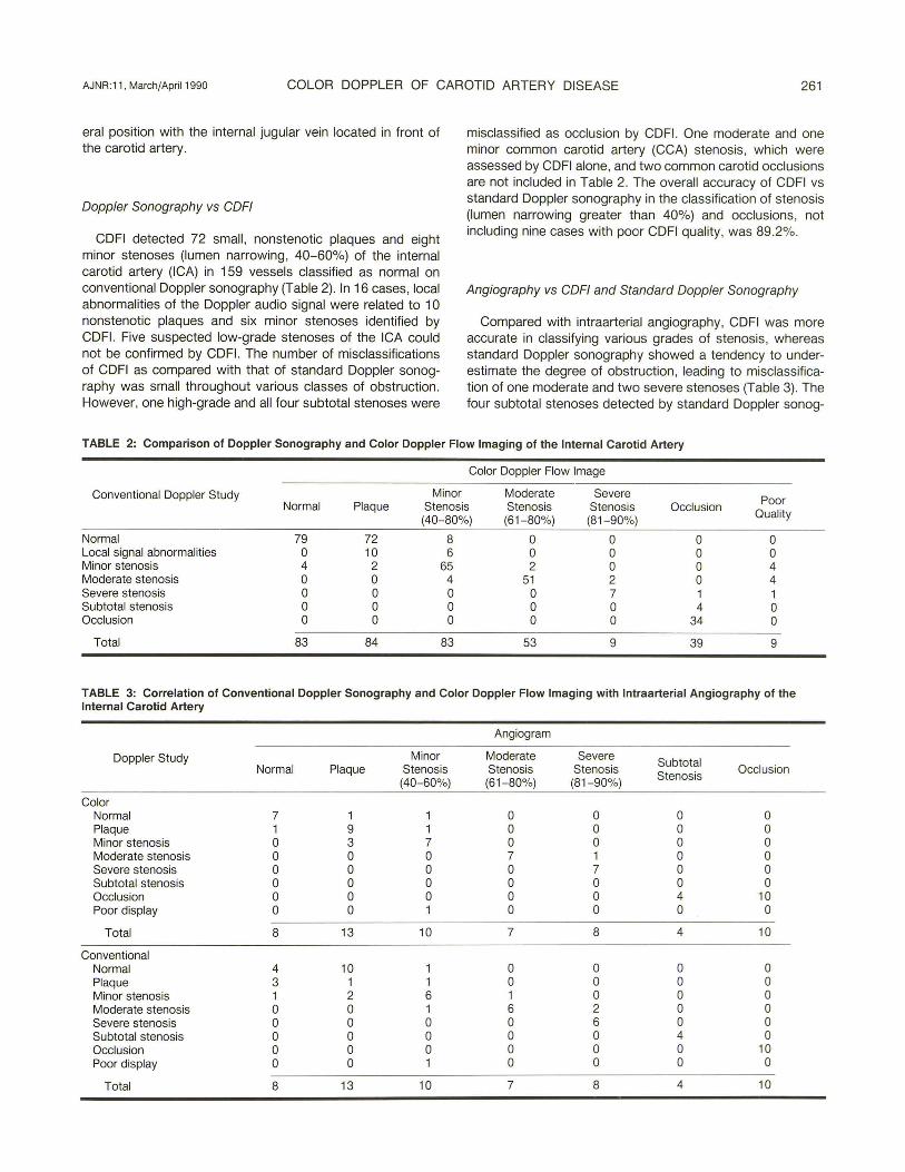
CDFI detected 72 small, nonstenotic plaques and eight minor stenoses (lumen narrowing, 40-60%) of the internal carotid artery (ICA) in 159 vessels classified as normal on conventional Doppler sonography (Table 2). In 16 cases, local abnormalities of the Doppler audio signal were related to 1 0 nonstenotic plaques and six minor stenoses identified by CDFI. Five suspected low-grade stenoses of the ICA could not be confirmed by CDFI. The number of misclassifications of CDFI as compared with that of standard Doppler sonography was small throughout various classes of obstruction . However, one high-grade and all four subtotal stenoses were
misclassified as occlusion by CDFI. One moderate and one minor common carotid artery (CCA) stenosis, which were assessed by CDFI alone, and two common carotid occlusions are not included in Table 2. The overall accuracy of CDFI vs standard Doppler sonography in the classification of stenosis (lumen narrowing greater than 40%) and occlusions, not including nine cases with poor CDFI quality, was 89 .2%.
Angiography vs CDFI and Standard Doppler Sonography
Compared with intraarterial angiography, CDFI was more accurate in classifying various grades of stenosis, whereas standard Doppler sonography showed a tendency to underestimate the degree of obstruction , leading to misclassification of one moderate and two severe stenoses (Table 3). The four subtotal stenoses detected by standard Doppler sonog-
TABLE 2: Comparison of Doppler Sonography and Color Doppler Flow Imaging of the Internal Carotid Artery
Color Doppler Flow Image
Conventional Doppler Study Minor Moderate Severe Poor Normal Plaque Stenosis Stenosis Stenosis Occlusion
Quality (40-80%) (61-80%) (81-90%)
Normal 79 72 8 0 0 0 0 Local signal abnormalities 0 10 6 0 0 0 0 Minor stenosis 4 2 65 2 0 0 4 Moderate stenosis 0 0 4 51 2 0 4 Severe stenosis 0 0 0 0 7 1 1 Subtotal stenosis 0 0 0 0 0 4 0 Occlusion 0 0 0 0 0 34 0
Total 83 84 83 53 9 39 9
TABLE 3: Correlation of Conventional Doppler Sonography and Color Doppler Flow Imaging with Intraarterial Angiography of the Internal Carotid Artery
Angiogram
Doppler Study Minor Moderate Severe Subtotal Normal Plaque Stenosis Stenosis Stenosis Stenosis Occlusion
(40-60%) (61-80%) (81-90%)
Color Normal 7 1 1 0 0 0 0 Plaque 1 9 1 0 0 0 0 Minor stenosis 0 3 7 0 0 0 0 Moderate stenosis 0 0 0 7 1 0 0 Severe stenosis 0 0 0 0 7 0 0 Subtotal stenosis 0 0 0 0 0 0 0 Occlusion 0 0 0 0 0 4 10 Poor display 0 0 1 0 0 0 0
Total 8 13 10 7 8 4 10
Conventional Normal 4 10 1 0 0 0 0 Plaque 3 1 1 0 0 0 0 Minor stenosis 1 2 6 1 0 0 0 Moderate stenosis 0 0 1 6 2 0 0 Severe stenosis 0 0 0 0 6 0 0 Subtotal stenosis 0 0 0 0 0 4 0 Occlusion 0 0 0 0 0 0 10 Poor display 0 0 1 0 0 0 0
Total 8 13 10 7 8 4 10
262 STEINKE ET AL. AJNR:11 , March/Apri11990
raphy but not displayed by CDFI were confirmed by angiography. CDFI identified nine nonstenotic plaques corresponding to the angiographic findings , but in three of 13 plaques CDFI demonstrated increased blood-flow velocity at the lesion site and hence suggested a low-grade stenosis. Standard Doppler sonography. however, failed to diagnose 1 0 of the small plaques. Overall accuracy of CDFI compared with angiography was 80%; the total sensitivity for the detection of carotid artery lesions was 1 00%. Statistical evaluation of the CDFI and conventional Doppler results vs angiography showed that CDFI also had a higher sensitivity, specificity, and accuracy for minor, moderate, and severe stenoses (Table 4).
TABLE 4: Sensitivity, Specificity, and Accuracy of Color Doppler (CDFI) vs Conventional Doppler Study and Angiography and Conventional Doppler Study vs Angiography of Minor (40-60%), Moderate (61-80%), and Severe (81-90%) Stenoses
Comparison 0/o % o/o
Sensitivity Specificity Accuracy
CDFI vs conventional Doppler (n = 360)
Minor 89.0 93.8 91 .3 Moderate 89.5 97.5 94.2 Severe 87.5 98.5 97 .8
CDFI vs angiography (n = 60)
Minor 77.8 100 91 .7 Moderate 100 94.1 95.8 Severe 87.5 100 95.8
Conventional Doppler vs angiography (n = 60)
Minor 66.7 93.3 83.3 Moderate 85.7 82 .3 83.3 Severe 75.0 100 91 .7
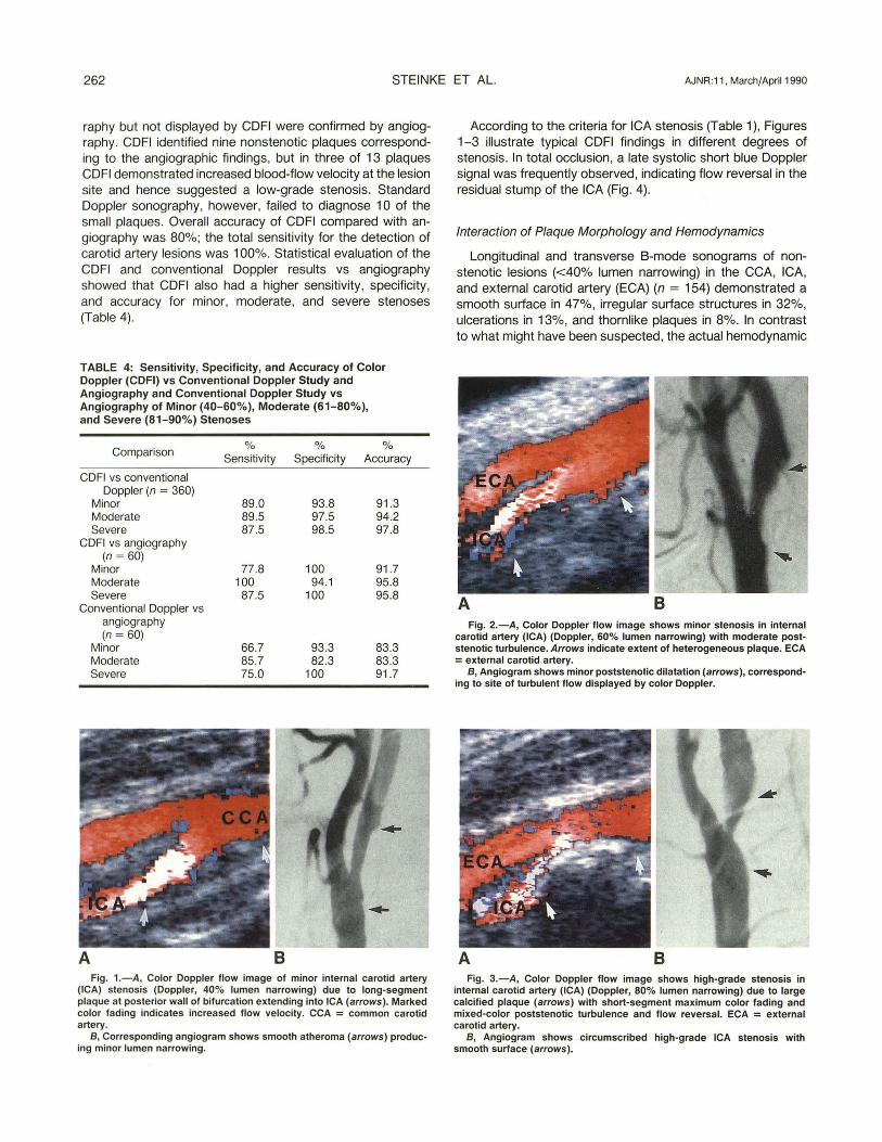
A Fig. 1.-A, Color Doppler flow image of minor internal carotid artery
(ICA) stenosis (Doppler, 40% lumen narrowing) due to long-segment plaque at posterior wall of bifurcation extending into ICA (arrows). Marked color fading indicates increased flow velocity. CCA = common carotid artery.
B, Corresponding angiogram shows smooth atheroma (arrows) producing minor lumen narrowing.
According to the criteria for ICA stenosis (Table 1), Figures 1-3 illustrate typical CDFI findings in different degrees of stenosis. In total occlusion, a late systolic short blue Doppler signal was frequently observed, indicating flow reversal in the residual stump of the ICA (Fig . 4).
Interaction of Plaque Morphology and Hemodynamics
Longitudinal and transverse 8-mode sonograms of nonstenotic lesions (<40% lumen narrowing) in the CCA, ICA, and external carotid artery (EGA) (n = 154) demonstrated a smooth surface in 47%, irregular surface structures in 32%, ulcerations in 13%, and thornlike plaques in 8%. In contrast to what might have been suspected, the actual hemodynamic
A 8 Fig. 2.-A, Color Doppler flow image shows minor stenosis in internal
carotid artery (ICA) (Doppler, 60% lumen narrowing) with moderate post-stenotic turbulence. Arrows indicate extent of heterogeneous plaque. ECA =external carotid artery.
B, Angiogram shows minor poststenotic dilatation (arrows), correspond-ing to site of turbulent flow displayed by color Doppler.
A 8 Fig. 3.-A , Color Doppler flow image shows high-grade stenosis in
internal carotid artery (ICA) (Doppler, 80% lumen narrowing) due to large calcified plaque (arrows) with short-segment maximum color fading and mixed-color poststenotic turbulence and flow reversal. ECA = external carotid artery.
B, Angiogram shows circumscribed high-grade ICA stenosis with smooth surface (arrows).
AJNR:11, March/April1990 COLOR DOPPLER OF CAROTID ARTERY DISEASE 263
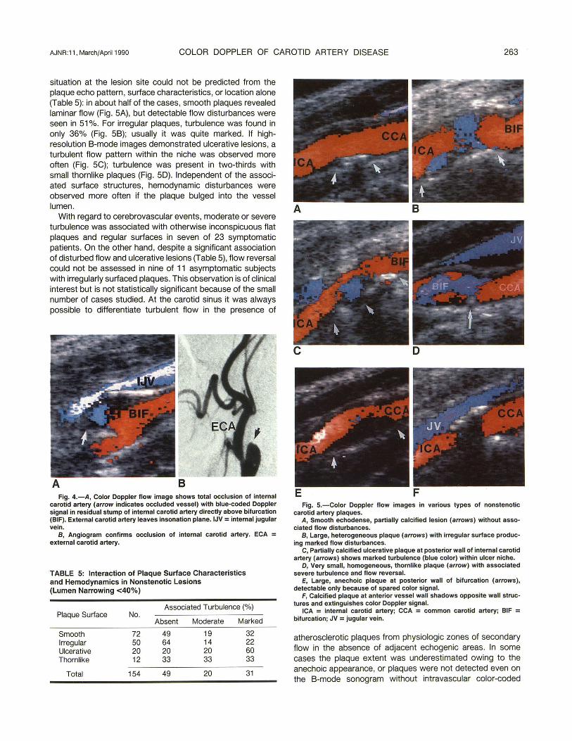
situation at the lesion site could not be predicted from the plaque echo pattern, surface characteristics, or location alone (Table 5): in about half of the cases, smooth plaques revealed laminar flow (Fig . 5A), but detectable flow disturbances were seen in 51%. For irregular plaques, turbulence was found in only 36% (Fig. 5B); usually it was quite marked. If highresolution B-mode images demonstrated ulcerative lesions, a turbulent flow pattern within the niche was observed more often (Fig . 5C); turbulence was present in two-thirds with small thornlike plaques (Fig. 5D). Independent of the associated surface structures, hemodynamic disturbances were observed more often if the plaque bulged into the vessel lumen.
With regard to cerebrovascular events, moderate or severe turbulence was associated with otherwise inconspicuous flat plaques and regular surfaces in seven of 23 symptomatic patients. On the other hand, despite a significant association of disturbed flow and ulcerative lesions (Table 5), flow reversal could not be assessed in nine of 11 asymptomatic subjects with irregularly surfaced plaques. This observation is of clinical interest but is not statistically significant because of the small number of cases studied. At the carotid sinus it was always possible to differentiate turbulent flow in the presence of
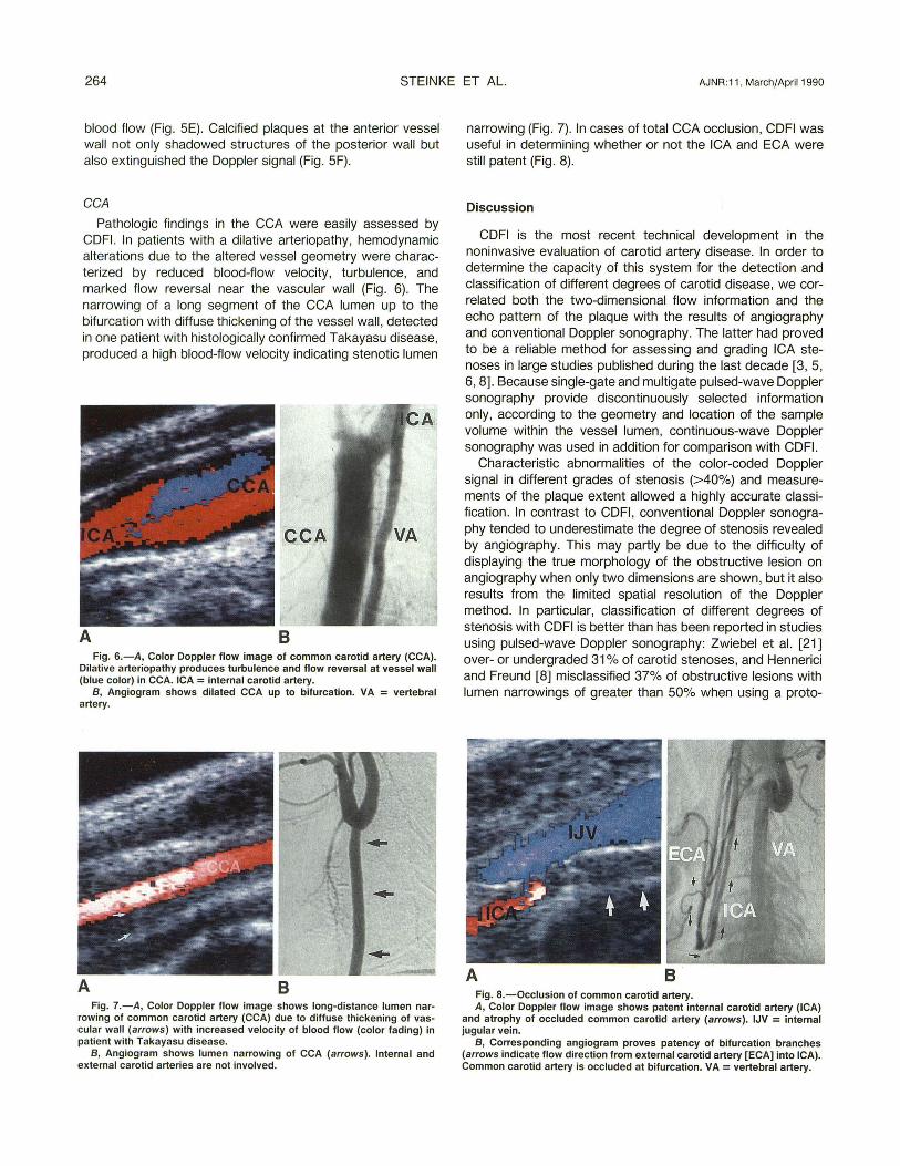
A B Fig. 4.-A, Color Doppler flow image shows total occlusion of internal
carotid artery (arrow indicates occluded vessel) with blue-coded Doppler signal in residual stump of internal carotid artery directly above bifurcation (BIF). External carotid artery leaves insonation plane. IJV = internal jugular vein.
B, Angiogram confirms occlusion of internal carotid artery. ECA = external carotid artery.
TABLE 5: Interaction of Plaque Surface Characteristics and Hemodynamics in Nonstenotic Lesions (Lumen Narrowing <40%)
Associated Turbulence (%) Plaque Surface No.
Absent Moderate Marked
Smooth 72 49 19 32 Irregular 50 64 14 22 Ulcerative 20 20 20 60 Thornlike 12 33 33 33
Total 154 49 20 31
A B
E F Fig. 5.-Color Doppler flow images in various types of nonstenotic
carotid artery plaques. A, Smooth echodense, partially calcified lesion (arrows) without asso
ciated flow disturbances. B, Large, heterogeneous plaque (arrows) with irregular surface produc
ing marked flow disturbances. C, Partially calcified ulcerative plaque at posterior wall of internal carotid
artery (arrows) shows marked turbulence (blue color) within ulcer niche. D, Very small, homogeneous, thornlike plaque (arrow) with associated
severe turbulence and flow reversal. E, Large, anechoic plaque at posterior wall of bifurcation (arrows),
detectable only because of spared color signal. F, Calcified plaque at anterior vessel wall shadows opposite wall struc
tures and extinguishes color Doppler signal. ICA = internal carotid artery; CCA = common carotid artery; BIF =
bifurcation; JV = jugular vein.
atherosclerotic plaques from physiologic zones of secondary flow in the absence of adjacent echogenic areas. In some cases the plaque extent was underestimated owing to the anechoic appearance, or plaques were not detected even on the B-mode sonogram without intravascular color-coded
264 STEINKE ET AL. AJNR:11 , March/April1990
blood flow (Fig. 5E). Calcified plaques at the anterior vessel wall not only shadowed structures of the posterior wall but also extinguished the Doppler signal (Fig. 5F).
CCA
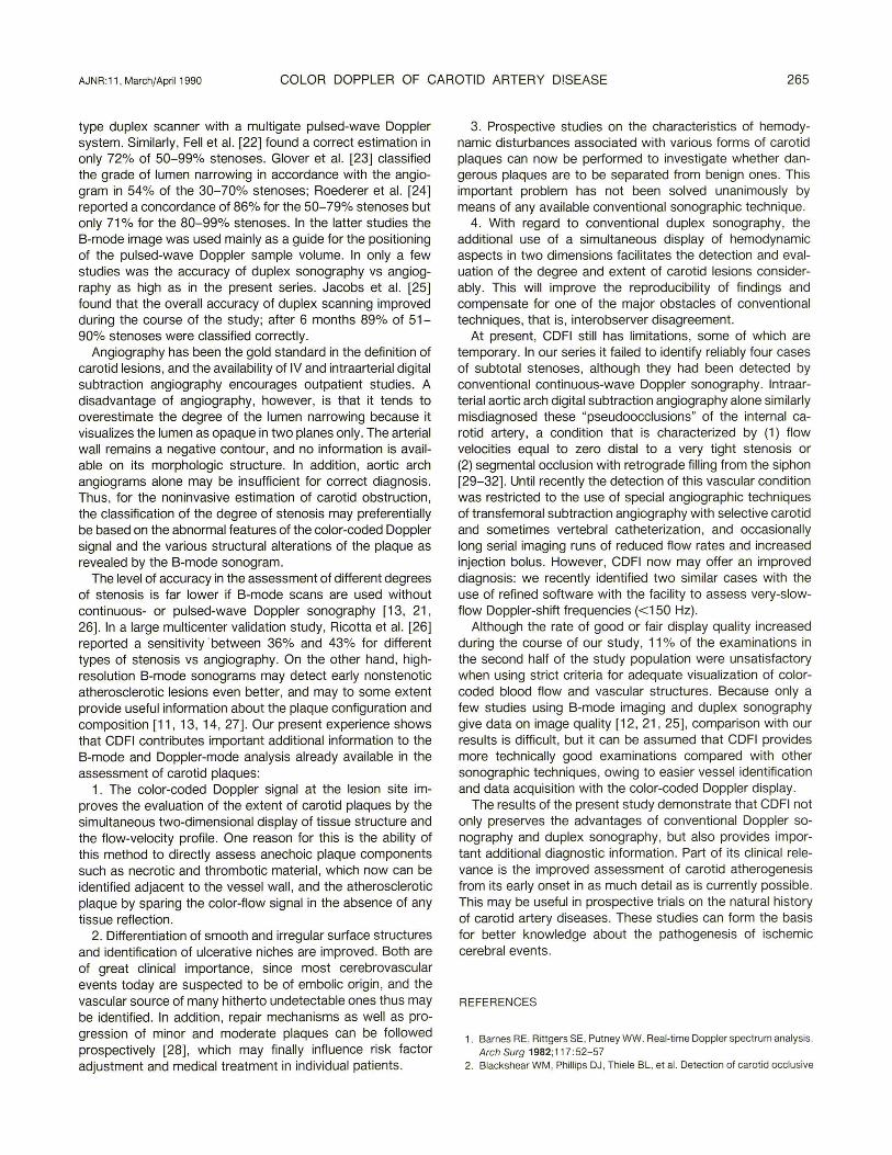
Pathologic findings in the CCA were easily assessed by CDFI. In patients with a dilative arteriopathy, hemodynamic alterations due to the altered vessel geometry were characterized by reduced blood-flow velocity , turbulence, and marked flow reversal near the vascular wall (Fig . 6). The narrowing of a long segment of the CCA lumen up to the bifurcation with diffuse thickening of the vessel wall , detected in one patient with histologically confirmed Takayasu disease, produced a high blood-flow velocity indicating stenotic lumen
8 Fig. 6.-A, Color Doppler flow image of common carotid artery (CCA).
Dilative arteriopathy produces turbulence and flow reversal at vessel wall (blue color) in CCA. ICA = internal carotid artery.
B, Angiogram shows dilated CCA up to bifurcation. VA = vertebral artery.
A 8 Fig. 7.-A , Color Doppler flow image shows long-distance lumen nar
rowing of common carotid artery (CCA) due to diffuse thickening of vascular wall (arrows) with increased velocity of blood flow (color fading) in patient with Takayasu disease.
B, Angiogram shows lumen narrowing of CCA (arrows). Internal and external carotid arteries are not involved.
narrowing (Fig . 7). In cases of total CCA occlusion, CDFI was useful in determining whether or not the ICA and EGA were still patent (Fig. 8).
Discussion
CDFI is the most recent technical development in the noninvasive evaluation of carotid artery disease. In order to determine the capacity of this system for the detection and classification of different degrees of carotid disease, we correlated both the two-dimensional flow information and the echo pattern of the plaque with the results of angiography and conventional Doppler sonography. The latter had proved to be a reliable method for assessing and grading ICA stenoses in large studies published during the last decade [3, 5, 6, 8]. Because single-gate and multigate pulsed-wave Doppler sonography provide discontinuously selected information only, according to the geometry and location of the sample volume within the vessel lumen, continuous-wave Doppler sonography was used in addition for comparison with CDFI.
Characteristic abnormalities of the color-coded Doppler signal in different grades of stenosis (>40%) and measurements of the plaque extent allowed a highly accurate classification. In contrast to CDFI , conventional Doppler sonography tended to underestimate the degree of stenosis revealed by angiography. This may partly be due to the difficulty of displaying the true morphology of the obstructive lesion on angiography when only two dimensions are shown, but it also results from the limited spatial resolution of the Doppler method. In particular, classification of different degrees of stenosis with CDFI is better than has been reported in studies using pulsed-wave Doppler sonography: Zwiebel et al. [21] over- or undergraded 31% of carotid stenoses, and Hennerici and Freund [8] misclassified 37% of obstructive lesions with lumen narrowings of greater than 50% when using a proto-
Fig. a.-Occlusion of common carotid artery. A, Color Doppler flow image shows patent internal carotid artery (ICA)
and atrophy of occluded common carotid artery (arrows). IJV = internal jugular vein.
B, Corresponding angiogram proves patency of bifurcation branches (arrows indicate flow direction from external carotid artery [ECA] into ICA). Common carotid artery is occluded at bifurcation. VA= vertebral artery.
AJNR :11 , March/April1990 COLOR DOPPLER OF CAROTID ARTERY DISEASE 265
type duplex scanner with a multigate pulsed-wave Doppler system. Similarly, Fell et al. [22) found a correct estimation in only 72% of 50-99% stenoses. Glover et al. [23] classified the grade of lumen narrowing in accordance with the angiogram in 54% of the 30-70% stenoses; Roederer et al. [24) reported a concordance of 86% for the 50-79% stenoses but only 71% for the 80-99% stenoses. In the latter studies the B-mode image was used mainly as a guide for the positioning of the pulsed-wave Doppler sample volume. In only a few studies was the accuracy of duplex sonography vs angiography as high as in the present series. Jacobs et al. [25) found that the overall accuracy of duplex scanning improved during the course of the study; after 6 months 89% of 51-90% stenoses were classified correctly.
Angiography has been the gold standard in the definition of carotid lesions, and the availability of IV and intraarterial digital subtraction angiography encourages outpatient studies. A disadvantage of angiography, however, is that it tends to overestimate the degree of the lumen narrowing because it visualizes the lumen as opaque in two planes only. The arterial wall remains a negative contour, and no information is available on its morphologic structure. In addition, aortic arch angiograms alone may be insufficient for correct diagnosis. Thus, for the noninvasive estimation of carotid obstruction, the classification of the degree of stenosis may preferentially be based on the abnormal features of the color-coded Doppler signal and the various structural alterations of the plaque as revealed by the B-mode sonogram.
The level of accuracy in the assessment of different degrees of stenosis is far lower if B-mode scans are used without continuous- or pulsed-wave Doppler sonography [13, 21, 26). In a large multicenter validation study, Ricotta et al. [26] reported a sensitivity ·between 36% and 43% for different types of stenosis vs angiography. On the other hand, highresolution B-mode sonograms may detect early nonstenotic atherosclerotic lesions even better, and may to some extent provide useful information about the plaque configuration and composition [11 , 13, 14, 27). Our present experience shows that CDFI contributes important additional information to the B-mode and Doppler-mode analysis already available in the assessment of carotid plaques:
1. The color-coded Doppler signal at the lesion site improves the evaluation of the extent of carotid plaques by the simultaneous two-dimensional display of tissue structure and the flow-velocity profile. One reason for this is the ability of this method to directly assess anechoic plaque components such as necrotic and thrombotic material, which now can be identified adjacent to the vessel wall , and the atherosclerotic plaque by sparing the color-flow signal in the absence of any tissue reflection.
2. Differentiation of smooth and irregular surface structures and identification of ulcerative niches are improved. Both are of great clinical importance, since most cerebrovascular events today are suspected to be of embolic origin, and the vascular source of many hitherto undetectable ones thus may be identified. In addition, repair mechanisms as well as progression of minor and moderate plaques can be followed prospectively [28] , which may finally influence risk factor adjustment and medical treatment in individual patients.
3. Prospective studies on the characteristics of hemodynamic disturbances associated with various forms of carotid plaques can now be performed to investigate whether dangerous plaques are to be separated from benign ones . This important problem has not been solved unanimously by means of any available conventional sonographic technique.
4. With regard to conventional duplex sonography, the additional use of a simultaneous display of hemodynamic aspects in two dimensions facilitates the detection and evaluation of the degree and extent of carotid lesions considerably. This will improve the reproducibility of findings and compensate for one of the major obstacles of conventional techniques, that is, interobserver disagreement.
At present, CDFI still has limitations, some of which are temporary. In our series it failed to identify reliably four cases of subtotal stenoses, although they had been detected by conventional continuous-wave Doppler sonography. Intraarterial aortic arch digital subtraction angiography alone similarly misdiagnosed these "pseudoocclusions" of the internal carotid artery, a condition that is characterized by (1) flow velocities equal to zero distal to a very tight stenosis or (2) segmental occlusion with retrograde filling from the siphon [29-32] . Until recently the detection of this vascular condition was restricted to the use of special angiographic techniques of transfemoral subtraction angiography with selective carotid and sometimes vertebral catheterization , and occasionally long serial imaging runs of reduced flow rates and increased injection bolus. However, CDFI now may offer an improved diagnosis: we recently identified two similar cases with the use of refined software with the facility to assess very-slowflow Doppler-shift frequencies (< 150Hz).
Although the rate of good or fair display quality increased during the course of our study, 11 % of the examinations in the second half of the study population were unsatisfactory when using strict criteria for adequate visualization of colorcoded blood flow and vascular structures. Because only a few studies using B-mode imaging and duplex sonography give data on image quality [12, 21 , 25) , comparison with our results is difficult, but it can be assumed that CDFI provides more technically good examinations compared with other sonographic techniques, owing to easier vessel identification and data acquisition with the color-coded Doppler display.
The results of the present study demonstrate that CDFI not only preserves the advantages of conventional Doppler sonography and duplex sonography, but also provides important additional diagnostic information. Part of its clinical relevance is the improved assessment of carotid atherogenesis from its early onset in as much detail as is currently possible. This may be useful in prospective trials on the natural history of carotid artery diseases. These studies can form the basis for better knowledge about the pathogenesis of ischemic cerebral events.
REFERENCES
1. Barnes RE, Rittgers SE, Putney WW. Real-time Doppler spectrum analysis. Arch Surg 1982;11 7:52- 57
2. Blackshear WM, Phillips DJ, Thiele BL, et al. Detection of carotid occlusive
266 STEINKE ET AL. AJNR:11 , March/April1990
disease by ultrasonic imaging and pulsed Doppler spectrum analysis. Surgery 1979;86 :698-706
3. Hennerici M, Aulich A, Sandmann W, Freund HJ. Incidence of asymptomatic extracranial arterial disease. Stroke 1981 ;12:750-758
4. Johnston KW, Baker WH , Burnham SJ, Hayes AC, Kupper CA, Poole MA. Quantitative analysis of continuous-wave Doppler spectral broadening for the diagnosis of carotid disease: results of a multicenter study. J Vase Surg 1986;4:493-504
5. Reneman RS, Spencer MP. Local Doppler audio spectra in normal and stenosed carotid arteries in man. Ultrasound Med Biol1979;5: 1-11
6. Trockel U, Hennerici M, Aulich A, Sandmann W. The superiority of combined continuous wave Doppler examinations over periorbital Doppler for the detection of extracranial carotid disease. J Neural Neurosurg Psychiatry 1984;47:43-50
7. Arbeille P, Lapierre F, Patat F, et al. Evaluation du degre des stenoses carotidiennes par l'analyse spectrale du signal Doppler. Arch Mal Coeur 1984;77 :1097- 1107
8. Hennerici M, Freund HJ. Efficacy of cw-Doppler and duplex system examinations for the evaluation of extracranial carotid disease. JCU 1984; 12:155-161
9. Keagy BA, Pharr WF, Thomas D, Bowles DE. A quantitative method for the evaluation of spectral analysis patterns in carotid artery stenosis. Ultrasound Med Biol1982;8:625-630
10. Rittgers SE, Thornhill BM, Barnes RW. Quantitative analysis of carotid artery spectral waveforms: diagnostic value of parameters. Ultrasound Med Biol1983;9:255-264
11 . Bluth El , Kay D, Merritt CRB, et al. Sonographic characterization of carotid plaque: detection of hemorrhage. AJR 1986;146: 1061-1065
12. Camerata AJ, Cranley JJ , Cook SE. Real-time B-mode carotid imaging in diagnosis of cerebrovascular disease. Surgery 1981;89:718-729
13. Hennerici M, Reifschneider G, Trockel U, Aulich A. Detection of early atherosclerotic lesions by duplex scanning of the carotid artery. JCU 1984;12 :455-464
14. Reilly LM, Lusby RJ, Hughes L, Ferrell LD, Stoney RJ, Ehrenfeld WK. Carotid plaque histology using real-time ultrasonography. Am J Surg 1983;146: 188-189
15. Middleton WD, Foley WD, Lawson TL. Flow reversal in the normal carotid bifurcation: color Doppler flow imaging analysis. Radiology 1988;167: 207-210
16. Steinke W, Kloetzsch C, Hennerici M. Variability of flow patterns in the normal carotid bifurcation. Atherosclerosis (in press)
17. LoGerfo FW. Hemodynamics and the arterial wall. J Vase Surg 1989;9: 380-381
18. Nicholls SC, Phillips DJ , Primozich JF, et al. Diagnostic significance of flow
separation in the carotid bulb. Stroke 1989;20 : 175-182 19. Middleton WD, Foley WD, Lawson TL. Color-flow Doppler imaging of
carotid artery abnormalities. AJR 1988; 1 50 : 419-425 20. Hallam MJ, Reid JM, Cooperberg PL. Color-flow Doppler and conventional
duplex scanning of the carotid bifurcation: prospective, double-blind, correlative study. AJR 1989;152: 1101-1105
21. Zwiebel WJ, Austin CW, Sackett JF, Strother CM. Correlation of highresolution B-mode and continuous-wave Doppler sonography with arteriography in the diagnosis of carotid stenosis. Radiology 1983;149 : 523-532
22. Fell G, Phillips DJ, Chikos PM, Harley JD, Thiele BL, Strandness DE Jr. Ultrasonic duplex scanning for disease of the carotid artery. Circulation 1981;64 : 1191-1195
23. Glover JL, Bendick PJ , Jackson VP, Becker GJ, Dilley RS, Holden RW. Duplex ultrasonography, digital subtraction angiography and conventional angiography in assessing carotid atherosclerosis. Arch Surg 1984;119: 664-669
24. Roederer GO, Langlois YE, Jager KA, et al. The natural history of carotid arterial disease in asymptomatic patients with cervical bruits. Stroke 1984;15: 605-613
25. Jacobs NM, Grant EG, Schellinger D, Byrd MC, Richardson JD, Cohan SL. Duplex carotid sonography: criteria for stenosis, accuracy, and pitfalls. Radiology 1985;154:385-391
26. Ricotta JJ, Bryan FA, Bond G, et al. Multicenter validation study of realtime (B-mode) ultrasound, arteriography, and pathologic examination. J Vase Surg 1987;6:512-520
27. O'Donnell TF, Erdoes L, Mackey WC, et al. Correlation of B-mode ultrasound imaging and arteriography with pathologic findings at carotid endarterectomy. Arch Surg 1985;120:443-449
28. Hennerici M, Rautenberg W, Trockel U, Kladetzky RG. Spontaneous progression and regression of small carotid atheroma. Lancet 1985;1: 1415-1419
29. Countee RW, Vijayanathan T. Reconstruction of "totally" occluded internal carotid arteries. Angiographic and technical considerations. J Neurosurg 1979;50:747-757
30. Heros RC, Sekhar LN. Diagnostic and therapeutic alternatives in patients with symptomatic "carotid occlusion" referred for extracranial-intracranial bypass surgery. J Neurosurg 1981;54:790-796
31. Gabrielsen TO, Seeger JF, Knake JE, Burke DP, Stilwill EW. The newly occluded internal carotid artery. A diagnostic trap. Radiology 1981; 138:611-618
32. Ringelstein EB, Zeumer H, Angelou D. The pathogenesis of strokes from internal carotid artery occlusion. Diagnostic and therapeutical implications. Stroke 1983; 14: 867-87 4