
Cardiovascular System Nursing 1120 By: Diana Blum RN MSN Metropolitan Community College 1.
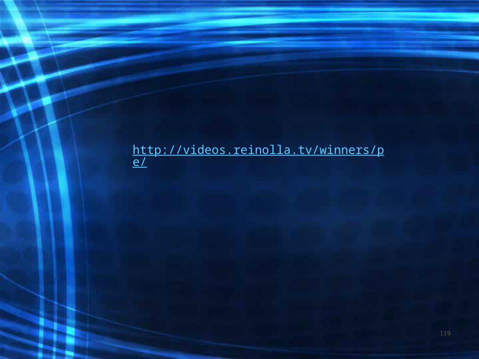
176
Cardiovascular System Nursing 1120 By: Diana Blum RN MSN Metropolitan Community College 1
-
Upload
tobias-barnett -
Category
Documents
-
view
214 -
download
0
Transcript of Cardiovascular System Nursing 1120 By: Diana Blum RN MSN Metropolitan Community College 1.

Cardiovascular System
Nursing 1120By: Diana Blum RN MSN
Metropolitan Community College
1
2

3

Coronary Arteries
• Two major coronary arteries– arise from the aorta beyond the aortic
valve. – Blood flows to the coronary arteries
during diastole• Left main, LAD, Circumflex feeds most of
Left side of the heart• Right feeds SA node, AV node, RA, RL
4

5

6
Collateral circulation is a network of tiny blood vessels, and, under normal conditions, not open. When the coronary arteries narrow to the point that blood flow to the heart muscle is limited (coronary artery disease), collateral vessels may enlarge and become active. This allows blood to flow around the blocked artery to another artery nearby or to the same artery past the blockage, protecting the heart tissue from injury.
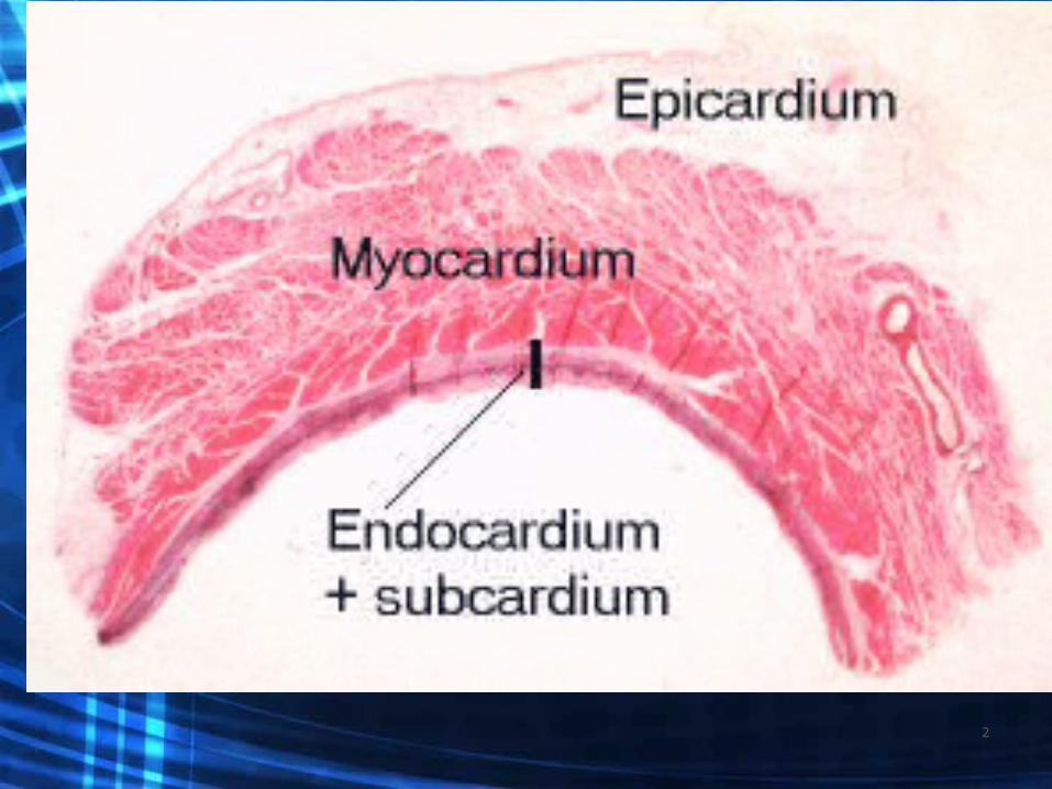
Conduction Continued
7
Cardiac Cycle
• Contraction and relaxation of the heart: • Diastole:• Systole:
8
• Video Mysterious Heart Volume 2 chapter 3
9

Cardiac Output
• the volume of blood ejected by the heart each minute and is determined by stroke volume and the heart rate.
• Normal stroke volume is 60-100 ml• Normal cardiac output is 4 to 8 L / min• (CO = HR X SV)
10

Factors affecting Stroke Volume• Preload: the amount of blood remaining in
the ventricles at the end of diastole or the pressure generated at the end of diastole
• Contractility: is the ability of the cardiac muscle fibers to shorten and produce a muscle contraction. (Inotropic, + or -)
• Afterload: amount of pressure the Ventricle must overcome to eject blood volume out
11
Heart Rate
• SA node : pacemaker of heart 60-100 bpm
• AV node : 40 -60 bpm• Heart is innervated by sympathetic and
parasympathetic nervous system– Sympathetic: speeds HR, and increases force
of contraction– Parasympathetic: slows HR and force
12

Heart Tones• Murmur: Produced by turbulent sounds across
valves– Rub: inflamed pericardium-best heard along left
sternal border– S3 murmur: sounds like “Kentucky”– S4 murmur: sounds like “Mississippi”
http://www.blaufuss.org/ http://www.med.ucla.edu/wilkes/Rubintro.htm
13

Health History
14

Present Illness
15

Past Medical History
16

Family History:
17

Review of systems• weight gain• fatigue• dyspnea• cough• orthopnea• palpitations• chest pain• fainting• concentrated urine• edema
18
Functional assessment• effects of illness on ADLs and rest patterns• smoker• diet• stress• coping
19
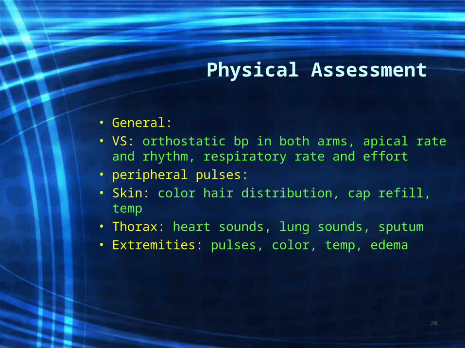
Physical Assessment
• General:• VS: orthostatic bp in both arms, apical rate
and rhythm, respiratory rate and effort• peripheral pulses:• Skin: color hair distribution, cap refill, temp• Thorax: heart sounds, lung sounds, sputum• Extremities: pulses, color, temp, edema
20

Age Related Changes
• Heart less able to adapt to changing needs related to activity
• Valves thicken and stiffen• # of pacemaker cells decrease• Nerve fibers decrease• Frequent dysrhythmias
21

Diagnostic Tests
• EKG: rate, rhythm, ischemia (T-inverted), injury (ST segment elevation), arrhythmias, strain, infarction (q wave)
• Echocardiogram: (TEE) sound wave test detects size of chambers, valve integrity, flow, wall motion, Cardiac Output
22
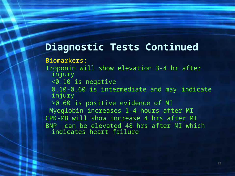
Diagnostic Tests ContinuedBiomarkers: Troponin will show elevation 3-4 hr after injury
<0.10 is negative0.10-0.60 is intermediate and may indicate
injury>0.60 is positive evidence of MI
Myoglobin increases 1-4 hours after MI CPK-MB will show increase 4 hrs after MIBNP can be elevated 48 hrs after MI which
indicates heart failure
23
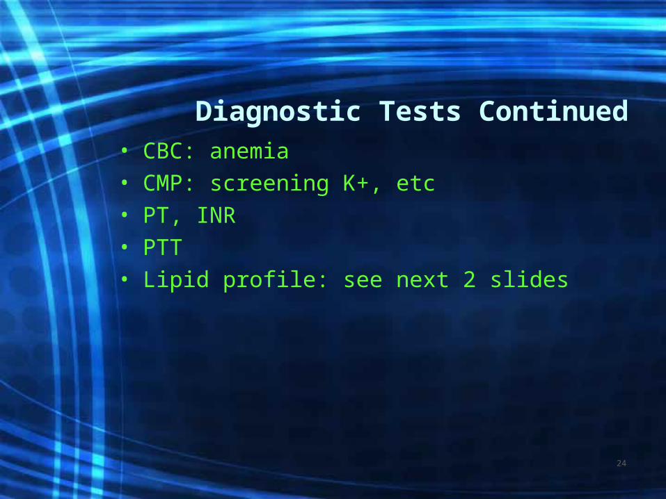
Diagnostic Tests Continued• CBC: anemia • CMP: screening K+, etc• PT, INR• PTT• Lipid profile: see next 2 slides
24
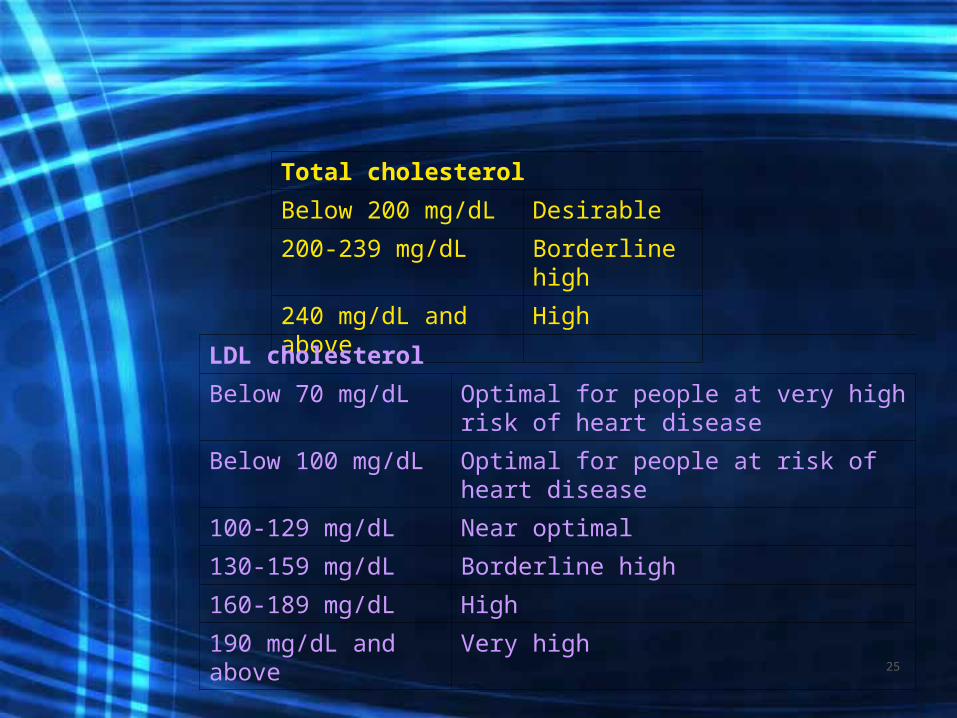
Total cholesterol
Below 200 mg/dL Desirable
200-239 mg/dL Borderline high
240 mg/dL and above
High
LDL cholesterol
Below 70 mg/dL Optimal for people at very high risk of heart disease
Below 100 mg/dL Optimal for people at risk of heart disease
100-129 mg/dL Near optimal
130-159 mg/dL Borderline high
160-189 mg/dL High
190 mg/dL and above
Very high25

HDL cholesterol
Below 40 mg/dL Poor
40-59 mg/dL Better
60 mg/dL and above
Best
Triglycerides
Below 150 mg/dL
Desirable
150-199 mg/dL
Borderline high
200-499 mg/dL
High
500 or above Very high26

Diagnostic Tests Continued• ABG: assess acid/base levels• Pulse Oximetry: generally >92%• Holter monitoring: 24+ hr of EKG + events• Stress test: treadmill or pharmacological• Cardiac Catheterization: invasive, NPO 6-8h,
consent. Visualizes chambers, valves, arteries, pressures, CO
• Heart-CT scan: assesses CAD, MRI• Nuclear scans: assess heart muscle viability• EPS: NPO, consent, IV, assess electrical activity
27

• http://preop.medselfed.com/asp/center.asp?centerId=heart&partnerId=preop&id=&cachedate=&emailId=&affId=&campId=&hideNav=
28

CADVideo-mysterious heart volume 3 chapter 2
29

Etiology of CAD
• CAD occurs when the intimal lining of the coronaries begin to plaque resulting in jagged edges and narrowed passageway for blood flow
• Atherosclerosis results in impaired blood flow to the heart muscle
30

Risk Factors for CAD
Non-controllable-
Controllable-
31

s/s of CAD
• Angina which results from a lack of 0xygen to the heart muscle– 4Es=
• Weakness, diaphoresis, SOB• N/V
32

MI: Myocardial Infarction• Occlusion of a coronary artery resulting in
necrosis of the heart muscle.• Risk factors: same as for CAD• Pathophysiology: AMI-over 4-6 hrs ischemia
injury and infarction develop. Ischemia=lack of 02 to heart muscle, if not relieved=injury. After 20 min of ischemia=infarction
• Main S/S: chest pain and accompanying S/S
33
• Within 24 hours after infarction, healing begins, collateral circulation begins.
• 10-14 days after MI=extension of MI may occur due to myocardial tissue vulnerability to stress
• Complete scar formation and healing takes about 6 weeks
• Video- mysterious heart volume 1 chapter 2
34
Data Collection
Same as for CAD but will assess symptom of chest pain with accompanying s/s
May have EKG changes with or with-out ST-T wave changes or Q wave changes
Cardiac Bio-markers (Troponin, Myoglobin, CPK, CKMB)
May proceed with Echocardiogram to assess if wall motion sluggish
May go to cath lab
35

Angina or MI• Angina without MI} often relieved with rest and NTG• Angina with MI } may be relieved with rest, NTG, 02, MS, rescue
angioplasty, etc.• Think MONA
– Morphine – Oxygen– Nitroglycerin– Aspirinhttp://www.youtube.com/watch?v=4GlQmTlP2jE&feature=related
http://www.youtube.com/watch?v=rEqw3AK-M_g
36

Treatment continued…video mysterious heart volume 3 chapter 3-7
ASA:MS:Beta-blockerACE inhibitor:
37
Treatment continued– May need antiarrhythmic meds
• like what???
– Stool softeners to reduce valsalva maneuver and prevent constipation r/t narcotic use and bed rest
– Treat: HTN, DM other co-morbid illnesses– Cardiac Rehab to follow
38

Treatments
• Low fat low cholesterol diet• Prescribed exercise program 5-7 days a
week• Knows correct use of NTG for angina• Management of DM, HTN• Stop smoking• Medications to reduce work load or dilate
39

Low salt diet (<2000mg) does not include:
• Soups:
• Snacks:
40

Low fat <30% Low cholesterol <200mg• Lean meat: skinless• Dairy limited: egg beaters, skim milk• Olive oil, canola oil• Avoid: fried, fatty or heavily marbled meats,
sausage, lunch meat, spareribs, frankfurters, salt pork, canned fish in oil, yolks, duck. Cream sauces, gravy, buttered vegetables, sweet rolls, other processed foods
41

Exercise
• 5-7 X week is goal to include stretches with warm-up, progressive walking program, light weights, stretches with cool down.
• Strengthens heart muscle, reduces BP, BS, weight, stress, tension, appetite, LDLs.
• Increases HDLs, energy and self esteem and improves immune system
42
Principles of Exercise
• Practice on regular basis• Know how to do own pulse• Strive for target heart rate• Stop if chest pain occurs• Complications: CHF &
Dysrhythmias43
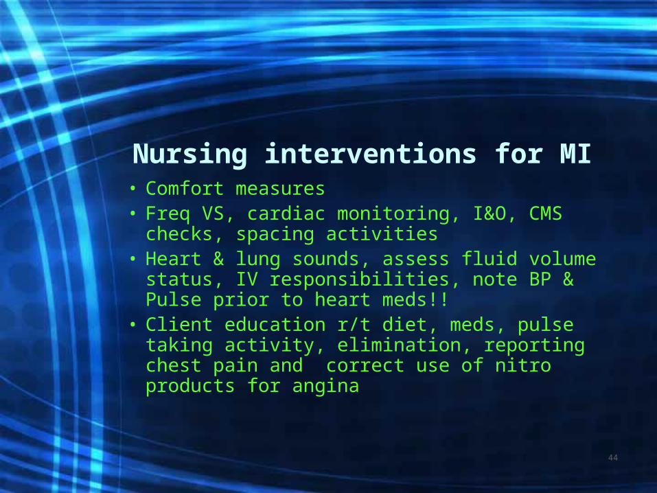
Nursing interventions for MI• Comfort measures• Freq VS, cardiac monitoring, I&O, CMS
checks, spacing activities• Heart & lung sounds, assess fluid volume
status, IV responsibilities, note BP & Pulse prior to heart meds!!
• Client education r/t diet, meds, pulse taking activity, elimination, reporting chest pain and correct use of nitro products for angina
44

Medications for Heart Disease
• Anti-Anginal:• Anti-Hypertensive:• Anti-Arrhythmic: l• Cardiac glycoside:
45
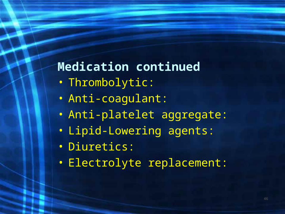
Medication continued• Thrombolytic:• Anti-coagulant:• Anti-platelet aggregate:• Lipid-Lowering agents:• Diuretics:• Electrolyte replacement:
46
Medication continued
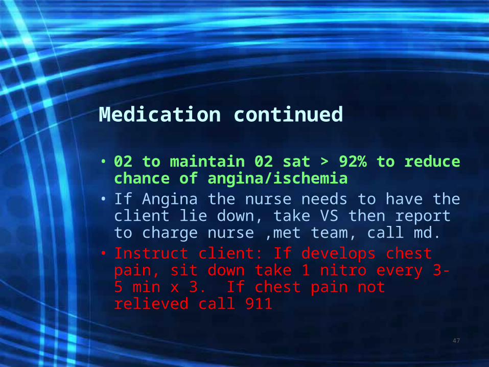
• 02 to maintain 02 sat > 92% to reduce chance of angina/ischemia
• If Angina the nurse needs to have the client lie down, take VS then report to charge nurse ,met team, call md.
• Instruct client: If develops chest pain, sit down take 1 nitro every 3- 5 min x 3. If chest pain not relieved call 911
47

•POST MI Complications
48
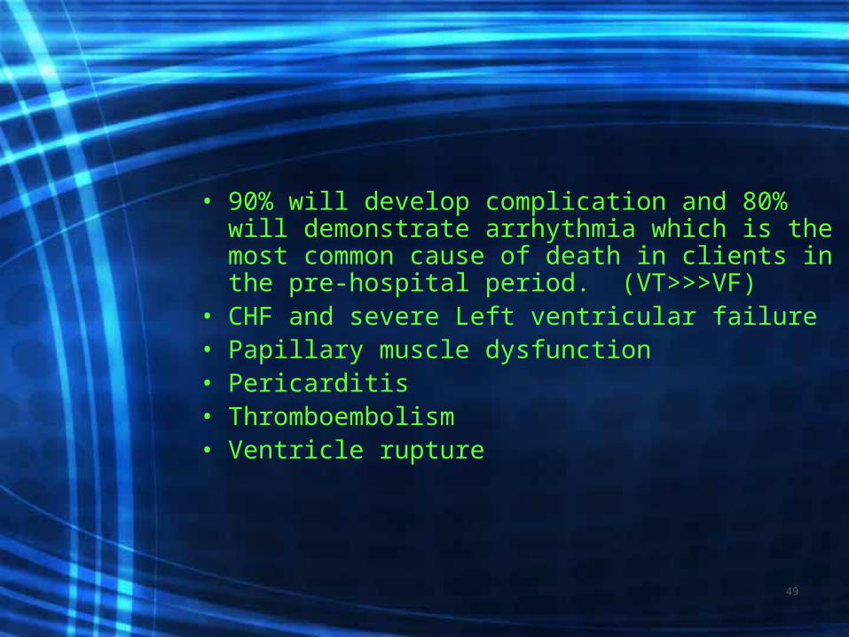
• 90% will develop complication and 80% will demonstrate arrhythmia which is the most common cause of death in clients in the pre-hospital period. (VT>>>VF)
• CHF and severe Left ventricular failure• Papillary muscle dysfunction• Pericarditis• Thromboembolism • Ventricle rupture
49

Nursing Diagnosis
• Decreased cardiac output r/t Dysrhythmias
• Acute Pain r/t lack of 02 to myocardium
• Anxiety r/t to feeling of doom, lack of understanding of medical diagnosis
50
•Surgical Procedures
51
• New in 1958 having purpose to restore the regular rhythm and to improve tissue perfusion and cardiac output.
• Temporary / permanent• Single chamber or double chamber• Teach client how to take 1 min pulse, s/s
to report to MD-dizzy, angina, dyspnea• Carry card and know precautions
Pacemaker
52

Video- mysterious heart volume 2 chapter 4
53
AICD
• Implantable defibrillator to correct a life threatening rhythm disturbance.– Has pacemaker back-up.
• check battery q2months• Instruct on how to take pulse for 1 min• s/s to report to MD: firing, s/s of dizziness,
dyspnea, weakness, carry card and wear bracelet, CPR for family. Know precautions
54

• Nursing care for client with newly implanted pacemaker or AICD.– Assess cardiac monitor for capture/pacing
(pacer)– VS post-op then q 4 hours, IV, bed rest till am– Dressing dry and intact until AM then often
may remove. Increase activity progressively– Instruct client not to raise arm above shoulder
for 5days. May shower in 5 days
55

Angioplasty with Stent
• Procedure done at the time of cardiac cath.
• Balloon angioplasty is accomplished to widen or open specific coronary vessel-stent is inserted to maintain patency of the vessel.
• pre-procedure Plavix given with follow up Plavix
56
http://preop.medselfed.com/asp/center.asp?centerId=heart&partnerId=preop&id=&cachedate=&emailId=&affId=&campId=&hideNav=
EP with Ablation
• Mapping of myocardial tissue to determine irritable focus.
• Low voltage current delivered to ablate tissue causing SVT or VT
• 90% effective• http://video.google.com/videoplay?do
cid=5590000557631435292
57

Nursing Care
• NPO prior• Coumadin stopped 4 days prior, Heparin 4
hours prior• Post – procedure same as heart cath
– Cardiac monitoring– Muscloskeletal and groin checks– VS– Ambulate prior to discharge
58
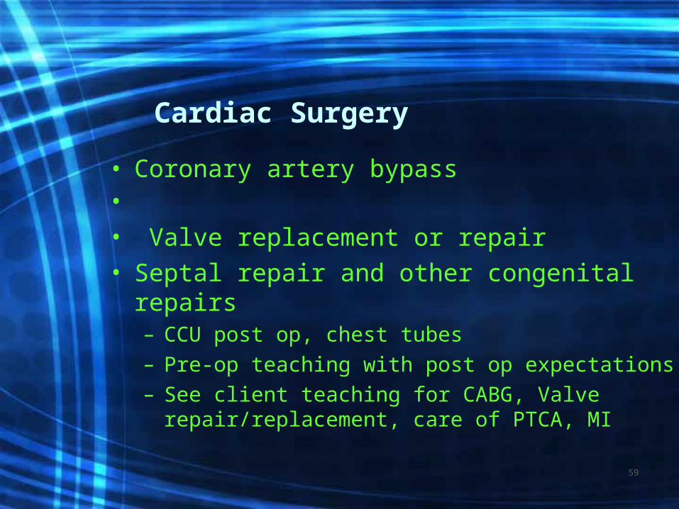
Cardiac Surgery
• Coronary artery bypass• • Valve replacement or repair• Septal repair and other congenital repairs
– CCU post op, chest tubes– Pre-op teaching with post op expectations– See client teaching for CABG, Valve
repair/replacement, care of PTCA, MI
59

• http://preop.medselfed.com/asp/center.asp?centerId=heart&partnerId=preop&id=&cachedate=&emailId=&affId=&campId=&hideNav=
60

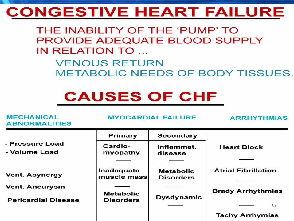
Congestive Heart Failure
• Video..the mysterious heart volume1 chapter 3 61
62

• DEFINITION:EF < ____% or when the myocardium is no longer able to pump efficiently
63

• Systolic failure= most common
64

Causes of CHF
• CAD, advancing age• HTN is a major factor > CHF x 3 • DM, Smoking, Obesity• Valvular incompetency, alcohol
or other chemicals, idiopathic,(unknown)
65
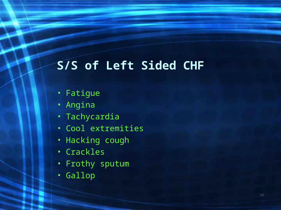
S/S of Left Sided CHF
• Fatigue• Angina• Tachycardia• Cool extremities• Hacking cough• Crackles• Frothy sputum• Gallop
66
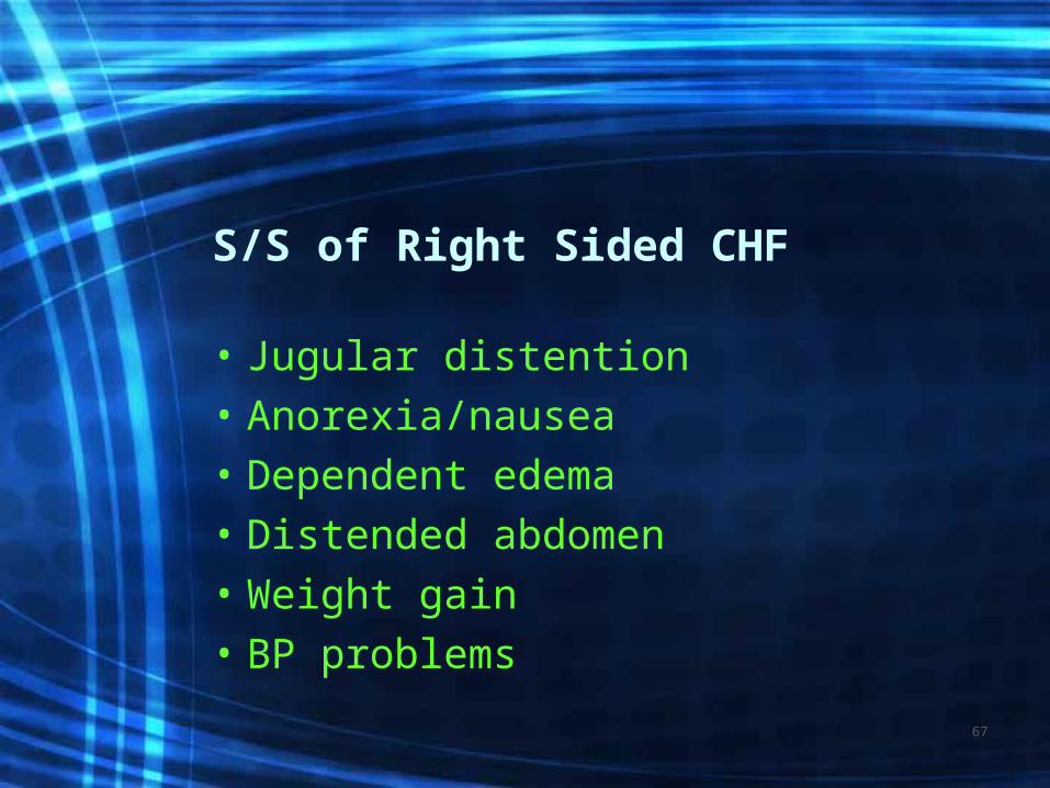
S/S of Right Sided CHF
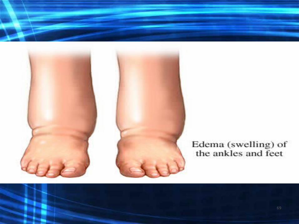
• Jugular distention• Anorexia/nausea• Dependent edema• Distended abdomen• Weight gain• BP problems
67

Assessment Findings
• C/O SOB, weakness, dry cough, fatigue, can not lie down must sit up to breath, has gained weight
• Auscultation of the heart} rapid HR, extra heart sounds
• Auscultation of the lungs} rales, wheezing• Examination of the extremities for
peripheral edema
68
69
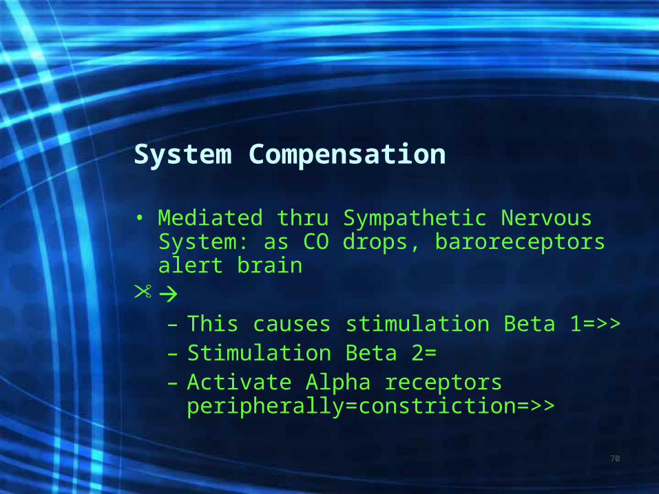
System Compensation
• Mediated thru Sympathetic Nervous System: as CO drops, baroreceptors alert brain
– This causes stimulation Beta 1=>>– Stimulation Beta 2=– Activate Alpha receptors
peripherally=constriction=>>
70
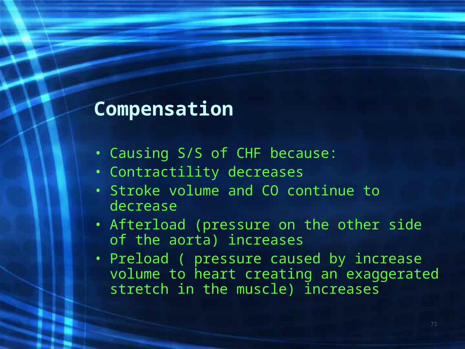
Compensation
• Causing S/S of CHF because: • Contractility decreases• Stroke volume and CO continue to decrease• Afterload (pressure on the other side of the
aorta) increases• Preload ( pressure caused by increase
volume to heart creating an exaggerated stretch in the muscle) increases
71

Renal Compensation
• CO drops initiating renin-angiotensin mechanism– Results in powerful
vasoconstrictor
72

Ventricular Hypertrophy
• The heart enlarges which results in strain
• The increase in volume causes the ventricles to dilate
• Eventually remodeling will occur
73
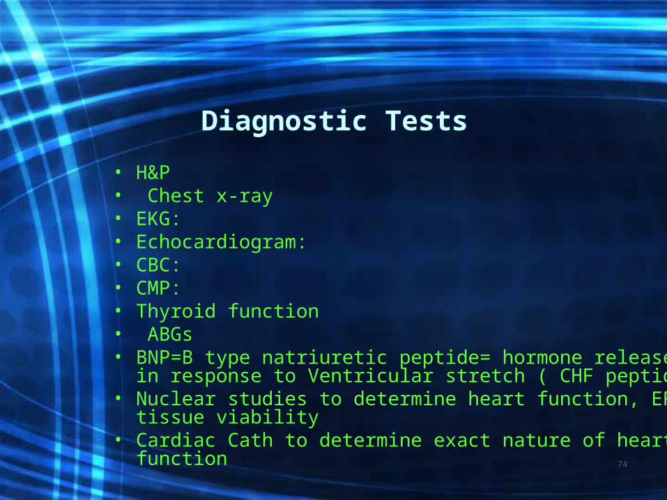
Diagnostic Tests
• H&P• Chest x-ray• EKG:• Echocardiogram:• CBC:• CMP:• Thyroid function • ABGs• BNP=B type natriuretic peptide= hormone released in
response to Ventricular stretch ( CHF peptide) • Nuclear studies to determine heart function, EF, tissue
viability• Cardiac Cath to determine exact nature of heart
function74

75

CHF Management
• Directed at: Improving LV function (Contractility) by decreasing intravascular volume and decreasing vascular resistance
• Decreasing venous return (Preload)• Decreasing BP (Afterload)• Improving gas exchange and 02• Increasing the CO and reducing anxiety
76

continued
• ACE inhibitors to < afterload by dilating vessels and < BP (ARBs)
• Beta blockers to < 02 demand by reducing the contractility of the heart and HR (not given in acute period)
• Diuretics <preload by reducing volume returning to the heart-Lasix & (Aldosterone Antagonists) K+ supplement
77
continued
• ASA in low doses or Plavix to help prevent blood clot formation
• Anticoagulants for those with poor EFs to prevent CVA
• Antiarrhythmics to control ectopy• Biventricular pacing (CRT=cardiac
resynchronization therapy) to improve CO• Digoxin to increase contractility of
myocardial fibers and improving cardiac output. +inotropic agent 78
Treatment of CHF
• Treat underlying cause• Rest and hi Fowlers to reduce work
load and improve ventilation• 02 at 2-6 L/min with 02 sats >92% to
increase available 02 and prevent hypoxemia
• Freq VS and cardiac monitoring79
Treatment continued• I & O q shift• Daily am weights before breakfast and after
voiding. 2-3# weight gain in 1-4 days call MD• Sodium restricted diet• Medications: to decrease intravascular volume
thus reducing venous return, dilate and reduce BP and improve contractility
• http://chfsolutions.com/zip_how_aquapheresis_works.html#
80

Educating the CHF Client
• Education re: heart failure– Explanation of heart failure– Expected S/S and when to call MD– Self monitoring of daily weights– Know medications and need to take them– 2000mg sodium restricted diet – Importance of low level daily exercise
program (energy conservation)– Prognosis / advanced directives
81

• Telemetry Interpretation/ Dysrhythmias
82

Lead Placement
83

• A dysrhythmia is a disturbance of the rhythm of the heart caused by a problem in the conduction system.
• Categorized by site of origin: atrial , AV nodal, ventricular
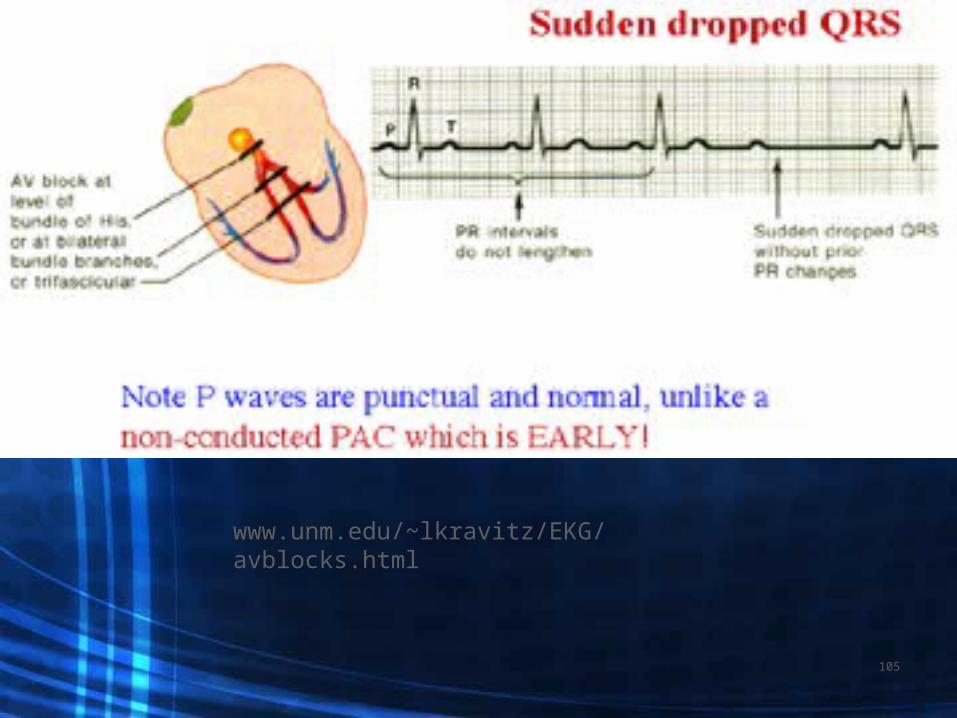
• Blocks are interruptions in impulse conduction: 1st, 2nd type 1&2, 3rd or complete heart block
84

Each small box measures 0.041 big box (5 small boxes) is equal to a HR of 3002 big boxes is hr of 1503 big boxes is hr of 1004 big boxes is hr of 755 big boxes is hr of 606 big boxes is hr of 507 big boxes is hr of 438 big boxes is hr of 38
85

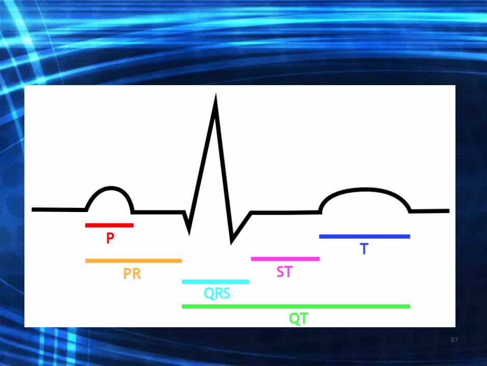
• P-wave = atrial electrical activity
• QRS= ventricular electrical activity
• T wave= resting phase of ventricle
86
87

P wave
Measures: 0.12-0.20
88
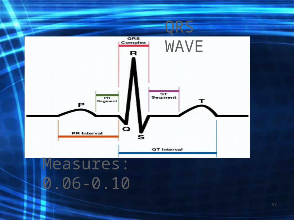
Measures: 0.06-0.10
QRS WAVE
89
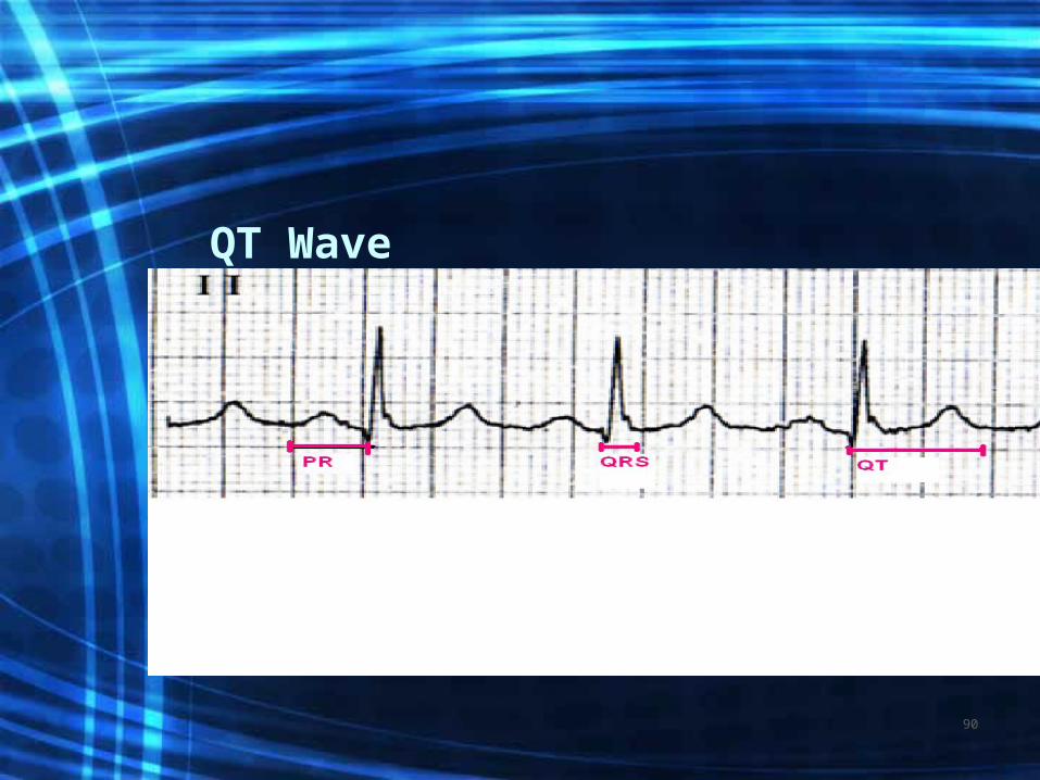
QT Wave
90

Heart rates
• NSR: heart rate is ___bpm• ST: heart rate ____ bpm• SB: heart rate ____bpm
91

NSR
92

Sinus rhythm
•PR interval- 0.12-0.20sec• QRS-0.06-0.10sec• QT segment 0.36-0.44
sec •Heart rate 60-100
93
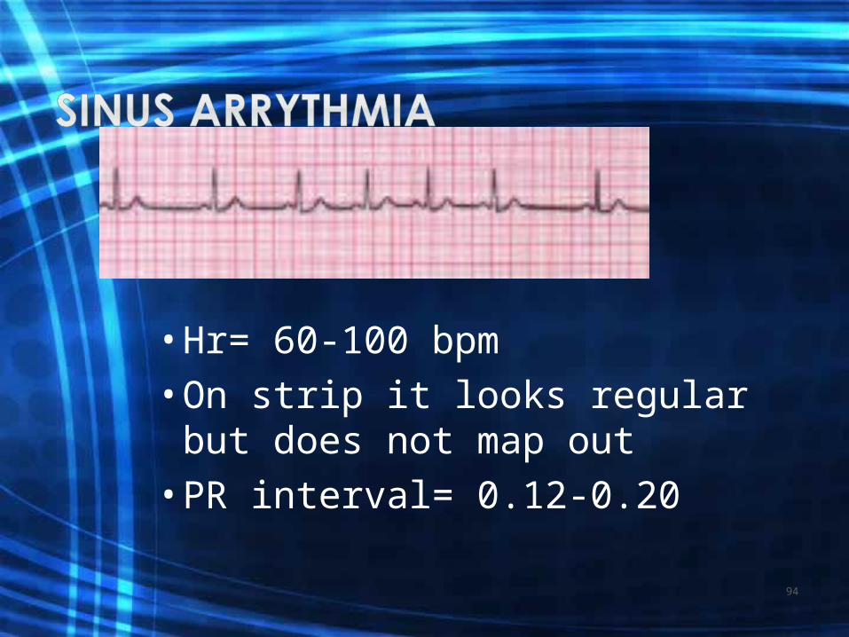
•Hr= 60-100 bpm•On strip it looks regular but
does not map out•PR interval= 0.12-0.20
94
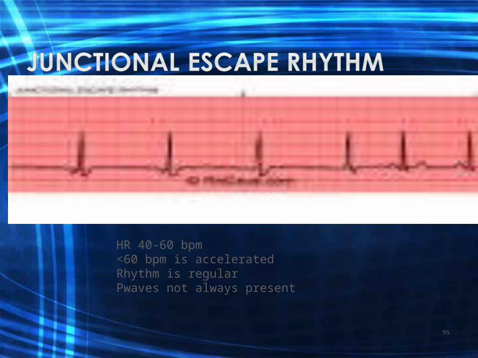
HR 40-60 bpm<60 bpm is acceleratedRhythm is regularPwaves not always present
95

SB
96

Sinus Bradycardia
• All criteria same except rate < 60bpm• S/S: dizziness, syncope, angina,
hypotension, sweating, nausea, dyspnea
• Sometimes no S/S• Treat underlying cause• IV atropine, pacemaker
97

ST
98

Sinus Tachycardia
• All criteria same as with NSR except rate >100• Causes: fever, dehydration, hypovolemia,
increased sympathetic nervous system stimulation, stress, exercise, AMI
• S/S: Palpations #1, angina and < CO from < V filling time
• Treatment: correct cause, eliminate caffeine, nicotine, alcohol. Beta blockers may be ordered
99

Rate is usually WNLRhythm is regularPwaves are normal in size and shape The PR interval is prolonged (>0.20 sec) but constant
100

101
www.unm.edu/~lkravitz/EKG/avblocks.html
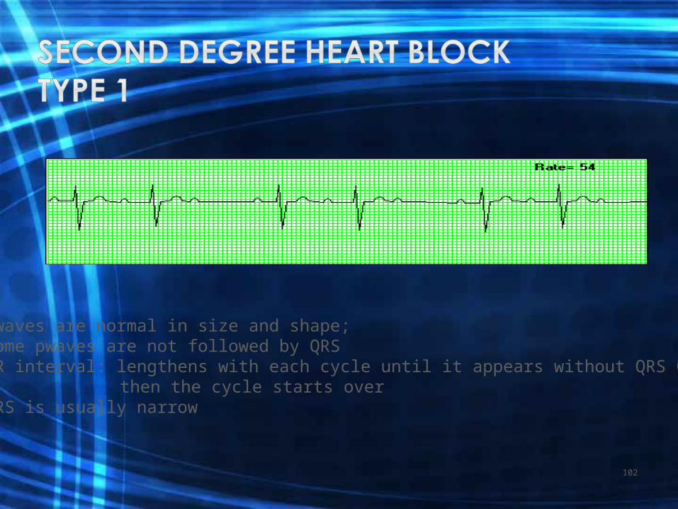
Pwaves are normal in size and shape; Some pwaves are not followed by QRSPR interval: lengthens with each cycle until it appears without QRS Complex
then the cycle starts overQRS is usually narrow
102

103
www.unm.edu/~lkravitz/EKG/avblocks.html
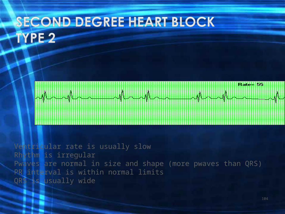
Ventricular rate is usually slowRhythm is irregularPwaves are normal in size and shape (more pwaves than QRS)PR interval is within normal limitsQRS is usually wide
104
105
www.unm.edu/~lkravitz/EKG/avblocks.html

Ventricular rate is regular but there is no correlation between pwaves and QRSPwaves are normal in size and shapeNo true PR interval
106

107
www.unm.edu/~lkravitz/EKG/avblocks.html
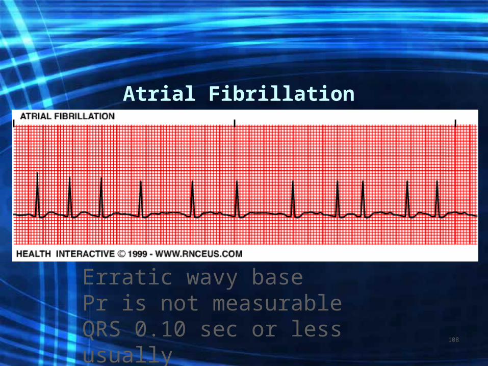
Atrial Fibrillation
Erratic wavy basePr is not measurableQRS 0.10 sec or less usuallyhttp://www.youtube.com/watch?v=VKxQgjj2yVU&feature=related
108

A fib continued
• Atrial rate > 400 bpm with a varying Ventricular rate
• Overall rhythm irregular• No P waves, unable to measure PR interval• QRS=normal: Twave undeterminable• Causes: Rheumatic fever, mitral valve
stenosis, cad. HTN, MI, hyperthyroidism, COPD, CHF see pp. 604
109

A fib continued
• Concern with A fib is the development of atrial thrombus and loss of atrial kick from ineffective atrial function.
• Treatment: Ca channel blockers and anti- arrhythmics to convert, beta blockers to < HR, anticoagulants to prevent embolization.
• Synchronized cardioversion
110
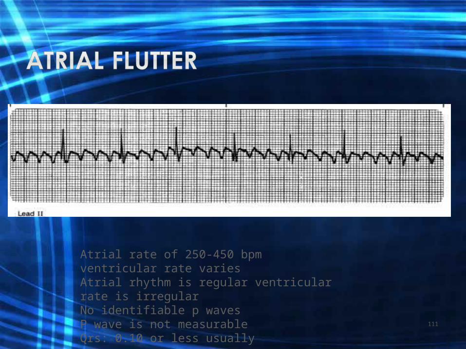
Atrial rate of 250-450 bpm ventricular rate variesAtrial rhythm is regular ventricular rate is irregularNo identifiable p wavesP wave is not measurableQrs: 0.10 or less usually
111

Pacer spike should fall before the P wave unless a dualChamber pacemaker; if it does not there could be a problem
112
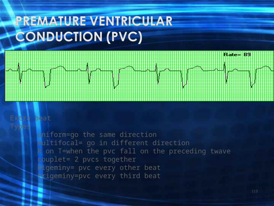
Extra beat Types
uniform=go the same directionmultifocal= go in different directionR on T=when the pvc fall on the preceding twavecouplet= 2 pvcs togetherbigeminy= pvc every other beattrigeminy=pvc every third beat
113

114
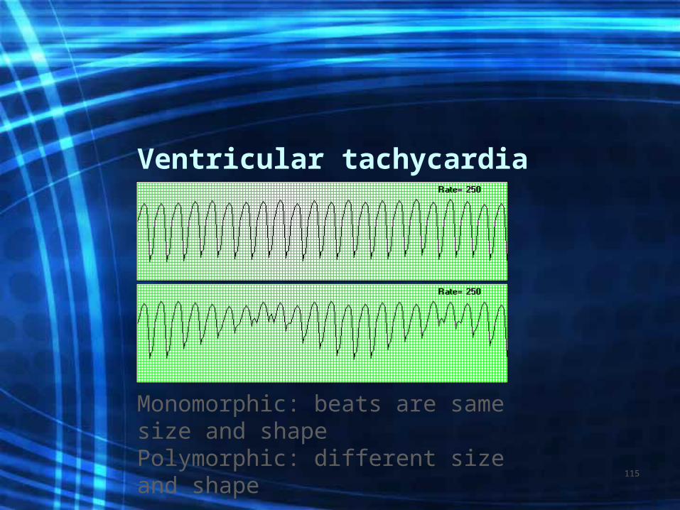
Ventricular tachycardia
Monomorphic: beats are same size and shapePolymorphic: different size and shape
115

This is a polymorphic VTUsually electrical imbalance in nature r/t NA+ or K+
116
Ventricular Fibrillation
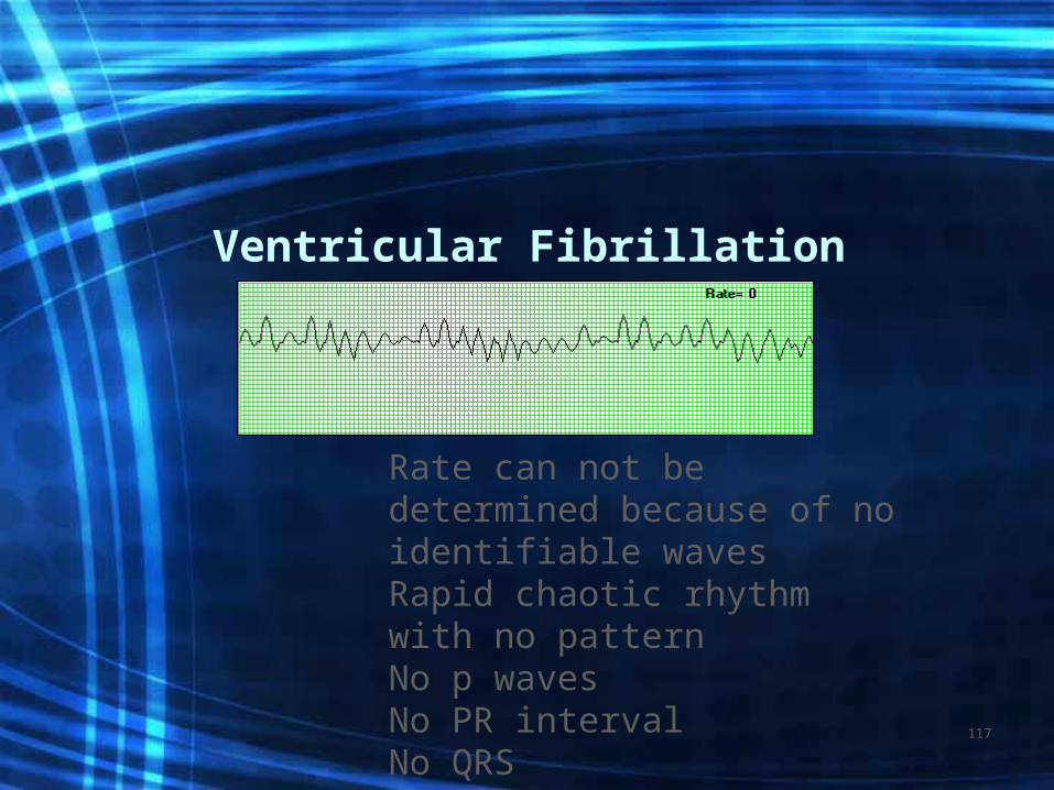
Rate can not be determined because of no identifiable wavesRapid chaotic rhythm with no patternNo p wavesNo PR intervalNo QRS
117

Vtach/Vfib
• Both can be life threatening• VT= V HR 100-250 bpm• Causes: AMI, CAD, hypokalemia, dig toxic• S/S: palpitations, dizzy, angina, <LOC• Treatment: assess for pulse, if none, defib• VF=Rate undeterminable Cause: same• Treatment: CPR
118
Hypertension
120

• HTN is described as persistent elevation of arterial blood pressure greater than 140/90 on at least 2 or more readings on different dates.
• The Joint National Committee, Detection, Evaluation, and Treatment of High Blood Pressure defines normal: – BP as S < 120 mm Hg and D < 80 mm
Hg– PreHTN: SBP 120-139 DBP 80-89– Stage 1: SBP 140-159 DBP 90-99– Stage 2: SBP >160 DBP >100
121

Types of Hypertension
• Essential HTN: (Primary) which is the most common 90-95% of population
• Secondary HTN: is a result of another disease, kidney, pregnancy.
122

Factors that determine arterial pressure
• Cardiac output which is the volume of blood pumped by the heart in 1 minute
• Peripheral vascular resistance which is the force in the peripheral blood vessels that the left ventricular must overcome to eject blood out of the heart
123

Possible Causes of PVR
• Narrowing of blood vessels, PVD, CAD, kidney disease: > renin/angiotensin =vasoconstriction
• Release of catecholamine (epinephrine and adrenalin) = vasoconstriction
• > blood volume= more work to pump• > Blood viscosity=harder to pump• Ability of blood vessel to stretch
124
Causative Factors of HTN
• Hyperlipidemia Obesity• Atherosclerosis Sedentary• DM Family Hx• Cigarette smoking• Age > 60• Men• Post menapausal women
125

S/S
• Often none• Occipital headache more severe
on rising• Lightheadedness• Epistaxis• Known as the ‘Silent Killer’
126
Complications
• Damage to blood vessels of the eyes, heart, kidney, brain resulting in:
• Stroke• CHF• AMI• Renal failure • Blindness
127

Lifestyle Change Education
• Exercise, dash diet, stop smoking, weight management and control, stress reduction, medications and recording BP frequently
• Avoid OTC meds• Instruct on how to do postural BPs
128

Valvular Disorders
129
• STENOSIS:
• INSUFFICIENCY:
130

Stenosis
• Narrowing of the opening of the valves. Limits the amount of blood which is ejected from one chamber to the next.
131

132

Mitral Stenosis
• Mitral valve leaflets become thickened and fibrotic. Affect women age 20-40
• CHF may develop• TX if failure develops: Digoxin, Lasix, beta
blockers, and anti arrhythmics, lo Na diet, etc• Will monitor with yearly echocardiogram• Surgery if worsens• Prophylactic antibiotics prior to invasive
procedure or dental work
133

Insufficiency
• The inability of the valves to close completely.
• Allows the blood to backflow. • Mitral valve is the most commonly
affected
134
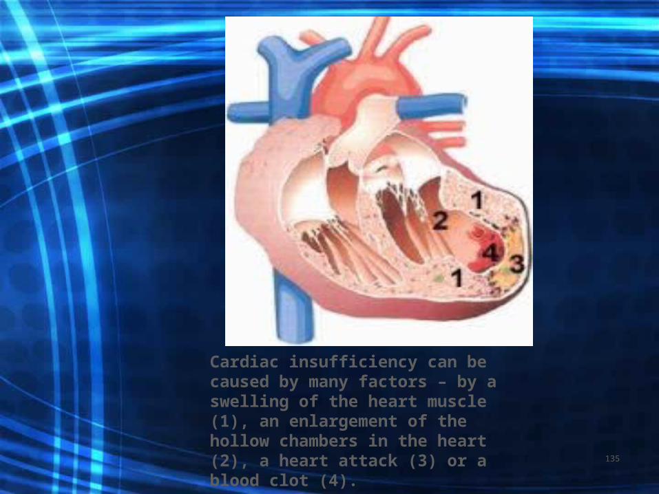
Cardiac insufficiency can be caused by many factors – by a swelling of the heart muscle (1), an enlargement of the hollow chambers in the heart (2), a heart attack (3) or a blood clot (4).
135

Mitral Insufficiency
• Often accompanies mitral stenosis as a result of rheumatic fever.
• Valve leaflet become rigid and shorten, prevents closure of valve.
• Hypertrophy of Left Atrium and Ventricle = L sided heart failure occurs
• Murmur heard. F/U with echocardiogram• TX: vasodilators, same as for stenosis
136

Mitral Valve Prolapse
• When the Left ventricle leaflets become enlarged, and protrude into the left atrium during systole.
• Benign but may progress to Mitral insufficiency
• More common in women age 20-55
137
S/S of mitral prolapse• Often none• Others experience chest pain, palpitations,
dizziness, syncope, dysrhythmias• Monitor with echocardiogram• May do heart catherization• Manage stress, beta blockers if
tachycardia
138

Aortic Stenosis• Occurs when valve cusps become
fibrotic and calcify. • Most commonly caused by aging and
atherosclerosis. • Occurs most predominantly in men• Untreated will lead to Left sided CHF
139

Aortic Insufficiency
• Caused primarily by rheumatic fever• May also be caused by chronic HTN• Predominantly in men• Hypertrophy of the Left ventricle and
eventually to left sided CHF• Blood may eventually back up into the
pulmonary system and lead to Right Ventricle failure
140
S/S and Treatment
• Aortic murmur, tachycardia, palpitations, CHF with fatigue, SOB, ascites
• Monitored with echocardiogram assessing L ventricular dilatation
• Chest X-ray-enlargement of heart• May do cardiac cath• May need valve repair or replacement
141

•Inflammatory Diseases of the Heart
142
• Inflammation of the heart most often results from systemic infections and may include any layer of the heart:What are they???
143

Endocarditis
• Inner layer: tends to affect the valves (Mitral=L). Organisms (Bacterial or fungal) present in blood stream and collect (colonize) on the valves: Rheumatic heart disease, congenital defects or mitral valve prolapse
• IV drug users or invasive procedures
144

• Clients with known valvular disease need to be treated with prophylactic antibiotics prior to any invasive procedure including dental. Immunosuppression and any source of contamination places clients at risk
145

Pathophysiology
• Bacteria may enter blood stream:• Bacteria collect on valves and
vegetate• Complications: Ventricular septal
defect, CHF(#1 cause of death) and embolization
146

S/S• Fever- (99-105)• Chills and night sweats may accompany• Malaise, fatigue and weight loss• Appearance of petechiae in the mouth,
conjunctiva and legs• Chest and abdominal pain indicating
embolization
147
Treatment and Diagnostics
• H&P and Lab testsCBC with diff with leukocytosis, > sed rate, blood cultures
• May have heart murmur Echocardiogram to visualize valves and vegetation
• Chest x-ray: CHF• Long term antibiotics, rest, limited activity,
prophylactic anticoagulants, valve replacement after inflammation treated
148

Nursing Assessment
• Frequent VS and assess for fever• Assess for heart murmur• Note cough• Assess peripheral edema• Rest with limited activity, administer
meds in a timely manner
149

Nursing Diagnosis
150

• myocarditis
151

• Muscle layer: Local or diffuse inflammation of the myocardium. May be viral or bacterial, an autoimmune process or drug toxicity.
• May result in cardiomyopathy=
152

Pathophysiology• Characterized by degeneration and
necrosis of myocardial tissue that is different of that caused by MI
• Tissue next to necrosed area hypertrophies, loses elasticity, results in CHF and arrhythmias
153
S/S
• Asymptomatic• May have fever, fatigue, sore
throat, dyspnea, muscle aches• Lymph nodes may be enlarged• Chest pain 7-10 days after virus• CHF S/S
154

Diagnosis• Based on Hx, S/S, and testing-enzymes> • May hear friction rub, rales• Jugular vein distention• Chest x-ray, echocardiogram=hypertrophy• EKG=arrhythmias• Biopsy (RV) shows lymphatic infiltration
and cell necrosis
155
Treatment
• Bed rest• 02• Meds: cardiac glycoside-Lanoxin,
anticoagulants, antiarrhythmic, antibiotics, steroids
• Cardiac monitoring• NI same as for endocarditis • ND same as for endocarditis
156

• Pericarditis
157

• Outer-surrounds heart• Inflammation of the pericardium. • Primary or secondary• Acute or chronic• Acute: virus, bacteria, fungi,
chemotherapy, MI• Chronic: TB, radiation or metastases
158
Pathophysiology
• Inflammation causes an increase in the amount of pericardial fluid and inflammation of surrounding tissues.
• Fluid accumulates in the pericardial space• Adhesions may occur which causes loss of
elasticity which causes constriction and prevents adequate filling of ventricles.
• May lead to tamponade==pericardiocentesis
159
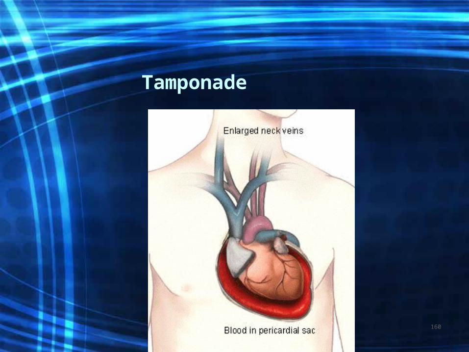
Tamponade
160

S/S
• Chest pain is hallmark• Most severe on inspiration, sharp,
stabbing, or dull and burning.• Pain is relieved by sitting up or
leaning forward• Dyspnea, chills and fever
161

Diagnosis • WBC elevated• Serial EKG show that ST segment increases
and resolves in several weeks. A fib may occur
• Echocardiogram to see pericardial thickening and effusion
• Enzymes can be increased• Blood cultures to ID organism
162

Treatment
• Analgesics• Antipyretics• Anti-inflammatory agents• Antibiotics• May need OR to create a pericardial
window to allow for drainage of fluid• NI and ND same as for endocarditis
163
• Nursing the Heart Client
164

Assessment
• Heart rate and rhythm, color, temperature, cognition
• Circulation: peripheral CMS checks• Vital signs to include 02 saturations
and telemetry interpretation• Subjective: c/o chest pain, SOB,
fatigue, lightheadedness, dizziness
165

Nursing Diagnosis
166

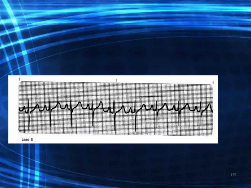
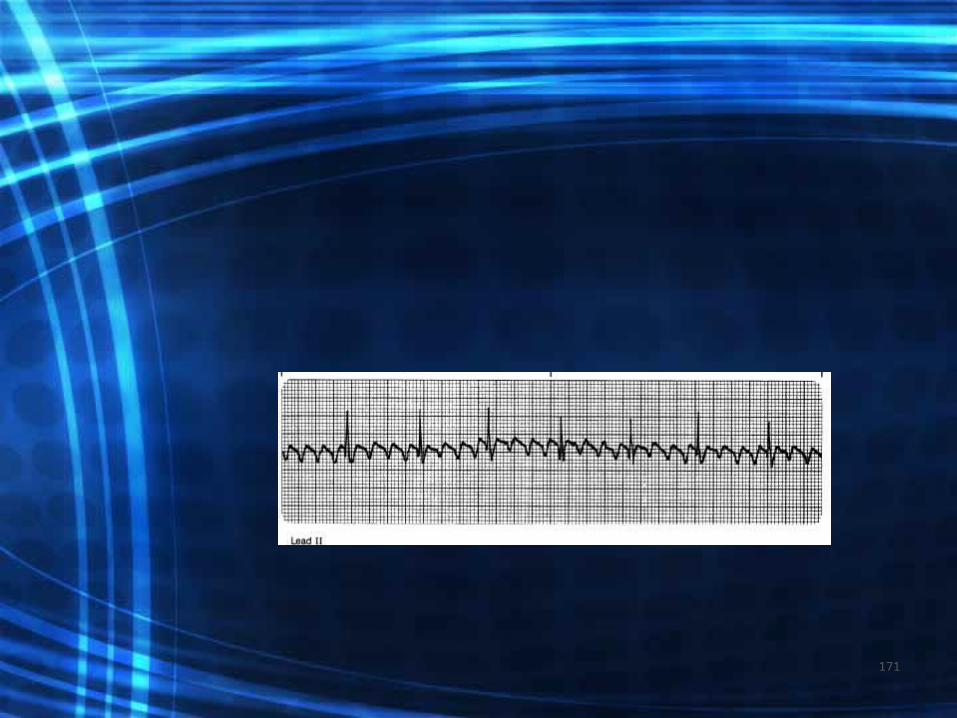
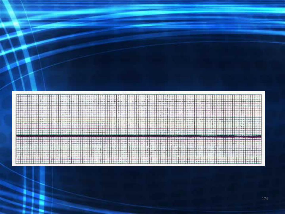
Ekg practice
167

168
169

170
171

172

173
174

•www.mirule.com retrieved on 4/8/07.•Images found at www.aol.com.
Retrieved on 4/8/07.• Aehlert, B. RN BSPA (2006). EKGs
Made Easy. Mosby (3rd ed). St Louis.
175

•The End
176