
Cardiovascular, skeletal, and renal defects in mice with a ...
6
Cardiovascular, skeletal, and renal defects in mice with a targeted disruption of the Pkd1 gene Catherine Boulter*, Sharon Mulroy* , Sandra Webb ‡ , Stewart Fleming § , Kevin Brindle ¶ , and Richard Sandford i Departments of *Genetics, ¶ Biochemistry, and ² Medical Genetics, University of Cambridge, Cambridge CB2 1TN, United Kingdom; ‡ Department of Anatomy and Developmental Biology, St. George’s Hospital Medical School, London 5W17 ORE, United Kingdom; and § Department of Molecular and Cellular Pathology, University of Dundee, Dundee DD195Y, United Kingdom Edited by Oliver Smithies, University of North Carolina at Chapel Hill, Chapel Hill, NC, and approved August 8, 2001 (received for review April 18, 2001) Autosomal dominant polycystic kidney disease (ADPKD) is charac- terized by cyst formation in the kidney, liver, and pancreas and is associated often with cardiovascular abnormalities such as hyper- tension, mitral valve prolapse, and intracranial aneurysms. It is caused by mutations in PKD1 or PKD2, encoding polycystin-1 and -2, which together form a cell surface nonselective cation ion channel. Pkd22y2 mice have cysts in the kidney and pancreas and defects in cardiac septation, whereas Pkd1 del34 2y2 and Pkd1 L 2y2 mice have cysts but no cardiac abnormalities, although vascular fragility was reported in the latter. Here we describe mice carrying a targeted mutation in Pkd1 (Pkd1 del17–21bgeo ), which defines its expression pattern by using a lacZ reporter gene and may identify novel functions for polycystin-1. Although Pkd1 del17–21bgeo 1y2 adult mice develop renal and hepatic cysts, Pkd1 del17–21bgeo 2y2 embryos die at embryonic days 13.5–14.5 from a primary cardio- vascular defect that includes double outflow right ventricle, dis- organized myocardium, and abnormal atrio-ventricular septation. Skeletal development is also severely compromised. These abnor- malities correlate with the major sites of Pkd1 expression. During nephrogenesis, Pkd1 is expressed in maturing tubular epithelial cells from embryonic day 15.5. This expression coincides with the onset of cyst formation in Pkd1 del34 2y2, Pkd1 L 2y2, and Pkd22y2 mice, supporting the hypothesis that polycystin-1 and polycystin-2 interact in vivo and that their failure to do so leads to abnormalities in tubule morphology and function. A utosomal dominant polycystic kidney disease (ADPKD) is a common inherited disorder that affects 1 in 800 people and accounts for ’8% of patients with end-stage renal failure. It is characterized by the formation of multiple cysts in the kidneys and liver and, less frequently, in the pancreas. Cardio- vascular abnormalities including hypertension, mitral valve pro- lapse, and intracranial aneurysms are also frequently recog- nized. Extensive characterization of the cellular defects in cyst- lining epithelial cells derived from kidneys affected by ADPKD and from a variety of rodent models of renal cystic disease has demonstrated generalized abnormalities in cell proliferation, differentiation, and apoptosis (1–3). More specific defects in cell polarity and extracellular matrix production are also seen and have been implicated directly in the process of cyst formation (4, 5). However, the primary events that give rise to this cystic phenotype have not been elucidated. The cloning of PKD1 and PKD2, the genes mutated in almost all cases of ADPKD, has provided the opportunity to investigate the molecular basis of cyst formation and develop appropriate model systems to study the functions of their protein products, polycystin-1 and -2. Both are predicted to be polytopic mem- brane proteins, with polycystin-1 having a large predicted ex- tracellular region comprising multiple discrete domains while polycystin-2 has homology to ion channels (6, 7). An interaction mediated by the C-terminal regions of both proteins results in the formation of calcium-permeable nonselective cation chan- nels in vitro, suggesting that extracellular signals can be trans- duced by the polycystin complex to regulate diverse cellular processes (8). Indeed, the cytoplasmic tail of polycystin-1 has been shown to activate several signal transduction pathways (9, 10), whereas Madin-Darby canine kidney cells transfected with PKD1 are resistant to apoptosis and undergo spontaneous tubulogenesis (11). The nature of the extracellular signals or protein ligands that activate polycystin-1 signaling have not been determined. The formation of a polycystin complex suggests that Pkd1 and Pkd2 should have considerable overlap in their expression patterns, and detailed analysis of the cellular and subcellular distribution has been performed. Expression of polycystin-2 has been defined in renal tubular epithelial cells with widespread expression reported in other tissues including the heart and vasculature (12–14). Unfortunately, considerable differences have been reported in the expression pattern of polycystin-1 by using both antibodies directed against different epitopes and RNA in situ hybridization (15–23). This has made meaningful comparisons of Pkd1 and Pkd2 expression difficult. Mice carrying targeted mutations in Pkd1 and Pkd2 or a PKD1 transgene have been reported (24 –28). They all have renal cysts, suggesting that alterations in the level of polycystin-1 lead to cyst formation. Both Pkd1 del34 2y2 and Pkd1 L 2y2 mice develop renal, hepatic, and pancreatic cystic disease. However Pkd1 L 2y2 embryos also develop gross edema and s.c. hemorrhage, which may be caused by a defect in vascular wall integrity (24). Unlike these models, Pkd2 mutant mice also have major defects in cardiac development manifested by septal abnormalities in addition to the cystic phenotype (27). Here we describe a mouse model of ADPKD that allows the accurate description of Pkd1 expression by using a lacZ reporter gene and identifies a major function for polycystin-1 in cardio- vascular and skeletal development in addition to its role in embryonic and adult kidney. Materials and Methods Pkd1 Gene Targeting. A 14-kb mouse genomic fragment contain- ing Pkd1 exons 15–33 was isolated from a l2001 129ySv genomic DNA library derived from the CCB embryonic stem (ES) cell line (constructed by A. Smith) by screening with a human PKD1 cDNA probe. To construct the targeting vector (pPkd1 del17–21bgeo ), a 6.5-kb XbaI fragment (with one XbaI site derived from the l2001 polylinker) containing exons 15–21 was cloned into pBS (Stratagene). A 1.5-kb HindIII–XbaI fragment including exons 17–21 was deleted and replaced with a promo- torless cassette containing a 59 donor engrailed-2 intron and splice acceptor site, an internal ribosome entry site (IRES) This paper was submitted directly (Track II) to the PNAS office. Abbreviations: ADPKD, autosomal dominant polycystic kidney disease; ES, embryonic stem; IRES, internal ribosome entry site; WT, wild type; En, embryonic day n; bgeo, lacZ-neomy- cin R fusion gene; X-Gal, 5-bromo-4-chloro-3-indolyl-b-D-galactoside; DORV, double outlet right ventricle. i To whom reprint requests should be addressed: Addenbrooke’s Hospital, Cambridge, CB2 2XY, UK. E-mail: [email protected]. The publication costs of this article were defrayed in part by page charge payment. This article must therefore be hereby marked “advertisement” in accordance with 18 U.S.C. §1734 solely to indicate this fact. 12174 –12179 u PNAS u October 9, 2001 u vol. 98 u no. 21 www.pnas.orgycgiydoiy10.1073ypnas.211191098 Downloaded by guest on October 24, 2021
Transcript of Cardiovascular, skeletal, and renal defects in mice with a ...
Cardiovascular, skeletal, and renal defects in micewith a targeted disruption of the Pkd1 geneCatherine Boulter*, Sharon Mulroy*†, Sandra Webb‡, Stewart Fleming§, Kevin Brindle¶, and Richard Sandford†i
Departments of *Genetics, ¶Biochemistry, and †Medical Genetics, University of Cambridge, Cambridge CB2 1TN, United Kingdom; ‡Department of Anatomyand Developmental Biology, St. George’s Hospital Medical School, London 5W17 ORE, United Kingdom; and §Department of Molecular and CellularPathology, University of Dundee, Dundee DD195Y, United Kingdom
Edited by Oliver Smithies, University of North Carolina at Chapel Hill, Chapel Hill, NC, and approved August 8, 2001 (received for review April 18, 2001)
Autosomal dominant polycystic kidney disease (ADPKD) is charac-terized by cyst formation in the kidney, liver, and pancreas and isassociated often with cardiovascular abnormalities such as hyper-tension, mitral valve prolapse, and intracranial aneurysms. It iscaused by mutations in PKD1 or PKD2, encoding polycystin-1 and-2, which together form a cell surface nonselective cation ionchannel. Pkd22y2 mice have cysts in the kidney and pancreas anddefects in cardiac septation, whereas Pkd1del34 2y2 and Pkd1L 2y2
mice have cysts but no cardiac abnormalities, although vascularfragility was reported in the latter. Here we describe mice carryinga targeted mutation in Pkd1 (Pkd1del17–21bgeo), which defines itsexpression pattern by using a lacZ reporter gene and may identifynovel functions for polycystin-1. Although Pkd1del17–21bgeo 1y2
adult mice develop renal and hepatic cysts, Pkd1del17–21bgeo 2y2
embryos die at embryonic days 13.5–14.5 from a primary cardio-vascular defect that includes double outflow right ventricle, dis-organized myocardium, and abnormal atrio-ventricular septation.Skeletal development is also severely compromised. These abnor-malities correlate with the major sites of Pkd1 expression. Duringnephrogenesis, Pkd1 is expressed in maturing tubular epithelialcells from embryonic day 15.5. This expression coincides with theonset of cyst formation in Pkd1del34 2y2, Pkd1L 2y2, and Pkd22y2
mice, supporting the hypothesis that polycystin-1 and polycystin-2interact in vivo and that their failure to do so leads to abnormalitiesin tubule morphology and function.
Autosomal dominant polycystic kidney disease (ADPKD) isa common inherited disorder that affects 1 in 800 people
and accounts for '8% of patients with end-stage renal failure.It is characterized by the formation of multiple cysts in thekidneys and liver and, less frequently, in the pancreas. Cardio-vascular abnormalities including hypertension, mitral valve pro-lapse, and intracranial aneurysms are also frequently recog-nized. Extensive characterization of the cellular defects in cyst-lining epithelial cells derived from kidneys affected by ADPKDand from a variety of rodent models of renal cystic disease hasdemonstrated generalized abnormalities in cell proliferation,differentiation, and apoptosis (1–3). More specific defects in cellpolarity and extracellular matrix production are also seen andhave been implicated directly in the process of cyst formation (4,5). However, the primary events that give rise to this cysticphenotype have not been elucidated.
The cloning of PKD1 and PKD2, the genes mutated in almostall cases of ADPKD, has provided the opportunity to investigatethe molecular basis of cyst formation and develop appropriatemodel systems to study the functions of their protein products,polycystin-1 and -2. Both are predicted to be polytopic mem-brane proteins, with polycystin-1 having a large predicted ex-tracellular region comprising multiple discrete domains whilepolycystin-2 has homology to ion channels (6, 7). An interactionmediated by the C-terminal regions of both proteins results inthe formation of calcium-permeable nonselective cation chan-nels in vitro, suggesting that extracellular signals can be trans-duced by the polycystin complex to regulate diverse cellularprocesses (8). Indeed, the cytoplasmic tail of polycystin-1 has
been shown to activate several signal transduction pathways (9,10), whereas Madin-Darby canine kidney cells transfected withPKD1 are resistant to apoptosis and undergo spontaneoustubulogenesis (11). The nature of the extracellular signals orprotein ligands that activate polycystin-1 signaling have not beendetermined.
The formation of a polycystin complex suggests that Pkd1 andPkd2 should have considerable overlap in their expressionpatterns, and detailed analysis of the cellular and subcellulardistribution has been performed. Expression of polycystin-2 hasbeen defined in renal tubular epithelial cells with widespreadexpression reported in other tissues including the heart andvasculature (12–14). Unfortunately, considerable differenceshave been reported in the expression pattern of polycystin-1 byusing both antibodies directed against different epitopes andRNA in situ hybridization (15–23). This has made meaningfulcomparisons of Pkd1 and Pkd2 expression difficult.
Mice carrying targeted mutations in Pkd1 and Pkd2 or a PKD1transgene have been reported (24–28). They all have renal cysts,suggesting that alterations in the level of polycystin-1 lead to cystformation. Both Pkd1del34 2y2 and Pkd1L 2y2 mice developrenal, hepatic, and pancreatic cystic disease. However Pkd1L
2y2 embryos also develop gross edema and s.c. hemorrhage,which may be caused by a defect in vascular wall integrity (24).Unlike these models, Pkd2 mutant mice also have major defectsin cardiac development manifested by septal abnormalities inaddition to the cystic phenotype (27).
Here we describe a mouse model of ADPKD that allows theaccurate description of Pkd1 expression by using a lacZ reportergene and identifies a major function for polycystin-1 in cardio-vascular and skeletal development in addition to its role inembryonic and adult kidney.
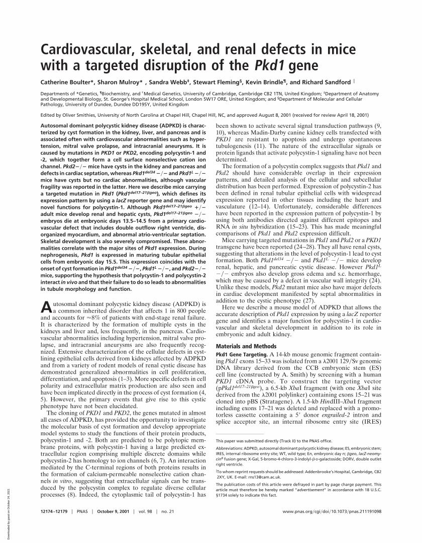
Materials and MethodsPkd1 Gene Targeting. A 14-kb mouse genomic fragment contain-ing Pkd1 exons 15–33 was isolated from a l2001 129ySv genomicDNA library derived from the CCB embryonic stem (ES)cell line (constructed by A. Smith) by screening with a humanPKD1 cDNA probe. To construct the targeting vector(pPkd1del17–21bgeo), a 6.5-kb XbaI fragment (with one XbaI sitederived from the l2001 polylinker) containing exons 15–21 wascloned into pBS (Stratagene). A 1.5-kb HindIII–XbaI fragmentincluding exons 17–21 was deleted and replaced with a promo-torless cassette containing a 59 donor engrailed-2 intron andsplice acceptor site, an internal ribosome entry site (IRES)
This paper was submitted directly (Track II) to the PNAS office.
Abbreviations: ADPKD, autosomal dominant polycystic kidney disease; ES, embryonic stem;IRES, internal ribosome entry site; WT, wild type; En, embryonic day n; bgeo, lacZ-neomy-cinR fusion gene; X-Gal, 5-bromo-4-chloro-3-indolyl-b-D-galactoside; DORV, double outletright ventricle.
iTo whom reprint requests should be addressed: Addenbrooke’s Hospital, Cambridge, CB22XY, UK. E-mail: [email protected].
The publication costs of this article were defrayed in part by page charge payment. Thisarticle must therefore be hereby marked “advertisement” in accordance with 18 U.S.C.§1734 solely to indicate this fact.
12174–12179 u PNAS u October 9, 2001 u vol. 98 u no. 21 www.pnas.orgycgiydoiy10.1073ypnas.211191098
Dow
nloa
ded
by g
uest
on
Oct
ober
24,
202
1
coupled to a lacZ-neomycinR fusion gene (bgeo), and a 39-simianvirus 40 polyadenylation site (29). The 39 region of homology wascloned as a 5.8-kb XbaI–HindIII fragment, and a phosphoglyc-erate kinase promoter-herpes simplex virus thymidine kinasegene (HSVTK) negative selection cassette was ligated down-stream. The targeting construct is shown in Fig. 1. NotI-linearized pPkd1del17–21bgeo was electroporated into CCB EScells. G418-resistant clones were selected and screened bySouthern blotting using EcoRI-cut genomic DNA and the 39probe. Of 266 ES cell clones screened, 11 clones containing thecorrectly targeted mutant allele (Pkd1del17–21bgeo) were identi-fied. Chimeric mice were generated by C57yBl6 blastocystinjection and germline transmission determined by glucosephosphate isomerase assay (30). Chimeric males were bred to129ySv females, and offspring were genotyped by Southernblotting (Fig. 1b) and PCR. PCR primers were directed againstthe IRES and Pkd1 exons 18 and 19 (sequences and PCRparameters are available on request). Wild type (WT) animalswere positive for the exon 18–19 PCR and negative for the IRESPCR; Pkd1del17–21bgeo 1y2 animals were positive for both, andPkd1del17–21bgeo 2y2 animals were positive only for the IRESPCR.
Analysis of RNA and Protein. Total RNA was prepared from E12.5embryos by first grinding snap frozen embryos in liquid nitrogenand then by using the method of Chomczynski and Sacchi (31).Northern blot analysis of 20 mg of total RNA was carried outaccording to standard protocols by using Pkd1 exon 15 or theneoR gene as probes. Total protein (10–15 mg) from whole mouseembryo homogenates was used to detect polycystin-2 and b-
galactosidase expression by Western blot analysis under reducingconditions. The primary antibodies used were PKD2-CFP (12)at a dilution of 1:1,000 and a mouse monoclonal antibody againstb-galactosidase at 1:5,000 dilution (Promega catalog numberZ3781).
Staining for b-Galactosidase Activity in Tissues and Frozen Sections.Staining for b-galactosidase activity was carried out accordingto published protocols (32). Fixation and staining times varieddepending on tissue and sample size. Samples were postfixedin 4% paraformaldehyde in PBS and stored in 70% EtOH at4°C. Samples were transferred to 70% methanol, dehydratedthrough graded methanols, and cleared in benzylalcoholybenzylbenzoate (1:1) before photography. Frozen sectionswere counterstained with nuclear fast red before permanentmounting.
Histological and Skeletal Analysis. Tissues and embryos for histo-logical analysis were fixed overnight in 4% paraformaldehyde inPBS and stored in 70% ethanol before paraffin embedding.Paraffin sections of 6 mm were cut and stained with hematoxylinand eosin. Skeletal preparations and cartilage staining werecarried out as described (32).
Nuclear Magnetic Resonance Imaging. Magnetic resonance imageswere acquired at a magnetic field of 9.4 T (400-MHz protonfrequency) by using an unshielded gradient set and a 25 mmdiameter 1H probe (Varian Ltd). The images were acquired byusing a spin echo pulse sequence (time to echo 5 60 msec, timeto return 5 2 sec) with 256 phase-encode increments and 16
Fig. 1. Generation of a targeted disruption of Pkd1. (a) Pkd1 exons 17–21 were replaced with a lacZ-neomycinR fusion gene (bgeo) located downstreamof an engrailed-2 gene donor intron (En-2) and splice acceptor site (SA), an IRES, and upstream of a simian virus 40 polyadenylation signal (SVpA). Thepositions of the 59 and 39 external probes are indicated. HSVtk, herpes simplex virus thymidine kinase gene; B, BamHI; RI, EcoRI; H, HindIII; X, XbaI; S, SalI.(b) A Southern blot of EcoRI-digested DNA from a cross of Pkd1del17–21bgeo 1y2 mice hybridized with an external 39 probe showing the WT (1y1) (9 kb)and mutant (7.5 kb) alleles; this result was confirmed with the 59-external probe (data not shown). (c) Northern analysis using a Pkd1 exon 15 probedemonstrated the '14-kb Pkd1 transcript in WT (1y1) and Pkd1del17–21bgeo 1y2 embryos and the predicted 12.5-kb mutant transcript in Pkd1del17–21bgeo
1y2 and 2y2 embryos; different intensities between RNA samples reflected different RNA loading are shown. (d) A neoR probe confirmed the presenceof the 12.5-kb mutant transcript in Pkd1del17–21bgeo 1y2 and 2y2 embryos. (e) Independent translation of the bgeo gene was demonstrated by using ananti-b-galactosidase antibody to detect the predicted 146-kDa b-galactosidase-neomycin fusion protein. ( f) Levels of polycystin-2 (110 kDa) were unalteredin Pkd1del17–21bgeo 1y2 and 2y2 E12.5 embryos compared with WT (1y1) littermates.
Boulter et al. PNAS u October 9, 2001 u vol. 98 u no. 21 u 12175
MED
ICA
LSC
IEN
CES
Dow
nloa
ded
by g
uest
on
Oct
ober
24,
202
1
transients per increment. The echoes were acquired into 512 datapoints, and the data set zero was filled to 1,024 3 1,024 datapoints before two-dimensional Fourier transformation. The slicethickness was 500 mm, and the in-plane resolution was 78 378 mm.
ResultsGeneration of Knockout Mice Carrying a Truncating Mutation in Pkd1and an Inserted lacZ Gene. We have generated mice carrying atruncating mutation in the Pkd1 gene (Pkd1del17–21bgeo) by usinggene targeting to replace exons 17–21 of the Pkd1 gene with alacZ-neomycin fusion gene (bgeo) downstream of a splice ac-ceptor site and an IRES (Fig. 1 a and b) (29). The resultingtranscript, containing the first 16 exons of Pkd1 spliced to bgeo,is predicted to encode a truncated form of polycystin-1, whichincludes only the extracellular domains up to and including thePKD repeats, and thus represents a common class of mutationfound in ADPKD patients (33). Northern analysis demonstratedexpression of the expected 12.5-kb Pkd1-bgeo fusion transcriptand loss of the WT 14.5-kb Pkd1 transcript in Pkd1del17–21bgeo
2y2 E12.5 embryos (Fig. 1 c and d). Western blotting confirmedthat the IRES was used efficiently to produce a b-galactosidase-neomycin fusion protein (Fig. 1e) and indicated that the expres-sion level of Pkd2 was unaffected in Pkd1del17–21bgeo 1y2 and2y2 E12.5 embryos (Fig. 1f ). The efficient translation of bgeomRNA allowed Pkd1 expression to be visualized by staining withX-Gal and correlations to be made with the phenotype of mutantmice.
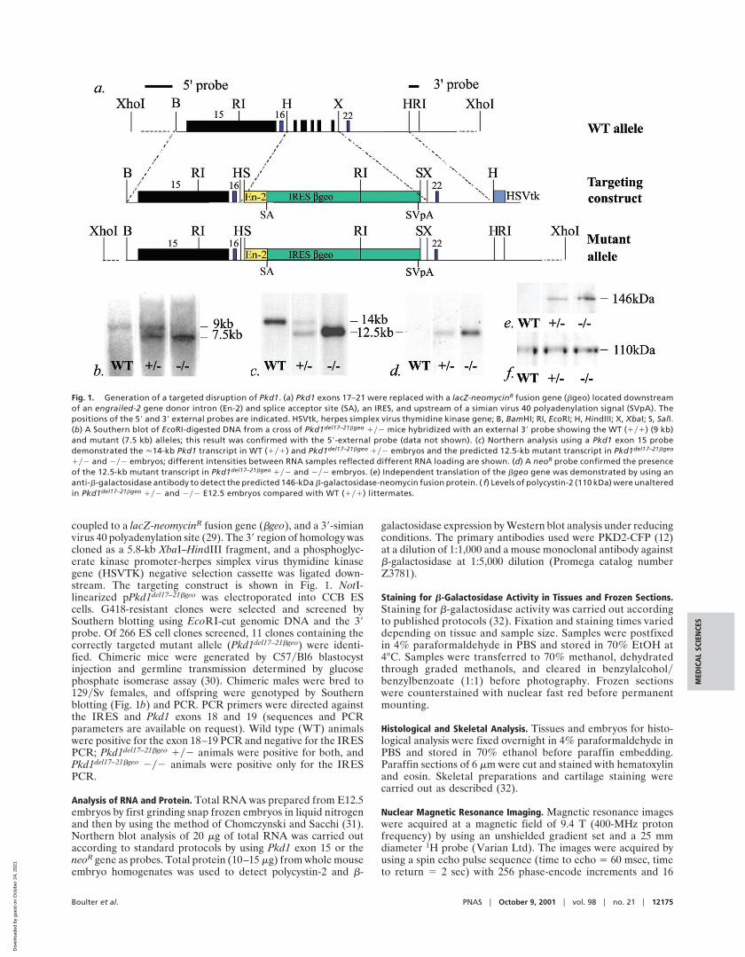
Kidney and Liver Cysts in Pkd1del17–21bgeo Heterozygotes and Expres-sion of Pkd1. Microscopic renal cysts were observed in '50% ofPkd1del17–21bgeo 1y2 mice analyzed before 9 months of age (n 515) and were detected as early as 3 months, with occasionalmacroscopic cysts seen in animals of 4–19 months (n 5 8y18).Cysts arose throughout the nephron and were often lined withhyperplastic cells or apoptotic cells (Fig. 2 a and b). X-Galstaining of kidneys from adult Pkd1del17–21bgeo heterozygotesrevealed activity of the Pkd1 promoter in cyst-lining epithelia(Fig. 2e) but no significant up-regulation of the mutant allelecompared with normal tubule epithelia. Low levels of Pkd1expression were detected throughout the nephron, except inpodocytes, with the highest levels seen in the medullary andpapillary collecting ducts, ureter, and renal vessels (Fig. 2 b–d).All WT tissues were negative or showed only weak backgroundstaining with X-Gal.
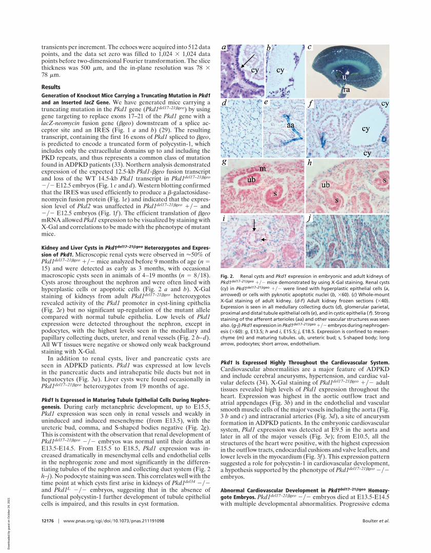
In addition to renal cysts, liver and pancreatic cysts areseen in ADPKD patients. Pkd1 was expressed at low levelsin the pancreatic ducts and intrahepatic bile ducts but not inhepatocytes (Fig. 3a). Liver cysts were found occasionally inPkd1del17–21bgeo heterozygotes from 19 months of age.
Pkd1 Is Expressed in Maturing Tubule Epithelial Cells During Nephro-genesis. During early metanephric development, up to E15.5,Pkd1 expression was seen only in renal vessels and weakly inuninduced and induced mesenchyme (from E13.5), with theureteric bud, comma, and S-shaped bodies negative (Fig. 2g).This is consistent with the observation that renal development ofPkd1del17–21bgeo 2y2 embryos was normal until their deaths atE13.5-E14.5. From E15.5 to E18.5, Pkd1 expression was in-creased dramatically in mesenchymal cells and endothelial cellsin the nephrogenic zone and most significantly in the differen-tiating tubules of the nephron and collecting duct system (Fig. 2h–j). No podocyte staining was seen. This correlates well with thetime point at which cysts first arise in kidneys of Pkd1del34 2y2and Pkd1L 2y2 embryos, suggesting that in the absence offunctional polycystin-1 further development of tubule epithelialcells is impaired, and this results in cyst formation.
Pkd1 Is Expressed Highly Throughout the Cardiovascular System.Cardiovascular abnormalities are a major feature of ADPKDand include cerebral aneurysms, hypertension, and cardiac val-vular defects (34). X-Gal staining of Pkd1del17–21bgeo 1y2 adulttissues revealed high levels of Pkd1 expression throughout theheart. Expression was highest in the aortic outflow tract andatrial appendages (Fig. 3b) and in the endothelial and vascularsmooth muscle cells of the major vessels including the aorta (Fig.3 b and c) and intracranial arteries (Fig. 3d), a site of aneurysmformation in ADPKD patients. In the embryonic cardiovascularsystem, Pkd1 expression was detected at E9.5 in the aorta andlater in all of the major vessels (Fig. 3e); from E10.5, all thestructures of the heart were positive, with the highest expressionin the outflow tracts, endocardial cushions and valve leaflets, andlower levels in the myocardium (Fig. 3f ). This expression patternsuggested a role for polycystin-1 in cardiovascular development,a hypothesis supported by the phenotype of Pkd1del17–21bgeo 2y2embryos.
Abnormal Cardiovascular Development in Pkd1del17–21bgeo Homozy-gote Embryos. Pkd1del17–21bgeo 2y2 embryos died at E13.5-E14.5with multiple developmental abnormalities. Progressive edema
Fig. 2. Renal cysts and Pkd1 expression in embryonic and adult kidneys ofPkd1del17–21bgeo 1y2 mice demonstrated by using X-Gal staining. Renal cysts(cy) in Pkd1del17–21bgeo 1y2 were lined with hyperplastic epithelial cells (a,arrowed) or cells with pyknotic apoptotic nuclei (b, 360). (c) Whole-mountX-Gal staining of adult kidney. (d-f ) Adult kidney frozen sections (340).Expression is seen in all medullary collecting ducts (d), glomerular parietal,proximal and distal tubule epithelial cells (e), and in cystic epithelia ( f). Strongstaining of the afferent arterioles (aa) and other vascular structures was seenalso. (g-j) Pkd1 expression in Pkd1del17–21bgeo 1y2 embryos during nephrogen-esis (360): g, E13.5; h and i, E15.5; j, E18.5. Expression is confined to mesen-chyme (m) and maturing tubules. ub, ureteric bud; s, S-shaped body; longarrow, podocytes; short arrow, endothelium.
12176 u www.pnas.orgycgiydoiy10.1073ypnas.211191098 Boulter et al.
Dow
nloa
ded
by g
uest
on
Oct
ober
24,
202
1
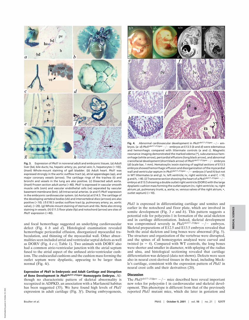
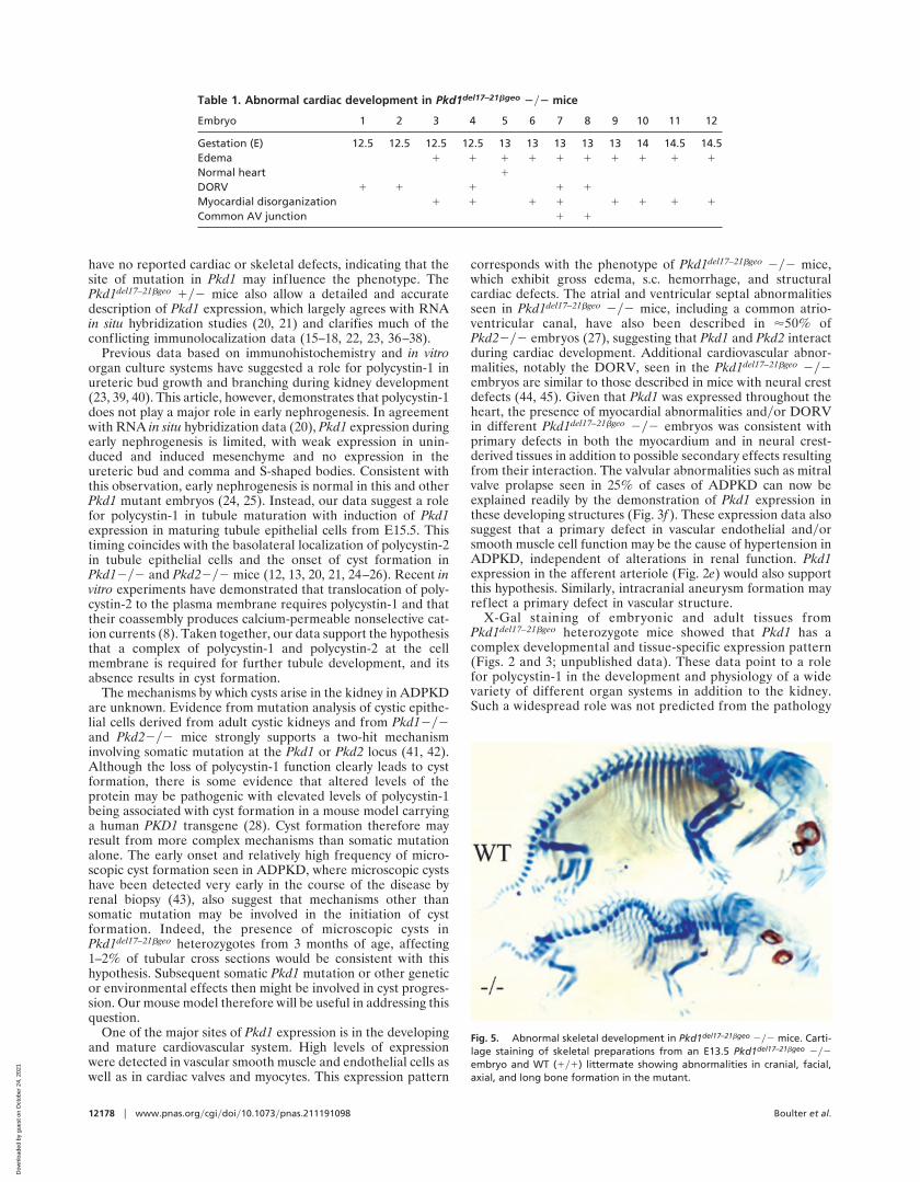
and focal hemorrhage suggested an underlying cardiovasculardefect (Fig. 4 b and d). Histological examination revealedhemorrhagic pericardial effusion, disorganized myocardial tra-beculation, and thinning of the myocardial wall. Other abnor-malities seen included atrial and ventricular septal defects as wellas DORV (Fig. 4 e–i; Table 1). Two animals with DORV alsohad a common atrio-ventricular junction with the atrial septumfused to the atrial aspect of the unfused atrio-ventricular cush-ions. The endocardial cushions and the cushion mass forming theoutlet septum were dysplastic, appearing to be larger thannormal (Fig. 4i).
Expression of Pkd1 in Embryonic and Adult Cartilage and Disruptionof Bone Development in Pkd1del17–21bgeo Homozygote Embryos. Al-though no characteristic pattern of skeletal abnormality isrecognized in ADPKD, an association with a Marfanoid habitushas been suggested (35). We have found high levels of Pkd1expression in adult cartilage (Fig. 3f ). During embryogenesis,
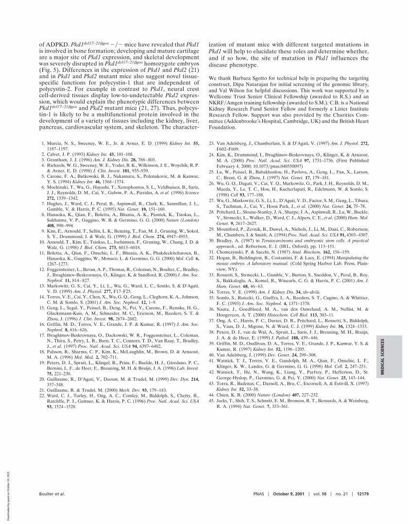
Pkd1 is expressed in differentiating cartilage and somites andearlier in the notochord and floor plate, which are involved insomite development (Fig. 3 e and h). This pattern suggests apotential role for polycystin-1 in formation of the axial skeletonand in cartilage differentiation. Indeed, skeletal developmentwas compromised severely in Pkd1del17–21bgeo 2y2 embryos.Skeletal preparations of E12.5 and E13.5 embryos revealed thatboth the axial skeleton and long bones were abnormal (Fig. 5).The structure and organization of the vertebrae were disrupted,and the spines of all homozygotes analyzed were curved andtwisted (n 5 4). Compared with WT controls, the long boneswere shorter and smaller in diameter, with splaying of the radiusand ulna, and histological sectioning revealed that cartilagedifferentiation was delayed (data not shown). Defects were seenalso in neural crest-derived tissues in the head, including Meck-le’s cartilage, consistent with the expression pattern of Pkd1 inneural crest cells and their derivatives (20).
DiscussionThe Pkd1del17–21bgeo 2y2 mice described here reveal importantnew roles for polycystin-1 in cardiovascular and skeletal devel-opment. This phenotype is different from that of the previouslyreported Pkd1 mutant mice, which die later in gestation and
Fig. 3. Expression of Pkd1 in nonrenal adult and embryonic tissues. (a) Adultliver [bd, bile ducts; ha, hepatic artery; pv, portal vein; h, hepatocytes (310)].(Inset) Whole-mount staining of gall bladder. (b) Adult heart. Pkd1 wasexpressed strongly in the aortic outflow tract (a), atrial appendages (ap), andmajor coronary vessels (arrow). The cartilage rings of the trachea (t) andbronchi and vessels in the lung are also positive. (c) Dissected adult aorta.(Inset) Frozen section adult aorta (340). Pkd1 is expressed in vascular smoothmuscle cells (vsm) and vascular endothelial cells (ve) separated by vascularbasement membrane (bm). (d) Intracranial arteries. (e and f ) Pkd1 expressionin the embryonic cardiovascular system. (e) Aorta (a) at E14.5. The cartilage ofthe developing vertebral bodies (vb) and intervertebral discs (arrows) are alsopositive (310). ( f) E18.5 cardiac outflow tract (p, pulmonary artery; av, aorticvalve), (320). (g) Whole-mount staining of sternum and ribs. Note also strongstaining in vessels. (h) E11.5 floor plate (fp) and notochord (arrow) are sites ofPkd1 expression (340).
Fig. 4. Abnormal cardiovascular development in Pkd1del17–21bgeo 2y2 em-bryos. (a–d) Pkd1del17–21bgeo 2y2 embryos at E13.5 (b and d) were edematousand hemorrhagic compared with littermate controls (a and c). Magneticresonance imaging demonstrated the marked edema (*), subcutaneous hem-orrhage (white arrow), pericardial effusions (long black arrow), and abnormalcraniofacial development (short black arrow) of Pkd1del17–21bgeo 2y2 embryos(d) (scale bar, 1 mm). Hematoxylinyeosin staining of sagittal sections of E13.5embryos showed hemorrhagic effusion and disorganization of the myocardialwall and ventricular septum in Pkd1del17–21bgeo 2y2 embryos ( f and h) but notin WT littermates (e and g). lv, left ventricle; rv, right ventricle. e and f, 310;g and h, 340. (i) Transverse section showing the heart of a Pkd1del17–21bgeo 2y2embryo at E13.5 showing a double outlet right ventricle (DORV) with the largedysplastic cushion mass forming the outlet septum (rv, right ventricle; ra, rightatrium; pt, pulmonary trunk; a, aorta; vv, venous valves of the right atrium; *,outlet septum) (310).
Boulter et al. PNAS u October 9, 2001 u vol. 98 u no. 21 u 12177
MED
ICA
LSC
IEN
CES
Dow
nloa
ded
by g
uest
on
Oct
ober
24,
202
1
have no reported cardiac or skeletal defects, indicating that thesite of mutation in Pkd1 may influence the phenotype. ThePkd1del17–21bgeo 1y2 mice also allow a detailed and accuratedescription of Pkd1 expression, which largely agrees with RNAin situ hybridization studies (20, 21) and clarifies much of theconflicting immunolocalization data (15–18, 22, 23, 36–38).
Previous data based on immunohistochemistry and in vitroorgan culture systems have suggested a role for polycystin-1 inureteric bud growth and branching during kidney development(23, 39, 40). This article, however, demonstrates that polycystin-1does not play a major role in early nephrogenesis. In agreementwith RNA in situ hybridization data (20), Pkd1 expression duringearly nephrogenesis is limited, with weak expression in unin-duced and induced mesenchyme and no expression in theureteric bud and comma and S-shaped bodies. Consistent withthis observation, early nephrogenesis is normal in this and otherPkd1 mutant embryos (24, 25). Instead, our data suggest a rolefor polycystin-1 in tubule maturation with induction of Pkd1expression in maturing tubule epithelial cells from E15.5. Thistiming coincides with the basolateral localization of polycystin-2in tubule epithelial cells and the onset of cyst formation inPkd12y2 and Pkd22y2 mice (12, 13, 20, 21, 24–26). Recent invitro experiments have demonstrated that translocation of poly-cystin-2 to the plasma membrane requires polycystin-1 and thattheir coassembly produces calcium-permeable nonselective cat-ion currents (8). Taken together, our data support the hypothesisthat a complex of polycystin-1 and polycystin-2 at the cellmembrane is required for further tubule development, and itsabsence results in cyst formation.
The mechanisms by which cysts arise in the kidney in ADPKDare unknown. Evidence from mutation analysis of cystic epithe-lial cells derived from adult cystic kidneys and from Pkd12y2and Pkd22y2 mice strongly supports a two-hit mechanisminvolving somatic mutation at the Pkd1 or Pkd2 locus (41, 42).Although the loss of polycystin-1 function clearly leads to cystformation, there is some evidence that altered levels of theprotein may be pathogenic with elevated levels of polycystin-1being associated with cyst formation in a mouse model carryinga human PKD1 transgene (28). Cyst formation therefore mayresult from more complex mechanisms than somatic mutationalone. The early onset and relatively high frequency of micro-scopic cyst formation seen in ADPKD, where microscopic cystshave been detected very early in the course of the disease byrenal biopsy (43), also suggest that mechanisms other thansomatic mutation may be involved in the initiation of cystformation. Indeed, the presence of microscopic cysts inPkd1del17–21bgeo heterozygotes from 3 months of age, affecting1–2% of tubular cross sections would be consistent with thishypothesis. Subsequent somatic Pkd1 mutation or other geneticor environmental effects then might be involved in cyst progres-sion. Our mouse model therefore will be useful in addressing thisquestion.
One of the major sites of Pkd1 expression is in the developingand mature cardiovascular system. High levels of expressionwere detected in vascular smooth muscle and endothelial cells aswell as in cardiac valves and myocytes. This expression pattern
corresponds with the phenotype of Pkd1del17–21bgeo 2y2 mice,which exhibit gross edema, s.c. hemorrhage, and structuralcardiac defects. The atrial and ventricular septal abnormalitiesseen in Pkd1del17–21bgeo 2y2 mice, including a common atrio-ventricular canal, have also been described in '50% ofPkd22y2 embryos (27), suggesting that Pkd1 and Pkd2 interactduring cardiac development. Additional cardiovascular abnor-malities, notably the DORV, seen in the Pkd1del17–21bgeo 2y2embryos are similar to those described in mice with neural crestdefects (44, 45). Given that Pkd1 was expressed throughout theheart, the presence of myocardial abnormalities andyor DORVin different Pkd1del17–21bgeo 2y2 embryos was consistent withprimary defects in both the myocardium and in neural crest-derived tissues in addition to possible secondary effects resultingfrom their interaction. The valvular abnormalities such as mitralvalve prolapse seen in 25% of cases of ADPKD can now beexplained readily by the demonstration of Pkd1 expression inthese developing structures (Fig. 3f ). These expression data alsosuggest that a primary defect in vascular endothelial andyorsmooth muscle cell function may be the cause of hypertension inADPKD, independent of alterations in renal function. Pkd1expression in the afferent arteriole (Fig. 2e) would also supportthis hypothesis. Similarly, intracranial aneurysm formation mayreflect a primary defect in vascular structure.
X-Gal staining of embryonic and adult tissues fromPkd1del17–21bgeo heterozygote mice showed that Pkd1 has acomplex developmental and tissue-specific expression pattern(Figs. 2 and 3; unpublished data). These data point to a rolefor polycystin-1 in the development and physiology of a widevariety of different organ systems in addition to the kidney.Such a widespread role was not predicted from the pathology
Fig. 5. Abnormal skeletal development in Pkd1del17–21bgeo 2y2 mice. Carti-lage staining of skeletal preparations from an E13.5 Pkd1del17–21bgeo 2y2embryo and WT (1y1) littermate showing abnormalities in cranial, facial,axial, and long bone formation in the mutant.
Table 1. Abnormal cardiac development in Pkd1del17–21bgeo 2y2 mice
Embryo 1 2 3 4 5 6 7 8 9 10 11 12
Gestation (E) 12.5 12.5 12.5 12.5 13 13 13 13 13 14 14.5 14.5Edema 1 1 1 1 1 1 1 1 1 1
Normal heart 1
DORV 1 1 1 1 1
Myocardial disorganization 1 1 1 1 1 1 1 1
Common AV junction 1 1
12178 u www.pnas.orgycgiydoiy10.1073ypnas.211191098 Boulter et al.
Dow
nloa
ded
by g
uest
on
Oct
ober
24,
202
1
of ADPKD. Pkd1del17–21bgeo 2y2 mice have revealed that Pkd1is involved in bone formation; developing and mature cartilageare a major site of Pkd1 expression, and skeletal developmentwas severely disrupted in Pkd1del17–21bgeo homozygote embryos(Fig. 5). Differences in the expression of Pkd1 and Pkd2 (21)and in Pkd1 and Pkd2 mutant mice also suggest novel tissue-specific functions for polycystin-1 that are independent ofpolycystin-2. For example in contrast to Pkd1, neural crestcell-derived tissues display low-to-undetectable Pkd2 expres-sion, which would explain the phenotypic differences betweenPkd1del17–21bgeo and Pkd2 mutant mice (21, 27). Thus, polycys-tin-1 is likely to be a multifunctional protein involved in thedevelopment of a variety of tissues including the kidney, liver,pancreas, cardiovascular system, and skeleton. The character-
ization of mutant mice with different targeted mutations inPkd1 will help to elucidate these roles and determine whether,and if so how, the site of mutation in Pkd1 inf luences thedisease phenotype.
We thank Barbara Sgotto for technical help in preparing the targetingconstruct, Dipa Natarajan for initial screening of the genomic library,and Val Wilson for helpful discussions. This work was supported by aWellcome Trust Senior Clinical Fellowship (awarded to R.S.) and anNKRFyAmgen training fellowship (awarded to S.M.). C.B. is a NationalKidney Research Fund Senior Fellow and formerly a Lister InstituteResearch Fellow. Support was also provided by the Charities Com-mittee (Addenbrooke’s Hospital, Cambridge, UK) and the British HeartFoundation.
1. Murcia, N. S., Sweeney, W. E., Jr. & Avner, E. D. (1999) Kidney Int. 55,1187–1197.
2. Calvet, J. P. (1993) Kidney Int. 43, 101–108.3. Grantham, J. J. (1996) Am. J. Kidney Dis. 28, 788–803.4. Richards, W. G., Sweeney, W. E., Yoder, B. K., Wilkinson, J. E., Woychik, R. P.
& Avner, E. D. (1998) J. Clin. Invest. 101, 935–939.5. Carone, F. A., Butkowski, R. J., Nakamura, S., Polenakovic, M. & Kanwar,
Y. S. (1994) Kidney Int. 46, 1368–1374.6. Mochizuki, T., Wu, G., Hayashi, T., Xenophontos, S. L., Veldhuisen, B., Saris,
J. J., Reynolds, D. M., Cai, Y., Gabow, P. A., Pierides, A. et al. (1996) Science272, 1339–1342.
7. Hughes, J., Ward, C. J., Peral, B., Aspinwall, R., Clark, K., Sanmillan, J. L.,Gamble, V. & Harris, P. C. (1995) Nat. Genet. 10, 151–160.
8. Hanaoka, K., Qian, F., Boletta, A., Bhunia, A. K., Piontek, K., Tsiokas, L.,Sukhatme, V. P., Guggino, W. B. & Germino, G. G. (2000) Nature (London)408, 990–994.
9. Kim, E., Arnould, T., Sellin, L. K., Benzing, T., Fan, M. J., Gruning, W., Sokol,S. Y., Drummond, I. & Walz, G. (1999) J. Biol. Chem. 274, 4947–4953.
10. Arnould, T., Kim, E., Tsiokas, L., Jochimsen, F., Gruning, W., Chang, J. D. &Walz, G. (1998) J. Biol. Chem. 273, 6013–6018.
11. Boletta, A., Qian, F., Onuchic, L. F., Bhunia, A. K., Phakdeekitcharoen, B.,Hanaoka, K., Guggino, W., Monaco, L. & Germino, G. G. (2000) Mol. Cell. 6,1267–1273.
12. Foggensteiner, L., Bevan, A. P., Thomas, R., Coleman, N., Boulter, C., Bradley,J., Ibraghimov-Beskrovnaya, O., Klinger, K. & Sandford, R. (2000) J. Am. Soc.Nephrol. 11, 814–827.
13. Markowitz, G. S., Cai, Y., Li, L., Wu, G., Ward, L. C., Somlo, S. & D’Agati,V. D. (1999) Am. J. Physiol. 277, F17–F25.
14. Torres, V. E., Cai, Y., Chen, X., Wu, G. Q., Geng, L., Cleghorn, K. A., Johnson,C. M. & Somlo, S. (2001) J. Am. Soc. Nephrol. 12, 1–9.
15. Geng, L., Segal, Y., Peissel, B., Deng, N., Pei, Y., Carone, F., Rennke, H. G.,Glucksmann-Kuis, A. M., Schneider, M. C., Ericsson, M., Reeders, S. T. &Zhou, J. (1996) J. Clin. Invest. 98, 2674–2682.
16. Griffin, M. D., Torres, V. E., Grande, J. P. & Kumar, R. (1997) J. Am. Soc.Nephrol. 8, 616–626.
17. Ibraghimov-Beskrovnaya, O., Dackowski, W. R., Foggensteiner, L., Coleman,N., Thiru, S., Petry, L. R., Burn, T. C., Connors, T. D., Van Raay, T., Bradley,J., et al. (1997) Proc. Natl. Acad. Sci. USA 94, 6397–6402.
18. Palsson, R., Sharma, C. P., Kim, K., McLaughlin, M., Brown, D. & Arnaout,M. A. (1996) Mol. Med. 2, 702–711.
19. Peters, D. J., Spruit, L., Klingel, R., Prins, F., Baelde, H. J., Giordano, P. C.,Bernini, L. F., de Heer, E., Breuning, M. H. & Bruijn, J. A. (1996) Lab. Invest.75, 221–230.
20. Guillaume, R., D’Agati, V., Daoust, M. & Trudel, M. (1999) Dev. Dyn. 214,337–348.
21. Guillaume, R. & Trudel, M. (2000) Mech. Dev. 93, 179–183.22. Ward, C. J., Turley, H., Ong, A. C., Comley, M., Biddolph, S., Chetty, R.,
Ratcliffe, P. J., Gattner, K. & Harris, P. C. (1996) Proc. Natl. Acad. Sci. USA93, 1524–1528.
23. Van Adelsberg, J., Chamberlain, S. & D’Agati, V. (1997) Am. J. Physiol. 272,F602–F609.
24. Kim, K., Drummond, I., Ibraghimov-Beskrovnaya, O., Klinger, K. & Arnaout,M. A. (2000) Proc. Natl. Acad. Sci. USA 97, 1731–1736. (First PublishedFebruary 4, 2000; 10.1073ypnas.040550097)
25. Lu, W., Peissel, B., Babakhanlou, H., Pavlova, A., Geng, L., Fan, X., Larson,C., Brent, G. & Zhou, J. (1997) Nat. Genet. 17, 179–181.
26. Wu, G. Q., Dagati, V., Cai, Y. Q., Markowitz, G., Park, J. H., Reynolds, D. M.,Maeda, Y., Le, T. C., Hou, H., Kucherlapati, R., Edelmann, W. & Somlo, S.(1998) Cell 93, 177–188.
27. Wu, G., Markowitz, G. S., Li, L., D’Agati, V. D., Factor, S. M., Geng, L., Tibara,S., Tuchman, J., Cai, Y., Hoon Park, J., et al. (2000) Nat. Genet. 24, 75–78.
28. Pritchard, L., Sloane-Stanley, J. A., Sharpe, J. A., Aspinwall, R., Lu, W., Buckle,V., Strmecki, L., Walker, D., Ward, C. J., Alpers, C. E., et al. (2000) Hum. Mol.Genet. 9, 2617–2627.
29. Mountford, P., Zevnik, B., Duwel, A., Nichols, J., Li, M., Dani, C., Robertson,M., Chambers, I. & Smith, A. (1994) Proc. Natl. Acad. Sci. USA 91, 4303–4307.
30. Bradley, A. (1987) in Teratocarcinoms and embryonic stem cells. A practicalapproach., ed. Robertson, E. J. (IRL, Oxford), pp. 113–151.
31. Chomczynski, P. & Sacchi, N. (1987) Anal. Biochem. 162, 156–159.32. Hogan, B., Beddington, R., Costantini, F. & Lacy, E. (1994) Manipulating the
mouse embryo. A laboratory manual. (Cold Spring Harbor Lab. Press, Plain-view, NY).
33. Rossetti, S., Strmecki, L., Gamble, V., Burton, S., Sneddon, V., Peral, B., Roy,S., Bakkaloglu, A., Komel, R., Winearls, C. G. & Harris, P. C. (2001) Am. J.Hum. Genet. 68, 46–63.
34. Torres, V. E. (1999) Am. J. Kidney Dis. 34, xlv-xlviii.35. Somlo, S., Rutecki, G., Giuffra, L. A., Reeders, S. T., Cugino, A. & Whittier,
F. C. (1993) J. Am. Soc. Nephrol. 4, 1371–1378.36. Nauta, J., Goedbloed, M. A., van den Ouweland, A. M., Nellist, M. &
Hoogeveen, A. T. (2000) Histochem. Cell Biol. 113, 303–11.37. Ong, A. C., Harris, P. C., Davies, D. R., Pritchard, L., Rossetti, S., Biddolph,
S., Vaux, D. J., Migone, N. & Ward, C. J. (1999) Kidney Int. 56, 1324–1333.38. Peters, D. J., van de Wal, A., Spruit, L., Saris, J. J., Breuning, M. H., Bruijn,
J. A. & de Heer, E. (1999) J. Pathol. 188, 439–446.39. Griffin, M. D., Osullivan, D. A., Torres, V. E., Grande, J. P., Kanwar, Y. S. &
Kumar, R. (1997) Kidney Int. 52, 1196–1205.40. Van Adelsberg, J. (1999) Dev. Genet. 24, 299–308.41. Watnick, T. J., Torres, V. E., Gandolph, M. A., Qian, F., Onuchic, L. F.,
Klinger, K. W., Landes, G. & Germino, G. G. (1998) Mol. Cell. 2, 247–251.42. Watnick, T., He, N., Wang, K., Liang, Y., Parfrey, P., Hefferton, D., St.
George-Hyslop, P., Germino, G. & Pei, Y. (2000) Nat. Genet. 25, 143–144.43. Torra, R., Badenas, C., Darnell, A., Bru, C., Escorsell, A. & Estivill, X. (1997)
Kidney Int. 52, 33–38.44. Chien, K. R. (2000) Nature (London) 407, 227–232.45. Jacks, T., Shih, T. S., Schmitt, E. M., Bronson, R. T., Bernards, A. & Weinberg,
R. A. (1994) Nat. Genet. 7, 353–361.
Boulter et al. PNAS u October 9, 2001 u vol. 98 u no. 21 u 12179
MED
ICA
LSC
IEN
CES
Dow
nloa
ded
by g
uest
on
Oct
ober
24,
202
1


