Cardiovascular and Respiratory Systems: Getting Oxygen From Air to Muscle Integration of...
103
Cardiovascular and Respiratory Systems: Getting Oxygen From Air to Muscle Integration of Ventilation, Heart, and Circulation
-
date post
20-Dec-2015 -
Category
Documents
-
view
213 -
download
0
Transcript of Cardiovascular and Respiratory Systems: Getting Oxygen From Air to Muscle Integration of...
Cardiovascular and Respiratory Systems: Getting Oxygen From
Air to Muscle Integration of Ventilation, Heart,
and Circulation
Cardiorespiratory System
Functions of cardiorespiratory system: transportation of O2 and CO2
transportation of nutrients/waste products distribution of hormones thermoregulation maintenance of blood pressure
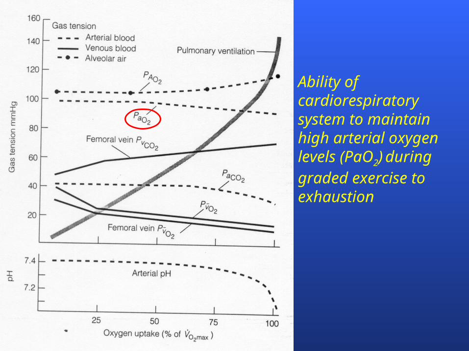
Ability of cardiorespiratory system to maintain high arterial oxygen levels (PaO2) during graded exercise to exhaustion

Critical elements of O2 Transport Pathway
Ventilation– Moving air in/out of lungs
External respiration – Gas exchange between alveoli and blood
Heart and circulation O2 diffusion into mitochondria

Role of the Heart
Moving O2 from lungs to muscle

Oxygen Delivery Determines VO2
(Fick Principle)
VO2 = Q (CaO2 – CvO2)
VO2 = [HR SV] (CaO2 – CvO2)
VO2 = [BP TPR] (CaO2 – CvO2)

Cardiac Cycle
systole diastole cardiac output (Q) = stroke volume (SV)
heart rate (HR)
examples– rest: SV = 75 ml; HR = 60 bpm; Q = 4.5 Lmin-1
– exercise: SV = 130 ml; HR = 180 bpm; Q = 23.4 Lmin-1

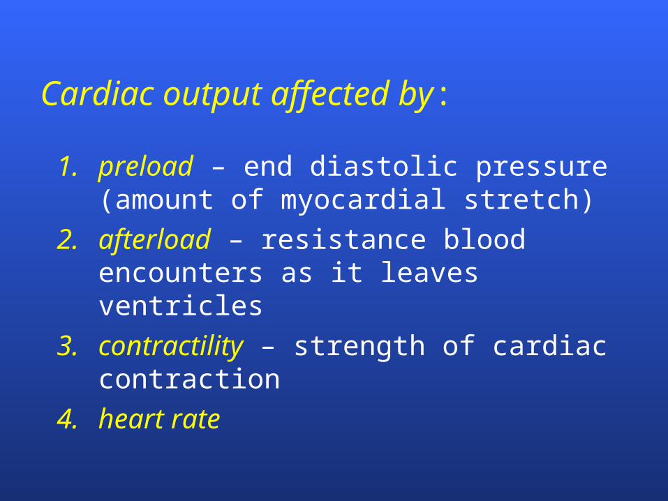
Cardiac output affected by:
1. preload – end diastolic pressure (amount of myocardial stretch)
2. afterload – resistance blood encounters as it leaves ventricles
3. contractility – strength of cardiac contraction
4. heart rate

Muscle pump and one-way valves assist venous return
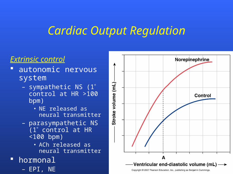
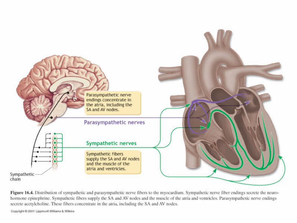
Cardiac Output Regulation
Extrinsic control autonomic nervous
system– sympathetic NS (1
control at HR >100 bpm)• NE released as neural
transmitter
– parasympathetic NS (1 control at HR <100 bpm)
• ACh released as neural transmitter
hormonal– EPI, NE
Ventilation and External Respiration
Getting O2 from air into blood

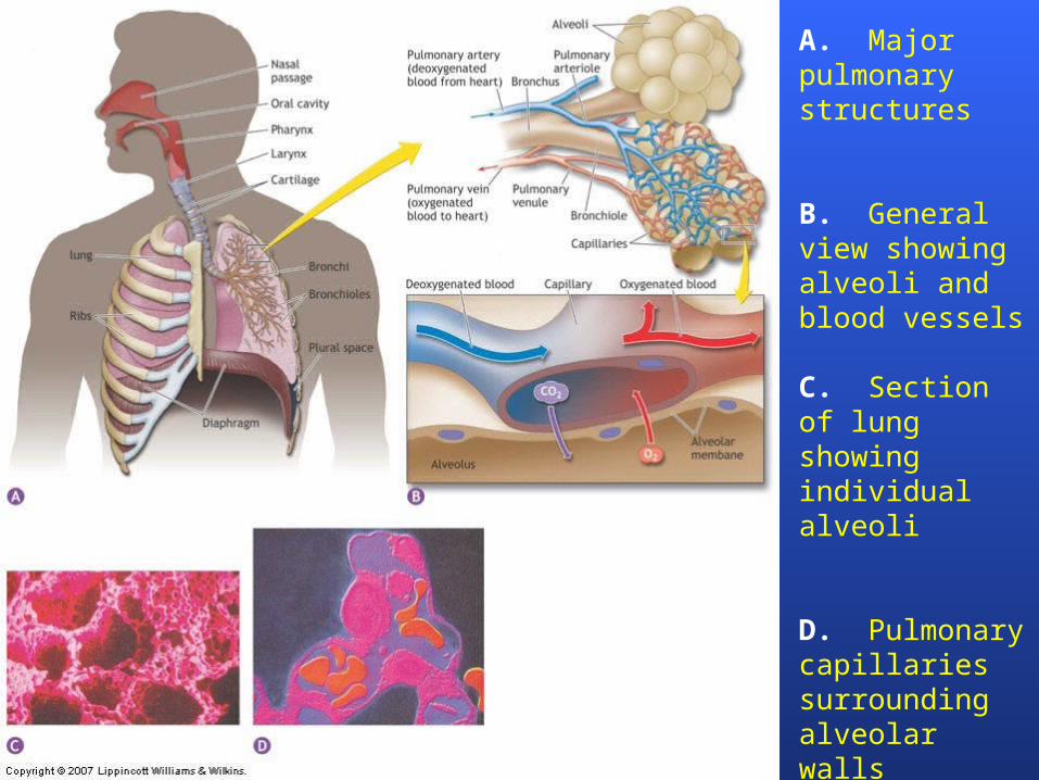
A. Major pulmonary structures
B. General view showing alveoli and blood vessels
C. Section of lung showing individual alveoli
D. Pulmonary capillaries surrounding alveolar walls

RBC
Single alveoli at rest showing individual RBCs
Single alveoli under high flow showing increased RBCs

Lungs and Pulmonary Circulation
alveolar membrane thickness is ~ 0.1 µm
total alveolar surface area is ~75 m2
80-90% of alveoli are covered by capillaries
pulmonary circulation varies with cardiac output and matched to ventilation rate

Gases Move Down Pressure Gradients

O2 and CO2 transit time in lungs (left) and tissue (right) at rest
Notice rapid saturation with O2 by the time RBCs have traveled ⅓ around alveolus
PO2 in blood returning to the lungs is ____ PO2 in the alveoli.
A. greater than
B. less than
C. similar to
PO2 in arterial blood is ____ PO2 in the mitochondria.
A. greater than
B. less than
C. similar to

PCO2 in venous blood is ____ PCO2 in the alveoli.
A. greater than
B. less than
C. similar to
What would be the effect on the saturation of arterial blood with O2 (SaO2) when pulmonary
blood flow is faster than the diffusion rate of O2?
A. SaO2 would remain unchanged
B. SaO2 would be decreased
C. SaO2 would be increased
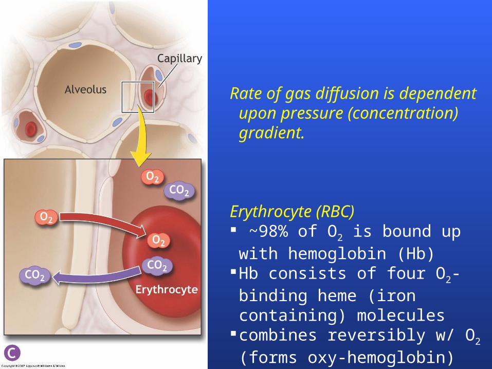
Rate of gas diffusion is dependent upon pressure (concentration) gradient.
Erythrocyte (RBC) ~98% of O2 is bound up with hemoglobin (Hb)
Hb consists of four O2-binding heme (iron containing) molecules
combines reversibly w/ O2 (forms oxy-hemoglobin)
1-2% of O2 is dissolved in plasma
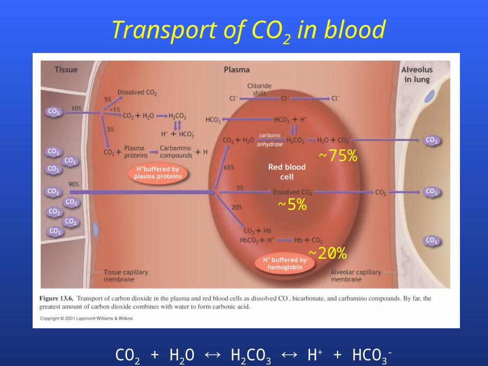
CO2 + H2O H2CO3 H+ + HCO3-
Transport of CO2 in blood
~75%
~5%
~20%

Oxygen is transported from lungs to muscle primarily
A. dissolved in blood.
B. bound to hemoglobin.
C. as a bicarbonate ion.

Carbon dioxide is transported from muscle to the lungs
A. dissolved in blood.
B. bound to hemoglobin.
C. as a bicarbonate ion.
D. all of the above are transport mechanisms for CO2
Ventilatory Response to Exercise and Control of Blood pH
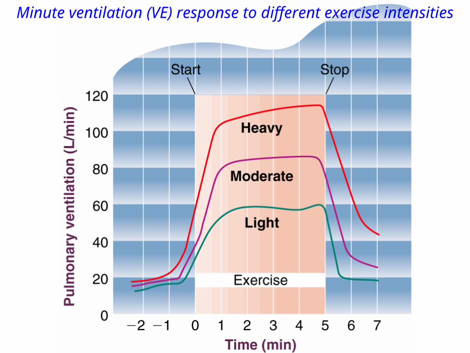
Minute ventilation (VE) response to different exercise intensities
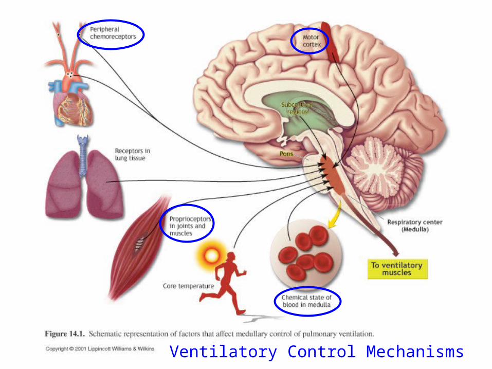
Ventilatory Control Mechanisms
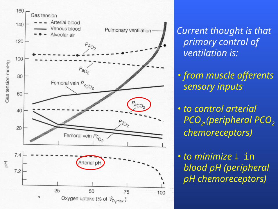
Current thought is that primary control of ventilation is:
• from muscle afferentssensory inputs
• to control arterial PCO2,(peripheral PCO2 chemoreceptors)
• to minimize in blood pH (peripheral pH chemoreceptors)
VE vs VO2
0
20
40
60
80
100
120
140
160
180
200
0 1 2 3 4 5 6 7
VO2 (L/min)
VE
(L/m
in)
VCO2 vs VO2
0
0.5
1
1.5
2
2.5
3
3.5
4
4.5
5
0 1 2 3 4 5 6
VO2 (L/min)
VC
O2
(L/m
in)
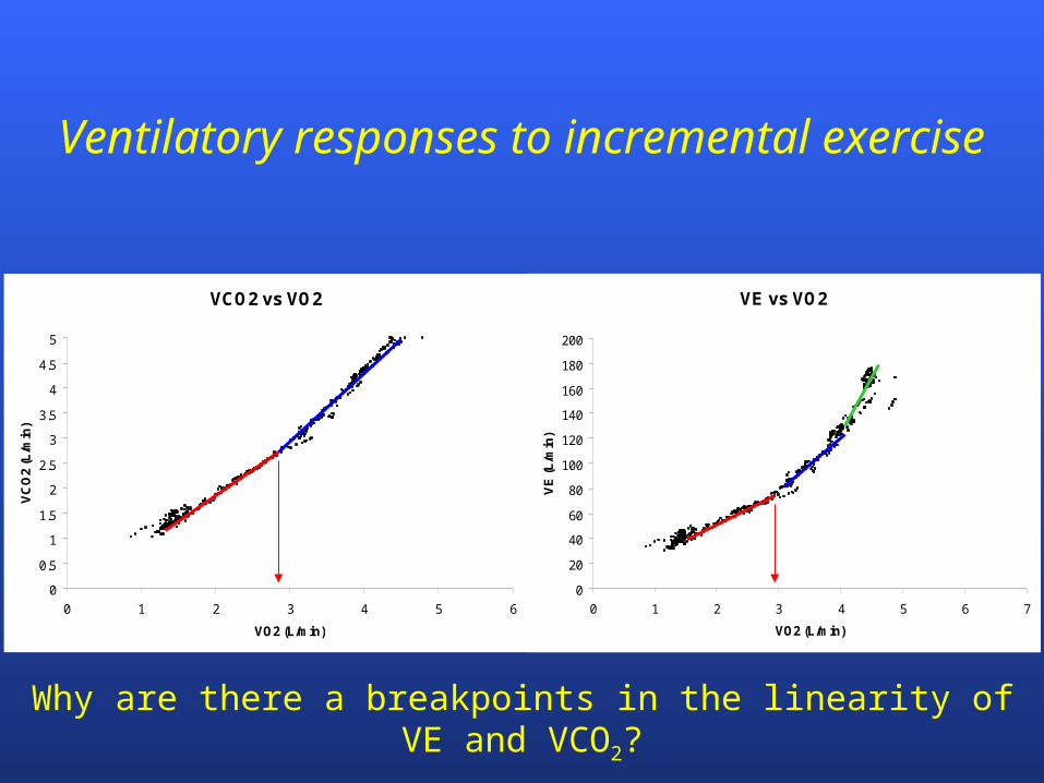
Ventilatory responses to incremental exercise
Why are there a breakpoints in the linearity of VE and VCO2?

Ventilatory Regulation of Acid-Base Balance
CO2 + H2O H2CO3 H+ + HCO3-
at low-intensity exercise, source of CO2 is entirely from substrate metabolism
bicarbonate (HCO3-) buffers H+ produced during high-
intensity exercise at high-intensity exercise, bicarbonate ions also
contribute to CO2 production– source of CO2 is from substrates and bicarbonate ions (HCO3
-)
blood [H+] stimulates VE to rid excess CO2 (and H+)
Can RER ever exceed 1.0? When? Explain

Blood Lactate
02468
1012
50 100 150 200 250 300 350
Treadmill Speed (m/min)
Lacta
te (m
M)

Blood pH
7.05
7.10
7.15
7.20
7.25
7.30
7.35
7.40
7.45
4 5 6 7 8 9 10 11 12 13 14 15
Treadmill Speed (mph)
pH
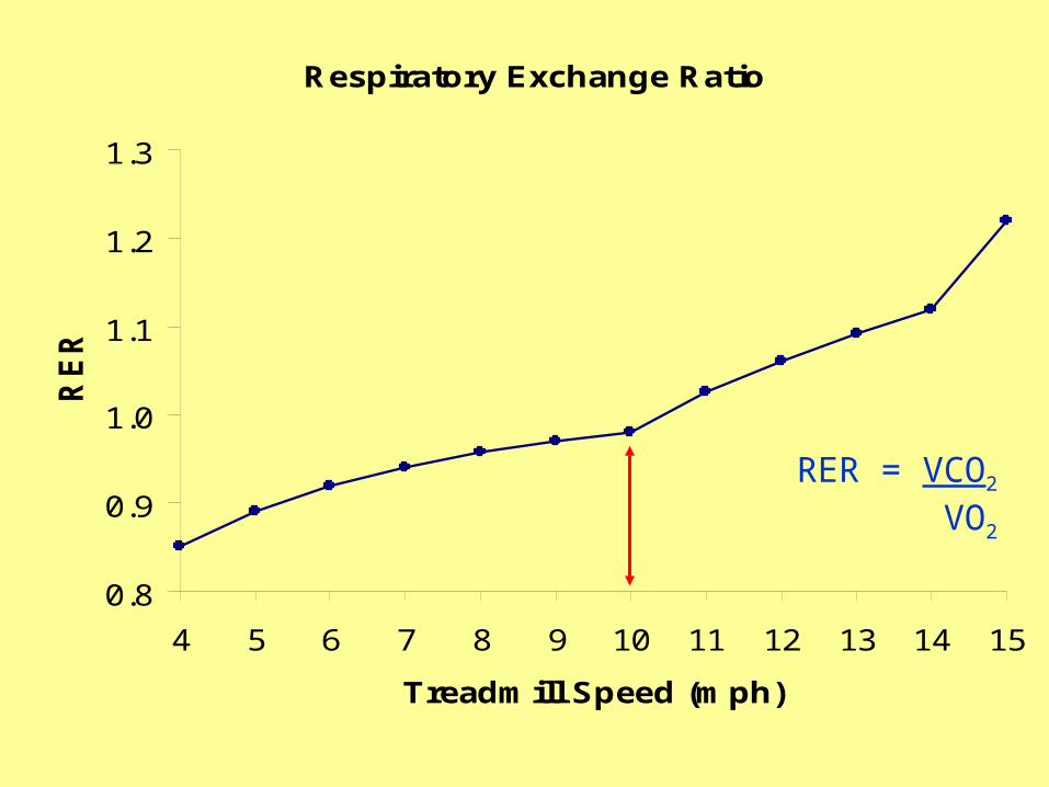
Respiratory Exchange Ratio
0.8
0.9
1.0
1.1
1.2
1.3
4 5 6 7 8 9 10 11 12 13 14 15
Treadmill Speed (mph)
RE
R
RER = VCO2
VO2

CO2 Production
0
10
20
30
40
50
60
70
80
90
2 3 4 5 6 7 8 9 10 11 12 13 14 15
Treadmill Speed (mph)
VC
O2
(m
l/k
g/m
in)
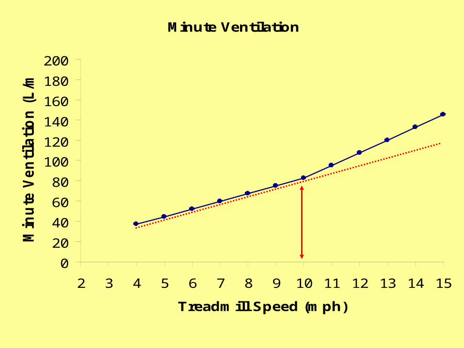
Minute Ventilation
0
20
40
60
80
100
120
140
160
180
200
2 3 4 5 6 7 8 9 10 11 12 13 14 15
Treadmill Speed (mph)
Min
ute
Ven
tila
tio
n (
L/m
in)

What is the primary cause of hyperventilation during incremental exercise?
A. muscles cannot get enough O2
B. sympathetic innervationC. accumulation of lactate ions in bloodD. conscious desire to breath harder
E. additional stimulation of PCO2 chemoreceptors
VE vs VO2
0
20
40
60
80
100
120
140
160
180
200
0 1 2 3 4 5 6 7
VO2 (L/min)
VE
(L/m
in)
VCO2 vs VO2
0
0.5
1
1.5
2
2.5
3
3.5
4
4.5
5
0 1 2 3 4 5 6
VO2 (L/min)
VC
O2
(L/m
in)

Ventilation Questions
1. Describe how ventilation regulates blood pH.
2. Explain why the ventilatory threshold is related to the lactate threshold
3. Can RER ever exceed 1.0? Under what circumstances? Explain.

Young cowboys in the old west

Matching O2 delivery to muscle O2 needs
Regulation of cardiorespiratory system
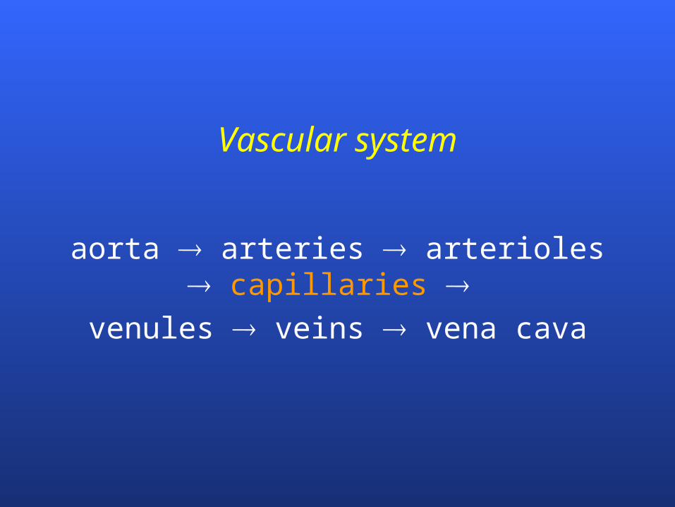
Vascular system
aorta arteries arterioles capillaries
venules veins vena cava
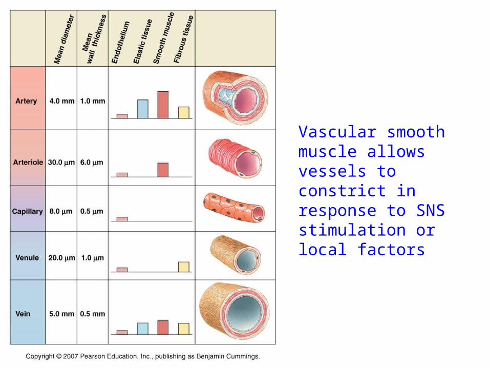
Vascular smooth muscle allows vessels to constrict in response to SNS stimulation or local factors

Arterioles and Capillaries
arterioles terminal arterioles (TA) capillaries collecting venules (CV)
arterioles regulate circulation into tissues– under sympathetic and local control
precapillary sphincters fine tune circulation within tissue– under local control
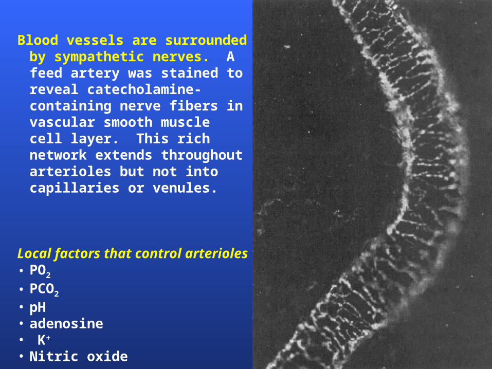
Blood vessels are surrounded by sympathetic nerves. A feed artery was stained to reveal catecholamine-containing nerve fibers in vascular smooth muscle cell layer. This rich network extends throughout arterioles but not into capillaries or venules.
Local factors that control arterioles• PO2
• PCO2
• pH• adenosine• K+
• Nitric oxide

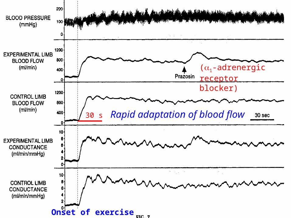
Onset of exercise
(1-adrenergic receptor blocker)
30 s Rapid adaptation of blood flow
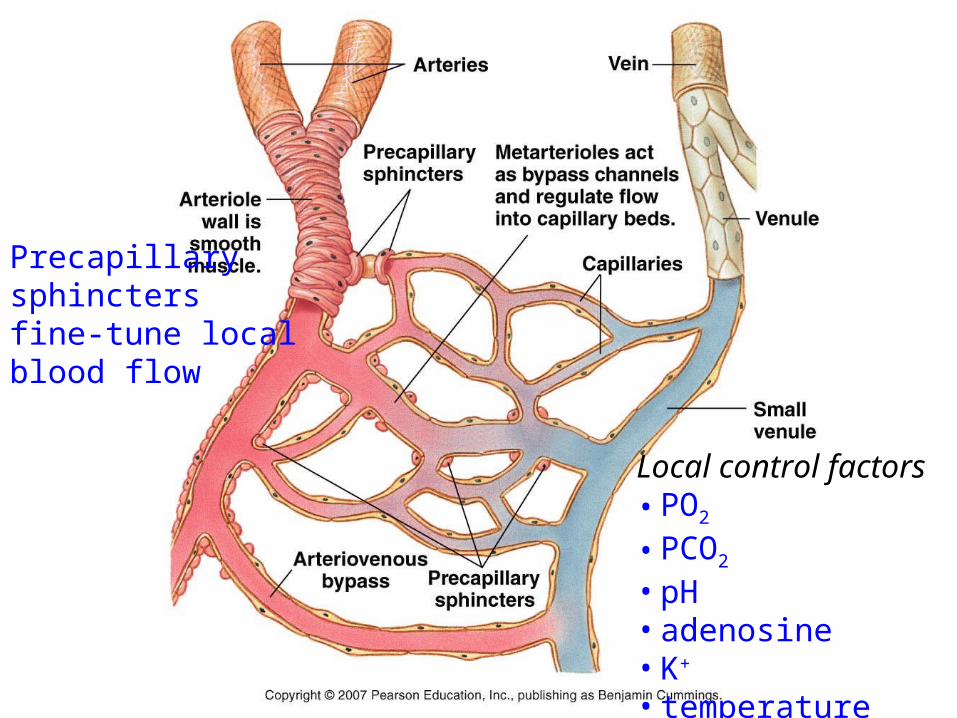
Precapillary sphincters fine-tune local blood flow
Local control factors• PO2
• PCO2
• pH• adenosine• K+
• temperature

Blood Distribution During Rest

Blood Flow Redistribution During Exercise
At rest, most blood is found in the ______ while at exercise most blood is in _____.
A. venous system; active muscle
B. pulmonary circulation; heart
C. arterioles; capillaries
D. heart; heart
E. liver; active muscle
What is the primary mechanism to increase blood flow to working muscle?
A. baroreceptors
B. sympathetic innervation
C. local factors
D. epinephrine
E. central command

What effect would these local conditions (from resting values) have on arteriole blood flow?
PO2, PCO2, pH, temperature
A. increase flow
B. decrease flow
C. no effect on flow
D. cannot be determined


O2 Extraction
Moving O2 from blood into muscle
Factors affecting Oxygen Extraction
Fick equation
VO2 = Q (aO2 – vO2)

O2 extraction response to
exercise
Represents mixed venous blood returning
to right heart

a-v O2 difference
Bohr Effect: effect of local environment on oxy-hemoglobin binding strength
amount of O2 released to muscle depends on local environment– PO2, pH, PCO2, temperature, 2,3 DPG
2,3 diphosphoglycerate (DPG)– produced in RBC during prolonged, heavy
exercise– binds loosely with Hb to reduce its affinity for O2
which increases O2 release
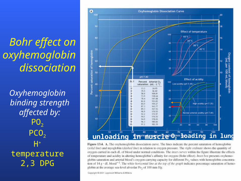
Bohr effect on oxyhemoglobin
dissociation
O2 loading in lungsO2 unloading in muscle
Oxyhemoglobin binding strength
affected by:PO2
PCO2
H+
temperature2,3 DPG
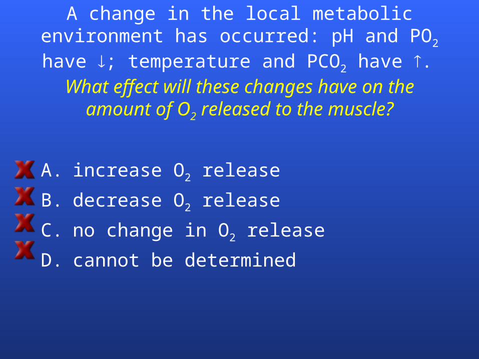
A change in the local metabolic environment has occurred: pH and PO2 have ; temperature
and PCO2 have . What effect will these changes have on the
amount of O2 released to the muscle?
A. increase O2 release
B. decrease O2 release
C. no change in O2 release
D. cannot be determined

A change in the local metabolic environment has occurred: pH and PO2 have ; temperature
and PCO2 have .
What do these changes in local environmental suggest has occurred?
A. the muscles changed from an exercise to a resting state
B. the muscles began to exercise
C. no change
D. cannot be determined

During graded exercise,
A. VCO2 increases linearly
B. A breakpoint occurs in VCO2 that coincides with lactate threshold
C. A breakpoint occurs in VE that is caused by increased VO2
D. A breakpoint occurs in VCO2 that results from increased epinephrine release
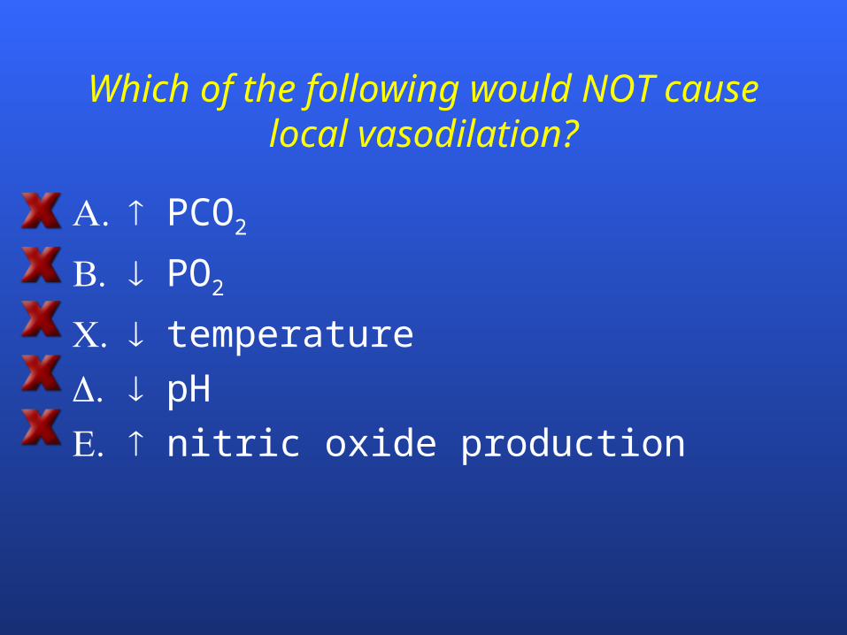
Which of the following would NOT cause local vasodilation?
A. PCO2
B. PO2
C. temperature
. pH
E. nitric oxide production
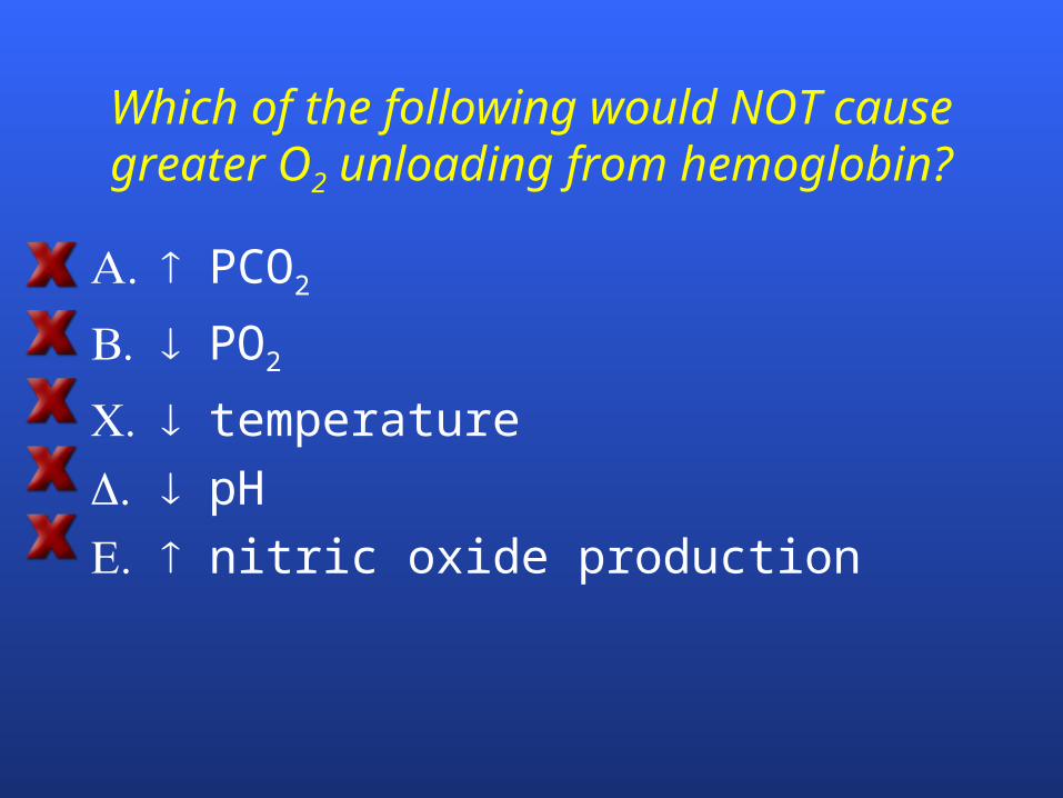
Which of the following would NOT cause greater O2 unloading from hemoglobin?
A. PCO2
B. PO2
C. temperature
. pH
E. nitric oxide production

Which of the following adaptations likely had the LEAST influence for explaining why VO2max
increased 12% after completing a cross country season?
A. cardiac output
B. blood volume
C. mitochondrial volume
. capillary density
E. number of RBC
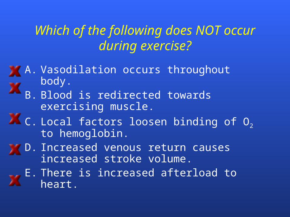
Which of the following does NOT occur during exercise?
A. Vasodilation occurs throughout body.B. Blood is redirected towards exercising
muscle.
C. Local factors loosen binding of O2 to hemoglobin.
D. Increased venous return causes increased stroke volume.
E. There is increased afterload to heart.
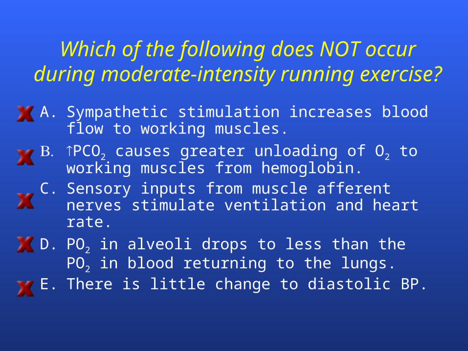
Which of the following does NOT occur during moderate-intensity running exercise?
A. Sympathetic stimulation increases blood flow to working muscles.
B. PCO2 causes greater unloading of O2 to working muscles from hemoglobin.
C. Sensory inputs from muscle afferent nerves stimulate ventilation and heart rate.
D. PO2 in alveoli drops to less than the PO2 in blood returning to the lungs.
E. There is little change to diastolic BP.
Control of cardiac function and ventilation
Parallel activations
What would be the effect of local arteriole dilation on BP?
A. Decrease BP
B. Increase BP
C. No effect on BP
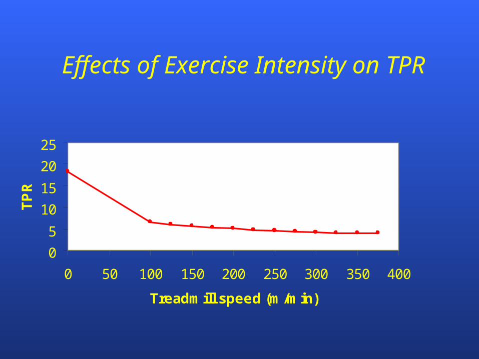
During running exercise, total peripheral
resistance ____ because of _____.
A. increases; sympathetic stimulation
B. increases; local control factors
C. decreases; vasoconstriction
D. decreases; local control factors

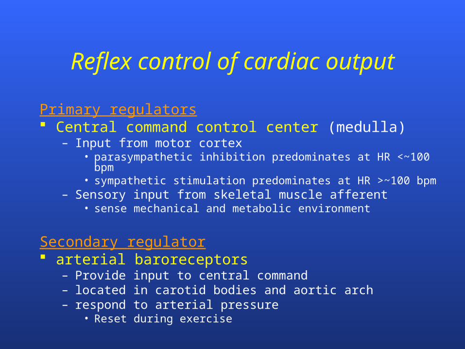
Reflex control of cardiac output
Primary regulators Central command control center (medulla)
– Input from motor cortex• parasympathetic inhibition predominates at HR <~100 bpm• sympathetic stimulation predominates at HR >~100 bpm
– Sensory input from skeletal muscle afferent• sense mechanical and metabolic environment
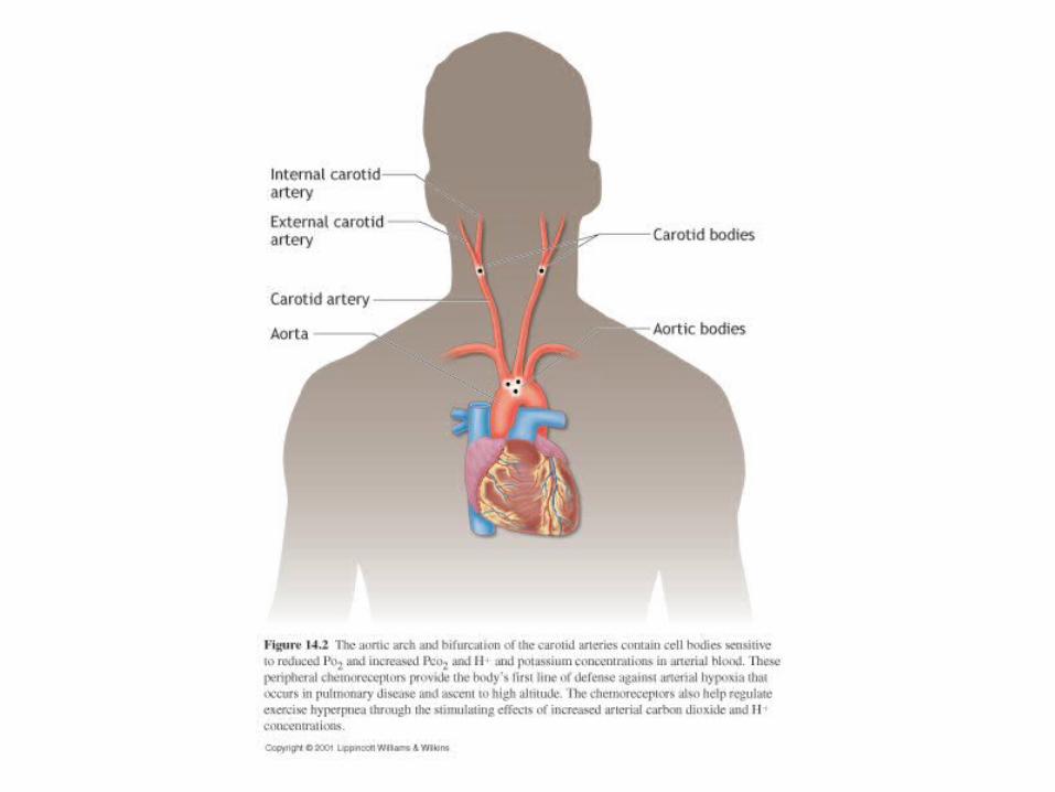
Secondary regulator arterial baroreceptors
– Provide input to central command– located in carotid bodies and aortic arch– respond to arterial pressure
• Reset during exercise
Maintaining Blood Pressure
Pressure is Necessary for Blood Flow
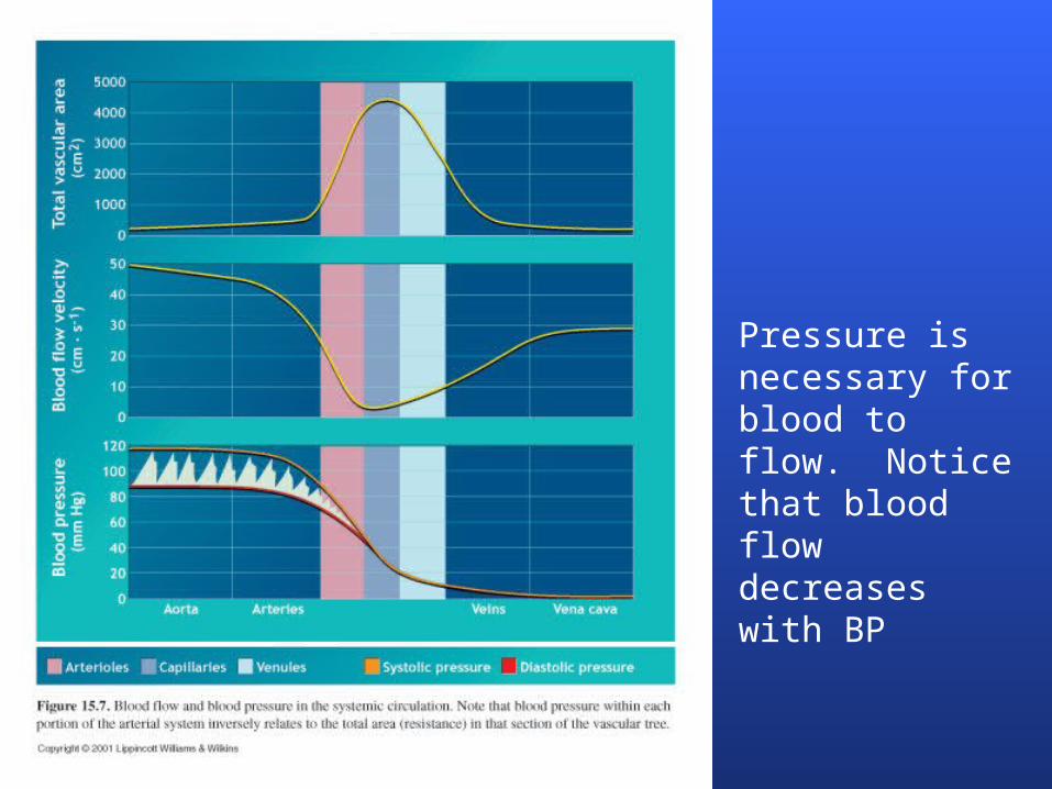
Pressure is necessary for blood to flow. Notice that blood flow decreases with BP

Regulation of Blood Flow and Pressure
Blood flow and pressure determined by:
arterioles
B. Pressure difference between two ends
A. Vessel resistance (e.g. diameter) to blood flow
A
A BB
cardiac output
BP = Q TPR

Regulation of Blood Flow and Pressure
Time
120
Pressure(mm Hg)
80
BP = Q TPR

At what level is peripheral resistance greatest?

Effects of Exercise on Cardiac Output
0
5
10
15
20
25
0 50 100 150 200 250 300 350 400
Treadmill speed (m/min)
TP
R
Effects of Exercise Intensity on TPR

Effects of Incremental Exercise on BP
0
25
50
75
100
125
150
175
200
225
250
0 50 100 150 200 250 300
Workload (W)
Blo
od
pre
ssu
re (
mm
Hg
)
Systolic BP
Diastolic BP

Cardiovascular Response to Exercise
Fick equation
VO2 = Q (aO2 – vO2)
VO2 = [HR SV] (aO2 – vO2)
VO2 = [BP TPR] (aO2 – vO2)
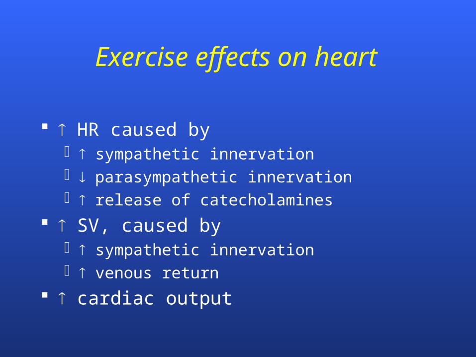
Exercise effects on heart
HR caused by sympathetic innervation parasympathetic innervation release of catecholamines
SV, caused by sympathetic innervation venous return
cardiac output

Cardiorespiratory adaptations to endurance training
How does endurance training affect VO2max?

Maximal oxygen consumption (VO2max)
VO2max
– highest VO2 attainable– maximal rate at which aerobic system
utilizes O2 and synthesizes ATP– single best assessment of CV fitness
intensity
VO2VO2max
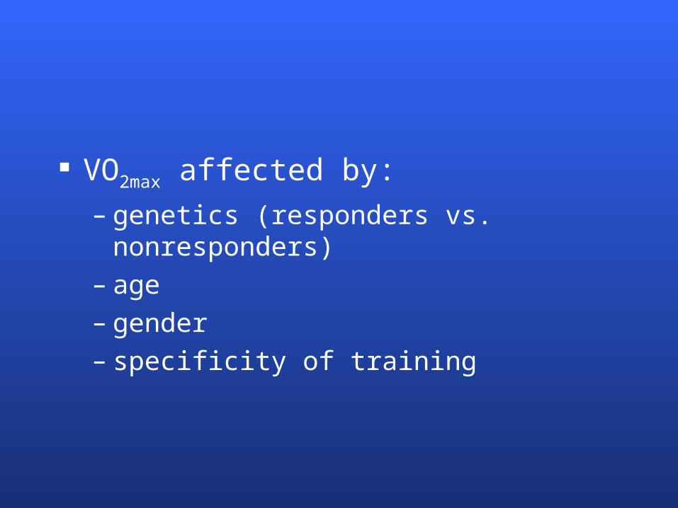
VO2max affected by:
– genetics (responders vs. nonresponders)– age– gender– specificity of training

Cardiorespiratory training adaptations
VO2max ~15% with training
ventilation? – training has no effect on ventilation capacity
O2 delivery?– CO ( ~15%) plasma volume SV
O2 utilization?– mitochondrial volume >100%

1995 marathon training data (women)
VO2 Pre-training Post-training 5 mph 30.7 29.8 6 mph 35.5 34.6
RER 5 mph 0.92 0.88* 6 mph 0.95 0.92*
HR 5 mph 168 151* 6 mph 182 167*
VO2max 54.4 58.5* HRmax 206 198*
*P < 0.05
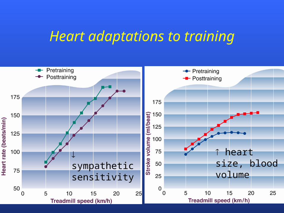
Heart adaptations to training
sympathetic sensitivity
heart size, blood volume
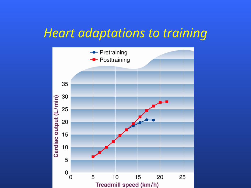
Heart adaptations to training
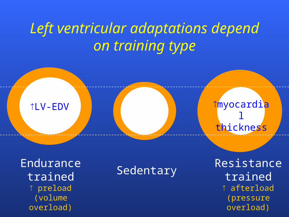
Left ventricular adaptations depend on training type
Endurance trained preload
(volume overload)
SedentaryResistance
trained afterload
(pressure overload)
LV-EDV myocardial thickness
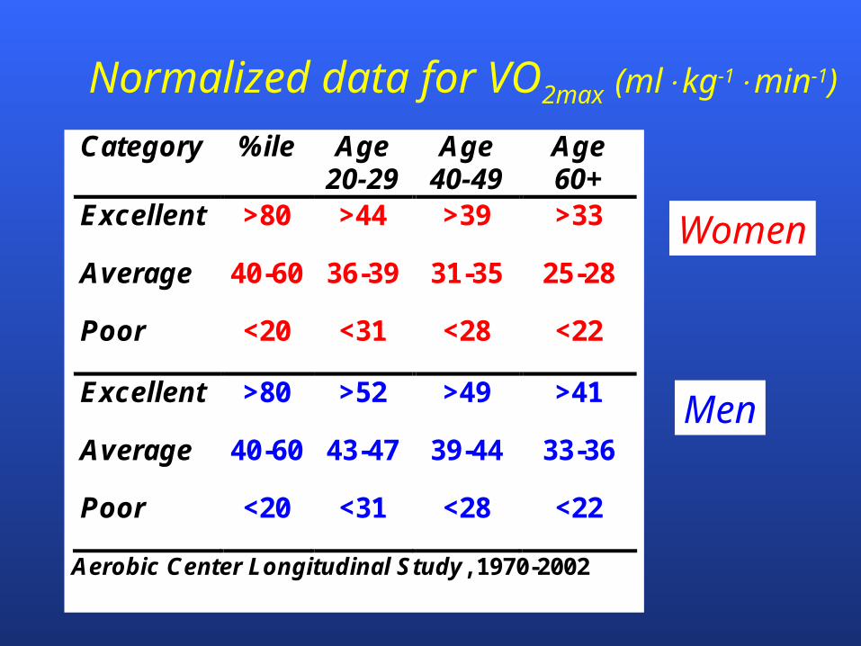
Normalized data for VO2max (mlkg-1min-1)
Category %ile Age 20-29
Age 40-49
Age 60+
Excellent >80 >44 >39 >33
Average 40-60 36-39 31-35 25-28
Poor <20 <31 <28 <22
Excellent >80 >52 >49 >41
Average 40-60 43-47 39-44 33-36
Poor <20 <31 <28 <22
Aerobic Center Longitudinal Study, 1970-2002
Women
Men

Which of the following would likely result in an increase of VO2max?
A. breathing faster and deeper during maximal exercise
B. faster HR at maximal exercise
C. ability to deliver more O2 to muscles during maximal exercise
D. more mitochondria
Which of the following does NOT occur following endurance training?
A. blood volume
B. HRmax
C. SVmax
. COmax
E. mitochondrial volume
F. maximal ventilatory capacity
How would you evaluate a VO2max of 28.9 mL/kg/min for a 22-year-old man?
A. excellent
B. above average
C. average
D. very low
E. dead
Which of the following exercises would likely decrease TPR the LEAST?
A. jogging
B. fast walking
C. shoveling snow
D. cycling
E. the above would decrease TPR similarly
What is the mechanism for the sudden increase in VE when the lactate threshold is reached
during an incremental exercise test?
A. greater muscle afferent input
B. greater stimulation of peripheral baroreceptors
C. greater stimulation of peripheral PCO2 chemoreceptors
D. greater stimulation of peripheral PO2 chemoreceptors
E. greater stimulation from motor cortex