CARDIAC VALVE REPLACEMENT - Home - Springer978-1-4613-2601...CARDIAC VALVE REPLACEMENT CURRENT...
32
CARDIAC VALVE REPLACEMENT
Transcript of CARDIAC VALVE REPLACEMENT - Home - Springer978-1-4613-2601...CARDIAC VALVE REPLACEMENT CURRENT...

CARDIAC VALVE REPLACEMENT

CARDIAC VALVE REPLACEMENT
CURRENT STATUS
PROCEEDINGS OF THE FOURTH INTERNATIONAL SYMPOSIUM ON THE ST. JUDE MEDICAL@ VALVE, MARCH 11-14, 1984
JACK M. MATLOFF, EDITOR
UI
" MARTINUS NIJHOFF PUBLISHING
A MEMBER OF THE KLUWER ACADEMIC PUBLISHERS GROUP
BOSTON DORDRECHT LANCASTER
Copyright 1985 @ by Martinus Nijhoff Publishing, Boston
Softcover reprint of the hardcover 1st edition 1985
All rights reserved. No part of this publication may be reproduced, stored in a retrieval system, or transmitted in any form or by any means, mechanical, photocopying, recording, or otherwise, without written permission of the publisher, Martinus Nijhoff Publishing, 190 Old Derby Street, Hingham, Massachusetts 02043.
Distrihutors for North America: Kluwer Academic Publishers 190 Old Derby Street Hingham, MA 02043
Distrihutors for all other countries: Kluwer Academic Publishers Group Distribution Centre P.O. Box 322 3300 AH Dordrecht The Netherlands
Library of Congress Cataloging in Publication Data
International Symposium on the St. Jude Medical Valve (4th: 1984 : Montego Bay, Jamaica) Cardiac valve replacement.
Includes bibliographies. 1. Heart valve prosthesis-Congresses. 1. Matloff,
Jack M. II. Title. III. Title: St. Jude Medical Valve. [DNLM: 1. Heart Valve Prosthesis-congresses. W3 IN924NU 4th 1984c I WG 169 1608 1984c] RD598.I54 1984 617'.412 85-5059 ISBN- 13: 978-1-4612-9629-4 e-ISBN-I3: 978-1-4613-2610-4 DOl: 10.1007/978-1-4613-2610-4
CONTENTS
Registered trademarks used in this book IX
Contributing authors xi Dedication by Jack M. Matloff xvii Preface by Jack M. Matloff xix Keynote address by Denton A. Cooley xxiii
I COMPLICATIONS OF CARDIAC VALVE REPLACEMENTS AND THEIR TREATMENT
1. Following the patient with prosthetic heart valves 3 SHAHBUDIN RAHIMTOOLA
2. Thromboembolism after cardiac valve replacement 9 LAWRENCE H. COHN
3. Nonprosthetic factors producing thromboembolism in patients with cardiac valve substitutes: their nature and the problems of assessing their role 17 R. LEIGHTON FISK
4. Anticoagulant therapy and cardiac valvular surgery: Coumadin® and other alternatives 25 SHAHBUDIN RAHIMTOOLA
5. Reoperation after cardiac valve surgery: elective, urgent and emergent 29 JAMES R. PLUTH
Discussion 33 LAWRENCE H. COHN, MODERATOR
v
vi Contents
II CARDIAC VALVE SUBSTITUTES: CURRENT STATUS
6. Clinical experience with the caged-ball Starr-Edwards® prosthesis 37 JAMES R. PLUTH
7. Overview of experience with the Bjork-Shiley® valve 41 DONALD B. DOTY
8. Experience with standard and supra-annular Carpentier-Edwards® porcine bioprostheses 45 w. R. ERIC JAMIESON, MICHAEL T. JANUSZ, G. FRANK O. TYERS, ALFRED N. GEREIN,
DONALD R. RICCI, LAWRENCE H. BURR, ROBERT T. MIYAGISHIMA
9. Aortic valve replacement with the Ionescu-Shiley® bovine pericardial valve: an 81-month experience 57 LORENZO GONZALEZ-LAVIN, SEONG CHI, T. CALVIN BLAIR, J. Y. JUNG, A. G. FABAZ,
BETTY LEWIS, GEORGE DAUGHTERS
Discussion 67 DIETER HORSTKOTTE, MODERATOR
III CARDIAC VALVE REPLACEMENT IN SPECIAL CIRCUMSTANCES
10. Annuloaortic ectasia: surgical repair using a composite St. Jude Medical® valve and Dacron ® tube graft 73 DENTON A. COOLEY
11. Valve replacement in the geriatric patient using the St. Jude Medical® prosthesis 83 DEMETRE M. NICOLOFF, WILLIAM G. LINDSAY, KIT V. AROM, WILLIAM F. NORTHRUP, III,
THOMAS E. KERSTEN
12. Tricuspid valve replacement-a comparative experience with different valve substitutes 91 FRANCIS WELLENS, JEAN-LOUIS LECLERC, F. DEUVAERT, G. VAN NOOTEN, J. GOLDSTEIN,
G. PRIMO
13. Double valve replacement 99 RICHARD J. GRAY, LAWRENCE S. C. CZER, AURELIO CHAUX, TIMOTHY M. BATEMAN,
MICHELE DEROBERTIS, JACK M. MATLOFF
14. Cardiac valve replacement in the presence of coronary atherosclerosis 111 JACK M. MATLOFF AND LAWRENCE S. C. CZER
Discussion 123 DONALD B. DOTY AND EUGENE M. BAUDET, MODERATORS
IV CARDIAC VALVE REPLACEMENT IN PEDIATRIC PRACTICE: EXPERIENCE WITH THE ST. JUDE MEDICAL® VALVE
15. St. Jude Medical® valve replacement in infants and children 129 A. MICHAEL BORKON, BRUCE A. REITZ, JAMES S. DONAHOO, TIMOTHY J. GARDNER
16. Pediatric use of the St. Jude Medical® prosthesis 137 ROBERT M. SADE, FRED A. CRAWFORD, JR., HARVEY I. PASS, JOHN M. KRATZ
17. Mitral valve replacement in a child using the St. Jude Medical® valve 141 JOHN W. MACK
18. Thromboembolism in children with St. Jude Medical® valves maintained on aspirin and Persantine® 145
STUART L. BOE, KATHLEEN W. MCNICHOLAS, HENRY F. OLIVER,
FAUSTINO N. NIGUIDULA, GEORGE J. MAGOVERN, GERALD M. LEMOLE
Discussion 155
EUGENE M. BAUDET AND DONALD B. DOTY, MODERATORS
vii
V INTERMEDIATE FOLLOW-UP OF PATIENTS WITH ST. JUDE MEDICAL® PROSTHESES: 52 TO 72 MONTHS
19. A 52-month experience with the St. Jude Medical® cardiac prosthesis at the Medical University of South Carolina 163
FRED A. CRAWFORD, JR., JOHN M. KRATZ, ROBERT M. SADE, MARTHA P. STROUD,
DAVID M. BARTLES
20. A 57-month experience with the St. Jude Medical® cardiac prosthesis at Hahnemann University Hospital 167
ELDRED D. MUNDTH
21. Incidence of complication with the St. Jude Medical® prosthesis: a 58-month study at Hamot Medical Center 173
GEORGE J. D'ANGELO, G. F. KISH, P. G. SARDESAI, W. S. TAN
22. Mitral valve replacement with St. Jude Medical® prostheses: a 60-month study of 350 cases at Centre Hospitalier Universitaire 179
HENRI DUPON, J. L. MICHAUD, D. DUVEAU, PH. DESPINS, M. TRAIN
23. A 60-month experience with the St. Jude Medical® prosthesis at Cedars-Sinai Medical Center 189
RICHARD J. GRAY, AURELIO CHAUX, LAWRENCE S. C. CZER, MICHELE DEROBERTIS,
JACK M. MATLOFF
24. A 60-month experience with the St. Jude Medical® prosthesis at University Hospital, Brugmann 195
JEAN-LOUIS LECLERC, FRANCIS WELLENS, FRANK E. DEUVAERT, MARTINE ANTOINE,
JACQUES DEPAEPE, GEORGES PRIMO
25. A 72-month clinical experience with the St. Jude Medical® cardiac valve prosthesis at Newark Beth Israel Medical Center 201
ISAAC GIELCHINSKY, MARK S. HOCHBERG, S. MANSOOR HUSSAIN, VICTOR PARSONNET,
DANIEL FISCH, JOHN C. NORMAN
26. A 72-month experience with the St. Jude Medical® cardiac valve prosthesis at the Minneapolis Heart Institute and United Hospitals, St. Paul, Minnesota 205
WILLIAM G. LINDSAY, DEMETRE NICOLOFF, KIT V. AROM, WILLIAM F. NORTHRUP III,
THOMAS E. KERSTEN
27. A 72-month clinical experience with the St. Jude Medical® prosthesis at Tucson Medical Center 209
CHRISTOPHER T. MALONEY
Discussion 215
SHAHBUDIN RAHIMTOOLA, MODERATOR

viii Contents
VI CLINICAL EXPERIENCE WITH CURRENT CARDIAC VALVE SUBSTITUTES: COMPARATIVE OBSERVATIONS
28. Late complications in patients with Bjork-Shiley® and St. Jude Medical® prostheses 225 DIETER HORSTKOTTE
29. Experience with the St. Jude Medical® valve and the Ionescu-Shiley® bovine pericardial valve at the Texas Heart Institute 233 J. MICHAEL DUNCAN, DENTON A. COOLEY, GEORGE J. REUL, DAVID A. OTT,
JAMES J. LIVESAY, O. H. FRAZIER, WILLIAM E. WALKER, PHILLIP R. ADAMS
30. Comparative assessment of single Bjork-Shiley®, Hancock® and St. Jude Medical® valves at 43 months after operation 247 LESTER R. SAUVAGE, MARY A. O'BRIEN
Discussion 261 DEMETRE M. NICOLOFF, MODERATOR
VII CLINICAL FORUM
31. Noninvasive assessment of prosthetic heart valve function by continuous-wave doppler ultrasound 269 ANDREAS HOFFMAN, PETER STULZ, ERICH GRADEL, DIETER BURCKHARDT
32. Potential for immobilization of the valve occluder in various valve prostheses 281 OSCAR BAEZA
33. Escape of a leaflet from a St. Jude Medical® prosthesis in the mitral position 285 E. HJELMS, M.D.
34. Prophylaxis against thromboembolism using aspirin and dipyridamole in patients with the St. Jude Medical® aortic prosthesis 291 JOSEPH LOCICERO, III
Discussion 293 RICHARD J. GRAY, MODERATOR
VIII CLOSING OBSERVATIONS
35. Summary and concluding remarks 301 SHAHBUDIN RAHIMTOOLA
36. Summary and concluding observations 305 JACK M. MATLOFF

REGISTERED TRADEMARKS USED IN THIS BOOK
BJORK-SHILEY®-Shiley Corporation CARPENTIER-EDW ARDS®-American Edwards Laboratories COUMADIN®-The DuPont Corporation DACRON®-The DuPont Corporation DELRIN®-The DuPont Corporation HANCOCK®-Vascor, Inc. IONESCU-SHILEY® -Shiley Corporation KEFLIN®-Eli Lilly and Company LILLEHEI-KASTER ®-Medical Incorporated MEADOX-COOLEY®-Meadox Medicals, Inc. MEDTRONIC HALLTM-Blood Systems, Inc. OMNISCIENCE®-Medical Incorporated PERSANTINE®-Boehringer Ingelheim SILASTIC®-Dow Coming ST. JUDE MEDICAL®-St. Jude Medical, Inc. STARR-EDW ARDS®-American Edwards Laboratories STELLITE®-Cabot Corporation SWAN-GANZ®-Edwards Laboratories TEFLON®-DuPont Corporation
ix

CONTRIBUTING AUTHORS
Oscar Baeza, M.D. Passaic General Hospital Passaic, NJ 07055 Address for correspondence: 350 Boulevard Passaic, NJ 07055
Eugene M. Baudet Dept. of Cardiovascular Surgery Hopital Cardiologique A venue de Magellan 33604 Bordeaux France
Stuart L. Boe, M.D. Suite 609 3661 South Miami Avenue Miami, FL 33133
A. Michael Borkon, M.D. Division of Cardiac Surgery The Johns Hopkins Hospital 600 North Walfe Street Baltimore, MD 21205
xi

xii Contributing authors
Lawrence H. Cohn, M.D. Brigham & Women's Hospital 75 Francis Street Boston, MA 02115
Denton A. Cooley, M.D. P.O. Box 20345 Texas Heart Institute of St. Luke's Episcopal and Texas Children's Hospitals Houston, Texas 77225
Fred A. Crawford, Jr., M.D. The Medical University of South Carolina 171 Ashley Avenue Charleston, SC 29425
George J. D'Angelo, M.D. Hamot Medical Center Erie, PA Address for correspondence: 104 East Second Street Erie, P A 16507
Donald B. Doty, M.D. LDS Hospital Primary Children's Medical Center Salt Lake City, UT Address for correspondence: 324 - 10th A venue Salt Lake City, UT 84103
J. Michael Duncan, M.D. Texas Heart Institute P.O. Box 20345 Houston, TX 77225
Prof. Henri Dupon, M.D. 35, rue Paul Bert 44035 Nantes France
R. Leighton Fisk, M.D. Phoenix Foundation for Cardiovascular Research, St. Luke's Hospital Heart-Lung Center 525 North 18th Street Suite 5 Phoenix, AZ 85006
Isaac Gielchinsky, M.D. Dept. of Cardiothoracic Surgery Newark Beth Israel Medical Center 201 Lyons Avenue Newark, NJ 17112
Lorenzo Gonzalez-Lavin, M.D. Chairman, Department of Surgery Deborah Heart and Lung Center Browns Mills, NJ 08015
Richard J. Gray, M.D. Cedars Sinai Medical Center 8700 Beverly Blvd. Los Angeles, CA 90048
prof. Andreas Hoffmann, M.D. Lange Gasse 78 4052 Basel Switzerland
Dieter Horstkotte, M.D. Department of Medicine B University Hospital Duesseldorf Moorenstr. 5 4000 Duesseldorf West Germany
E. Hjelms, M.D. Department of Cardiothoracic Surgery Rigshospitalet University of Copenhagen Blegdamsvej 9, DK-2100 Copenhagen, Denmark
xiii

xiv Contributing authors
W. R. Eric Jamieson, M.D. University of British Columbia Vancouver, B.C. Canada Address for correspondence: 750 West Broadway Vancouver, B.C. V5Z 1HL Canada
Jean-Louis LeClerc, M.D. University Hospital Brugmann Cardiac Surgery 808 route de Lennik 1070 Brussels Belgium
William G. Lindsay, M.D. Cardiac Surgical Assoc. 2545 Chicago Avenue Suite 730 Minneapolis, MN 55404
Joseph LoCicero, III, M.D. Northwestern University Div. of Cardiothoracic Surgery Ward Building 9-105 303 East Chicago A venue Chicago, IL 60611
John W. Mack, M.D. Clinical Associate Professor University of Texas Health Sciences Center San Antonio, TX Address for correspondence: 13702 Wilderness Point San Antonio, TX 78231
Christopher T. Maloney, M.D. Southwest Cardio-Thoracic Surgery, Ltd. 5200 East Grant Road Suite 200-A Tucson, AZ 85712
Jack M. Matloff, M.D. Cedars Sinai Medical Center 8700 Beverly Blvd. Los Angeles, CA 90048
Eldred D. Mundth, M.D. The Hahnemann Medical College and Hospital 230 North Broad Street Suite 6328 Philadelphia, PA 19102
Demetre M. Nicoloff, M.D. Cardiac Surgical Assoc., P.A. 2545 Chicago Avenue Suite 730 Minneapolis, MN 55404
James R. Pluth, M.D. Mayo Clinic 200 - 1st Street S.W. W6-B Rochester, MN 55901
Shahbudin Rahimtoola, M.D. Professor of Medicine Chief, Section of Cardiology University of Southern California 2025 Zonal Avenue Los Angeles, CA 90033
Robert M. Sade, M.D. The Medical University of South Carolina Div. of Cardiothoracic Surgery 171 Ashley Avenue Charleston, SC 29025
Lester R. Sauvage, M.D. 528 - 18th Avenue Seattle, W A 98122
xv

xvi Contributing authors
Francis Wellens, M.D. Hopital Erasme 808, Route de Lennik 1070 Bruxelles Belgium

DEDICATION
Cardiac Valve Replacement: Current Status is dedicated to all those patients and their families who have had cardiac valve replacement surgery. Their story has been one of personal courage because they decided to accept such therapy when a precise definition of their future with one valve substitute or another was not clear. This courageous decision was initiated with the first implantations of the Harken and STARR-EDWARDS® caged-ball valves in 1960 and has continued to exist as each new valve substitute has been implanted, because laboratory testing of such devices, no matter how sophisticated the in vitro or animal test, has never been able to precisely define the device performance characteristics in humans. In this sense, the ultimate determination of each valve's clinical record has only been defined during its clinical use in patients. Thus, each patient who undergoes valve replacement owes a debt to those who previously had such surgical therapy; each repays that debt by contributing to the learning experience that makes the valve safer and easier for those who benefit later.
All of us-patients, their families, cardiologists and cardiac surgeons-therefore should recognize the contributions of Mary Richardson and her pioneering surgeon, Dwight E. Harken, M.D. They shared this initial experience with total aortic valve excision and replacement in the anatomic position in February 1960 at the Peter Bent Bingham Hospital in Boston.
With regard to the experience with the ST. JUDE MEDICAL® valve, 2 patients are deserving of particular note for their special contributions. Mrs. Helen Heikkinen underwent the first ST. JUDE MEDICAL valve replacement in Octo-
xvii

xviii Dedication
ber 1977. She has to be recognized for her courage in accepting the recommendation for this valve's initial use, at a time when there were other valves available, with well-defined clinical track records. She continues to be well and establishes the standard for durability of the valve.
Henry Jaffe also must be recognized for his exquisite understanding of this process of shared patient responsibility. Because of his sensitivity for this sequence and his innate intellectual curiosity, he and his wife, Florence have personally funded more clinical investigations of the ST. JUDE MEDICAL valve than any other individual or agency, to my knowledge.
While the medical contributions to this symposium are identified, all of us must also acknowledge the contributions of the unci ted coauthors, our patients. On behalf of my colleagues, we thank you.
Jack M. Matloff

PREFACE
Cardiac Valve Replacement: Current Status is the proceedings of the Fourth International Symposium on the ST. JUDE MEDICAL@ valve. The first three symposia on this topic were held primarily for designated investigators involved in clinical trials of the ST. JUDE MEDICAL valve. The last meeting, chaired by Michael E. DeBakey, M.D., was held in November 1982 [1], immediately before the valve was released for general clinical use in the United States by the Food and Drug Administration. These proceedings then are the first comprehensive compilation of clinical data since that time; and they include, particularly in the discussions, the experience of physicians other than the original clinical investigators.
Over the past 5 years the character of these symposia has changed. Whereas the first two dealt almost entirely with the ST. JUDE MEDICAL valve, the last two have evolved into a more generic cardiac valvular surgery meeting, focusing primarily on valve replacement rather than valve repair [2]. Thus, these proceedings contain a wide spectrum of topics, including a keynote presentation on criteria for selection of cardiac valve substitutes in 1984, complications of cardiac valve replacement and their treatment, a review of the current status of cardiac valve substitutes other than the ST. JUDE MEDICAL valve and a consideration of cardiac valve replacement in special circumstances. Among these special circumstances are four presentations on pediatric use of the ST. JUDE MEDICAL valve. Finally, there are three presentations by groups who have had experience with BJORK-SHILEY@ spherical, HANCOCK@ porcine, IONESCU-SHILEY@ pericardial and ST. JUDE MEDICAL valves and who attempt to define their
xix

xx Preface
comparative results. That these presentations are from a number of groups around the world establishes this as a truly international symposium and reflects the fact that the ST. JUDE MEDICAL valve has become the most widely used cardiac prosthesis in the world.
Chapters that constitute each of the sections are followed by the recorded discussions that took place after each session of the symposium. For these, I am indebted to the moderators who chaired the various sections; they are Lawrence H. Cohn, Dieter Horstkotte, Donald B. Doty, Eugene M. Baudet, Demetre M. Nicoloff, Shahbudin Rahimtoola and Richard J. Gray. These discussions have been included in an attempt to make these proceedings a current progress report. The medical literature is replete with somewhat dated abstracts and articles in a format that does not allow for a ready opportunity to resolve the multiplicity of opinions. I have felt these discussions should be included because they are an interaction between the presenters and other participants, are an attempt to share their experiences, and are an attempt to resolve apparent and real differences and to generally learn from each other. I, therefore, would like to thank each of the discussants for their participation as well.
It is apparent from this forum that there is not an international, national or even local standard of terminology for valvular heart surgery. While editing this volume I have tried to achieve some degree of standardization in the terminology. Thus, valve repair and valve replacement with substitutes are used in this volume to reflect the two basic methods of valvular heart surgery. Unfortunately, there is no term for the issue of whether a valve is deserving of surgical intervention. This decision has become one of the most difficult issues of valvular heart surgery to resolve, especially when so many patients are now studied for considerations other than valvular and are found to have varying degrees of concomitant valvular or prosthetic dysfunction. The term bioprosthesis is used in this volume to include those valves fabricated from tissue or biological components; and the term prosthesis includes all mechanical valves fabricated from materials other than tissue. Mobile element refers to functional components whether they are tissue leaflets, mechanical leaflets, poppets, occluders, balls or discs. I have also tried to standardize the reporting of mortalities as operative or less than 30 days if a patient was discharged from the hospital and late if mortality occurred more than 30 days after surgery and after discharge. In the same manner, thromboembolism, thrombotic occlusions and valve-related events (dysfunction, hemolysis and infection) are reported as percent (%) per patient year of follow-up when possible.
I would be remiss if I did not thank St. Jude Medical on behalf of all who attended the symposium that was the basis of this volume. Even though St. Jude Medical sponsored this symposium, they did not and do not desire that this book be a testimonial to any valve substitute, including their own. Rather, it was their intention that this be a forum for initiating exchanges about the valve-related topics presented. To their credit, they did not, in any way, intrude on the choice of topics discussed, the choice of papers presented or the scientific organization of the symposium. If there is a bias projected, it is an unintended reflection of my thoughts

xxi
about valvular heart surgery. Given the widespread use of the ST. JUDE MEDICAL valve and its seemingly increasing popularity, I have felt that there has been a disparity between its use reflected at this symposium and what has been presented previously in the medical literature. My choices, therefore, are an attempt to correct this disparity.
Finally, I want to thank all of the presenters for their efforts. They are the individuals who are responsible for whatever this volume mayor may not be. In particular, I would like to thank Denton Cooley, M.D. for his active participation. of all of the illustrious trainees of Alfred Blalock, M.D. at the Johns Hopkins Hospital, none has attained as much in clinical cardiac surgery as Dr. Cooley. He personally has performed more cardiac surgery than anyone else ever has and probably ever will in the future. From this experience, he has distilled the essence of cardiac surgery-constantly innovating, introducing, revising and improving existing procedures, to become the virtuoso technician and consummate teacher that he is. Even more amazingly, he has done this in the three major areas of cardiac surgery: congenital heart disease, coronary heart disease and valvular heart disease. In the keynote presentation, "Cardiac Valvular Surgery, 1984-Criteria for Selection of Cardiac Valve Substitutes," by Dr. Cooley established the outline for the presentations that follow.
Jack M. Matloff, M.D.
REFERENCES
1. DeBakey ME (ed): Advances in Cardiac Valves: Clinical Perspectives. New York, Yorke Medical Books, 1983.
2. Oury J: Recent Progress in Mitral Valve Disease. Kent, England, Butterworths, Sevenoaks, 1984.
KEYNOTE ADDRESS: CARDIAC VALVULAR SURGERY, 1984-CRITERIA FOR SELECTION OF CARDIAC VALVE SUBSTITUTES
DENTON A. COOLEY
Thirty years have elapsed since the introduction of the fIrst artifIcial heart valves. Since that time, many improvements have been made in the design and fabrication of prosthetic valves, but the perfect valve remains elusive. Although much has been learned, more remains to be clarifIed.
The earliest valvular substitute was the Hufnagel valve, which was introduced into the descending thoracic aorta in 1952 [1]. Early investigators were confused, however, by the misconception that the prosthetic valve should closely resemble the human anatomy. Attempts to apply this concept in the early years of replacement met with limited success. The valves of Hufnagel, McGoon, Bahnson, and others came to an abrupt end when thrombosis, fracture and disruption occurred.
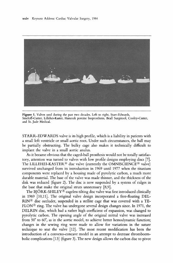
In the early 1960s, Harken [2] and Starr [3] devised caged-ball prostheses that aroused objections from advocates of the anatomic design concept (fIgure 1). This caged-ball design had been in use a l~ng time and, in fact, was illustrated in 1858 by a request for a bottle stopper patent [4]. The Harken and STARREDWARDS® valves were placed in the normal anatomic position, or the subcoronary and mitral annular positions, respectively. The STARR-EDWARDS valve design is still practical, although a number of design modifIcations and improvements have been made since the early models. These included a method of "curing" the silicone ball in 1965 that eliminated the "ball variance" problem and an extension of the cloth covering of the sewing ring to reach the inlet orifIce to lessen the incidence of thromboembolic events [5,6]. The primary disadvantage of the
xxiii
xxiv Keynote Address: Cardiac Valvular Surgery, 1984
Figure 1. Valves used during the past two decades. Left to right, Starr-Edwards, Smeloff-Cutter, Lillehei-Kaster, Hancock porcine bioprosthesis, Beall Surgitool, Cooley-Cutter, and St. Jude Medical.
STARR-EOW ARDS valve is its high profile, which is a liability in patients with a small left ventricle or small aortic root. Under such circumstances, the ball may be partially obstructing. The bulky cage also makes it technically difficult to implant the valve in a small aortic anulus.
As it became obvious that the caged-ball prosthesis would not be totally satisfactory, attention was turned to valves with low profile designs employing discs [7]. The LILLEHEI-KASTER ® disc valve (currently the OMNISCIENCE® valve) survived unchanged from its introduction in 1969 until 1977 when the titanium components were replaced by a housing made of pyrolytic carbon, a much more durable material. The base of the valve was made thinner, and the thickness of the disk was reduced (figure 2) . The disc is now suspended by a system of ridges in the base that make the original struts unnecessary [8,9]'
The BJORK-SHILEY® cageless tilting disc valve was first introduced clinically in 1969 [10,11] . The original valve design incorporated a free-floating DELRIN® disc occluder, suspended in a stellite cage that was covered with a TEFLON® ring. The valve has undergone several design changes since. In 1971, the OELRIN disc, which had a rather high coefficient of expansion, was changed to pyrolytic carbon. The opening angle of the original mitral valve was increased from 50° to 60°, as in the aortic model, to achieve better hemodynamic function; changes in the sewing ring were made to allow for variations in the suture technique to seat the valve [12] . The most recent modification has been the introduction of a convexo-concave model in an attempt to decrease thromboembolic complications [13] (figure 3). The new design allows the carbon disc to pivot

xxv
Figure 2. Omniscience disc valve.
Figure 3. Bjork-Shiley convex-concave disc valve.

xxvi Keynote Address: Cardiac Valvular Surgery, 1984
2.5 mm downstream during opening, to allow for a washing effect at the pivot points and increase flow through the minor orifice.
Also during the 1960s, valve replacement with biologic tissue was tried, including formaldehyde-treated porcine xenografts, stented and unstented fascia lata, freeze-dried homografts, pulmonary valve autografts and various chemical treatments to stented and unstented homografts. Many of these valves sounded perfect, but most deteriorated rapidly. Ionescu, however, persevered and developed a pericardial valve, cured with glutaraldehyde, which is still in use today [14,15].
Besides the IONE3CU-SHILEY® valve, the two other bioprostheses commercially available today are the HANCOCK® porcine xenograft and the CARPENTIER-EDVV' ARDS® porcine xenograft. Both are preserved with glutaraldehyde and mounted on cloth-covered flexible stents [16,17] (figures 4 and 5). Both valves are silent, do not cause hemolysis and are relatively free of thromboembolic complications [18,19]. The main disadvantages of the bioprostheses are restricted hemodynaIT'ic performance and durability. The original HANCOCK valve, which had a muscle bar across the noncoronary leaflet, was partially ob~tructive [20,21]. In an attempt to rectify this hemodynamic problem, Hancock developed a modified orifice valve. Although the hemodynamic characteristics of this new valve are better, transvalvular gradients in the smaller valve sizes are still higher than those found with some mechanical valves. A new HANCOCK bovine pericardial valve (figure 6) is currently under clinical investigation.
The IONESCU-SHILEY valve is made from bovine pericardium treated with
Figure 4. Hancock porcine bioprosthesis.

xxvii
Figure 5. Carpentier-Edwards porcine xenograft valve.
Figure 6. Hancock bovine pericardial bioprosthesis currently undergoing clinical investigation.

xxviii Keynote Address: Cardiac Valvular Surgery, 1984
glutaraldehyde [22] (figure 7). Each of the valve leaflets is cut and sutured on a cloth-covered titanium frame. Hydraulic characteristics are good since there is no muscle bar at the base of any leaflet and a very low flow gradient across the valve [23]. A lower profile design is currently undergoing evaluation (figure 8). The IONESCU-SHILEY valve has been used in 2680 patients since 1978 at the Texas Heart Institute. Although the rate of valve failure in this series was 1.76% per patient-year, the failure rate is time-related and increases after five years. We are concerned that the failure rate of this group may be unacceptably high in the future [24-26].
The 1970s also saw the development of additional tilting disc valves, including the ST. JUDE MEDICAL® valve (figure 9). It is our current valve of choice in all patients, unless long-term anticoagulation is contraindicated [27]. The ST. JUDE MEDICAL valve is a low-profile, bileaflet, central-flow prosthesis made entirely of pyrolytic carbon [28,29]. The valve has an excellent sewing ring, fabricated of velour-knitted DACRON® fabric. The valve has a wide opening angle with low resistance to forward flow, and the low profile makes the valve very satisfactory in both the mitral and aortic positions as well as in the tricuspid anulus. It is easy to insert, even in a small anulus [30]. We began using the ST. JUDE MEDICAL valve in 1978. In a follow-up of 615 patients, we have not found any instances of valve dysfunction and few instances of systemic embolism [31]. Its low profile is a distinct advantage.
Figure 7. Valve prostheses in mock circulation showing differing degrees of opening and obstruction to forward flow. The largest orifice is found in the Ionescu-Shiley valve (bottom row).

xxix
Figure 8. Recently modifted low proftle Ionescu-Shiley valve prosthesis.
Figure 9. St. Jude Medical tilting disc valve.
xxx Keynote Address: Cardiac Valvular Surgery, 1984
COMPLICATIONS
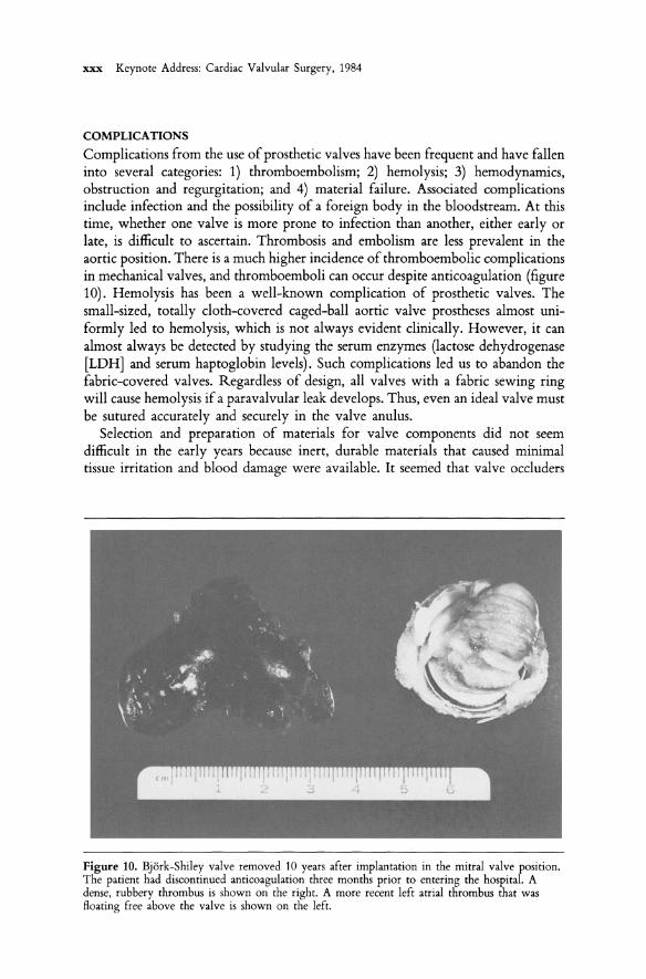
Complications from the use of prosthetic valves have been frequent and have fallen into several categories: 1) thromboembolism; 2) hemolysis; 3) hemodynamics, obstruction and regurgitation; and 4) material failure. Associated complications include infection and the possibility of a foreign body in the bloodstream. At this time, whether one valve is more prone to infection than another, either early or late, is difficult to ascertain. Thrombosis and embolism are less prevalent in the aortic position. There is a much higher incidence of thromboembolic complications in mechanical valves, and thromboemboli can occur despite anticoagulation (figure 10). Hemolysis has been a well-known complication of prosthetic valves. The small-sized, totally cloth-covered caged-ball aortic valve prostheses almost uniformly led to hemolysis, which is not always evident clinically. However, it can almost always be detected by studying the serum enzymes (lactose dehydrogenase [LDH] and serum haptoglobin levels). Such complications led us to abandon the fabric-covered valves. Regardless of design, all valves with a fabric sewing ring will cause hemolysis if a paravalvular leak develops. Thus, even an ideal valve must be sutured accurately and securely in the valve anulus.
Selection and preparation of materials for valve components did not seem difficult in the early years because inert, durable materials that caused minimal tissue irritation and blood damage were available. It seemed that valve occluders
Figure 10. Bjork-Shiley valve removed 10 years after implantation in the mitral valve position. The patient had discontinued anticoagulation three months prior to entering the hospital. A dense, rubbery thrombus is shown on the right. A more recent left atrial thrombus that was floating free above the valve is shown on the left.

xxxi
made of SILASTIC® would last almost indefinitely. However, a slight flaw in the curing and processing of Silastic caused the occluders to deteriorate when exposed to the enzymes and fatty acids in the blood. As a result, swelling, fracture and occasional dislodgement and embolization of the poppet into the circulation occurred [32,33] (figure 11). SILASTIC was not the only material that failed. TEFLON occluders used in the Beall and Wada valves also deteriorated, sometimes with fatal consequences. Fascia lata and dura mater were both destroyed very quickly [34,35,36]. As noted earlier, covering surfaces with cloth to encourage ingrowth of tissue proved unsatisfactory [37]. Metal wear or fatigue was also a problem.
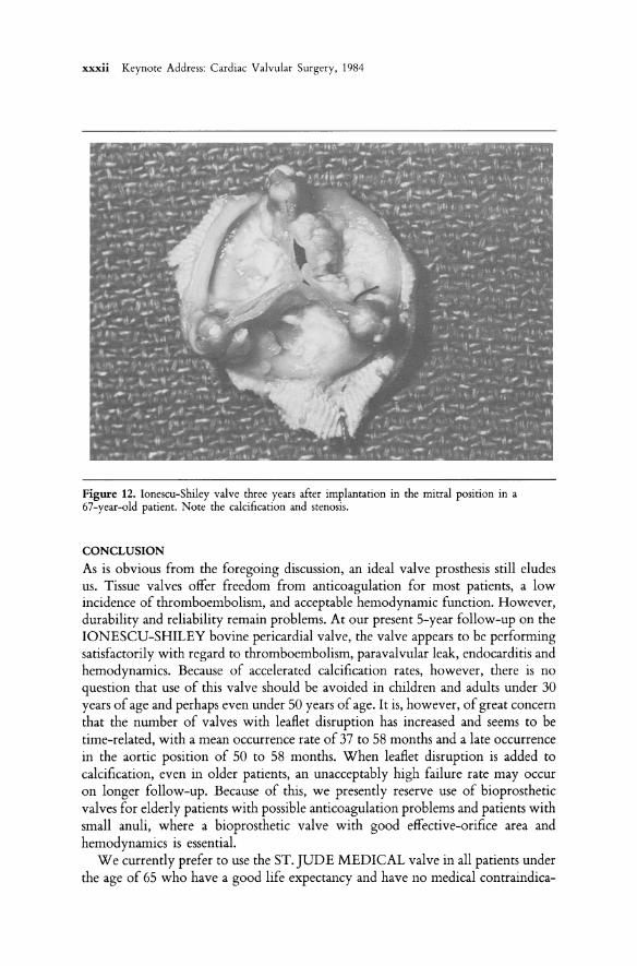
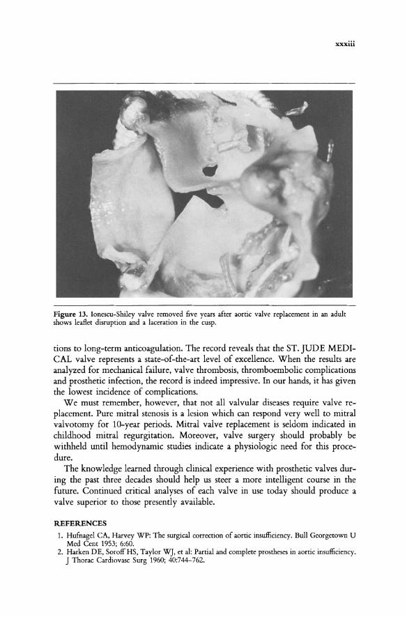
Obviously, more durable materials were needed; and new designs were tried using glutaraldehyde-treated tissue and pyrolytic carbon components. Tissue valves, however, thicken with age and are subject to calcification, especially in young children [38,39] (figure 12). Although glutaraldehyde prolongs the life of a tissue valve, it will eventually undergo degeneration and possible leaflet disruption [40,41] (figure 13). It would seem reasonable, then, to use tissue valves only when anticoagulants are contraindicated. Surfaces of pyrolytic carbon seem most encouraging from the standpoint of wear and the non thrombogenic nature of this material. Optimum healing is obtained with this material. Apparently, the negatively charged pyrolytic carbon inhibits the overgrowth of fibrous tissue.
Peripheral versus central flow is still an issue in design of prosthetic heart valves. Advantages of the full orifice principle in reducing the flow pressure gradient are obvious, but the self-washing action is another distinct advantage.
Figure 11. Starr-Edwards prosthesis with swollen poppet.
xxxii Keynote Address: Cardiac Valvular Surgery, 1984
Figure 12. Ionescu-Shiley valve three years after implantation in the mitral position in a 67-year-old patient. Note the calcification and stenosis.
CONCLUSION
As is obvious from the foregoing discussion, an ideal valve prosthesis still eludes us. Tissue valves offer freedom from anticoagulation for most patients, a low incidence of thromboembolism, and acceptable hemodynamic function. However, durability and reliability remain problems. At our present 5-year follow-up on the IONESCU-SHILEY bovine pericardial valve, the valve appears to be performing satisfactorily with regard to thromboembolism, paravalvular leak, endocarditis and hemodynamics. Because of accelerated calcification rates, however, there is no question that use of this valve should be avoided in children and adults under 30 years of age and perhaps even under 50 years of age. It is, however, of great concern that the number of valves with leaflet disruption has increased and seems to be time-related, with a mean occurrence rate of 37 to 58 months and a late occurrence in the aortic position of 50 to 58 months. When leaflet disruption is added to calcification, even in older patients, an unacceptably high failure rate may occur on longer follow-up. Because of this, we presently reserve use of bioprosthetic valves for elderly patients with possible anticoagulation problems and patients with small anuli, where a bioprosthetic valve with good effective-orifice area and hemodynamics is essential.
We currently prefer to use the ST. JUDE MEDICAL valve in all patients under the age of 65 who have a good life expectancy and have no medical contraindica-
xxxiii
Figure 13. Ionescu-Shiley valve removed five years after aortic valve replacement in an adult shows leaflet disruption and a laceration in the cusp.
tions to long-term anticoagulation. The record reveals that the ST. JUDE MEDICAL valve represents a state-of-the-art level of excellence. When the results are analyzed for mechanical failure, valve thrombosis, thromboembolic complications and prosthetic infection, the record is indeed impressive. In our hands, it has given the lowest incidence of complications.
We must remember, however, that not all valvular diseases require valve replacement. Pure mitral stenosis is a lesion which can respond very well to mitral valvotomy for 10-year periods. Mitral valve replacement is seldom indicated in childhood mitral regurgitation. Moreover, valve surgery should probably be withheld until hemodynamic studies indicate a physiologic need for this procedure.
The knowledge learned through clinical experience with prosthetic valves during the past three decades should help us steer a more intelligent course in the future. Continued critical analyses of each valve in use today should produce a valve superior to those presently available.
REFERENCES
1. Hufnagel CA, Harvey WP: The surgical correction of aortic insufficiency. Bull Georgetown U Med Cent 1953; 6:60.
2. Harken DE, SoroffHS, Taylor WJ, et al: Partial and complete prostheses in aortic insufficiency. J Thorac Cardiovasc Surg 1960; 40:744-762.

xxxiv Keynote Address: Cardiac Valvular Surgery, 1984
3. Starr A, Edwards ML: Mitral replacement: Clinical experience with a ball-valve prosthesis. Ann Surg 1961; 154:726-740.
4. Williams JB: US Patent No 19323, February 9, 1858. 5. Starr A, Pierie WR, Raible DA, et al: Cardiac valve replacement: Experience with the durability
of silicone rubber. Circulation 1966; 33 (Suppl 1):115-123. 6. Starr A, Grunkemeier GL, Lambert LE: Aortic valve replacement: A ten-year follow-up on
noncloth-covered vs. cloth-covered caged-ball prostheses. Circulation 1977; 56(3) (Part II): Abstracts 133-139.
7. Lillehei CW, Kaster RL, Coleman M, et al: Heart-valve replacement with Lillehei-Kaster pivoting disk prosthesis. NY State Med J 1974; 74:1426-1438.
8. Lillehei CW: Heart valve replacement with the pivoting disc prosthesis: Appraisal of results and description of a newall-carbon model. J Assoc Advance Med Instr 1977; 11 :85-94.
9. Nitter-Hauge S, Hall KV, Froysaker T, et al: Aortic valve replacement, one year results with Lillehei-Kaster and Bjork-Shiley disc prostheses: A comparative clinical study. Am Heart J 1974; 88:23.
10. Bjork VO: A new tilting disc valve prosthesis. Scand J Thorac Cardiovasc Surg 1969; 3:1-10.
11. Bjork VO: The central-flow tilting disc valve prosthesis (Bjork-Shiley) for mitral valve replacement. Scand J Thorac Cardiovoasc Surg 1970; 4:15-23.
12. Bjork VO, Henze A: Ten years' experience with the Bjork-Shiley tilting disc valve. J Thorac Cardiovasc Surg 1979; 78:331-342.
13. Aberg B, Henze A: Comparison between the in-vitro flow dynamics of the standard and the convexo-concave Bjork-Shiley tilting disc valve prosthesis. Scand J Thorac Cardiovasc Surg 1979; 13:177-189.
14. Ionescu MI, Pakrashi BC, Holden MP, et al: Results of aortic valve replacement with framesupported fascia lata and pericardial grafts. J Thorac Cardiovasc Surg 1972; 64:340--353.
15. Ionescu MI, Tandon AP, Mary DAS, et al: Heart valve replacement with the Ionescu-Shiley pericardial xenograft. J Thorac Cardiovasc Surg 1977; 73:31-42.
16. Reis RL, Hancock WD, Yarbrough JW, et al: The flexible stent: A new concept in the fabrication of tissue valve prostheses. J Thorac Cardiovasc Surg 1971; 62:683-689.
17. Carpentier A, Dubost A: From xenograft to bioprosthesis: Evolution of concepts and techniques of valvular xenografts, in Ionescu MI, Ross DN, Wooler GH (eds): Biological Tissue in Heart Valve Replacement. London, Butterworths, 1971, pp 515-541.
18. Stinson EB, Griepp RB, Oyer PE, et al: Long-term experience with porcine aortic valve xenografts. J Thorac Cardiovasc Surg 1977; 73:54--63.
19. Oyer PE, Stinson EB, Reitz BA, et al: Long-term evaluation of the porcine xenograft bioprosthesis. J Thorac Cardiovasc Surg 1979; 78:343-350.
20. Kaiser GA, Hancock WD, Lukban SB, et al: Clinical use of a new design stented xenograft heart valve prosthesis. Surg Forum 1969; 20:137-138.
21. Lurie AJ, Miller RR, Maxwell K, et al: Postoperative hemodynamic assessment of the glutaraldehyde-preserved porcine heterograft in the aortic and mitral positions. Circulation 1976; 53--54 (Suppl II) :II-148.
22. Ionescu MI, Tandon AP: The Ionescu-Shiley pericardial xenograft heart valve, in Ionescu MI (ed). Tissue Heart Valves. London, Butterworths, 1979, pp 201-252.
23. Ionescu MI, Tandon AP, Saunders NR, et al: Clinical durability of the pericardial xenograft valve: 11 years' experience, in Cohn LH, Gallucci V (eds). Cardiac Bioprostheses. New York, Yorke Medical Books, 1982, pp 42-60.
24. Ott DA, Coelho AT, Cooley DA, et al: Ionescu-Shiley pericardial xenograft valve: Hemodynamic evaluation and early clinical follow-up of 326 patients. Cardiovasc Dis, Bull Texas Heart Inst 1980; 7:137-148.
25. Reul GJ, Cooley DA, DuncanJM, et al: Valve failure with the Ionescu-Shiley bovine pericardial bioprosthesis: Analysis of 2,680 patients. J Vasc Surg 1985; 2:192-204.
26. Gabbay S, Bortolotti U, Wasserman F, et al: Fatigue-induced failure of the Ionescu-Shiley pericardial xenograft in the mitral position. J Thorac Cardiovasc Surg 1984; 87:836-844.
27. Duncan JM, Cooley DA, Livesay JJ, et al: The St. Jude Medical valve: Early clinical results in 253 patients. Texas Heart Institute Journal 1983; 10:11-16.
28. Emery R W, Nicoloff OM: St. Jude Medical cardiac valve prosthesis: In-vitro studies. J Thorac Cardiovasc Surg 1979; 78:269-276.

xxxv
29. NicoloffDM, Emery R W, Arom KV, et al: Clinical and hemodynamic results with the St. Jude Medical cardiac valve prosthesis. J Thorac Cardiovasc Surg 1981; 82:674--683.
30. Wortham DC, Tri TB, Bowen TE: Hemodynamic evaluation of the St. Jude Medical valve prosthesis in the small aortic annulus. J Thorac Cardiovasc Surg 1981; 81:615-620.
31. Duncan JM, Cooley DA, Reul GJ, et al: Experience with the St. Jude Medical valve and the Ionescu-Shiley bovine pericardial valve at the Texas Heart Institute in MadoffJM (ed). Cardiac Valve Replacement: Cu"ent Status. Boston, Martinus Nijhoff Publishers, 1985, pp 233-245.
32. Hylen JC, Hodam RP, Kiester FE: Changes in the durability of silicone rubber in ball-valve prostheses. Ann Thorac Surg 1972; 13:324--329.
33. Leatherman LL, Leachman RD, McConn RG, et al: Malfunction of mitral ball-valve prostheses due to swollen poppet. J Thorac Cardiovasc Surg 1969; 57:160-163.
34. Olsen EGJ, Al-Janabi N, Salamao CS, et al: Fascia lata valves: A clinicopathological study. Thorax 1975; 30:528-534.
35. Highison GJ, Allen DJ, Didio LJA, et al: Ultrastructural morphology of dura mater aortic allografts after 44--73 months of implantation in humans. J Submicrosc Cytol 1980; 12:165-187.
36. Liotta D, Messmer BJ, Hallman GL, et al: Prosthetic and fascia lata valves: Hydrodynamics and clinical results. Trans Amer Soc Artif Intern Organs 1970; 16:244--251.
37. Wukasch DC, Sandiford FM, Reul GJ, et al: Complications of cloth-covered prosthetic valves: Results with a new mitral prosthesis. J Thorac Cardiovasc Surg 1975; 69:107-116.
38. Barnhart GT, Jones M, Ishihara T, et al: Degeneration and calcification of bioprosthetic cardiac valves. Am J Patho11982; 106:136-139.
39. Walker WE, Duncan JM, Frazier OH, et al: Early experience with the Ionescu-Shiley pericardial xenograft valve: Accelerated calcification in children. J Thorac Cardiovasc Surg 1983; 86: 570--575.
40. Broom ND: Fatigue-induced damage in glutaraldehyde-preserved heart valve tissue. J Thorac Cardiovasc Surg 1978; 76:202-211.
41. Ferrans VJ, Spray TL, Billingham ME, et al: Structural changes in glutaraldehyde-treated porcine heterografts used as substitute cardiac valves. Am J Cardiol 1978; 41:1159-1184.