
CARDIAC TROPONIN I - Tulip Group Troponin I (cTnI) New generation cardiac marker of choice ....
38
T E C H N I C A L S E R I E S T E C H N I C A L S E R I E S ...Setting trends Cardiac Troponin I (cTnI) New generation cardiac marker of choice
Transcript of CARDIAC TROPONIN I - Tulip Group Troponin I (cTnI) New generation cardiac marker of choice ....
TEC
HN
ICA
LSE
RIE
STE
CH
NIC
AL
SERIE
S
...Setting trends
Cardiac Troponin I (cTnI) New generation cardiac marker of choice

ForewordZephyr Biomedicals is a part of the innovative TULIP Group
of companies based at Goa, India.
The group’s commitment in building products of international
standards, through indigenous R&D has accorded the
company virtual leadership in most product segments in the
Indian marketplace. Its state-of-art manufacturing facility
conforms to the strictest FDA (India) and GMP regulations. In
its efforts to build world-class Quality products, the group has
recently received the ISO 9001(2000) certification from TUV.
It is this commitment to Quality, which has given the group
international acclaim.
The Group products are now exported to over 45 countries
globally with an ever-increasing user base. With decades of
experience in in-vitro diagnostics (IVD), TULIP has created a
strong knowledge base. TULIP believes that in the stknowledge-based society of the 21 century, regular
upgradation of knowledge is essential not only for better
diagnosis and patient care, but also to improve the overall
quality of life.
Publishing of Technical Series is one such initiative to make
available to the Laboratory professionals and clinicians
updated knowledge that is vital for them to set trends in their
day-to-day practice.

Other Technical Series published by TULIP Group
1. Monitoring oral anticoagulant therapy – Concepts & Practice
2. Quality Assurance for routine Haemostasis Laboratory
3. Lupus Anticoagulants – Basic concepts and Laboratory Diagnosis
4. Syphilis Diagnosis.
5. Anti Human Globulin Reagent - Basic Concepts and Practice
6. Mycobacterium tuberculosis-AFB staining, culture and sensitivity
7. Turbidimetry : An insight
8. Human Immunodeficiency Virus - Perspectives
9. Malaria and its Diagnosis - Rapid Diagnostic Tests for Malaria
10. CK and its isoenzymes - The time tested biomarker for diagnosis
and monitoring of MI
11. Hepatitis C Virus - Perspectives
12. Glycated Haemoglobin (GHb)- The marker for retrospective glycemic
control
TUV SUDDEUTSCHILAND AG
IntroductionCoronary artery disease (CAD) is the most important cause of morbidity and mortality in
the industrialized world. In western countries approximately 15 million people are
affected by heart failure. The W.H.O.-MONICA project on myocardial infarction and
coronary death in 38 populations from 21 countries in 4 continents revealed that with
regards to both morbidity and mortality significant regional differences exist. The risk of
coronary artery disease is significantly higher in Northern Europe than in Central and
Southern Europe. This is the so-called 'French Paradox' (low rate of coronary artery
disease with high caloric nutrition), which can be best explained by differences in
drinking, eating patterns and genetic factors. The risk of coronary artery disease is
especially high in Eastern Europe, whereas Canada, USA and Australia show a midlevel
risk. Also the risk of CAD is comparatively higher in males as compared to females.
In the more recently reported SHARE study the overall prevalence of coronary artery
disease was 10.7% among South Asians as against 4.6% in Europeans and 1.7% in
Chinese population. Projections based on global burden of disease estimate that by year
2020, the burden of atherothrombotic cardiovascular disease in India would surpass that
in any other region in the world.
Hence during preselection of patients for further cardiological examinations sensitive
and specific laboratory tests can play an important role in diagnosing acute and chronic
heart diseases.
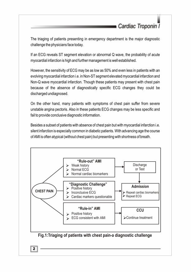
Triaging of patients with or without AMI - A diagnostic challengeAccording to W.H.O. criteria, diagnosis of Acute Myocardial infarction (AMI) is based on
the detection of at least two out of three infarction specific findings:
Ø Chest pain > 20 minutes, resistant to nitro derivatives.
Ø Infarction specific ECG changes (ST segment elevation, development of
abnormal Q wave) in at least two leads of the standard 12 lead ECG
within the same vascular area.
Ø Serial enzyme changes (cardiac markers) with initial rise and subsequent
reduction in level of concentration.
1
Cardiac Troponin I
2
The triaging of patients presenting in emergency department is the major diagnostic
challenge the physicians face today.
If an ECG reveals ST segment elevation or abnormal Q wave, the probability of acute
myocardial infarction is high and further management is well established.
However, the sensitivity of ECG may be as low as 50% and even less in patients with an
evolving myocardial infarction i.e. in Non-ST segment elevated myocardial infarction and
Non-Q wave myocardial infarction. Though these patients may present with chest pain
because of the absence of diagnostically specific ECG changes they could be
discharged undiagnosed.
On the other hand, many patients with symptoms of chest pain suffer from severe
unstable angina pectoris. Also in these patients ECG changes may be less specific and
fail to provide conclusive diagnostic information.
Besides a subset of patients with absence of chest pain but with myocardial infarction i.e.
silent infarction is especially common in diabetic patients. With advancing age the course
of AMI is often atypical (without chest pain) but presenting with shortness of breath.
Fig.1:Triaging of patients with chest pain-a diagnostic challenge
“Rule-out” AMI Weak history Normal ECG Normal cardiac biomarkers
Discharge or Test
“Diagnostic Challenge” Positive history Inconclusive ECG Cardiac markers questionable
“Rule-in” AMI Positive history ECG consistent with AMI
Admission
Repeat cardiac biomarkers Repeat ECG
CCU
Continue treatment
CHEST PAIN
ØØØ
ØØØ
ØØ Ø
ØØ

3
Importance of accurate triagingCoronary ischaemia is the root cause of acute myocardial infarction, hence early and
reliable detection of myocardial ischaemia is a prerequisite for appropriate triage
decision in emergency room so as to initiate the right therapy. It has been observed that
only 10-15% of patients presenting in emergency room with the cardinal symptom of
chest pain develop acute myocardial infarction. Hence early diagnosis of AMI has to
be so sensitive that all suitable patients with myocardial infarction can be treated
with thrombolytic therapy and yet so specific that patients with chest pain but
without myocardial infarction are not unnecessarily exposed to the risks of such
therapy. This is the most important decision the clinicians have to take when the patients
present in the emergency rooms.
Role of cardiac markersFollowing are the important applications of cardiac markers for management of patients
with acute coronary syndrome:
Ø Confirm the diagnosis of AMI in the presence of diagnostically specific ECG
changes.
Ø Diagnosis of AMI in the absence of unequivocal ECG changes (NSTEMI and Non-Q
wave MI).
Ø Identification of high-risk patients with unstable angina pectoris in the absence of
unequivocal ECG changes.
Ø For monitoring patients with AMI undergoing thrombolytic therapy i.e. success of
therapy for reperfusion.
From a clinical point of view, an ideal cardiac marker that detects myocardial injury
should satisfy the following properties:
Ø It should be present in myocardium in high concentration and absent in other
tissues thereby ensuring high cardiac specificity.
Ø It should be released rapidly in blood stream after myocardial injury, so as to
achieve optimal sensitivity in early phase after the onset of myocardial injury.
Ø It should remain abnormal for several days thereby offering wide diagnostic
window time.
Ø It should be assayed with a rapid turnaround time.
4
Cardiac Troponin I
Cardiac Markers
Creatine Kinase (CK) / Creatine Kinase MB (CK-MB) activityThree different isoenzymes of CK exist namely CK-MM, CK-MB and CK-BB. Skeletal
muscle approximately consists of CK-MM (97-99%) and CK-MB (1-3%). The cardiac
muscle approximately contains CK-MM (95%) and CK-MB (5%). CK-BB is found
primarily in brain and contributes very little to the total CK level.
After myocardial infarction CK and CK-MB levels rise after 2-6 hours; peak levels are
observed at 12-24 hours. CK returns to normal levels after 3-4 days where as CK-MB
because of shorter half-life returns to normal level after 2-3 days.
The calculation of CK-MB/CK ratio improves the specificity of CK-MB for acute
myocardial infarction in patients accompanying with skeletal muscle damage. CK-MB
returns to normal levels within 2-3 days after myocardial infarction, hence it is useful in
detecting reinfarction.
Limitation:
Though CK-MB/CK ratio improves specificity of CK-MB, however small myocardial
necrosis may be missed (unstable angina pectoris may show the presence of micro
infarcts / minor myocardial injury).
CK-MB isoformsIn an attempt to improve sensitivity of CK-MB, high voltage electrophoresis technique
was developed to separate CK-MB into its two isoforms: CK-MB2 and CK-MB1. In serum
of healthy individuals CK-MB2/CK-MB1 ratio of approximately 1 is present. The
reference range of this ratio is 1.5. A higher ratio indicates acute myocardial infarction.
Limitation:
Ø This technique is labour intensive.
Ø Requires high level of technical skill.
Ø Long delay in reporting of results.
CK-MB mass
Here CK-MB is detected immunologically by using a combination of CK-B and CK-M
specific monoclonal antibodies or with CK-MB specific monoclonal antibodies.

5
Limitation:
Ø Interference in these assays is observed because of CK-MM, CK-BB, and CK-B
autoantibodies.
CK-MB immunoinhibition method
The theoretical basis for the clinical application of immunoinhibition method is the
assumption that only CK-MM and CK-MB are released into the blood stream after muscle
damage. The reagent contains anti CK-M antibodies, which completely inhibit all CK-M
activity i.e. both M subunits in CK-MM and the single M subunit in CK-MB. The remaining
non CK-M activity corresponding to the CK-B activity of CK-MB is measured. Since only
CK-B of the dimeric CK-MB molecule is measured, multiplication by a factor of 2 gives the
CK-MB activity in the specimen.
Limitation:Ø In case of macro CK, which contains no CK-M subunits immunoinhibition cannot
take place.
Common limitations of CK-MB assays:
Ø As CK-MB is also present in skeletal muscle it is not absolutely specific to cardiac
muscle damage.
Ø Evaluation of CK-MB levels may present problems in conditions such as
extensive skeletal muscle injury with small infarction, chronic skeletal muscle
injury and myocardial infarction after coronary artery bypass graft.
Ø Determination of CK and CK-MB activity alone is not suitable for assessment of
risk in patients with unstable angina pectoris (minor myocardial damage).
Lactate DehydrogenaseLactate Dehydrogenase is also an enzyme released by ischaemic heart muscle. Out of the 5 isoenzymes only two of them LD1 and LD2 are useful in the diagnosis of AMI. Usually in normal healthy individuals the amount of LD2 in blood is higher than LD1 but patients with AMI show more of LD1 than LD2.
Limitation:
Ø LD1 and LD2 are not cardiospecific markers
Ø Elevated levels of LD1 and LD2 are observed in leukemia, renal and hemolytic
diseases.

Cardiac MarkerHours after onset of pain
0 - 2 3 - 4 5 - 6
CK activity 15 35 70
CK-MB activity 10 25 55
CK-MB mass 30 70 90
CK-MB isoform ratio 25 60 90
Myoglobin 35 80 95
Fig.3: Average diagnostic sensitivities (%) of cardiac markers during early phase of AMI
6
Cardiac Troponin I
Myoglobin
Myoglobin, the oxygen binding haem protein constitutes about 2% in both skeletal and
cardiac muscle. The low molecular weight of Myoglobin (17.8 kDa) facilitates its rapid
release in circulation and is the first marker to exhibit rising levels after AMI. The
advantages of Myoglobin in early diagnosis of myocardial infarction are its high early
sensitivity and the possibility of rapidly assessing the success of thrombolytic therapy.
Limitation:
Ø Since Myoglobin is also present in skeletal muscle it is not a cardiospecific marker.
Ø The extremely short biological half life (10-20 minutes) restricts the usage of
Myoglobin to detect unstable angina pectoris (minor myocardial injury or micro
infarcts).
Cardiac Marker M.Wt (kDa)
Half life(hours)
Increase(hours)
Peak* (hours)
Normalization (days)
LD - 1 110 6 - 12 48 - 144 7 - 14135
17 3 - 12 12 - 24 3 - 4CK 86
86CK-MB 13 3 - 12 12 - 24 2 - 3
CK-MB mass 13 2 - 6 12 - 24 386
Myoglobin 17.8 0.25 2 - 6 - 16 12
* Strongly dependent on the timing of reperfusion of the infarct-related blood vessel
Fig.2: Typical characteristics of cardiac markers
7
The advent of cardiac Troponins T and I, unarguably the most sensitive and specific
markers encompass all the requirements that physicians and laboratarians require for
accurate triaging and better risk stratification of patients with acute coronary syndrome.
Role of Troponins in muscle contractionThe contractile apparatus of striated muscle fiber is composed of thick and thin filaments.
The thick filament is composed mainly of myosin. Actin, tropomyosin and Troponin
comprise of thin filament. Muscle contraction occurs when thick and thin filament slide
past each other. The interaction between thick and thin filament is regulated by Troponin
complex found on thin filaments. The Troponin complex is composed of three protein
subunits: Troponin I (TnI), Troponin T (TnT) and Troponin C (TnC). The calcium-
mediated contraction of striated muscle (fast-skeletal, slow-skeletal and cardiac muscle)
is regulated by the Troponin complex. Contraction of smooth muscle is regulated by
calmodulin (intracellular protein that combines with calcium and is involved in smooth
muscle contraction).
Troponins are proteins that are integral to the functioning of striated muscle. They exist
as a complex with actin and tropomyosin on thin filament of the contractile apparatus.
The Troponin complex consists of three protein subunits:
Ø Troponin C, binds with calcium and regulates activation of thin filaments during
contraction.
Ø Troponin T, binds the Troponin complex to tropomyosin.
Ø Troponin I, prevents the contraction of muscle in the absence of calcium and
Troponin C.
During the functioning of the contractile apparatus depolarization of muscle leads to
intracellular release of calcium, which binds with Troponin C. A conformational change
occurs in Troponin-Tropomyosin complex in such a way that actin molecules can then
interact with myosin, resulting in muscle contraction.
Cardiac Troponins (cTn)-emerging cardiac marker of choiceLimitations of existing cardiac markers led to the search for markers uniquely expressed
by the myocardium. The cardiac troponins T and I (cTnT and cTnI) have excellent
sensitivity and specificity and are superior to CK-MB in indicating minor myocardial
injury.

8
Cardiac Troponin I
Types of Cardiac Troponins
ØTroponin C exists as two isoforms, fast and slow. The fast isoform is found only in
skeletal muscle, but the slow isoform is found both in skeletal and cardiac muscles.
The molecular weight of cardiac isoform (cTnC) is 18 kDa.
Ø Troponin T is also found in fast and slow skeletal muscle, cardiac muscle.
Troponin T present in skeletal muscle exists as a slightly different subform. The
cardiac isoform (cTnT) has a molecular weight of 37 kDa.
Ø Three isoforms of Troponin I have been identified, one each in fast and slow
skeletal muscles and one isoform in cardiac muscle. The cardiac isoform of
Troponin I (cTnI) has a molecular weight of 22.5kDa. cTnI has an extra 30 amino
acid sequence at the N terminal portion of molecule making it absolutely specific to
cardiac muscle. cTnI is mostly bound to contractile apparatus in myocardium, but
about 8% is found free in cytoplasm.
Fig.4 : Diagram of muscle contraction depicting Troponin involvement
2+ 2+2+
2+
l
9
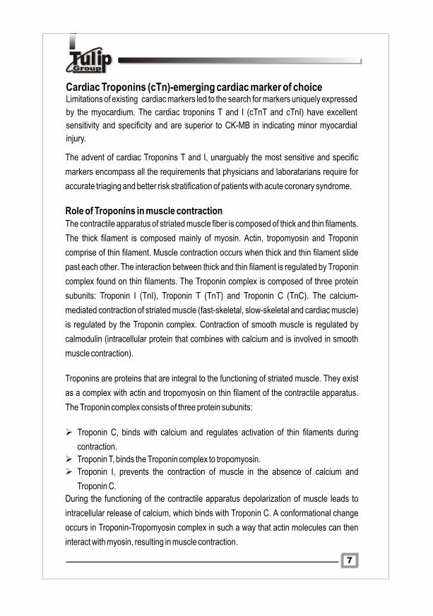
Cardiac Troponin M.Wt.(kDa)
Half Life(hours)
Increase(hours)
Peak* (hours)
Normalization (days)
22.5 2 - 4 3 - 8 12 - 24 7 - 10cTnl
37 2 - 4 3 - 8 12 - 96 7 - 14cTnT
* Strongly dependent on the timing of reperfusion of the infarct-related blood vessel
400 20 60 80 100 120 140 1601
2
3
4
5
6
7
x U
pper
lim
it of
nor
mal
Hours from onset of infarction
Troponin I
Myoglobin
Total CK
CK-MB LDH
Fig.5: Characteristics of cardiac troponin (I and T) in AMI
Fig.6: Graphical representation-Levels of cardiac markers in AMI
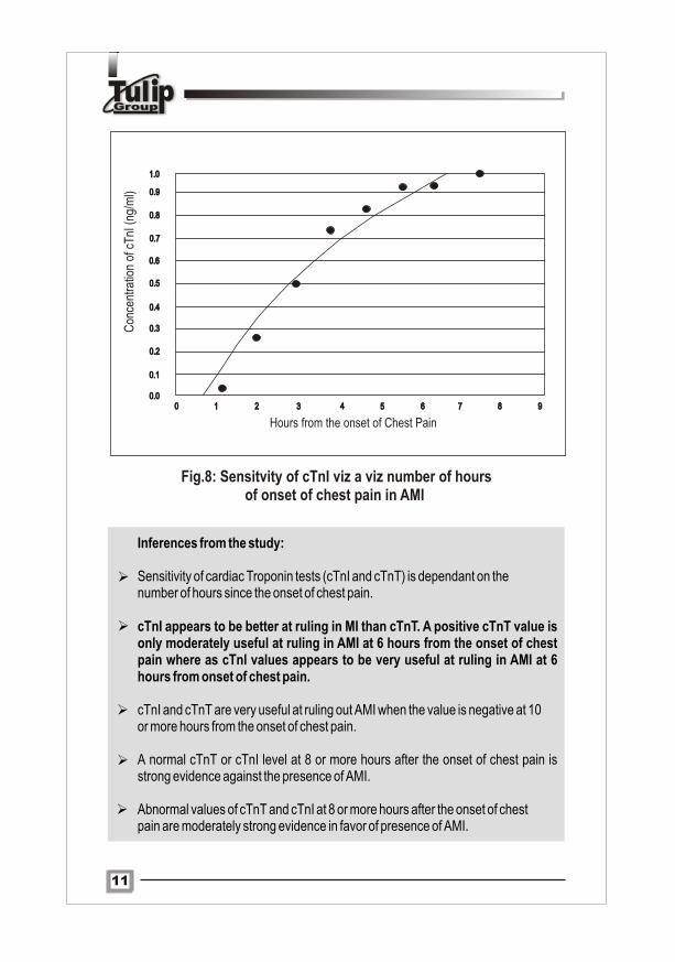
Cardiac Troponins (cTnI and cTnT) - sensitivity & specificityAny damage or injury to myocardial cells results in the release of cardiac Troponins into
the circulation. Concentration of cTnI and cTnT in the circulation initially increases with
the number of hours after the onset of chest pain and decreases as the enzymes are
cleared from the circulation. The most important take home message is that sensitivity of
cardiac Troponin tests, like any other cardiac marker is dependant on the number of
hours after the onset of chest pain.
Internationally a lot of scientific research work has been done to evaluate important
parameters of sensitivity, specificity and predictive values of cTnI and cTnT in clinical
settings.
10
Cardiac Troponin I
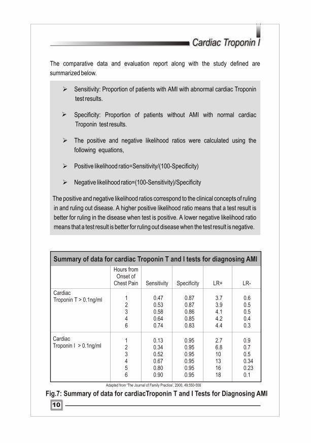
The comparative data and evaluation report along with the study defined are
summarized below.
Ø Sensitivity: Proportion of patients with AMI with abnormal cardiac Troponin
test results.
Ø Specificity: Proportion of patients without AMI with normal cardiac
Troponin test results.
Ø The positive and negative likelihood ratios were calculated using the
following equations,
Ø Positive likelihood ratio=Sensitivity/(100-Specificity)
Ø Negative likelihood ratio=(100-Sensitivity)/Specificity
The positive and negative likelihood ratios correspond to the clinical concepts of ruling
in and ruling out disease. A higher positive likelihood ratio means that a test result is
better for ruling in the disease when test is positive. A lower negative likelihood ratio
means that a test result is better for ruling out disease when the test result is negative.
Summary of data for cardiac Troponin T and I tests for diagnosing AMI
Hours from Onset of
Chest Pain Sensitivity Specificity LR+ LR-
Cardiac Troponin T > 0.1ng/ml 1 0.47 0.87 3.7 0.6
2 0.53 0.87 3.9 0.53 0.58 0.86 4.1 0.54 0.64 0.85 4.2 0.46 0.74 0.83 4.4 0.3
Cardiac Troponin I > 0.1ng/ml
1 0.13 0.95 2.7 0.92 0.34 0.95 6.8 0.73 0.52 0.95 10 0.54 0.67 0.95 13 0.345 0.80 0.95 16 0.236 0.90 0.95 18 0.1
Fig.7: Summary of data for cardiacTroponin T and I Tests for Diagnosing AMI
Adapted from ‘The Journal of Family Practice’, 2000, 49:550-556
11
00 11 22 33 44 55 66 77 88 990.00.0
0.10.1
0.20.2
0.30.3
0.40.4
0.50.5
0.60.6
0.70.7
0.80.8
0.90.9
1.01.0
Hours from the onset of Chest Pain
Fig.8: Sensitvity of cTnI viz a viz number of hours of onset of chest pain in AMI
Inferences from the study:
Sensitivity of cardiac Troponin tests (cTnI and cTnT) is dependant on the number of hours since the onset of chest pain.
cTnI appears to be better at ruling in MI than cTnT. A positive cTnT value is only moderately useful at ruling in AMI at 6 hours from the onset of chest pain where as cTnI values appears to be very useful at ruling in AMI at 6 hours from onset of chest pain.
cTnI and cTnT are very useful at ruling out AMI when the value is negative at 10 or more hours from the onset of chest pain.
A normal cTnT or cTnI level at 8 or more hours after the onset of chest pain is strong evidence against the presence of AMI.
Abnormal values of cTnT and cTnI at 8 or more hours after the onset of chest pain are moderately strong evidence in favor of presence of AMI.
Ø
Ø
Ø
Ø
Ø
Con
cent
ratio
n of
cT
nI (
ng/m
l)
12
Cardiac Troponin I
cTnI versus cTnTThe debate continues as to which of the two, cTnI or cTnT is better for management of
patients with acute coronary syndromes. Since assays of cTnT were commercially
available few years before cTnI, more peer-reviewed publications on the clinical utility of
cTnT might have appeared in the past.
However, recent studies have questioned the diagnostic specificity of cTnT assays in
patients with myocardial injury and chronic renal failure, muscular dystrophies and
skeletal muscle damage. cTnI indeed scores over cTnT in the specificity aspect because
cTnI is the only Troponin I expressed in myocardial cells during postnatal development.
cTnI is not expressed in normal skeletal muscle at any time including,during postnatal
development.
Thus cTnI determination promises higher diagnostic efficacy because of the following
unique characteristics,Ø Wide diagnostic window time with early appearance and prolonged presence in
circulation.Ø Allows detection of minor myocardial injury because cTnI levels is almost absent
in normal healthy individuals.Ø No cross reactivity with skeletal muscle isoformsØ Virtually absent in skeletal muscle tissue.
Inferences from international reports highlighting the superior diagnostic
efficacy of cTnI
Ø`cTnI values of less than 0.4 ng/ml are associated with a 42 day mortality of 1% and this risk increases progressively to a mortality of 7.5% at values of 9.0 ng/ml or more. Patients presenting with cardiac chest pain and ECG changes can be classified as Troponin positive or negative acute coronary syndromes, with consequent prognostic and therapeutic implications'.
-BMJ, 2002; 34.
Ø`The current study demonstrates that ACS patients who have increased cTnI measured on a point of care whole blood assay show a significant increase in risk over 30-180 days for all cause death, cardiac death and cardiac events in the presence or absence of ST-elevation. These findings add to the evidence based metaanalyses which demonstrates that increased cTnI predicts the risk of adverse outcomes in ACS'.
- Clinical Chemistry, 2002; 48.
Ø'Cardiac Troponin T, but not cardiac Troponin I, has been found to be re expressed in skeletal muscles of patients with renal failure and muscular dystrophy. cTnI appears to be more specific than cTnT, with discordant results more often being cTnT(+) /cTnI(-). cTnI is more specific for cardiac injury in settings of renal and muscle disease.'- The Journal of Emergency Medicine, Vol.23, Jan. 2002.
Ø'In patients with clinically documented acute coronary syndrome who are treated with glycoprotein IIb/IIIa inhibitors, even small elevations in cTnI identify high risk patients who derive a large clinical benefit from an early invasive strategy'.- JAMA, Vol. 286 No.19, November 21, 2001.
Ø'The prognostic power of cTnI testing in combination with ECG improves efficiency of low risk patient management and improved patient risk stratification. This study adds to the evidence favoring cTnI evaluation as part of the management of acute coronary syndromes'.- Q. J. Med. 2001; 94.
Ø'In the management of acute coronary syndromes and acute MI in clinical practice, cTnI is comparable in diagnostic and prognostic efficacy to cTnT. In renal impairment even against second generation cTnT assays, cTnI is superior'.- Heart, 2000; 83.
Ø'Early studies have questioned the clinical specificity of cTnT assays in patients with chronic renal failure. With the development of second generation assay for cTnT, the frequency of positive results in these patients is lower than first generation, although still higher than for cTnI'.- Clinical Chemistry, 45 No.7, 1999.
Ø'The goal of this prospective study was to assess whether cTnI could replace CK-MB mass as the serum biomarker for detection of AMI. Findings have strongly supported our clinical implementation of cTnI, replacing CK-MB mass as the preferred marker for detection of AMI'.- American Heart Journal, 1999 Feb.; 137 (2).
Ø'The first generation of cTnT assay lacked absolute specificity for the cardiac isoform and allowed interference by skeletal muscle Troponin T. However, cTnT is still detectable in many cases of end stage renal failure, to a lesser extent than first generation but to an extent far more frequent than cTnI'.- Clinical Chemistry, 44:7, 1998.
13

Ø'The cTnI determination is expected to reveal absolute myocardial specificity because cTnI is not expressed in fetal and healthy or diseased adult human skeletal muscle tissue, promising no false positive test results in patients with substantial skeletal muscle damage or renal failure. In conclusion the use of cTnI rapid device could improve efficacy and safety of decision making in patients with chest pain that might produce more cost effective use of intensive care facilities'.- Clinical Chemistry, 44, No.9, 1998.
Ø'Questions about the cardiac specificity of cTnT remain. Some subjects with musculoskeletal or renal disease have elevated levels of cTnT, thus this marker may not be as sensitive as cTnI for detection of myocardial injury'.- American Journal of Critical Care, Vol.7, No.6, Nov.1998.
Ø'cTnI is part of a new generation of biochemical markers that provide an additional clinical tool for assessment of acute coronary syndromes, a term that describes the continuum of myocardial injury ranging from angina, or so called reversible ischaemia to Q-wave MI and definite tissue necrosis. Studies have indicated that cTnI is a more specific marker in cases involving skeletal muscle injury and renal failure. Therefore cTnI may have an important role in real time strategies for evaluating acute coronary syndrome patients, an area that has been of intense interest, discussion and study over recent years'.- Clinical Chemistry, 44:1, 1998.
Ø`Routine use of cTnI bed side test in the emergency room improves decision making and is highly cost effective. cTnI values provide additional prognostic information over and above the level of critical illness in patients presenting to the emergency room'. - CAP, February 1998.
Ø`The slightly higher sensitivity of cTnI test as compared with the cTnT may be related to different release kinetics. The findings of false positive results for cTnT but not cTnI, in patients with renal failure may, however, represent a true difference between the two test'.- The New England Journal of Medicine, Dec.4, 1997
Ø'In conclusion, the results of our prospective study provide evidence that cTnI is an indicator of adverse outcome in patients with severe unstable angina. Our results are keeping in mind with the recent retrospective analysis of patients enrolled in the TIMI III B study. The use of cTnI in the immediate triage of patients with unstable angina appears warranted to identify those at greater risk for cardiac events'.- Circulation, 1997; 95.
14
Cardiac Troponin I
15
Ø'The study compared the diagnostic accuracy of measurement of serum cTnI with CK-MB mass in patients with minor myocardial injury whose measured total CK activity did not exceed twice the upper reference limit. The clinical sensitivity of myocardial injury for cTnI was 100% compared with 81.8% for CK-MB. Thus, cTnI was more sensitive than CK-MB mass for detection of myocardial injury in patients with small increases of total CK'.- Clinical Chemistry, 1997; 43.
Ø'cTnI was as sensitive and specific for AMI as was CK-MB in ED patients who presented within 24 hours of symptom onset. However, cTnI was more sensitive in patients who presented at 24 hours after symptom onset'.- Academic Emergency Medicine, Vol.4, 1997.
Ø 'cTnI could replace CK-MB and would facilitate the rapid and effective triage of patients with chest pain in the emergency department'.- American Journal of Clinical Pathology, 1997 Nov., 108 (5).
Ø'In patients with acute coronary syndromes, cardiac Troponin I levels provide useful prognostic information and permit early identification of patients with an increased risk of death'.- The New England Journal of Medicine, Vol. 335, No.18, 1996.
Ø`Elevations of cTnI are highly specific for myocardial injury. Use of cTnI should facilitate distinguishing whether elevations of CK-MB are due to myocardial or skeletal muscle injury'.- Circulation, Vol.88, 1993.
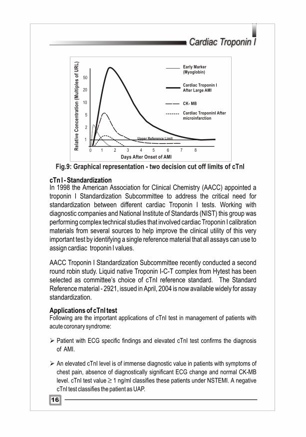
cTnI - Cut off levelsThe National academy of Clinical Biochemistry (NACB), USA and International
Federation of Clinical Chemistry (IFCC), Germany have recommended the use of two
decision cut off limits for cardiac Troponins. A low limit that establishes the presence of
myocardial injury and a high limit that establishes injury to the extent that qualifies as
AMI.
Based on literature data and clinical assessments cTnI levels greater than 0.1 ng/ml
places a patient with unstable angina in the high-risk category for short-term risk of death
or non-fatal MI. The cut off for the definition of AMI is taken to be greater than 1.2 ng/ml.
Thus cTnI levels with a cut off of 0.1 ng/ml identify patients at higher risk for very early
adverse outcomes.
16
Cardiac Troponin I
Applications of cTnI testFollowing are the important applications of cTnI test in management of patients with
acute coronary syndrome:
Ø Patient with ECG specific findings and elevated cTnI test confirms the diagnosis
of AMI.
Ø An elevated cTnI level is of immense diagnostic value in patients with symptoms of
chest pain, absence of diagnostically significant ECG change and normal CK-MB
level. cTnI test value 1 ng/ml classifies these patients under NSTEMI. A negative
cTnI test classifies the patient as UAP.
cTn I - StandardizationIn 1998 the American Association for Clinical Chemistry (AACC) appointed a troponin I Standardization Subcommittee to address the critical need for standardization between different cardiac Troponin I tests. Working with diagnostic companies and National Institute of Standards (NIST) this group was performing complex technical studies that involved cardiac Troponin I calibration materials from several sources to help improve the clinical utility of this very important test by identifying a single reference material that all assays can use to assign cardiac troponin I values.
AACC Troponin I Standardization Subcommittee recently conducted a second round robin study. Liquid native Troponin I-C-T complex from Hytest has been selected as committee’s choice of cTnI reference standard. The Standard Reference material - 2921, issued in April, 2004 is now available widely for assay standardization.
Fig.9: Graphical representation - two decision cut off limits of cTnI
1
2
5
10
20
50
10 2 3 4 5 6 7 8
Days After Onset of AMI
Upper Reference Limit
Rel
ativ
e C
on
cen
trat
ion
(M
ult
iple
s o
f U
RL
)
Early Marker(Myoglobin)
Cardiac Troponin IAfter Large AMI
CK- MB
Cardiac TroponinI Aftermicroinfarction
17
Ø Also elevated cTnI value provides prognostic value in identifying patients with
unstable angina pectoris. Symptoms of chest pain, absence of diagnostically
significant ECG change, normal CK-MB level and cTnI value >0.1 ng/ml are
classified under UAP. If the test is negative retesting probably at 12 hours after
post onset of chest pain is important to rule out diagnosis of acute coronary
syndrome.
Ø Patients presenting with chest pain after trauma or surgery and elevated CK-MB
assay value (to rule out true elevation of CK-MB).
Ø Patients presenting with chest pain 2 to 6 days prior to admission may have
sustained acute myocardial infarction but CK-MB would have returned to normal
levels. Superior diagnostic efficacy of cTnI over CK-MB in detecting microinfarcts
Patients with normal CK-MB levels and elevated cTnI levels could be attributed probably
to the low sensitivity and specificity of CK-MB in detecting micro infarction. Since CK-MB
is present in skeletal muscle and normal healthy individuals, diagnostic cut off values are
typically set above the upper limit of reference range for CK-MB assay. Cardiac troponin I
is not present in normal healthy individuals and is approximately 13 times more abundant
in myocardium than CK-MB on a weight basis. Hence the signal to noise ratio (increased
sensitivity) associated with cTnI is more favorable for detection of micro infarction
(NSTEMI and UAP).
LimitationsØ cTnI levels remain elevated for about 7 days hence for serial monitoring of
patients undergoing thrombolytic therapy cardiac markers such as CK-MB and
Myoglobin may be used for successful reperfusion. Also in cases of reinfarction
markers with shorter half-life such as CK-MB should be used for accurate
diagnosis.
Ø Since cardiac Troponins (both cTnI and cTnT) are sensitive markers for myocardial
damage, they are detected in many other cardiac conditions such as acute
pericarditis, acute myocarditis, congestive heart failure (CHF), perioperative
myocardial infarction and cardiac contusion.
18
Cardiac Troponin I
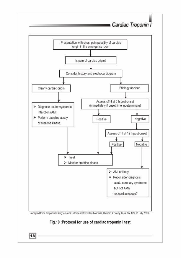
Presentation with chest pain possibly of cardiacorigin in the emergency room
Is pain of cardiac origin?
Consider history and electrocardiogram
Clearly cardiac origin Etiology unclear
Ø
Ø
Diagnose acute myocardial
infarction (AMI)
Perform baseline assay
of creatine kinase
Assess cTnl at 6 h post-onset(immediately if onset time indeterminate)
Positive Negative
Assess cTnl at 12 h post-onset
Ø
Ø
Treat
Monitor creatine kinase
Ø
Ø
AMI unlikely
Reconsider diagnosis
- acute coronary syndrome
but not AMI?
- not cardiac cause?
Positive Negative
Fig.10 :Protocol for use of cardiac troponin I test
(Adapted from: Troponin testing: an audit in three metropolitan hospitals, Richard X Davey, MJA, Vol.179, 21 July 2003)
19
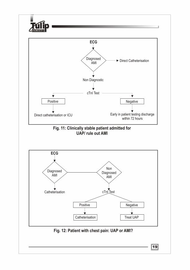
ECG
Diagnosed AMI
Direct Catheterisation
Non Diagnostic
cTnI Test
Direct catheterisation or ICU Early in patient testing discharge within 72 hours
Positive Negative
ECG
Diagnosed AMI
NonDiagnosed
AMI
Catheterisation cTnI Test
Positive Negative
Treat UAPCatheterisation
Fig. 11: Clinically stable patient admitted for UAP/ rule out AMI
Fig. 12: Patient with chest pain: UAP or AMI?
20
Cardiac Troponin I
Critical parameters for correct usage of cTnI test
Rising levels of cardiac markers are dependant on time elapsed since the onset of
myocardial necrosis. This is true regardless of whichever cardiac marker being used for
ruling in and ruling out AMI. Since patients present at varying times for testing following
the onset of chest pain in a cardiac event, it is necessary to perform sequential testing
of cTnI levels for optimal diagnostic accuracy.
Following are the important points to be considered regarding cTnI test when triaging
patients for acute coronary syndrome:Ø First test must be done preferably within onset of symptoms at baseline or within
3 hours of onset of chest pain along with ECG test. A single cTnI negative test
within 3 hours cannot safely rule out AMI. Ø If the first test is negative, second test for cTnI must be done at 6 hours from the
onset of chest pain accompanied by ECG test and if negative should be repeated
between 6-12 hours after the onset of chest pain. A third test at 8-9 hours after
onset of chest pain represents the best time for assessment of cTnI level and is
the most predictive for ruling in or ruling out acute coronary syndrome.
Ø If 6 hour cTnI test and ECG is normal it is unlikely that the patient will have an
adverse outcome in the next 30 days (1% chance).
Ø An elevated level of cTnI (positive cTnI test) in patients with normal ECG, but with
UAP or NSTEMI or Non Q wave MI identifies patient group at greater risk of
death.
Ø Any elevated level of cTnI is indicative of an increased short-term risk of death or
nonfatal myocardial infarction.
Ø The half life of cTnI is approximately 2-4 hours hence samples should
be tested preferably immediately. In case of delay in testing the samples may be
tested within 2 hours of blood collection.
21
ANNEXURE-I
The heart and how it works
The normal human heart is a strong muscular pump little larger than a fist. Each day an
average heart beats 1,00,000 times and pumps about 2000 gallons of blood. In a 70-year
lifetime, an average human heart beats more than 2.5 billion times.
The heart pumps blood continuously through the circulatory system. The circulatory
system is the network of elastic blood vessels that carries blood throughout the body. The
circulatory system comprises of heart, arteries, arterioles, veins and capillaries. The
arteries are the blood vessels which carry oxygen and nutrient rich blood to all parts of
the body. The veins and capillaries are the blood vessels that carry oxygen and nutrient
depleted blood back to heart and lungs. If all these blood vessels were laid end to end
they would extend for about 60,000 miles.
The circulating blood brings oxygen and nutrients to all the body's organ and tissues,
including the heart. It also picks up waste products from the body's cells. These waste
products are removed as they are filtered through the kidney, liver and lungs.
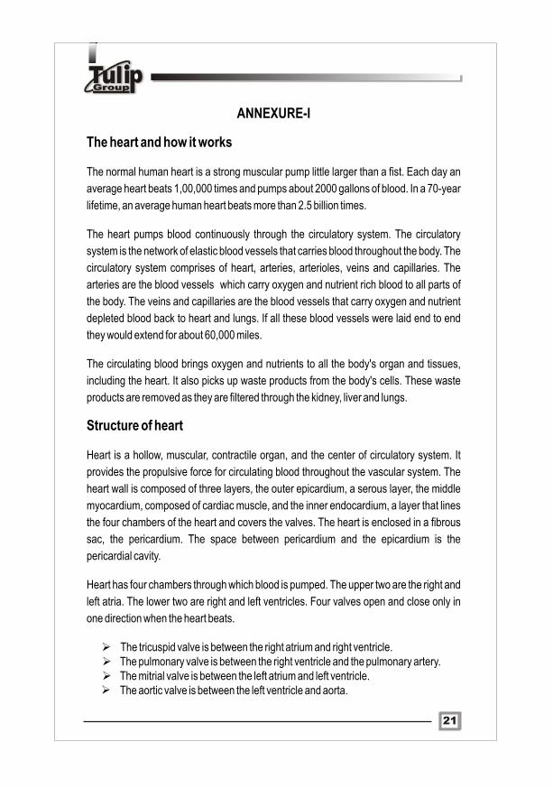
Structure of heart
Heart is a hollow, muscular, contractile organ, and the center of circulatory system. It
provides the propulsive force for circulating blood throughout the vascular system. The
heart wall is composed of three layers, the outer epicardium, a serous layer, the middle
myocardium, composed of cardiac muscle, and the inner endocardium, a layer that lines
the four chambers of the heart and covers the valves. The heart is enclosed in a fibrous
sac, the pericardium. The space between pericardium and the epicardium is the
pericardial cavity.
Heart has four chambers through which blood is pumped. The upper two are the right and
left atria. The lower two are right and left ventricles. Four valves open and close only in
one direction when the heart beats.
Ø The tricuspid valve is between the right atrium and right ventricle.Ø The pulmonary valve is between the right ventricle and the pulmonary artery.Ø The mitrial valve is between the left atrium and left ventricle.Ø The aortic valve is between the left ventricle and aorta.
22
Cardiac Troponin I
Each valve has a set of flaps, also known as leaflets or cusps. The mitral valve has twoflaps. The other valves have three flaps. Under normal conditions, these valves allow
blood flow in one direction. Blood flow occurs only when there is a difference in pressure
across the valves that cause them to open.
Head and Upper Extremity
Superior vena cava
Pulmonary valve
Right atrium
Tricuspid valve
Inferiorvena cava
Right ventricle
Trunk and Lower Extremity
Aorta Pulmonary artery
Lungs
Pulmonary vein
Left atrium
Aortic valve
Mitral valve
Left ventricle
Fig.1: Structure of heart
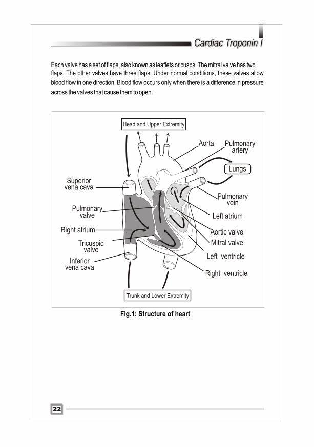
Atherosclerosis - Risk factorsEpidemiological studies indicates the following risk factors that potentiate
atherosclerosis,
Ø Hyperlipidaemia
Ø Hypertension
Ø Cigarette habituation
Ø Diabetes mellitus
Ø Age
Ø Sex
Signs and symptoms associated with atherosclerosisThe signs and symptoms of atherosclerosis are highly variable, but mainly present as
follows:
Ø Unstable angina pectoris
Ø Acute Myocardial Infarction
Ø Transient ischaemic attack
Ø Stroke
Ø Peripheral vascular disease
Ø Mesenteric angina
Ø Abdominal aortic aneurysm
Ø Atheroembolism
Fig.2: Depiction of Atherosclerosis
Progression of plaque build upin coronary artery
Plaque build up in
coronary artery
blocking blood flow
and oxygen to heart
Damage and death to
heart tissue
24
Cardiac Troponin I
Normal Tear in lining
of arteryAccumulation of
fat and cholesterol

23
ANNEXURE-II Atherosclerosis: major cause of cardio vascular disease
Atherosclerosis comes from the Greek word athero (meaning gruel or paste) and
sclerosis (meaning hardness). The inner lining of blood vessels namely the arteries
contains deposits of fatty substances, cholesterol, cellular waste products and calcium.
This build up is known as plaque.
Plaque formation results in luminal obstruction, abnormalities of blood flow, diminished
oxygen supply to target organs.
Atherosclerosis begins with damage to innermost layer of blood vessels namely the
endothelium. The probable cause of endothelial injury includes oxidized LDL cholesterol,
by products of cigarette smoking, hyperglycemia and hyperhomocystinaemia.
Circulating monocytes infiltrate the intima of the vessel wall and the tissue macrophages
act as scavenger cells forming the characteristic foam cell of early atherosclerosis.
These activated macrophages produce numerous factors that are injurious to
endothelium.
Injury to endothelium leads to increased platelet adhesion, increased tissue factor
release, increased plasminogen activator inhibitor, decreased plasminogen activator,
decreased thrombomodulin and alterations in heparan sulfate. Thus the sequence of
events results in procoagulant milieu and enhanced thrombus or clot formation.
Atherosclerotic plaques characteristically occur in regions of branching and marked
curvature at areas of geometric irregularity and where blood undergoes sudden changes
in velocity and direction of flow. Decreased shear stress and turbulence may promote
atherogenesis at these important sites within the coronary arteries, the major branches
of the thoracic and abdominal aorta and vessels of lower extremities of our body.
Generally plaques are static, but they can also become unstable and rupture. Those that
rupture can initiate thrombus formation and can totally block blood flow (occlusion) in the
artery. A blood clot that breaks off and travels to another part of the body is known as
emboli.
If a clot blocks a blood vessel, that supplies blood to heart it causes heart attack. If it
blocks a blood vessel supplying blood to brain, it causes stroke. And if blood supply to the
arms or legs is reduced, it can cause gangrene.
25
Angina pectoris
Angina pectoris is the medical term for chest pain or discomfort due to coronary heart
disease. Angina is a symptom of condition known as myocardial ischaemia. It occurs
when the heart muscle (myocardium) is deprived of required amount of blood it needs for
performing normal function. Myocardial ischaemia (insufficient blood supply to
myocardium) occurs due to narrowing or occlusion (blockage) of one or more arteries
that supply blood to heart.
Typical angina is characterized by uncomfortable pressure, fullness, squeezing pain in
center of chest. The discomfort also may be felt in the neck, jaw, and shoulder.
Angina is a sign that someone is at high risk of heart attack, cardiac arrest and sudden
cardiac death.
Stable angina pectoris
People with stable angina pectoris have episodes of chest pain that are usually
predictable. The chest pain episode occurs with exertion or mental or emotional stress.
Normally the chest discomfort is relieved with rest and/or sublingual nitroglycerin
administration.
Fig4.: Risk factors and associated signs in Atherosclerosis
Unstable angina pectorisPeople with unstable angina pectoris have unexpected chest pain that usually occurs
while even at rest. The discomfort may be more severe and prolonged than typical
angina.
People with unstable angina pectoris should be treated as an emergency because they
are at increased risk for acute myocardial infarction, severe cardiac arrhythmias and
cardiac arrest leading to sudden death.
Variant angina pectoris (prinzmetal angina pectoris)
Unlike typical angina, it nearly always occurs when a person is at rest. It does not follow a
period of physical exertion or emotional stress. Attacks can be very painful and usually
occur between midnight and 8 a.m. morning.
Ischaemic Heart Disease
Ischaemia is a condition where the flow of blood, and therefore oxygen, to a part of body
is restricted. Cardiac ischaemia refers to lack of blood flow and oxygen to heart muscle.
Ischaemic heart disease refers to heart problems caused by narrowing down of arteries
that supply blood to heart. When arteries are narrowed, less blood and oxygen reaches
the heart. This condition is also known as coronary artery disease or coronary heart
disease.
Acute Myocardial infarction
Acute myocardial infarction is defined as death or necrosis of myocardial cells. It is the
end diagnosis of myocardial ischaemia. Myocardial infarction occurs when myocardial
ischaemia exceeds a critical threshold and overwhelms myocardial cellular repair
mechanisms that are designed to maintain normal operating function.
Critical myocardial ischaemia may occur as a result of increased myocardial metabolic
demand, decreased supply of oxygen and nutrients to myocardium via the coronary
circulation. An interruption in the supply of myocardial oxygen and nutrients occurs when
a thrombus is superimposed on an unstable atherosclerotic plaque and results in
coronary occlusion. Conditions associated with increased myocardial metabolic
demand include extremes of physical exertion, severe hypertension and severe aortic
valve stenosis.
26
Cardiac Troponin I
The human body often creates small blood vessels known as collaterals to help
compensate for reduced blood flow. Collateral vessels normally are not open and normal
healthy individuals do show the presence of collateral vessels but in microscopic form.
But in people suffering from coronary artery disease or any other blood vessel disease
these collateral vessels grow and enlarge. When a collateral vessel enlarges, it allows
flow of blood from an open artery to either an adjacent artery or further downstream on
the same artery. Thus collateral vessels grow and form a detour around a blocked blood
vessel.
Types of Myocardial Infarction
Myocardial infarction can be subcategorized on the basis of anatomic, morphologic and
diagnostic clinical information.
From an anatomic or morphologic standpoint, the two types of myocardial infarction are
as follows,
27
Mechanism of Myocardial damageThe severity of myocardial infarction is dependant on three factors:
Ø Level of occlusion in coronary artery
Ø Length of time of occlusion
Ø Presence or absence of collateral circulation
Generally, more proximal the coronary occlusion, more extensive is the amount of
myocardial necrosis. Larger the myocardial infarct, greater is the chance of death due to
mechanical complication. Longer the time period of vessel occlusion, greater the
chances of irreversible myocardial damage distal to the occlusion.
The death of myocardial cells first occurs in the area of myocardium that is most distal to
arterial blood supply, the endocardium. As the duration of occlusion increases, the area
of myocardial cell death enlarges, extending from the endocardium to the myocardium
ultimately to the epicardium. Thus the extent of myocardial cell death defines the
magnitude of myocardial infarction.
28
Cardiac Troponin I
Transmural Myocardial infarction:
In transmural myocardial infarction the ischaemic necrosis affects muscle segment
extending from the endocardium through the myocardium to epicardium.
Non-Transmural Myocardial infarction:
In non-transmural myocardial infarction the area of ischaemic necrosis does not extend
through the full thickness of myocardial wall segments. The area of ischaemic necrosis is
limited to either endocardium or endocardium and myocardium. It is the endocardial and
subendocardial zones of myocardial wall segment that are least perfused regions of
heart and are most vulnerable to conditions of ischaemia.
An old sub classification of myocardial infarction based on clinical diagnostic criteria is
determined by the presence or absence of Q wave on ECG. But a more accepted clinical
diagnostic scheme based on ECG findings is the presence of ST segment elevation.
The presence of Q wave or ST segment elevation is associated with high early mortality
and morbidity. However the absence of these two findings does not necessarily confer
better long-term mortality and morbidity.
Signs and symptoms of AMIAcute myocardial infarction may have unique presentations in individual patients. The
degree of symptoms range from none to sudden cardiac death. Asymptomatic
myocardial infarction is not necessarily less severe than a symptomatic event, but
patients who experience asymptomatic myocardial infarction are more likely to be
diabetic. Despite the diverse presenting symptoms of myocardial infarction, there are
some characteristic typical symptoms,
Ø Chest pain described as pressure sensation, fullness or squeezing in the
midportion of thorax
Ø Chest pain radiating to jaw, teeth, shoulder, arm and back
Ø Associated dyspnoea or shortness of breath
Ø Associated epigastric discomfort with or without nausea and vomiting
Ø Associated diaphoresis or sweating
Ø Syncope or near syncope without other cause

29
Acute myocardial infarction may occur at any time of the day, but most appear to be
clustered around the early hours of morning and are associated with demanding physical
activity. Approximately 50% of patients have some warning symptoms prior to infarction.
Diagnosis of Myocardial Infarction
W.H.O. criteria for diagnosis of AMI
Twenty years ago, W.H.O. defined the diagnosis of AMI as a triad, two of which atleast
must be present for diagnosis,
Ø Typical history of severe and prolonged chest pain greater than 20 minutes
resistant to nitroglycerinØ Unequivocal electrocardiographic changes, with ST segment elevation and
development of abnormal Q wave.Ø Serial enzyme changes (cardiac markers) with initial rise and subsequent fall of
catalytic concentrations.
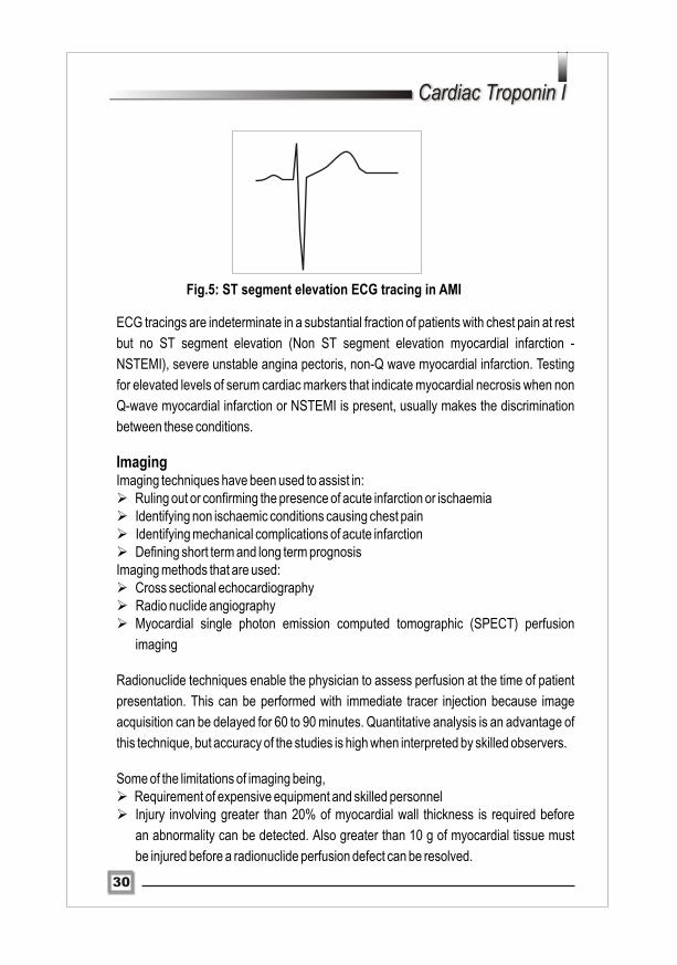
Electrocardiogram (ECG)ECG is usually the first diagnostic test performed. Diagnostic specificity is approximately
100%, and a positive tracing, signaled by an elevated ST segment, essentially confirms
diagnosis of AMI. The diagnostic sensitivity however has been estimated to range from
63-82%.
P
P
R
P-R
Seg Seg
ST
T
R S-TQ
S
Q RSInterval Interval
Q -T
U
Fig.4: Normal ECG tracing
30
Cardiac Troponin I
ECG tracings are indeterminate in a substantial fraction of patients with chest pain at rest
but no ST segment elevation (Non ST segment elevation myocardial infarction -
NSTEMI), severe unstable angina pectoris, non-Q wave myocardial infarction. Testing
for elevated levels of serum cardiac markers that indicate myocardial necrosis when non
Q-wave myocardial infarction or NSTEMI is present, usually makes the discrimination
between these conditions.
ImagingImaging techniques have been used to assist in:Ø Ruling out or confirming the presence of acute infarction or ischaemiaØ Identifying non ischaemic conditions causing chest painØ Identifying mechanical complications of acute infarctionØ Defining short term and long term prognosisImaging methods that are used:Ø Cross sectional echocardiographyØ Radio nuclide angiographyØ Myocardial single photon emission computed tomographic (SPECT) perfusion
imaging
Radionuclide techniques enable the physician to assess perfusion at the time of patient
presentation. This can be performed with immediate tracer injection because image
acquisition can be delayed for 60 to 90 minutes. Quantitative analysis is an advantage of
this technique, but accuracy of the studies is high when interpreted by skilled observers.
Some of the limitations of imaging being,Ø Requirement of expensive equipment and skilled personnel Ø Injury involving greater than 20% of myocardial wall thickness is required before
an abnormality can be detected. Also greater than 10 g of myocardial tissue must
be injured before a radionuclide perfusion defect can be resolved.
Fig.5: ST segment elevation ECG tracing in AMI
31
References and Suggested reading:
Ø Troponin I in patients without chest pain, Detlef Ritter, Paul A.Lee, James F. Taylor, Leo Hsu, Jerome D.Cohen, Hyung D.Chung, Katherene S. Virgo, Clinical Chemistry, 50:1, 112-119, 2004.
Ø National Institute of Standards and Technology, Certificate of Analysis, SRM 2921,Human Cardiac Troponin Complex, April, 2004.
Ø Evaluation of chest pain and heart failure in the emergency department: Impact of
Multimarker Strategies and B-type natriuretic peptide, W.Brian Gibler, Andra L.
Blomkalns, Sean P.Collins, Reviews in Cardiovascular medicine, Vol.4 Suppl.4, S47-
S55, 2003.
Ø Troponin testing: An audit in three metropolitan hospitals, Richard X Davey, JMA, stVol.179, 81-83, 21 July 2003.
Ø Cardiac Troponins, John Sarko, Charles V.Pollack, The Journal of Emergency
Medicine, Vol.23, No.1, 57-65,2003.
Ø Near Bedisde whole blood cardiac Troponin I assay for risk management of patients
with Acute Coronary Syndromes, Fred S. Apple, Mary Ann M. Murakami, Robert L.
Jesse, M. Andrew Levitt, Alan K. Berger, Lesly A. Pearce and Paul Collinson, Clinical
Chemistry 48: 1784-1787, 2002.
Ø Was it a heart attack? Editorial, BMJ; 324:337-338, 2002.
Ø A rapid Troponin I based protocol for assessing acute chest pain, N.J.Alp, J.A.Bell
and M.Shahi, Q J Med; 94:687-694, 2001
Ø Ability of Minor elevations of Troponin I and T to predict benefit from an early invasive
strategy in patients with unstable angina and Non-ST elevation myocardial infarction,
TACTICS-TIMI 18 investigators,JAMA, Vol. 286 No.19, Nov.21, 2001.
Ø Myocardial infarction redefined: Role of cardiac Troponin testing, Editorial, Fred S.
Apple, Alan H.B. Wu, Clinical Chemistry, 47, No.3, 2001.
Ø Troponin T or Troponin I as cardiac markers in ischaemic heart disease, Editorial, S.J.
Maynard, I.B.A. Menown, A.A.J.Adgey, Heart, 2000, 83, 371-373.
Ø Cardiac Markers into the new millennium, Paul O. Collinson, Ann. Clin. Biochemistry,
2000, 37:109-113.
Ø Troponin poised to trigger therapy, William Check, CAP, July 2000 cover story.
Ø ACC/AHA Guidelines for management of patients with UAP and NSTEMI: Executive
summary and recommendations, Circulation, 2000; 102:1193-1209.
Ø Predictors of outcome in patients with ACS without persistent ST-segment elevation:
Results from an International trial of 9461 patients (PURSUIT TRIAL), Circulation:
2000; 101: 2557-2567.
32
Cardiac Troponin I
Ø A systematic review of Troponin T and I for diagnosing AMI, Mark H. Ebell, Cheryll A. Flynn, The Journal of Family Practice: 2000; 49: 550-556.
Ø IFCC Committee on Standardization of Markers of Cardiac damage: Premises and Pro jec t P resen ta t ion (Abs t rac t ) , Vo l .11 No .2 , JIFCC, 1999:19-22.
Ø Implementation of serum cardiac Troponin I as marker for detection of acute myocardial infarction, Falahati A, Sharkley SW, Christensen D, McCoy M, Miller
EA, Murakamani MA, Apple FS, Am. Heart J., 137(2): 332-337, Feb. 1999.Ø Cardiac Troponin I for the diagnosis of acute myocardial infarction in emergency
department, D'Costa M, Fleming E, Patterson MC, Am.J Clin. Pathol., 108(5): 550-555, Nov.1997.
Ø Diagnostic Marker Cooperative Study for the diagnosis of Myocardial infarction, Jamice Zimmerman, Robert Fromn, Denise Meyer, Ann Bourdaeux, Chuan Chuan C. Wuu, Richard Smalling, Barry Davis, Gabriel Habib, Robert Roberts, Circulation, 1999; 99:1671-1677.
Ø National Academy of Clinical Biochemistry Standards of Laboratory Practice: Recommendations for the use of cardiac markers, Alan H.B.Wu, Fred S.Apple, W. Brian Gilber, Robert L.Jesse, Myron M.Warshaw, Roland Valdes. Jr, Clinical Chemistry, 45:7, 1104-1121, 1999.
Ø Cardiac Troponin T and Cardiac Troponin I: relative values in short term risk stratification of patients with ACS, Robert H. Christenson, Shru-Hong Duh, L. Kristin Newby, E.Magnus Ohmann, Robert M.Califf, Christopher B.Granger, Steven Peck, GUSTO-IIa investigators, Clinical Chemistry, 44:3, 494-501,1998.
Ø Characterisation of cardiac Troponin subunit release into serum after AMI and comparison of assays for Troponin I and T, Alan H.B.Wu, Yue Jin Feng, Robert Moore, Fred S.Apple for AACC subcommittee on cTnI standardization, Clinical Chemistry, 44:6, 1198-1208, 1998.
Ø Cardiac Troponins I and T in patients with suspected acute coronary syndrome: a comparative study in routine setting, Oywind Hetland, Kenneth Dickstein, Clinical Chemistry, 44:7, 1430-1436, 1998.
Ø Cardiac Biomarkers: Past, present and future, Jessie E.Adams, Vickie A.Miracle, American Journal of Critical Care, Vol.7 No.6, Nov.1998.
Ø Analytical Performance and Clinical application of new rapid bedside assay for detection of serum cardiac Troponin I, Christopher Heeschen, Britta U.Goldmann, Robert H.Moeller, Christian W.Hamm, Clinical Chemistry, 44:9, 1925-1930, 1998.
Ø Prognostic influence of Elevated cTnI in patients with UAP, Marcello Galvani, Fillipo Ottani, Donatella Ferrini, Jack H.Ladenson, Antonio Destro, Daniele Baccos, Franco Rusticali, Alan S. Jaffe, Circulation, 1997:95:2053-2059.
Ø Emergency room triage of Patients with acute chest pain by means of rapid testing for cTnI, Christian W. Hamm, Brita U. Goldmann, Christopher Heeschen, Georg Kreymann, Jorgen Berger, Thomas Meinertz, The New England Journal of Medicine, Vol.337, No.23, 1648-1653, Dec.4, 1997.
Ø Evaluation of a new assay for cardiac Troponin I vs. Creatine kinase-MB for the diagnosis of acute myocardial infarction. Biochemical Markers for Acute Myocardial Ischaemia (BAMI) study group, GX Brogan, CF McCuskey, HC Thode Jr, J Snow, A Sama, JL Bock, Academic Emergency Medicine, Vol.4, 6-12, 1997.
Ø Improved detection of minor ischaemic myocardial injury with measurement of serum cardiac Troponin I, Fred S.Apple, Alireza Falahati, Pamela R.Paulsen, Elizabeth A.Miller, Scott W.Sharkey, Clinical Chemistry, 43:2047-2051, 1997.
Ø Prognostic influence of elevated values of cardiac Troponin I in patients with Unstable angina, Marcello Galvani, Filippo Ottani, Donatella Ferrini, Jack H.Ladenson, Antonio Destro, Daniele Baccos, Franco Rusticali, Allan S.Jaffe, Circulation; 95:2053-2059, 1997.
Ø Cardiac specific Troponin I levels to predict the risk of mortality in patients with Acute Coronary Syndromes, Elliot M. Antman, Milenko J. Tanasijevic, Bruce Thompson, Mark Schactman, Carolyn H. McCabe, Christopher Cannon, George A. Fischer, Anthony Y. Fung, Christopher Thompson, Donald Wybenga, Eugene Braunwald, The New England Journal of Medicine, Vol.335, No.18, 1342-1349, Oct.31, 1996.
Ø Cardiac Troponin I. A marker with high specificity for cardiac injury, JE Adams, GS Bodor, VG Davila-Roman, JA Delmez, FS Apple, JH Ladenson, AS Jaffe,
Circulation, Vol.88, 101-106, 1993.thØ Text Book of Medical Physiology, Arthur C. Guyton, John E. Hall, 9 edition,
W.B.Saunders Company, 1996.Ø Clinical Laboratory Diagnostics, Use and Assessment of Clinical laboratory results,
Edited by Lothar Thomas, First Edition, TH Books Verlagsgesellschaft mbH, Frankfurt, Germany, 1998.
33

34
NOTES

For the use of Registered Medical Practitioners and Laboratories only
Gitanjali, Tulip Block, Dr.Rego Bagh, Alto Santacruz, Bambolim ComplexPost office, Goa - 403 202, India Tel:+91-832-2458546-51 Fax:+91-832-2458544
E- mail : [email protected], Website:www.tulipgroup.com
TUV SUDDEUTSCHILAND AG
Zephyr Biomedicals


![Chapter 21 Darapladib effect on circulating high sensitive ... · and cardiac troponin – cTn) as means of diagnosis of myocardial infarction [2]. Eleva-tions of serum cardiac troponin](https://static.fdocuments.net/doc/165x107/5f7bc4c1032dbf25d91e28ce/chapter-21-darapladib-effect-on-circulating-high-sensitive-and-cardiac-troponin.jpg)
