Cardiac-specific ablation of cypher leads to a severe form
42
1 © The Author 2008. Published by Oxford University Press. All rights reserved. For permissions, please e-mail: [email protected] Cardiac-specific ablation of cypher leads to a severe form of dilated cardiomyopathy with premature death Ming Zheng 1,2,§ , Hongqiang Cheng 1,§ , Xiaodong Li 1 , Jianlin Zhang 1 , Li Cui 1 , Kunfu Ouyang 1 , Liang Han 2 , Ting Zhao 2 , Yusu Gu 1 , Nancy D. Dalton 1 , Marie-Louise Bang 3,4 , Kirk L. Peterson 1 , and Ju Chen 1,* 1 Department of Medicine, University of California-San Diego, 9500 Gilman Drive, La Jolla, California 92093, USA; 2 Institute of Molecular Medicine, Peking University, Beijing 100871, China; 3 Dulbecco Telethon Institute at Istituto di Tecnologie Biomediche (ITB) – Consiglio nazionale delle ricerche (CNR) (CNR), Via Fratelli Cervi 93, 20090 Segrate, Milan, Italy; and 4 Istituti di ricovero e cura a carattere scientifico (IRCCS) Multimedia, Scientific and Technology Pole, Via Fantoli 16/15, 20138, Milan, Italy. * Corresponding author: Ju Chen, Ph.D. Department of Medicine University of California San Diego 9500 Gilman Drive La Jolla, CA 92093 Tel: (858) 822-4276; Fax: (858) 822-1355 [email protected] § Equal Contributors HMG Advance Access published November 21, 2008
Transcript of Cardiac-specific ablation of cypher leads to a severe form
1
© The Author 2008. Published by Oxford University Press. All rights reserved.
For permissions, please e-mail: [email protected]
Cardiac-specific ablation of cypher leads to a severe form of dilated
cardiomyopathy with premature death
Ming Zheng1,2,§
, Hongqiang Cheng1,§
, Xiaodong Li1, Jianlin Zhang
1, Li Cui
1, Kunfu Ouyang
1,
Liang Han2, Ting Zhao
2, Yusu Gu
1, Nancy D. Dalton
1, Marie-Louise Bang
3,4, Kirk L. Peterson
1,
and Ju Chen1,*
1Department of Medicine, University of California-San Diego, 9500 Gilman Drive, La Jolla,
California 92093, USA; 2 Institute of Molecular Medicine, Peking University, Beijing 100871,
China; 3Dulbecco Telethon Institute at Istituto di Tecnologie Biomediche (ITB) – Consiglio
nazionale delle ricerche (CNR) (CNR), Via Fratelli Cervi 93, 20090 Segrate, Milan, Italy; and
4Istituti di ricovero e cura a carattere scientifico (IRCCS) Multimedia, Scientific and Technology
Pole, Via Fantoli 16/15, 20138, Milan, Italy.
* Corresponding author:
Ju Chen, Ph.D.
Department of Medicine
University of California San Diego
9500 Gilman Drive
La Jolla, CA 92093
Tel: (858) 822-4276; Fax: (858) 822-1355
§ Equal Contributors
HMG Advance Access published November 21, 2008
1
ABSTRACT
Accumulating data suggest a link between alterations/deficiencies in cytoskeletal proteins and
the progression of cardiomyopathy and heart failure, although the molecular basis for this link
remains unclear. Cypher/ZASP is a cytoskeletal protein localized in the sarcomeric Z-line.
Mutations in its encoding gene have been identified in patients with isolated noncompaction of
the left ventricular myocardium (INLVM), dilated cardiomyopathy (DCM), and hypertrophic
cardiomyopathy (HCM). To explore the role of Cypher in the myocardium and better understand
the molecular mechanisms by which mutations in cypher cause cardiomyopathy, we utilized a
conditional approach to knockout Cypher specifically in developing and adult myocardium.
Cardiac-specific Cypher knockout mice developed a severe form of dilated cardiomyopathy with
disrupted cardiomyocyte ultrastructure and decreased cardiac function, which eventually led to
death before 23 weeks of age. A similar phenotype was observed in inducible cardiac-specific
Cypher knockout mice in which Cypher was specifically ablated in adult myocardium. In both
cardiac-specific Cypher knockout models, ERK and Stat3 signaling pathways were augmented.
Finally, we demonstrate the specific binding of Cypher’s PDZ domain to the C-terminal region
of both calsarcin-1 and myotilin within the Z-line. In conclusion, our studies suggest that (i)
Cypher plays a pivotal role in maintaining adult cardiac structure and cardiac function through
protein-protein interactions with other Z-line proteins, (ii) myocardial ablation of Cypher results
in DCM with premature death, and (iii) specific signaling pathways participate in Cypher mutant
mediated dysfunction of the heart, and may in concert facilitate the progression to heart failure.
2
INTRODUCTION
Dilated cardiomyopathy (DCM), which is characterized primarily by left ventricular dilation and
systolic dysfunction, is a leading cause of congestive heart failure in young patients. The
recognition that genetic defects may play pivotal roles in the pathogenesis of dilated
cardiomyopathy, especially familial DCM, has received increasing attention during the past
decade. Mutations in multiple cytoskeletal and sarcomeric genes, such as dystrophin (1), vinculin
(2), desmin (3), titin (4), actin (5), beta myosin heavy chain (6), and troponin T (7), have been
linked to the pathogenesis of familial DCM in both human and mouse models. The anatomical
and pathophysiologic phenotypes of these cytoskeletal and sarcomeric gene mutations are
associated variably with impaired structural maintenance, deficiencies in intracellular force
generation and intercellular force transmission, and alterations in stretch signaling (8-15).
However, the molecular mechanisms, linking the gene mutations to a specific DCM phenotype,
remain largely unknown.
Cypher is a cytoskeletal protein, which binds to α-actinin in the Z-line of both skeletal and
cardiac muscle (16,17). Mutations in Z-line alternatively spliced PDZ-motif protein (ZASP), a
human orthologue of Cypher, have been identified in patients with isolated noncompaction of the
left ventricular myocardium (INLVM), DCM, hypertrophic cardiomyopathy (HCM), as well as
skeletal myopathy (18-23).
Our previous studies of mice with global ablation of Cypher in all cells revealed a pivotal role of
Cypher in striated muscle structure and function. These mutant mice developed a severe form of
congenital myopathy and died from functional failure in multiple striated muscles within 1 week
3
after birth (17). The perinatal lethality of Cypher knockout mice prevented us from further
exploring the specific requirement for Cypher in adult myocardium. To overcome this problem
and better understand the molecular mechanisms by which mutations in Cypher cause
cardiomyopathy, we utilized a conditional approach to knockout Cypher specifically in the heart
by crossing floxed Cypher mice with the Mlc2v-Cre mouse line (24) and the myosin heavy chain
α-MHC mER-Cre-mER tamoxifen inducible Cre mouse line (25).
In the present study we report that cardiac-specific Cypher knockout (CKO) mice display
disrupted cardiomyocyte ultrastructure and decreased cardiac function, which eventually leads to
a severe form of dilated cardiomyopathy with premature death. A similar phenotype is observed
in inducible cardiac-specific Cypher knockout mice (ICKO) in which Cypher is specifically
ablated in adult myocardium, confirming our results showing an essential role of Cypher in adult
myocardium. In both cardiac-specific Cypher knockout models, cardiac dysfunction is
accompanied by augmentation of ERK and Stat3 signaling pathways. Furthermore, we
demonstrate specific interaction of Cypher’s PDZ domain with the C-terminal region of both
calsarcin-1 and myotilin.
Taken together, our studies suggest that (i) Cypher plays a pivotal role in maintaining adult
cardiac structure and cardiac function, through protein-protein interactions with other Z-line
proteins, (ii) myocardial ablation of Cypher results in DCM with premature death, and (iii)
specific signaling pathways participate in Cypher mutant mediated dysfunction of the heart and
may in concert facilitate the progression to heart failure.
4
RESULTS
Generation of Cardiac-Specific Cypher Knockout (CKO) Mice
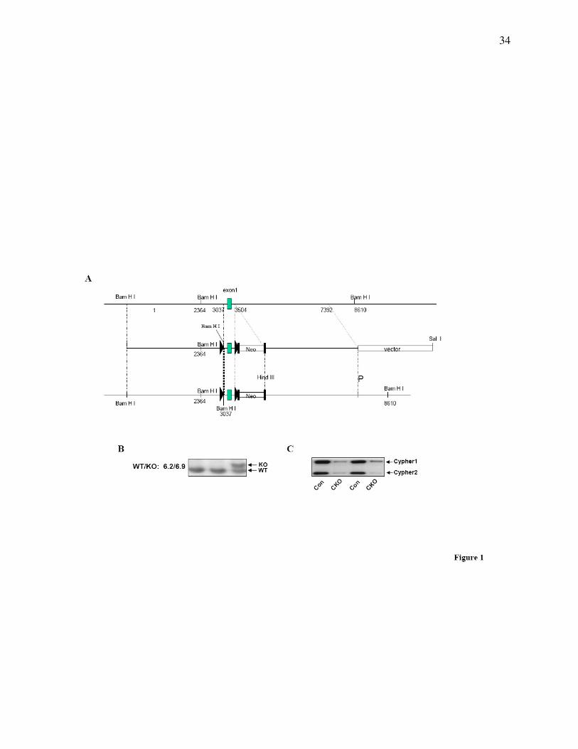
To specifically knockout Cypher in the heart, a targeting construct was generated containing
loxP sites (triangles) flanking exon 1 of the Cypher gene, which is conserved in all six Cypher
isoforms (Fig. 1A) (30). The neomycin (Neo) selection cassette was flanked by FRT sites,
allowing for subsequent excision with FLPase. Targeted ES cells were identified by Southern
blot analysis, and gave the expected 6.9kb mutant fragment compared to the 6.2kb wild type
fragment (Fig. 1B). Cypherf/f
mice were crossed with Mlc2v-Cre mice to generate cardiac-
specific Cypher knockout mice (CKO) in which Cypher is specifically deleted in ventricular
cardiomyocytes. Assessment of Cypher protein expression in CKO hearts, using a cardiac-
specific Cypher antibody, demonstrated the specific loss of Cypher in CKO hearts from one-
month-old mice as shown in Fig. 1C.
To further investigate the specificity and efficiency of Mlc2v-Cre mediated excision during
cardiac development, total protein was extracted from atria and ventricles separately and the
Cypher protein level was measured (data not shown). Consistent with previously published data
reporting Mlc2v expression only in ventricles (24), decreased expression of Cypher protein was
observed only in ventricles, but not in atria of CKO mouse hearts.
CKO Mice Develop Dilated Cardiomyopathy and Die within 5 Months
Cardiac-specific deletion of Cypher does not affect the Mendelian frequency of viable CKO
male and female mice produced at birth. CKO mutants began to die at 16 weeks of age and all
died before 23 weeks of age (Fig. 2A) as compared to 100% survival of control littermates,
5
indicating the essential functional role of Cypher in the heart. Histological analysis of CKO
hearts with H&E staining revealed enlarged right and left ventricular chambers with thinner
walls (Fig. 2B). Both ratios of heart weight to body weight and heart weight to tibia bone length
were significantly increased from 4.65 (mg/g) in control to 6.01 (mg/g) in CKO mice and 6.72
(mg/mm) in control to 9.3 (mg/mm) in CKO mice, respectively (Fig. 2C & D).
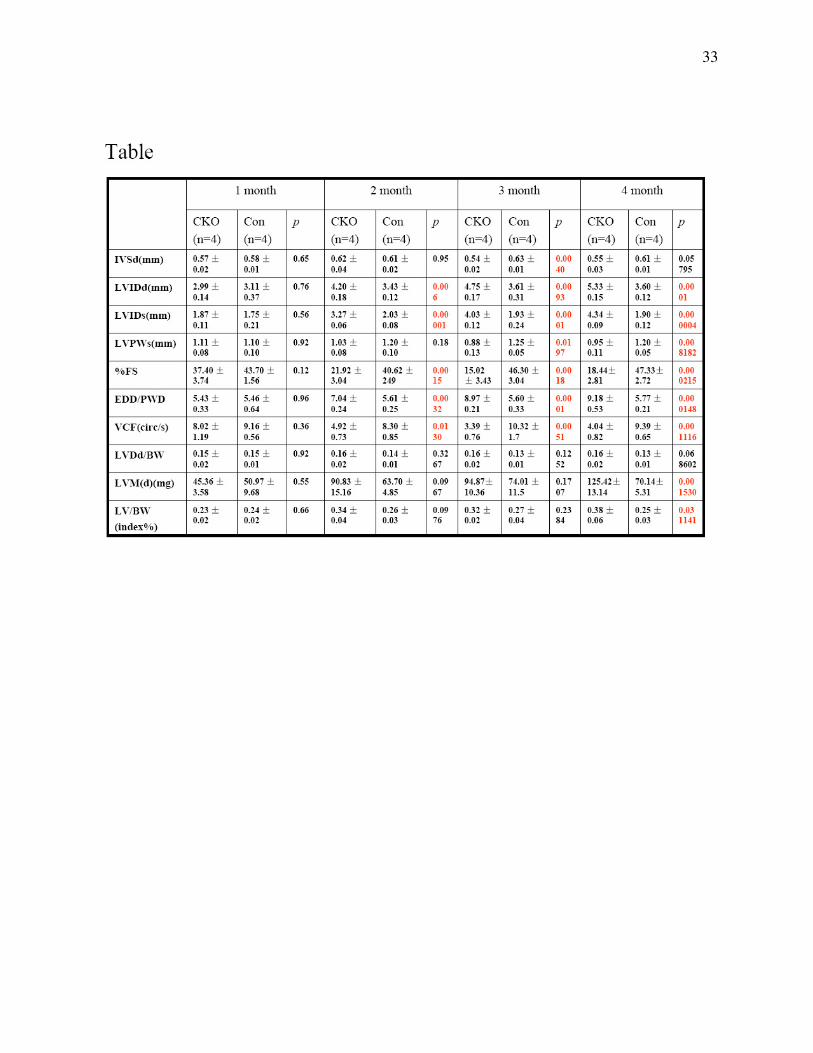
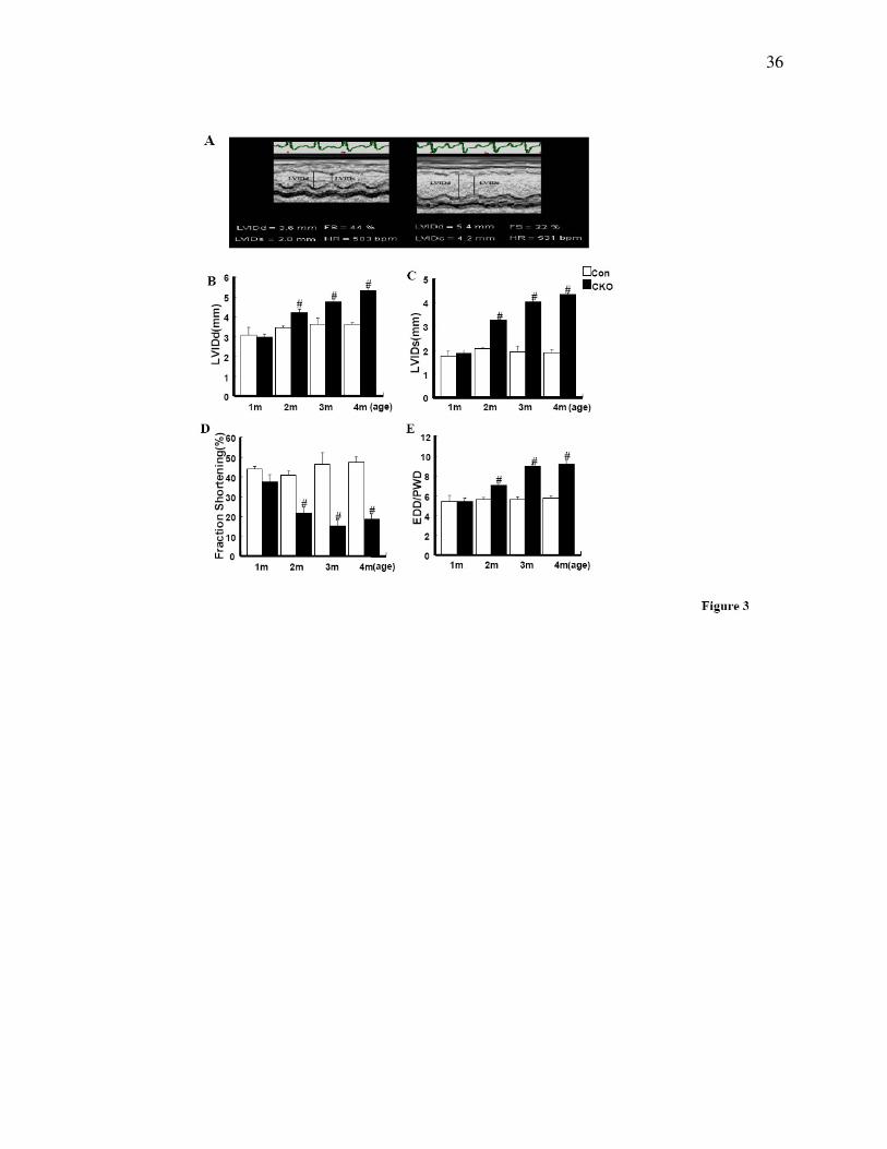
We next evaluated heart performance by echocardiography on CKO and control mice from 1 to 4
months of age. Whereas no significant differences were found between 1-month-old CKO and
control littermate mice, 2 to 4 months old CKO mice showed enlarged left ventricular chamber
size as evidenced by a significant increase in left ventricular internal dimension values at end
diastole (LVIDd) and end systole (LVIDs) (Fig. 3A, B & C) in agreement with the histological
observations (above). In addition, significant wall thinning was seen in CKO hearts as indicated
by a significant decrease in interventricular septal wall thickness (IVSd) at end-diastole
compared to controls (Table 1). More importantly, the changes in cardiac dimensions in CKO
mice were accompanied by a dramatic decrease in LV systolic function, showed by a significant
decrease in fraction shortening (%FS) and the velocity of circumferential fiber shortening (VCF)
as compared to littermate controls (Fig. 3D, Table 1). Despite wall thinning, the significant
increase in heart size led in CKO mice to an increase in calculated left ventricular mass (LVMd)
as well as left ventricular mass normalized to body weight (LV/BW) at 4 months of age
(Table1). ECG analyses also revealed AV block in CKO mice, which was further confirmed by
conscious telemetric monitoring of electrocardiograms (data not shown).
6
Inducible CKO Mice Exhibit Similar Dilated Cardiomyopathy
In CKO mouse heart, Cypher protein was knocked out by Cre recombinase, mediated by the
endogenous Mlc2v promoter, which drives Cre expression from embryonic day 8.75 (24). To
determine whether adult cardiac dysfunction in CKO mice was due to the effect of decreased
Cypher in the developing myocardium, we further generated an inducible cardiac-specific
Cypher knockout mouse strain (ICKO), α-MHC-mER-Cre-mER/Cypherf/f
, to specifically
explore the role of Cypher in adult myocardium. The ICKO mice were generated by crossing
Cypherf/f
mice with α-MHC-mER-Cre-mER mice. 1 month after tamoxifen injection for 5
consecutive days, expression of Cypher protein in α-MHC-mER-Cre-mER/Cypherf/f
mouse heart
was almost totally abolished as compared to controls of both α-MHC-mER-Cre-mER/Cypherf/f
mice injected with peanut oil and Cypherf/f
mice injected with tamoxifen (Fig. 4A).
ICKO mice began to die from 1 week after tamoxifen injection and all died before 16 weeks
after injection. In contrast, 100% were alive in both control groups at least to the time indicated
(Fig. 4B). Dilated cardiomyopathy could be readily determined by the manifestation of a huge
heart and severe dilated chambers of both left and right ventricles in ICKO mouse heart (Fig. 4C).
Consistent with CKO mouse, echocardiography performed 1 month after treatment showed
decreased cardiac function and increased chambers in ICKO mice compared to both control
groups (Fig. 4D, E, F, &G). Moreover, ECG analysis revealed a similar pattern in ICKO mouse
as in CKO mouse (data not shown). Thus, our data obtained from the inducible Cypher mutant
mice are in agreement with our observations in the CKO mouse model, showing that Cypher is
essential for adult cardiac function.

7
CKO Mice Develop Disruption of Z-line Structure
Since we have previously shown the localization of Cypher at the Z-line, we performed
ultrastructural transmission electron microscopy (TEM) to examine Z-line organization in LV
tissues from CKO and control littermate mice (Fig. 5). Whereas some Z-lines were still intact in
1-month-old CKO heart (Fig. 5B), cardiomyocytes from 3-month-old CKO mice had severely
disorganized and disrupted Z-lines with relatively normal M-lines (Fig. 5D & F). Abnormal
mitochondria were also observed in cardiac myocytes from both 1- and 3-month-old CKO mice
(Fig. 5B, D, F). These observations are consistent with previous studies in conventional Cypher
knockout mice, which showed ultrastuctural disorganized of both skeletal and cardiac muscle at
embryonic day 17.5 and postnatal day 1 (17). These data confirm the important role of Cypher in
maintaining cardiac structure and function.
Cypher Interacts with Calsarcin and Myotilin
To understand the molecular basis for the disrupted Z-line structure in CKO mouse hearts, we
next studied the effect of Cypher ablation on the protein level and localization of α-actinin,
Cypher’s interaction partner in the Z-line (16). As demonstrated by Western blot analysis and
immunostainings, neither the protein level, nor the localization of α-actinin was altered in CKO
mouse hearts (data not shown). To search for other Cypher interacting proteins, full-length
Cypher1C and Cypher2C were fused to the GAL4 DNA-binding domain and tested in the yeast
two-hybrid system. Unfortunately, cypher1C caused autoactivation of the GAL4-dependent
reporter genes, precluding its use as a bait. Using Cypher2C as a bait for screening of a
pretransformed human cardiac muscle cDNA library, six clones were identified, all of which
were confirmed by the α-galactosidase assay. Two clones encoded α-actinin-2, confirming the
8
previously identified interaction between Cypher and α-actinin (16), while two other clones
corresponded to calsarcin-1/FATZ-2/myozenin-2. The remaining two clones were leaky and
appeared to be false positive clones.
The interaction between Cypher with calsarcin-1 has previously been described (31), but the
specific domains essential for the interaction have not been identified. To determine the exact
interaction site for calsarcin-1 in Cypher2C, truncated constructs of Cypher2C were
cotransformed with calsarcin-1. This demonstrated that the PDZ domain of Cypher is sufficient
for interaction with calsarcin-1 (data not shown). It has been shown that PDZ can bind to the
carboxyl-terminal sequences of proteins. One C-terminal consensus sequences for PDZ domains
have been shown to be D/E-X-ψ where ψ is hydrophobic amino acid and X is an unspecified
amino acid (32). Consistent with a consensus PDZ motif, the last three amino acids of calsarcin-
1 are DDL. Correspondently, cotransformations of truncated constructs of calsarcin-1 with
Cypher2C demonstrated that the C-terminal 64 amino acids of calsarcin-1 are sufficient for its
binding to Cypher2C. Furthermore, deletion of the last amino acid of calsarcin-1 nearly
completely ablated the binding (Fig. 6A).
To search for other ligands of Cypher’s PDZ domain, we searched for consensus sequences
within known Z-line proteins. This revealed the presence of a PDZ binding consensus motif (the
last three amino acids are EEL) within myotilin, a Z-line protein known to interact with α-
actinin, calsarcin-1, calsarcin-2/FATZ-1/myozenin-1, and filamins (33-35). The potential binding
of myotilin to Cypher2C was tested in the yeast two-hybrid system, which revealed that

9
Cypher’s PDZ domain does indeed bind to myotilin. Consistently, deletion of the last amino acid
in myotilin nearly completely abolished its binding to Cypher (Fig. 6A).
To further confirm the interaction of Cypher’s PDZ domain with myotilin and calsarcin, HA-
tagged myotilin or calsarcin were cotransfected with Flag-tagged Cypher2C (Flag-Cypher),
Cypher’s PDZ domain (Flag-PDZ), or PDZ-deleted Cypher (Flag-∆PDZ) in HEK293 cells,
Following immunoprecipitation, Western blot analyses using anti-Flag antibody showed
coprecipitation of myotilin and calsarcin both with full-length Cypher2C and the PDZ domain
alone, but not with PDZ-deleted Cypher. Thus, Cypher’s 107 amino acid N-terminal PDZ
domain is sufficient and required for Cypher’s binding to both myotilin and calsarcin (Fig. 6B,
C).
To test the effect of Cypher deficiency on the expression and localization of calsarcin and
myotilin, Western blot analyses (Fig. 6D) and immunostainings (data not shown) were
performed on hearts from conventional Cypher knockout mice and control littermates at
embryonic day E17.5. This revealed no differences in the expression or localization of either
calsarcin or myotilin.
Altered Signals in the Hearts of Cypher Mutant Mice
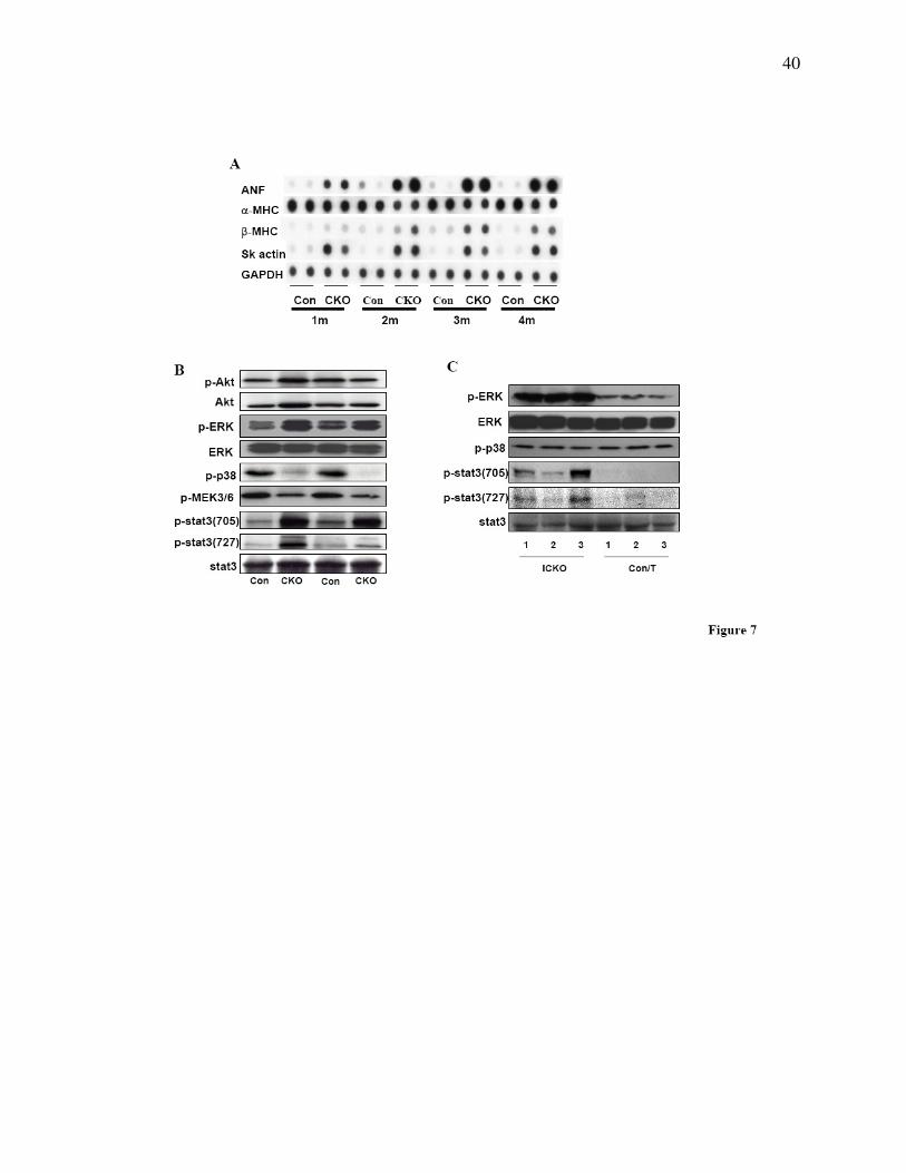
To examine cardiac hypertrophic biomarkers, dot blot analysis was performed using mRNA
isolated from hearts of CKO mice and control littermates at different ages (1, 2, 3, and 4 months
of age). As expected, levels of atrial natriuretic factor (ANF), β-myosin heavy chain (MHC) to
α-MHC ratio, and skeletal muscle actin were increased at all four ages (Fig. 7A).
10
We next investigated several growth-related signaling pathways. Although previous studies have
suggested that the phosphatidylinositol 3-kinase–Akt signaling pathway plays a pivotal role in
cardiac hypertrophy, we found no activation of Akt in CKO mouse hearts, showing that Akt is
not involved in cardiac hypertrophy due to ablation of Cypher. In contrast, phosphorylation
levels of two other growth-related proteins in the heart, extracellular signal-regulated kinase 1/2
(ERK1/2) and signal transducer and activator of transcription 3 (Stat3), were significantly
increased (Fig. 7B), whereas phosphorylation levels of MEK3/6 and p38 MAPK were decreased
in 2-month-old CKO mouse hearts (Fig. 7B). Surprisingly, in ICKO hearts one month after
tamoxifen injection, the phosphorylation level of p38 MAPK was unaltered compared to control
littermates, while phosphorylation of ERK1/2 and Stat3 were increased to levels similar to those
found in CKO mouse hearts (Fig 7C).

11
DISCUSSION
Cypher/ZASP is a striated muscle specific protein localized in the sarcomeric Z-line (16,36). Our
previous studies have demonstrated that conventional ablation of Cypher results in neonatal
lethality of mice within 1 week after birth, caused by multiple striated muscle failure with
symptoms that include decreased milk intake, limb muscle weakness, and cyanosis (17). To
further investigate the functional role of Cypher in the heart, we generated a conditional cardiac-
specific Cypher knockout model using the Cre-loxP system. Homozygous CKO/Cre mice
developed a severe form of dilated cardiomyopathy with disrupted cardiac ultrastructure,
increased heart weight-to-body weight and heart weight-to-tibia bone length ratios, and
decreased cardiac function, resulting in death of all CKO mice before 5 months of age. Similar
observations were made in an inducible cardiac-specific Cypher knockout mouse model. This
indicates an important role of Cypher in maintaining cardiac function.
Mutations in Cypher have been found in human patients with dilated cardiomyopathy, isolated
noncompaction of the left ventricular myocardium, hypertrophic cardiomyopathy, and skeletal
muscle myopathy. Consistent with the genetic and clinical observations in humans, our mouse
models in which Cypher has been specifically ablated in the heart developed dilated
cardiomyopathy, eventually leading to heart failure and premature death. The cardiac
abnormalities induced by Cypher deficiency were not only observed in Mlc2v-cre mediated
cardiac-specific Cypher knockout mouse but also in mice in which Cypher was specifically
ablated in adult myocardium, suggesting that Cypher is essential for the maintenance of cardiac
function and plays a critical role in the pathogenesis of cardiac diseases, especially dilated
cardiomyopathy.
12
A growing number of deficiencies in Z-line and Z-line associated proteins have been found to be
related to cardiomyopathy as well as skeletal muscle myopathies. In addition to Cypher/ZASP,
these proteins include desmin, α-actinin, myotilin, nebulin and others (3,18,19,37-39). Studies in
various model systems have revealed that a wide variety of deficiencies in distinct Z-line and Z-
line associated proteins lead to similar phenotypes, suggesting a converging mechanism shared
by all of these proteins, although the exact mechanisms are still largely unknown. The Z-line not
only serves as a mechanical stretch sensor that transmits structural or contractile changes to
downstream signaling pathways and initiates consequent dysfunctions (40), but is essential for
coordinating contraction and maintaining the cytoskeleton in muscle cells.
Previous studies have demonstrated that Cypher is localized at the Z-line where it binds to α-
actinin through its PDZ domain. In the current study we find that Cypher’s PDZ domain also
interacts with other two Z-line associated proteins, myotilin and calsarcin. However, although
ablation of Cypher results in severe DCM, it has no effect on the expression or localization of
myotilin, calsarcin, and α-actinin. It should be pointed out that α-actinin is the major component
of the Z-line that crosslinks and interacts with many other Z-line proteins, including myotinin
and calsarcin, which also bind to each other (34,41). This suggests that α-actinin, Cypher,
myotilin, and calsarcin are components of a Z-line macromolecular complex, which is essential
for the maintenance of Z-line structure during constant muscle contraction. Interestingly,
ablation of myotilin and calsarcin also has no effect on the expression or localization of other Z-
line proteins (42,43), suggesting that the Z-line macromolecular complex is different from the
Dystrophin-Dystroglycan complex in which ablation of one component down-regulates protein
13
levels of many other components of the complex (44). Nevertheless, we speculate that although
ablation of Cypher, calsarcin, or myotilin has no effect on protein levels of other components of
the Z-line complex, it may weaken the protein connections within the complex, thereby
disrupting the protein structure and lead to consequent cardiac dysfunction, maladaptive
remodeling, signaling changes, and heart failure.
Dilated cardiomyopathy and heart failure are usually accompanied by activation of intracellular
signaling mechanisms and cardiac remodeling. In our study, cardiac deletion of Cypher resulted
in increased expression of molecular markers for cardiomyocyte hypertrophy (ANP, β -MHC/α-
MHC, and SK actin) and consequently mild cardiac fibrosis (data not shown). Increasing
evidence shows that multiple intracellular signals are involved in the sarcomeric protein
deficiency-induced cardiomyopathy (45). Among them, MAPKs are implicated to play an
important role in the pathogenesis of heart failure (46). In the current study, constantly increased
ERK activation was observed in both CKO and ICKO mouse hearts. Surprisingly, p38 MAPK,
another MAPK family member which is usually found to be related with cardiac injury and
recently with cardiac development (47-49), shows distinct activation patterns with decreased
activation in CKO hearts and unaltered activation in ICKO hearts. The difference may reflect the
timing of the cardiac Cypher deletion, with Mlc2v-Cre driving Cypher deletion from an early
embryonic stage and α-MHC-mER-Cre-mER mediating Cypher depletion in adulthood; however,
the precise mechanism needs further elucidation.
A growing body of evidence also indicates a role of another important cardiac signaling protein,
Stat 3, in cardiomyopathy. Altered Stat3 activation has been observed in patients with end-stage
14
dilated cardiomyopathy (50). Consistent with the observations in human, transgenic mouse with
cardiac-Stat3 overexpression induces a protective signal against cardiomyopathy (51), whereas,
cardiac-specific knockout of Stat3 results in increased susceptibility to cardiac injury, reduced
cardiac function, and increased mortality (52). It has also recently been shown that the specific
activation of Stat3 is a potential signaling mechanism underlying the development of heart
failure in the TnI-203/MHC-403 double-mutant murine model of FHC (53). Consistent with this
observation, we found increased Stat3 activation, as indicated by increased phosphorylation of
Stat3, in both CKO and ICKO mouse hearts.
From the data presented here, intracellular signaling pathways associated with cardiac protection
appear to be increased in the cardiac Cypher ablated mouse heart, suggesting that these pathways
may play a common role in mediating cardiac compensation and subsequently the progression to
heart failure. Furthermore, the signal transduction pathways that participate in the pathogenesis
of cardiac dysfunction may provide potential targets for therapeutic interventions.
In summary, cardiac-specific knockout of Cypher in mouse leads to severe cardiac dysfunction,
including DCM and premature lethality from heart failure, with disrupted cellular architecture
and altered activities of multiple cellular signaling pathways. Together, the cardiac dysfunction
caused by Cypher deficiency likely reflects the integrated effects of impairment of sarcomeric
structure and alterations in intracellular signaling cascades.

15
MATERIALS AND METHODS
Generation of Gene Targeted Mice
Floxed Cypher mice were generated by standard techniques using a targeting vector containing a
neomycin selection cassette flanked by FRT sites (26). Briefly, exon 1 of Cypher was inserted
into 2 flanking LoxP sites (Fig. 1A). After electroporation of the targeting vector into R1
embryonic stem (ES) cells, G418-resistant ES cells were screened for homologous
recombination by Southern blot analysis as described below. One heterozyogous recombinant ES
clone was identified and microinjected into blastocysts from C57BL/6J mice to generate male
chimeras. Male chimeras were bred with female Black Swiss mice to generate germline
transmitted floxed heterozygous mice (Cypher f/+
). Cypherf/+
mice were then crossed with
FLPase deleter mice to remove the neomycin gene, and subsequently intercrosses of Cypherf/+
were used to generate homozygous Cypher floxed alleles (Cypherf/f
).
To generate cardiac-specific Cypher knockout mice, Cypherf/f
mice were bred with Mlc2v-Cre
mice in which Cre recombinase expression was controlled by the myosin light chain 2v promoter
(24) to generate double heterozygous Mlc2v-Cre and Cypher floxed mice (Mlc2v-Cre/Cypherf/+
).
The mice were then backcrossed with homozygous Cypherf/f
mice to generate Cypherf/f
Cre+ as
cardiac-specific Cypher knockout mice and Cypherf/f
Cre- mice as control littermates. In a subset
experiment to obtain inducible cardiac-specific Cypher knockout mice, Cypherf/f
mice were
crossed with α-MHC-mER-Cre-mER mice. α-MHC-mER-Cre-mER /Cypherf/f
and Cypherf/f
mice were treated with tamoxifen (Sigma) dissolved in peanut oil by intraperitoneal injection
once a day for 5 consecutive days at a dosage of 1mg/mouse per day, and used as inducible
cardiac-specific Cypher knockout mice and control littermates, respectively. An additional
16
control group was obtained by intraperitoneal injection of peanut oil in α-MHC-mER-Cre-
mER/Cypherf/f
mice.
DNA Analysis
Genomic DNA was extracted from G418-resistant ES cell clones and mouse tails, as previously
described (26). ES cell DNA was digested by BamHI, electrophorised on a 1% (wt/vol) agarose
gel, and subsequently blotted onto a nitrocellulose membrane. A 400-bp fragment was generated
by polymerase chain reaction (PCR) using mouse genomic DNA and specific Cypher primers
(forward, GAAGGCCCTGAAGAAAGGAGAG ; reverse, GACCCTCCTGTTTCTCCAGGTC).
The PCR product was subsequently radiolabeled using α-[32P]dATP by random priming
(Invitrogen). DNA blots were hybridized with the radiolabeled probe and visualized by
autoradiography. Mice were genotyped by PCR analysis using mouse tail DNA, Cypher primers
(forward, ATCAGTGTCGATGGCATTCA; reverse, TGCAGGCAAACAGTACGAAG) and
Cre primers (forward, GTTCGCAAGAACCTGATGGACA; reverse,
CTAGAGCCTGTTTTGCACGTTC) were used to detect floxed Cypher and Cre respectively.
Plasmid Construction for Yeast Two-hybrid Analysis
For yeast two-hybrid analysis, full-length cypher1C (Genbank: AF114378) and full-length
cypher2C (Genbank: AY206011) were amplified from a mouse heart cDNA library by PCR
using the primers: P1 (5’-cggcgaattcATGTCTTACAGTGTGACTCTGAC-3’) and P2 (5’-
ctcagtcgacgaGCTCTTAGCTTTTAACATTAAGG-3’) and cloned into yeast-two hybrid vector
pGBKT7. Full-length calsarcin-1 (Genbank: BC005195) was amplified from a positive clone
obtained from a yeast two-hybrid screen using the primers: P3 (5’-

17
aggccgaattcATGCTATCACATAATACTATGATGAAGC-3’) and P4 (5’-
aatgtcgacgagctcGCACATACAACTTTCTTTTCATAGG-3’), and full-length mouse myotilin
(Genbank: BC016214) was amplified from ATCC clone MCG-28747 using primers P5 (5’-
ccaagaattcATGTTTAACTACGAACGTCC-3’) and P6 (5’-
gtaaaggatccAAGTTCTTCACTTTCATAGAG-3’). All fragments were cloned into the yeast-two
hybrid vectors pGADT7 and/or pGADT7. Truncated constructs of calsarcin-1 and myotilin were
generated by PCR from full-length constructs and cloned into pGADT7.
For coimmunoprecipitations, full-length calsarcin and myotilin were cloned into the mammalian
expression vector pXJ40-HA, while full-length and truncated Cypher2C was inserted into the
pXJ40-Flag vector (both kindly provided by Dr. Tong Zhang). All constructs were checked by
sequencing.
Yeast Two-hybrid Studies
Full-length human cypher2C cDNA, fused to the GAL4 DNA-binding domain (pGBKT7-2C),
was used as a bait in a yeast two-hybrid screen of approximately 4 x 10⁶ clones from a
pretransformed human heart cDNA library (Matchmaker 638833, Clontech) according to the
instructions provided by the manufacturer. Briefly, positive clones growing on selective plates,
lacking histidine, leucine, adenine and tryptophan, were picked and screened for α-galactosidase
activity by plating onto selective plates containing X-α-galactosidase. Plasmid DNA from α-
galactosidase positive clones were isolated as described by the manufacturer and sequenced. To
confirm interactions, obtained clones were retransformed into yeast with cypher2C or empty
pGBKT7 vector. In addition, inserts of pGBKT7 and pGADT7 were swapped and cotransformed

18
into yeast. To pinpoint the interaction sites, truncated or mutant constructs of cypher2C,
calsarcin-1 and myotilin were cotransformed into yeast and positive clones were analyzed for α-
galactosidase activity in liquid culture using PNP-α-gal as a substrate (described in the yeast
protocols handbook, PT3024-1, Clontech).
Western Blot Analysis
To assess protein expression of cardiac isoforms of Cypher, Western blots analyses were
performed on heart lysates from CKO or ICKO mice and control mice using affinity purified
Cypher antibodies as described previously (17). In subset experiments, antibodies against
calsarcin-1 (Alpha diagnostic international), myotilin (kindly provided by Olli Carpen), α-actinin
(Sigma), Flag(Sigma), HA(Santa Cruz), phospho-p44/42 MAPK (Thr202/Tyr204), phospho-p38,
and phospho-stat3(Cell signaling technology) were used as described (17,27). GAPDH antibody
was used for normalization.
In vivo Coimmunoprecipitation
293 cells were harvested 36 hours after co-transfection with constructs generated in the pXJ40-
flag and pXJ40-HA vectors at a concentration of 16 µg/100mm plate, using the Hyfect reagent
according to the manufacturer’s instruction. Cell lysate (600µg total protein) was
immunoprecipitated with 2µg HA polyclonal antibody and 20 µl Protein G Sepharose 4 Fast
Flow beads (Amersham Biosciences), and subjected to Western blot analysis using anti-flag M2
monoclonal antibody (1:20,000).
19
Hematoxylin and Eosin Staining
Hearts were harvested, relaxed in 50 mM KCl in PBS, fixed in 4% paraformaldehyde, embedded
in paraffin, and sectioned into 7-µm thick sections. Hematoxylin and eosin (H & E) staining was
performed according to standard procedures.
Transmission Electron Microscopy (TEM)
Hearts were perfused with 50 mM KCL in tyrode solution followed by 2% PFA and 2%
glutaraldehyde in 0.15 M Na cacodylate buffer. The tissues were processed for transmission
electron microscopy as described previously (17).
RNA Dot Blot
Total RNA was isolated from ventricles using Trizol reagent (Invitrogen) following the
manufacturer’s protocol. RNA dot blot was performed as described (28). Briefly, 2 mg denatured
RNA of each sample was blotted on a nitrocellulose membrane and cross-linked by UV. The
membrane was then probed with α-[32P]dATP labeled probes and signals were visualized by
autoradiography. GAPDH mRNA levels were detected as RNA loading control.
Echocardiography Analysis
Mice were anesthetized with isoflurane and subjected to echocardiography as previously
described (29).
20
Electrocardiography (ECG) Analysis
Surface electrocardiogram recordings were performed on mice anesthetized with 1%-1.5%
isoflurane. Needle electrodes were used and placed in the conventional lead II position. A
differential amplifier (DP-304, Warner Instruments Corp., Hamden, CT) amplified the signals in
the bandwidth 0.1-1000Hz, and signals were filtered using an adaptive 60Hz filter (Humbug,
Quest Scientific, Vancouver, Canada). The signals were digitized at 3000Hz, and analyzed using
ECG Analysis Software (QRS phenotyping, Calgary, Canada).
Statistical Analysis
Statistical analyses were done using Student's unpaired t-Test. P<0.05 was considered
statistically significant. Data are represented as means ± standard errors of the means (SEM).
21
FUNDING
This work was supported by grants from NIH, MDA, and CCF to J.C., and partially by
National Science Foundation of China (30770870 & 30500182) and National Key Basic
Research Program of China (2007CB512100) to M.Z.
ACKNOWLEDGMENTS
We thank Dr. F Sheikh for critical reading of the manuscript.
DISCLOSURES
None.
22
REFERENCES
1. Ortiz-Lopez R, Li H, Su J, Goytia V, Towbin JA. (1997) Evidence for a dystrophin missense
mutation as a cause of X-linked dilated cardiomyopathy. Circulation, 5, 2434-2440.
2. Olson TM, Illenberger S, Kishimoto NY, Huttelmaier S, Keating MT, Jockusch BM. (2002)
Metavinculin mutations alter actin interaction in dilated cardiomyopathy. Circulation, 105,
431-437.
3. Li D, Tapscoft T, Gonzalez O, Burch PE, Quiñones MA, Zoghbi WA, Hill R, Bachinski LL,
Mann DL, Roberts R. (1999) Desmin mutation responsible for idiopathic dilated
cardiomyopathy. Circulation, 100, 461-464
4. Gerull B, Gramlich M, Atherton J, McNabb M, Trombitás K, Sasse-Klaassen S, Seidman
JG, Seidman C, Granzier H, Labeit S, et al. (2002) Mutations of TTN, encoding the giant
muscle filament titin, cause familial dilated cardiomyopathy. Nat. Genet., 30, 201-204
5. Olson TM, Michels VV, Thibodeau SN, Tai YS, Keating MT. (1998) Actin mutations in
dilated cardiomyopathy, a heritable form of heart failure. Science, 280, 750-752.
6. Villard E, Duboscq-Bidot L, Charron P, Benaiche A, Conraads V, Sylvius N, Komajda M.
(2005) Mutation screening in dilated cardiomyopathy: prominent role of the beta myosin
heavy chain gene. Eur. Heart J., 26, 794-803.
7. Morimoto S, Lu QW, Harada K, Takahashi-Yanaga F, Minakami R, Ohta M, Sasaguri T,
Ohtsuki I. (2002) Ca(2+)-desensitizing effect of a deletion mutation Delta K210 in cardiac
troponin T that causes familial dilated cardiomyopathy. Proc. Natl. Acad. Sci. U S A., 99,
913-918.
8. Knöll R, Hoshijima M, Hoffman HM, Person V, Lorenzen-Schmidt I, Bang ML, Hayashi T,
Shiga N, Yasukawa H, Schaper W, et al. (2002) The cardiac mechanical stretch sensor

23
machinery involves a Z disc complex that is defective in a subset of human dilated
cardiomyopathy. Cell, 111, 943-955.
9. Kamisago M, Sharma SD, DePalma SR, Solomon S, Sharma P, McDonough B, Smoot L,
Mullen MP, Woolf PK, Wigle ED, et al. (2000) Mutations in sarcomere protein genes as a
cause of dilated cardiomyopathy. N. Engl. J. Med., 343, 1688-1696.
10. Robinson P, Mirza M, Knott A, Abdulrazzak H, Willott R, Marston S, Watkins H, Redwood
C. (2002) Alterations in thin filament regulation induced by a human cardiac troponin T
mutant that causes dilated cardiomyopathy are distinct from those induced by troponin T
mutants that cause hypertrophic cardiomyopathy. J. Biol. Chem., 277, 40710-40716.
11. Chen J, Chien KR. (1999) Complexity in simplicity: monogenic disorders and complex
cardiomyopathies. J. Clin. Invest., 103, 1483-1485.
12. Seidman JG, Seidman C. (2001) The genetic basis for cardiomyopathy: from mutation
identification to mechanistic paradigms. Cell, 104, 557-567.
13. Towbin JA, Bowles NE. (2006) Dilated cardiomyopathy: a tale of cytoskeletal proteins and
beyond. J. Cardiovasc. Electrophysiol., 17, 919-926.
14. Lapidos KA, Kakkar R, McNally EM. (2004) The dystrophin glycoprotein complex:
signaling strength and integrity for the sarcolemma. Circ. Res., 94, 1023-1031.
15. Dellefave L, McNally EM. (2008) Sarcomere mutations in cardiomyopathy, noncompaction,
and the developing heart. Circulation, 117, 2847-2849.
16. Zhou Q, Ruiz-Lozano P, Martone ME, Chen J. (1999) Cypher, a striated muscle-restricted
PDZ and LIM domain-containing protein, binds to alpha-actinin-2 and protein kinase C. J.
Biol. Chem., 274, 19807-19813.

24
17. Zhou Q, Chu PH, Huang C, Cheng CF, Martone ME, Knoll G, Shelton GD, Evans S, Chen
J. (2001) Ablation of Cypher, a PDZ-LIM domain Z-line protein, causes a severe form of
congenital myopathy. J. Cell. Biol., 155, 605-612.
18. Vatta M, Mohapatra B, Jimenez S, Sanchez X, Faulkner G, Perles Z, Sinagra G, Lin JH, Vu
TM, Zhou Q, et al. (2003) Mutations in Cypher/ZASP in patients with dilated
cardiomyopathy and left ventricular non-compaction. J. Am. Coll. Cardiol., 42, 2014-2027
19. Xing Y, Ichida F, Matsuoka T, Isobe T, Ikemoto Y, Higaki T, Tsuji T, Haneda N, Kuwabara
A, Chen R, et al. (2006) Genetic analysis in patients with left ventricular noncompaction and
evidence for genetic heterogeneity. Mol. Genet. Metab., 88, 71-77
20. Arimura T, Hayashi T, Terada H, Lee SY, Zhou Q, Takahashi M, Ueda K, Nouchi T, Hohda
S, Shibutani M, et al. (2004) A Cypher/ZASP mutation associated with dilated
cardiomyopathy alters the binding affinity to protein kinase C. J. Biol. Chem., 279, 6746-
6752.
21. Theis JL, Bos JM, Bartleson VB, Will ML, Binder J, Vatta M, Towbin JA, Gersh BJ,
Ommen SR, Ackerman MJ. (2006) Echocardiographic-determined septal morphology in Z-
disc hypertrophic cardiomyopathy. Biochem. Biophys. Res. Commun., 351, 896-902.
22. Sheikh F, Bang ML, Lange S, Chen J. (2007) "Z"eroing in on the role of Cypher in striated
muscle function, signaling, and human disease. Trends. Cardiovas. Med., 17, 258-262.
23. Selcen D, Engel AG. (2005) Mutations in ZASP define a novel form of muscular dystrophy
in humans. Ann. Neurol., 57, 269-276.
24. Chen, J., S. W. Kubalak, S. Minamisawa, R. L. Price, K. D. Becker, R. Hickey, J. J. Ross,
and K. R. Chien. (1998) Selective requirement of myosin light chain 2v in embryonic heart
function. J. Biol. Chem., 273, 1252–1256.

25
25. Sohal DS, Nghiem M, Crackower MA, Witt SA, Kimball TR, Tymitz KM, Penninger JM,
Molkentin JD. (2001) Temporally regulated and tissue-specific gene manipulations in the
adult and embryonic heart using a tamoxifen-inducible Cre protein. Circ. Res., 89, 20-25
26. Li X, Zima AV, Sheikh F, Blatter LA, Chen J. (2005) Endothelin-1-induced arrhythmogenic
Ca2+ signaling is abolished in atrial myocytes of inositol-1,4,5-trisphosphate(IP3)-receptor
type 2-deficient mice. Circ. Res., 96, 1274-1281
27. Zheng M, Reynolds C, Jo SH, Wersto R, Han Q, Xiao RP. (2005) Intracellular acidosis-
activated p38 MAPK signaling and its essential role in cardiomyocyte hypoxic injury.
FASEB. J., 19, 109-111.
28. Jones WK, Grupp IL, Doetschman T, Grupp G, Osinska H, Hewett TE, Boivin G, Gulick J,
Ng WA, Robbins J. (1996) Ablation of the murine alpha myosin heavy chain gene leads to
dosage effects and functional deficits in the heart. J. Clin. Invest., 98, 1906-1917.
29. Tanaka N, Dalton N, Mao L, Rockman HA, Peterson KL, Gottshall KR, Hunter JJ, Chien
KR, Ross J Jr. (1996) Transthoracic echocardiography in models of cardiac disease in the
mouse. Circulation, 94, 1109-1117.
30. Huang C, Zhou Q, Liang P, Hollander MS, Sheikh F,Li X, Greaser M, Shelton GD, Evans S,
Chen J. (2003) Characterization and in vivo functional analysis of splice variants of Cypher.
J. Biol. Chem., 278, 7360-7365.
31. Frey N, Olson EN. (2002) Calsarcin-3, a novel skeletal muscle-specific member of the
calsarcin family, interacts with multiple Z-disc proteins. J. Biol. Chem., 277, 13998-14004.
32. Nourry C, Grant SG, Borg JP. (2003) PDZ domain proteins: plug and play. Sci. STKE., 179,
RE7.
26
33. Salmikangas P, Mykkänen OM, Grönholm M, Heiska L, Kere J, Carpén O. (1999) Myotilin,
a novel sarcomeric protein with two Ig-like domains, is encoded by a candidate gene for
limb-girdle muscular dystrophy. Hum. Mol. Genet., 8, 1329-1336.
34. Gontier Y, Taivainen A, Fontao L, Sonnenberg A, van der Flier A, Carpen O, Faulkner G,
Borradori L. (2005) The Z-disc proteins myotilin and FATZ-1 interact with each other and
are connected to the sarcolemma via muscle-specific filamins. J. Cell. Sci., 118, 3739-3749.
35. van der Ven PF, Wiesner S, Salmikangas P, Auerbach D, Himmel M, Kempa S, Hayess K,
Pacholsky D, Taivainen A, Schröder R, et al. (2000) Indications for a novel muscular
dystrophy pathway: gamma-filamin, the muscle-specific filamin isoform, interacts with
myotilin. J. Cell. Biol., 151, 235-248.
36. Faulkner G, Pallavicini A, Formentin E, Comelli A, Ievolella C, Trevisan S, Bortoletto G,
Scannapieco P, Salamon M, Mouly V, et al. (1999) ZASP: a new Z-band alternatively
spliced PDZ-motif protein. J. Cell. Biol., 146, 465-475.
37. Hauser MA, Horrigan SK, Salmikangas P, Torian UM, Viles KD, Dancel R, Tim RW,
Taivainen A, Bartoloni L, Gilchrist JM, et al. (2000) Myotilin is mutated in limb girdle
muscular dystrophy 1A. Hum. Mol. Genet., 9, 2141-2147.
38. Selcen D, Engel AG. (2004) Mutations in myotilin cause myofibrillar myopathy. Neurology.,
62, 1363-1371.
39. Lehtokari VL, Pelin K, Sandbacka M, Ranta S, Donner K, Muntoni F, Sewry C, Angelini C,
Bushby K, Van den Bergh P, et al. (2006) Identification of 45 novel mutations in the nebulin
gene associated with autosomal recessive nemaline myopathy. Hum. Mutat., 27, 946-956.
40. Chang AN, Potter JD. (2005) Sarcomeric protein mutations in dilated cardiomyopathy.
Heart. Fail. Rev., 10, 225-235.
27
41. Sanger JM, Sanger JW. (2008) The dynamic Z bands of striated muscle cells. Sci. Signal. 1,
pe37.
42. Moza M, Mologni L, Trokovic R, Faulkner G, Partanen J, Carpén O. (2007) Targeted
deletion of the muscular dystrophy gene myotilin does not perturb muscle structure or
function in mice. Mol. Cell. Biol., 27, 244-252.
43. Frey N, Barrientos T, Shelton JM, Frank D, Rütten H, Gehring D, Kuhn C, Lutz M,
Rothermel B, Bassel-Duby R, et al. (2004) Mice lacking calsarcin-1 are sensitized to
calcineurin signaling and show accelerated cardiomyopathy in response to pathological
biomechanical stress. Nat. Med., 10, 1336-1343.
44. Michele DE, Campbell KP. (2003) Dystrophin-glycoprotein complex: post-translational
processing and dystroglycan function. J. Biol. Chem., 278, 15457-15460.
45. Solaro RJ. (2008) Multiplex kinase signaling modifies cardiac function at the level of
sarcomeric proteins. J. Biol. Chem., 283, 26829-26833.
46. Ravingerová T, Barancík M, Strnisková M. (2003) Mitogen-activated protein kinases: a new
therapeutic target in cardiac pathology. Mol. Cell. Biochem., 247, 127-138.
47. Hernández-Torres F, Martínez-Fernández S, Zuluaga S, Nebreda A, Porras A, Aránega AE,
Navarro F. (2008) A role for p38alpha mitogen-activated protein kinase in embryonic
cardiac differentiation. FEBS. Lett., 582, 1025-1031.
48. Aouadi M, Bost F, Caron L, Laurent K, Le Marchand Brustel Y, Binétruy B. (2006) p38
mitogen-activated protein kinase activity commits embryonic stem cells to either
neurogenesis or cardiomyogenesis. Stem Cells., 24, 1399-1406.
49. Petrich BG, Wang Y. (2004) Stress-activated MAP kinases in cardiac remodeling and heart
failure; new insights from transgenic studies. Trends. Cardiovasc. Med., 14, 50-55.

28
50. Podewski EK, Hilfiker-Kleiner D, Hilfiker A, Morawietz H, Lichtenberg A, Wollert KC,
Drexler H. (2003) Alterations in Janus kinase (JAK)-signal transducers and activators of
transcription (STAT) signaling in patients with end-stage dilated cardiomyopathy.
Circulation, 107, 798-802.
51. Kunisada K, Negoro S, Tone E, Funamoto M, Osugi T, Yamada S, Okabe M, Kishimoto T,
Yamauchi-Takihara K. (2000) Signal transductor and activator of transcription 3 in the heart
transduces not only a hypertrophic signal but a protective signal against doxorubicin induced
cardiomyopathy. Proc. Natl. Acad. Sci. USA., 97, 315-319.
52. Jacoby JJ, Kalinowski A, Liu MG, Zhang SS, Gao Q, Chai GX, Ji L, Iwamoto Y, Li E,
Schneider M, et al. (2003) Cardiomyocyte-restricted knockout of STAT3 results in higher
sensitivity to inflammation, cardiac fibrosis, and heart failure with advanced age. Proc. Natl.
Acad. Sci. U S A., 100, 12929-12934.
53. Tsoutsman T, Kelly M, Ng DC, Tan JE, Tu E, Lam L, Bogoyevitch MA, Seidman CE,
Seidman JG, Semsarian C. (2008) Severe heart failure and early mortality in a double-
mutation mouse model of familial hypertrophic cardiomyopathy. Circulation, 117, 1820-
1831.

29
FIGURE LEGENDS
Figure 1. Generation of gene targeted mice. (A) Targeting construct for the generation of
floxed Cypher mice. In the targeting vector, cypher gene exon 1 is flanked by LoxP sites as
indicated with a black triangle. The neomyosin resistant gene is surrounded by FLP cassettes as
shown by black rectangles. (B) Southern blot analysis shows a 6.9 Kb band in heterozyogous
gene targeted ES cells as compared to a 6.2 Kb band in the wildtype allele. (C) Representative
Western blot results showing the effective deletion of Cypher in CKO mouse heart at 1 month of
age.
Figure 2. Survival curve and enlarged CKO mouse hearts. (A) Cumulative survival curve of
CKO and control littermate mice. CKO mice die from 16 weeks of age and are all dead before 23
weeks of age, n=29 for CKO mice and n=33 for control littermates. (B) Hematoxylin and eosin
(H&E) staining showing dilated ventricular chambers in 3-month-old CKO mouse heart
compared to control littermate. (C) Heart weight to body weight ratio of CKO mice versus
control littermates, n= 15 for CKO mice and n= 28 for control littermates. (D) Heart weight to
tibia bone length ratio of CKO mice versus control littermates, n=12 for CKO mice and n=12 for
control littermates. HW/BW, heart weight to body weight ratio; HW/BL, heart weight to tibia
bone length ratio; * P<0.05.
Figure 3. Echocardiography. (A) Typical example of echocardiography data showing LVIDd,
LVIDs, and FS in CKO and control littermate hearts. (B) LVIDd, (C) LVIDs, (D) FS, and (E)
EDD/PWD results in CKO mice and control littermates at various ages as indicated. n=4 for
30
each group. LVIDd, end-diastolic left ventricular internal dimension at end-diastole; LVIDs,
end-systolic left ventricular internal dimension; % FS, percentage of fractional shortening;
EDD/PWD, end-diastolic dimension/diastolic posterior wall thickness; # P<0.01.
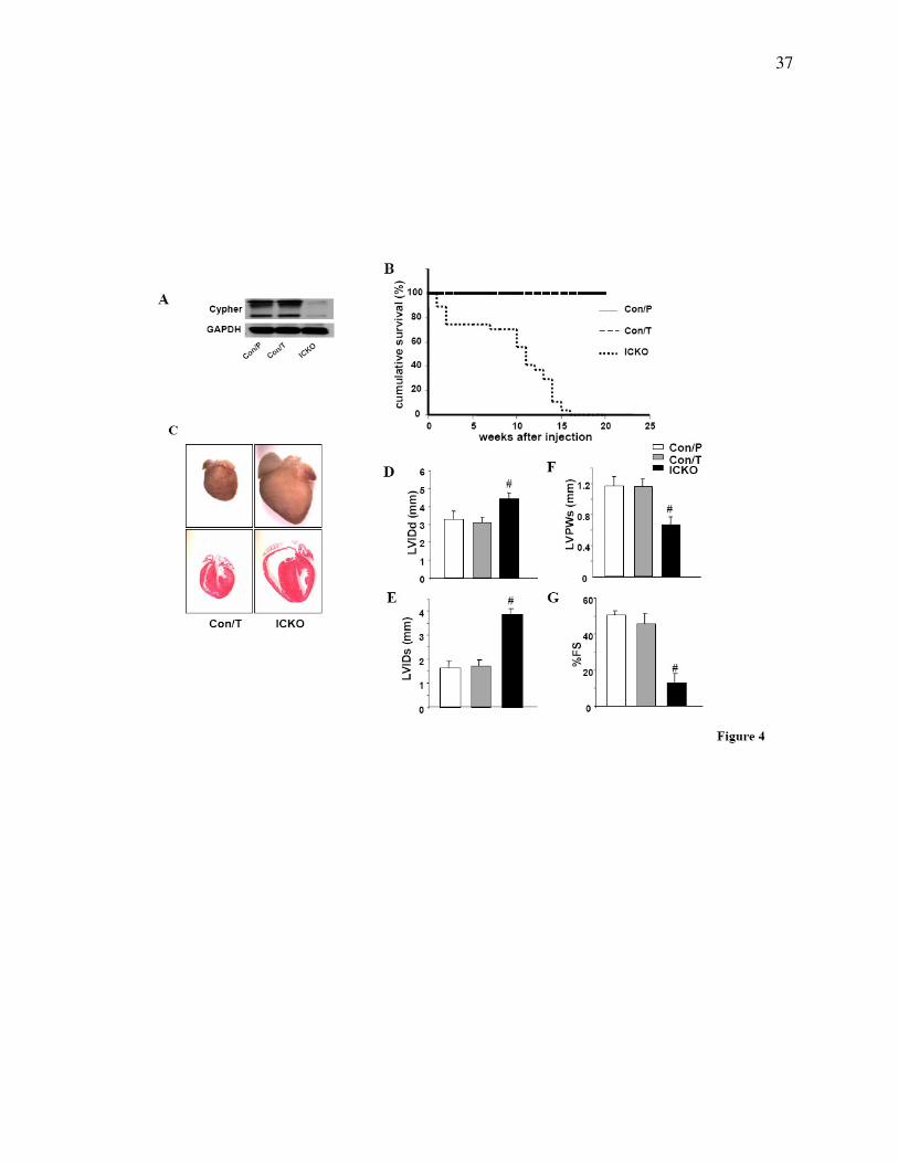
Figure 4. Inducible cardiac-specific deletion of Cypher. (A) Western blot results showing
inducible deletion of Cypher in the heart of Cypherf/f
/Cre mice 1 month after injection with
tamoxifen (ICKO) or Cypherf/f
/Cre and Cypherf/f
littermate mouse injected with peanut oil
(Con/P)and tamoxifen (Con/T), respectively as controls. (B) Accumulative survival curve
showing that ICKO mice die from 1 week after tamoxifen injection, and all die before 15 weeks
after injection, compared to 100% survival of both control groups. n=27 for ICKO group, n=20
for tamoxifen control group, and n=22 for peanut oil control group. (C) H&E staining showing
severe dilated ventricules in ICKO mouse heart 1month after injection, compared with tamoxifen
control mouse heart (Con/T). Echocardiography data showing (D) LVIDd, (E) LVIDs, (F)
LVPWs, and (G) FS results in ICKO and control littermate mice 1 month after tamoxifen or
peanut oil injection, n=4 for each group. LVIDd, end-diastolic left ventricular internal
dimension; LVIDs, end-systolic left ventricular internal dimension; LVPWs, systolic left
ventricular posterior wall thickness; % FS, percentage of fractional shortening; # P<0.01.
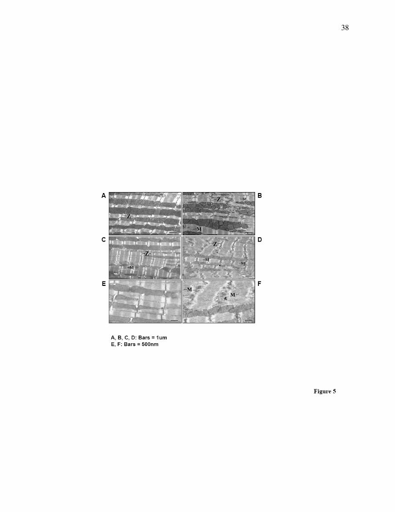
Figure 5. Disrupted ultrastructure in CKO mouse heart. Transmission electron microscopy
showing some disorganized Z-lines in cardiomyocytes from CKO mice at 1 month of age while
most Z-line structures remain intact (B), compared to the well-organized and condensed Z-line
structures in control littermates (A). At 3 months of age, Z-lines are completely disrupted in
cardiomyocytes from CKO mice (D) as compared to normal Z-lines in control littermates (C).
31
Magnified figures to better show the dispersed and punctured Z-lines in a 3-month-old CKO
mouse (F) compared to intact Z-lines in the control littermate (E).
Figure 6. Cypher’s PDZ domain interacts with calsarcin-1 and myotilin. (A) Deletion of the
last residue Leucine in calsarcin-1 (DL) and myotilin (DL) results in nearly complete ablation of
their interaction with Cypher’s PDZ domain in the yeast two-hybrid system. The strength of the
interactions was quantified in a liquid α-galactosidase assay using PNP-α-gal as a substrate. (B)
Schematic presentation of HA-tagged calsarcin-1 (HA-calsarcin) and myotilin (HA-myotilin)
and Flag-tagged fragments encoding Cypher2C (Flag-Cypher), truncated Cypher2C containing
the PDZ domain (Flag-PDZ), and PDZ-less Cypher2C (Flag-∆PDZ). (C) In vivo
immunoprecipitation of Cypher with calsarcin-1 and myotilin showing the interaction of Cypher
with calsarcin-1 and myotilin through its PDZ domain. (D) Western blot analysis showing
protein levels of Cypher, calsarcin-1, and myotilin in hearts from wildtype and conventional
Cypher knockout mice at embryonic day E17.5.
Figure 7. Alterations of signals in Cypher deficient heart. (A) Dot blot assay showing altered
expression of fetal genes in CKO mouse compared to control littermate mouse hearts at the
indicated ages. (B) Western blot analyses for activation of proteins involved in intracellular
signaling in mouse hearts from CKO and control littermates at 2 months of age as well as in (C)
ICKO and control littermates 1 month after tamoxifen injection.

32
TABLE
Echocardiographic assay of cardiac function in CKO and control mice. Echocardiographic
data were obtained both from CKO and control mice at the indicated ages. Results are expressed
as means ± standard errors.
33
34

35
36
37
38

39
40
41