Cardiac myocytes are short branched striated muscle...
10
Cardiac myocytes are short branched striated muscle cells Connected with gap junctions gap junctions transmit electrical activity between cells So, cardiac myocytes act as a single functional unit (syncitium)
Transcript of Cardiac myocytes are short branched striated muscle...

Cardiac myocytes are short
branched striated muscle cells
Connected with gap junctions
gap junctions transmit
electrical activity between cells
So, cardiac myocytes act as
a single functional unit
(syncitium)

1. Nodal fibers
2.Conducting
fibers
3. Contractile
fibers Atria
Ventricle

1. Rhythmicity
2. Excitability
3. Conductivity
4. Contractility

Rhythmicity means the ability of the heart to beat regularly without external stimulation.
It is myogenic in origin not neurogenic
The nodal fibres and conducting system are self-excitable.
Sinoatrial node (SAN)→110 b/min
Atrioventricular node (AVN)→ 90
Bundle of His (A-V bundle) → 45
Purkinje fibres → 35
Ventricular fibres → 25
The cells of SAN; (posterior wall of right atrium) is the primary pacemaker of the heart

The ability to conduct impulse from one cell to another---facilitated by the presence of gap junctions that transmit electrical currents
From SAN→ atrial muscle & atrioventricular node (AVN)
From AVN (slowest) → atrioventricular (AV) bundle (bundle of His) →left & right bundles →purkinje fibres (fastest)
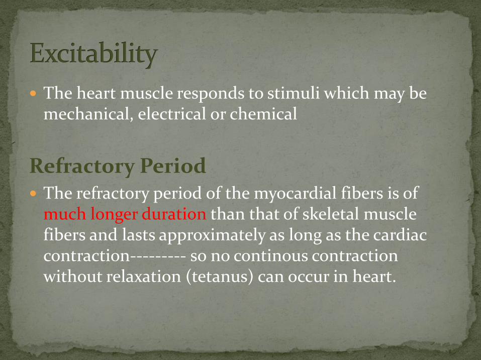
The heart muscle responds to stimuli which may be mechanical, electrical or chemical
Refractory Period The refractory period of the myocardial fibers is of
much longer duration than that of skeletal muscle fibers and lasts approximately as long as the cardiac contraction--------- so no continous contraction without relaxation (tetanus) can occur in heart.

The cardiac muscle contracts either maximally or not
at all (under constant conditions)
The Atria contract as one unit & the ventricles
contract as one unit
This is significant for efficient pumping of the blood

2- Staircase or Treppe
Phenomenon
Rapidly Repeated stimulation
of the cardiac muscle produce
gradual increase in the
strength of contraction
The earlier contractions
produce better conditions
(heat, less viscosity between
muscle fiber, more Ca) for the
following contraction

Within limits, the greater the initial length of cardiac
muscle fibre (stretch), the greater the force of
contraction
The initial length is determined by the volume of blood
filling ventricles at end of diastole (end-diastolic
volume; EDV)

A) Sympathetic supply:
1.↑es all cardiac properties
2. ↑es the coronary blood
flow.
B) Parasympathetic supply:
1.↓es all cardiac properties
except the ventricles (not
supplied by vagus nerve)
2.↓es the coronary blood flow